Haematology · General Medicine
Transfusion Medicine & Transfusion Reactions
Also known as Transfusion medicine · Blood transfusion · Transfusion reactions · TRALI · TACO · Acute haemolytic transfusion reaction · Massive transfusion · Patient blood management
Blood transfusion is a liquid transplant: each unit places donor cells, plasma proteins and donor leucocyte-fragments into a recipient, with benefits (oxygen carriage, haemostasis, volume) balanced against immune, infectious, and circulatory risks. The two universal safety principles are correct ABO/Rh matching (the commonest fatal error is clerical mislabelling of a sample or unit) and a restrictive transfusion strategy — red-cell threshold haemoglobin under 70 g/L (under 80 g/L in cardiac surgery, symptomatic anaemia, or active bleeding), one unit then reassess. Components: PRBCs (anaemia/haemorrhage), platelets (thrombocytopenia, prophylaxis under 10 x 10^9/L), fresh frozen plasma (INR over 1.5 with bleeding), cryoprecipitate (fibrinogen under 1.5 g/L), prothrombin complex concentrate (urgent warfarin reversal), albumin, IVIG, CMV-negative and irradiated products for the immunocompromised. Acute transfusion reactions (under 24 h): acute haemolytic (ABO mismatch — fever, flank pain, hypotension, haemoglobinuria, DIC; STOP), febrile non-haemolytic (cytokines from donor WBCs; commonest), allergic/urticarial (mild) to anaphylactic (anti-IgA in IgA-deficient patients), TRALI (donor anti-WBC antibodies, bilateral non-cardiogenic infiltrates within 6 h), TACO (circulatory overload), bacterial contamination (room-temperature platelets), and transfusion-associated graft-versus-host disease (immunocompromised — use irradiated). Delayed reactions (over 24 h): delayed haemolytic (anamnestic IgG, 3 to 14 days), post-transfusion purpura, transfusion-transmitted infection (HIV, HepB/C — now extremely rare with nucleic-acid testing), and iron overload (chronic transfusion; desferrioxamine/deferasirox). The universal reaction rule for ANY reaction: STOP, maintain IV access with normal saline, assess ABCDE, keep the unit and giving set, send fresh patient samples, return unit to blood bank, and report.
On this page & tools
Your progress
Saved locally on this device.
Exam tags
Red flags
Overview & Definition
Transfusion medicine is the prescription, processing, matching, and safe administration of blood components, and the recognition and management of the adverse reactions that follow them. A transfusion is best understood as a liquid transplant: every unit places donor cells, donor plasma proteins and residual donor leucocytes into a recipient, with the benefit (oxygen carriage in anaemia, platelets in thrombocytopenia, clotting factors in coagulopathy, volume in haemorrhage) weighed against the immune, infectious, and circulatory risks of that transplant.[1][2]
Three skills define safe practice. First, choosing the right component for the right deficit — anaemia is treated with red cells, not "blood"; coagulopathy is treated with FFP or cryoprecipitate, not red cells; thrombocytopenia is treated with platelets. Second, matching and checking rigorously — the commonest fatal error is ABO mismatch from clerical mislabelling of the sample, the unit, or the patient, and it is preventable by mandatory two-person bedside identification. Third, recognising reactions early — most are mild, but several are medical emergencies where the speed of recognition (the first five minutes) determines outcome.[1]
The cardinal rule for any transfusion reaction is to STOP, maintain IV access with normal saline, assess, return the unit and giving set with fresh patient samples to the blood bank, and report — then investigate the specific mechanism. A restrictive threshold (Hb under 70 g/L in most stable adults) is now the standard of care, supported by the TRICC, FOcus, TRISS, and AABB 2023 trials.[1][3][4][6]
The ABO and Rh Blood Group System
The ABO system is the single most important determinant of transfusion safety because anti-A and anti-B antibodies are naturally occurring IgM produced in the first year of life in response to environmental antigens (bacterial polysaccharides on gut flora). They activate complement efficiently, and ABO-incompatible transfusion is the commonest cause of fatal acute haemolytic transfusion reaction.[1]
The ABO antigens are carbohydrate moieties on the red-cell surface, produced by glycosyltransferases encoded on chromosome 9: [1]
- Group A — A antigen on the red cell; anti-B (IgM) in plasma.
- Group B — B antigen on the red cell; anti-A (IgM) in plasma.
- Group AB — both A and B antigens; neither anti-A nor anti-B in plasma — the universal plasma donor (no antibodies to attack a recipient).
- Group O — neither A nor B antigen; both anti-A and anti-B (IgM) in plasma — the universal red-cell donor (no A/B antigens to be attacked), but a dangerous plasma donor. [1]
Group O (universal RBC donor)
- No A or B antigen on red cells
- Recipient anti-A/anti-B has nothing to attack
- O negative used for emergency transfusion in females of childbearing potential
- Plasma contains anti-A AND anti-B (IgM) — dangerous as plasma donor
Group AB (universal plasma donor)
- Both A and B antigens on red cells
- No anti-A or anti-B in plasma
- AB plasma can be given to any ABO group
- AB red cells can ONLY go to AB recipients
Group A (40 percent of population)
- A antigen on red cells
- Anti-B (IgM) in plasma
- A red cells go to A or AB recipients only
- Most common ABO group in most populations
Group B
- B antigen on red cells
- Anti-A (IgM) in plasma
- B red cells go to B or AB recipients only
- Higher prevalence in South Asian and African populations
The Rhesus (Rh) system is the second most important. Of the ~50 Rh antigens, RhD is clinically dominant because RhD-negative individuals do not have naturally occurring anti-D — they form it only after exposure (pregnancy with an RhD-positive fetus, or transfusion of RhD-positive blood). Once sensitised, subsequent exposure triggers an IgG-mediated extravascular haemolysis and, in pregnancy, haemolytic disease of the fetus and newborn.[1]
The compatibility table below is reproduced exactly as examiners expect it: [1]
| Recipient ABO | Recipient Rh | Can receive RBC from | Can receive plasma from |
|---|---|---|---|
| O | + / − | O only | O, A, B, AB |
| A | + / − | A or O | A or AB |
| B | + / − | B or O | B or AB |
| AB | + / − | A, B, AB or O (universal recipient) | AB only |
Rh compatibility — RhD-negative recipients should receive RhD-negative red cells to avoid sensitisation; RhD-positive recipients can receive either. In emergencies, O negative red cells are given to females of childbearing potential and O positive to males and post-menopausal females to avoid depleting the limited O-negative stock.[1]
[1]Blood Components & Special Products
Selecting the right component for the deficit is the heart of transfusion medicine.[1]
- Whole blood — used in massive haemorrhage and military settings; rarely available in civilian hospitals where components are separated.
- Packed red blood cells (PRBCs) — one unit (~300 mL) raises Hb by roughly 10 g/L and haematocrit by 3 percent in a non-bleeding adult; stored in additive solution (SAG-M) at 1 to 6 degrees C for up to 35 to 42 days.
- Platelets — one adult dose (~200 to 300 mL, ~3 x 10^11 platelets) raises the platelet count by 20 to 40 x 10^9/L; stored at 20 to 24 degrees C with agitation for 5 to 7 days — this room-temperature storage is the reason platelets carry the highest bacterial-contamination risk of any component.
- Fresh frozen plasma (FFP) — 200 to 300 mL contains all coagulation factors; thawed over 20 to 30 minutes; dose 10 to 15 mL/kg (typically 4 units for an adult).
- Cryoprecipitate — 10 to 20 mL per unit, rich in fibrinogen, factor VIII, factor XIII, von Willebrand factor and fibronectin; given as a 10-unit pool (~150 mL) to raise fibrinogen by roughly 1 g/L.
- Prothrombin complex concentrate (PCC) — factors II, VII, IX and X; rapid (15 to 30 minute) warfarin reversal; 25 to 50 IU/kg with vitamin K 5 to 10 mg IV.
- Albumin 4 percent or 20 percent — volume expansion and hypoalbuminaemia; 20 percent in hepatorenal syndrome and severe ascites.
- Intravenous immunoglobulin (IVIG) — immune thrombocytopenia, Guillain-Barre, primary immunodeficiency, secondary immunodeficiency.
- Factor concentrates — recombinant factor VIII/IX for haemophilia, recombinant factor VIIa for refractory haemorrhage, von Willebrand factor concentrate. [1]
Special products for specific situations:[1][2]
- Leucodepleted (standard in most high-income countries) — fewer febrile non-haemolytic reactions, less CMV transmission, less alloimmunisation.
- CMV-negative — for pregnant women, neonates, intrauterine transfusion, stem-cell transplant recipients, and severely immunocompromised patients.
- Irradiated (25 Gy) — for stem-cell transplant recipients, haematological malignancy on fludarabine/cladribine/alemtuzumab, congenital immunodeficiency, intrauterine or neonatal exchange transfusion, and first-degree relatives as donors; prevents transfusion-associated graft-versus-host disease (TA-GVHD) by inactivating donor T-lymphocytes.
- Washed — for recurrent severe allergic reactions and confirmed IgA deficiency with anti-IgA; removes donor plasma.
- Methlene-blue or pathogen-reduced plasma — additional viral/bacterial kill.
- Volume-reduced — for severe volume intolerance, TACO-prone patients. [1]
PRBCs
- Anaemia (Hb under 70 g/L), haemorrhage, symptomatic anaemia
- 1 unit raises Hb by ~10 g/L; give one unit then reassess
- Stored 1-6 degrees C for 35-42 days
- Universal donor: O negative
Platelets
- Thrombocytopenia with bleeding or prophylaxis (under 10)
- Stored at 20-24 degrees C with agitation — bacterial risk
- Shelf life 5-7 days
- Adult dose raises count by 20-40 x 10^9/L
FFP
- Coagulopathy with INR over 1.5 and active bleeding
- Warfarin reversal if PCC unavailable
- Dose 10-15 mL/kg (4 units adult)
- Universal plasma donor: AB
Cryoprecipitate
- Fibrinogen under 1.5 g/L (DIC, massive haemorrhage, obstetric haemorrhage)
- 10-unit pool raises fibrinogen by ~1 g/L
- Rich in factor VIII, factor XIII, vWF, fibrinogen
- Thaw pooled; give within 4 h
PCC
- Urgent warfarin reversal (15-30 min, faster than FFP)
- Dose 25-50 IU/kg plus vitamin K 5-10 mg IV
- Carries thrombosis risk — weigh carefully
- Reverses within 30 min; vitamin K sustains
Albumin
- 4 percent for volume expansion; 20 percent for severe hypoalbuminaemia
- Hepatorenal syndrome, large-volume paracentesis
- No infection risk, no compatibility needed
- Expensive; do not use as first-line volume
Pre-Transfusion Testing
The aim is to deliver an ABO- and Rh-compatible unit that the recipient has no antibodies against.[1]
- Group and screen — ABO and RhD group of the recipient, plus an antibody screen (indirect antiglobulin test, IAT) against a panel of group O reagent red cells expressing the common clinically significant antigens (Rh, Kell, Duffy, Kidd). The antibody screen detects irregular antibodies formed by prior transfusion or pregnancy.
- Crossmatch — recipient serum is incubated with donor red cells to confirm compatibility. Electronic (computer) crossmatch is now standard when group-and-screen shows no antibodies; an immediate-spin crossmatch detects ABO incompatibility; a full IAT crossmatch is used when an antibody is present.
- Sample validity — group-and-screen samples are valid for 3 days (72 hours) in patients transfused or pregnant in the previous 3 months, and 7 to 14 days otherwise. Always label the sample at the bedside, in the presence of the patient, against two identifiers (full name and date of birth), and never pre-label.
- Bedside check — at administration, two clinicians independently confirm patient identity (name, date of birth, wristband), unit compatibility (recipient details on the bag match the patient), expiry date, and unit integrity (no clots, no discolouration). This single step prevents most fatal ABO-mismatch errors.[1]
Pre-transfusion testing — the rules
Indications & Transfusion Thresholds
Transfusion thresholds have tightened markedly over the past 20 years. The current international standard (AABB 2023, Carson et al) is restrictive.[1]
Red blood cells (PRBCs)[1][3][4][6]
- Hb under 70 g/L — most stable hospitalised adults, including ICU.
- Hb under 70 to 80 g/L — symptomatic anaemia (chest pain, orthostatic hypotension, tachycardia unresponsive to fluid, heart failure with low Hb).
- Hb under 80 g/L — postoperative cardiac surgery; hip fracture surgery in older patients (FOcus, AABB 2023).
- Hb under 70 g/L in septic shock — TRISS trial showed no benefit of Hb 90 vs Hb 70.
- Active haemorrhage — transfuse to clinical endpoints, not Hb targets; Hb can be falsely reassuring early in bleeding.
- Avoid transfusing above Hb 100 g/L — almost always inappropriate. [1]
Platelets — prophylactic for thrombocytopenia from bone-marrow failure:[1]
- under 10 x 10^9/L — prophylaxis in stable, afebrile chemotherapy/leukaemia patients.
- under 20 x 10^9/L — febrile or with risk factors (sepsis, mucositis, recent bleeding, high blast count).
- under 50 x 10^9/L — before invasive procedures (central line, lumbar puncture, transbronchial biopsy, epidural, surgery).
- Active bleeding or before surgery — keep above 50 (general surgery) or above 100 (neurosurgery, ophthalmic, retinal).
- Platelet dysfunction (antiplatelet drugs, uraemia, cardiopulmonary bypass) — transfuse despite a normal count if bleeding.
- Do NOT transfuse prophylactically in immune thrombocytopenia, heparin-induced thrombocytopenia, or thrombotic thrombocytopenic purpura — they are rapidly consumed and HIT/TTP are pro-thrombotic. [1]
Fresh frozen plasma[1]
- INR over 1.5 with active bleeding, or before emergency surgery/invasive procedure with multiple factor deficiency.
- Warfarin reversal if PCC unavailable (slower, larger volume).
- Massive haemorrhage as part of ratio-based resuscitation.
- Single-factor deficiency — use specific factor concentrate if available. [1]
Cryoprecipitate [1]
- Fibrinogen under 1.5 g/L with bleeding (DIC, massive haemorrhage, postpartum haemorrhage, liver disease, after cardiac bypass).
- Adult dose — 10-unit pool; recheck fibrinogen after infusion; aim to keep above 1.5 g/L. [1]
Prothrombin complex concentrate [1]
- Urgent warfarin reversal in life-threatening bleeding (intracranial haemorrhage, major GI bleed, haemorrhagic stroke).
- Dose 25 to 50 IU/kg based on INR and weight, plus vitamin K 5 to 10 mg IV; faster and lower-volume than FFP. [1]
Transfusion thresholds — the headline numbers
Classification of Transfusion Reactions
Transfusion reactions are classified by timing (acute vs delayed) and mechanism (immune vs non-immune). The classification drives both the differential and the treatment.[1][2]
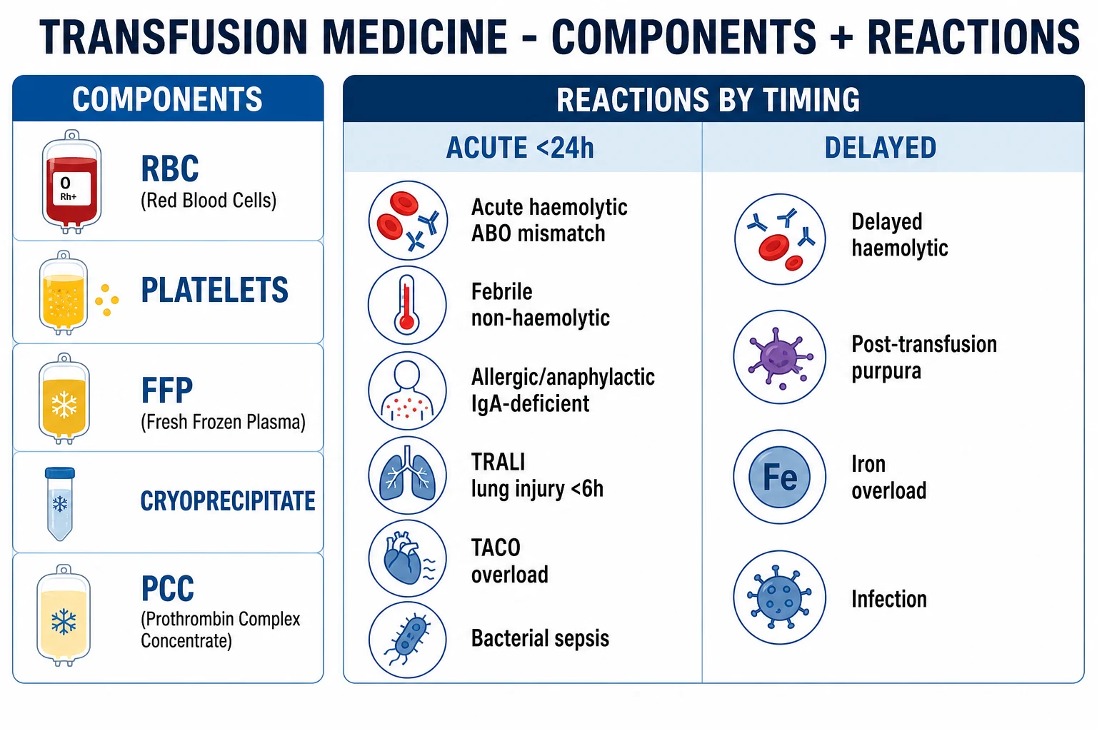
Acute (under 24 h)
- Acute haemolytic (ABO mismatch) — IgM, complement, intravascular haemolysis; medical emergency
- Febrile non-haemolytic — donor cytokines (IL-1, IL-6) and recipient anti-leukocyte antibodies; mild; commonest
- Allergic (urticaria) to anaphylactic — IgE to plasma proteins; IgA deficiency with anti-IgA for anaphylaxis
- TRALI — donor anti-HLA/neutrophil antibody; bilateral non-cardiogenic pulmonary oedema within 6 h
- TACO — volume overload in cardiac/renal/elderly patients; cardiogenic pulmonary oedema
- Bacterial contamination — room-temperature-stored platelets; endotoxin septic shock
Delayed (over 24 h)
- Delayed haemolytic — anamnestic IgG to Kidd/Duffy; 3 to 14 days; extravascular haemolysis
- Transfusion-transmitted infection — hepatitis B/C, HIV (now extremely rare with nucleic-acid testing)
- Post-transfusion purpura — severe thrombocytopenia 5 to 10 days; anti-HPA antibodies
- Transfusion-associated graft-versus-host disease — fatal; prevented by irradiated products in immunocompromised
- Iron overload — chronic transfusion (thalassaemia, MDS); cardiomyopathy, cirrhosis, diabetes
- Alloimmunisation — antibodies formed against donor antigens complicating future transfusion
Epidemiology & Risk Factors
- Roughly 1 in 70 hospitalised patients receives blood; in ICU and perioperative settings the proportion is far higher.
- ABO mismatch from mislabelling remains the commonest fatal error and is almost always preventable by two-person bedside checking. Incidence is estimated at 1 in 12 000 to 1 in 40 000 units transfused, with mortality of 5 to 10 percent.[1]
- Febrile non-haemolytic and mild allergic reactions are the most frequent acute reactions; with universal leucodepletion the FNHTR rate has fallen sharply.
- TRALI incidence fell dramatically after the introduction of male-predominant plasma donor strategies (avoiding multiparous female donors with anti-HLA antibodies); current incidence is approximately 1 in 50 000 to 1 in 100 000 units, mortality 5 to 10 percent.[2]
- TACO is now the leading cause of transfusion-related mortality reported to haemovigilance schemes (UK SHOT, US FDA), reflecting an ageing, comorbid inpatient population; incidence approximately 1 in 100 to 1 in 7000 units depending on definitions.[2]
- Severe IgA deficiency affects under 0.01 percent of the population and underlies anaphylactic reactions to plasma-containing products.
- Bacterial contamination (almost always platelets) is now the leading infectious cause of transfusion-related death in the United States; incidence approximately 1 in 50 000 platelet units.
Pathophysiology — Mechanism of Each Reaction
Each reaction's mechanism points directly to its treatment, so pathophysiology is high-yield for examiners.[1][2]
- Acute haemolytic (ABO) — preformed recipient IgM anti-A/anti-B binds ABO-incompatible donor red cells and potently activates the complement cascade (membrane attack complex), causing intravascular haemolysis with release of free haemoglobin, activation of coagulation (DIC), cytokine storm (TNF, IL-1, IL-6), hypotension, and acute kidney injury from haemoglobin cast nephropathy. Caused almost always by clerical mislabelling.[1]
- Febrile non-haemolytic — donor-derived cytokines (IL-1, IL-6, TNF) accumulated during storage, and/or recipient anti-leukocyte antibodies acting on the hypothalamus, cause a temperature rise of at least 1 degree C with rigors. Reduced by leucodepletion and pre-storage filtration.
- Allergic (urticarial) — donor plasma proteins (often complement components or other allergens) cross-link IgE on mast cells, releasing histamine; manifests as pruritus, urticaria, flushing. Mild.
- Anaphylactic — most often in patients with severe IgA deficiency (under 0.07 g/L) who have formed anti-IgA class antibodies; exposure to donor IgA in any plasma-containing product triggers massive mast-cell degranulation with bronchospasm, angio-oedema, hypotension, and cardiovascular collapse within minutes.
- TRALI — donor (rarely recipient) anti-HLA class I or II or anti-human neutrophil antigen (HNA) antibodies bind recipient neutrophils sequestered in pulmonary capillaries; neutrophil activation releases proteases and reactive oxygen species, causing capillary leak and bilateral non-cardiogenic pulmonary oedema within 6 hours. The two-event model also implicates the recipient's underlying inflammatory state (sepsis, surgery).[2]
- TACO — transfusion adds volume (each unit ~300 mL of crystalloid-like volume, often to a patient on concurrent maintenance fluids) to a patient with limited cardiac reserve (elderly, heart failure, renal failure), raising hydrostatic pressure and producing cardiogenic pulmonary oedema.[2]
- Bacterial contamination — room-temperature storage of platelets (20 to 24 degrees C) permits proliferation of contaminating bacteria (most often gram-positive skin flora like Staphylococcus epidermidis, or gram-negatives like Klebsiella, Serratia, Pseudomonas from donor bacteraemia); infused endotoxin causes acute septic shock with high fever, rigors, hypotension, and vomiting during or soon after the transfusion.
- Delayed haemolytic — an anamnestic IgG response to a previously sensitised red-cell antigen (most often Kidd (Jk), Duffy (Fy), or Kell (K)) reappears and rises 3 to 14 days post-transfusion; IgG-coated donor red cells are removed by splenic macrophages — extravascular haemolysis with falling Hb, mild jaundice, and positive direct antiglobulin test (IgG positive).[1]
- Transfusion-associated graft-versus-host disease (TA-GVHD) — viable donor T-lymphocytes in cellular components engraft in an immunocompromised recipient (or a recipient homozygous for an HLA haplotype shared with the donor) and attack host tissues — skin (rash), liver (hepatitis), gut (diarrhoea), and bone marrow (pancytopenia). Onset 4 to 30 days post-transfusion; mortality over 90 percent. Prevented only by irradiation (25 Gy) of all cellular products for at-risk recipients.
- Post-transfusion purpura (PTP) — abrupt severe thrombocytopenia (often under 10 x 10^9/L) 5 to 10 days post-transfusion in a previously sensitised recipient, due to anti-HPA-1a antibodies that paradoxically destroy the recipient's own platelets as well. Treat with IVIG.
- Iron overload — each unit of PRBCs delivers roughly 200 to 250 mg of iron; over years, transfusional haemosiderosis deposits in the heart (cardiomyopathy — the leading cause of death in transfusion-dependent thalassaemia), liver (cirrhosis, hepatocellular carcinoma), pancreas (diabetes), gonads (hypogonadism), and joints. Prevented by iron chelation.
Clinical Presentation
The tempo and constellation distinguish one reaction from another at the bedside.[1][2]
Acute haemolytic (under 24 h, often within minutes) — fever, chills, flank, chest or back pain, hypotension, tachypnoea, haemoglobinuria (dark red/coca-cola urine), nausea, flushing, bleeding from DIC, and a sense of impending doom. Under anaesthesia: diffuse bleeding, hypotension, dark urine in the catheter bag, unexplained rise in airway pressure. A medical emergency — speed of recognition determines outcome. [1]
TRALI (within 6 h) — acute dyspnoea and hypoxia, fever, hypotension, tachycardia with bilateral infiltrates on chest X-ray, normal or low cardiac filling pressures (non-cardiogenic), no fluid overload, no raised BNP. Often transient and resolves within 48 to 96 hours with supportive care; severe cases need ventilatory support.[2]
TACO (within 6 to 12 h) — dyspnoea, orthopnoea, hypertension, raised JVP, bilateral crackles, positive fluid balance, often in elderly or cardiac/renal patients; raised brain natriuretic peptide; responds to diuresis.[2]
Allergic (urticarial) — localised or generalised pruritic urticaria, flushing; no fever, no hypotension. Anaphylactic — bronchospasm (wheeze, stridor), angio-oedema, hypotension, abdominal cramping, often within minutes of starting the unit, in an IgA-deficient recipient. [1]
Febrile non-haemolytic — temperature rise of at least 1 degree C with rigors, often after the first unit; no haemolysis, no haemoglobinuria; the commonest reaction. [1]
Bacterial sepsis — very high fever (over 39 degrees C), severe rigors, hypotension, nausea and vomiting, often during or very soon after a platelet transfusion; rapid progression to septic shock. [1]
Delayed haemolytic (3 to 14 days) — unexplained fall in Hb with mild jaundice, occasionally mild fever; positive direct antiglobulin test; spherocytes on film. [1]
Post-transfusion purpura (5 to 10 days) — sudden severe thrombocytopenia with mucocutaneous bleeding. [1]
TA-GVHD (4 to 30 days) — fever, maculopapular rash, diarrhoea, hepatitis, pancytopenia; usually fatal. [1]
Iron overload (years) — fatigue, diabetes, cardiomyopathy, cirrhosis, hypogonadism, hyperpigmentation — in chronically transfused patients (thalassaemia major, MDS, sickle cell). [1]
Differential Diagnosis — The Two Recurring Questions
The bedside question is always one of two: TRALI or TACO? and which fever is it?[2]
- TRALI vs TACO — both cause acute dyspnoea and bilateral infiltrates within hours of transfusion. TRALI is non-cardiogenic: hypotension, normal or low filling pressures, no fluid overload, normal BNP. TACO is cardiogenic: hypertension, raised JVP, positive fluid balance, raised BNP, response to diuretic. The treatment is opposite: support TRALI (oxygen, no diuresis); diurese TACO.[2]
- Fever during transfusion — three main causes, all requiring the universal reaction rule: (1) febrile non-haemolytic (mild, no haemolysis, commonest), (2) acute haemolytic (haemoglobinuria, DIC, hypotension, flank pain — emergency), (3) bacterial sepsis (very high fever, rigors, hypotension, after platelets). Differentiate by LDH, haptoglobin, DAT, urine myoglobin, and blood cultures of patient and unit.
- Patient-driven fever — the underlying illness (infection, malignancy, drug reaction) may cause the fever rather than the transfusion; always reassess after stopping.
- Anaphylaxis during transfusion — consider IgA deficiency with anti-IgA (check serum IgA), latex allergy, drug given concurrently, or complement-mediated reaction to massively plasma-rich products.
Clinical & Bedside Assessment
For any reaction, the assessment sequence is fixed:[1]
- STOP the transfusion immediately; do not restart the unit.
- Maintain IV access with normal saline; keep the cannula open.
- ABCDE assessment — airway (stridor? angio-oedema?), breathing (oxygen saturation, wheeze or crackles, respiratory rate), circulation (BP, pulse, perfusion, capillary refill), disability (conscious level), exposure (skin — urticaria, flushing, rash).
- Look for the red flags of acute haemolysis — flank or back pain, bleeding, red urine (haemoglobinuria), hypotension.
- Look for the red flags of TRALI/TACO — bilateral crackles, raised JVP, hypertension (TACO) vs hypotension (TRALI), positive fluid balance, oxygen requirement.
- Review the prescription against the indication and threshold — was the restrictive strategy applied? Was the unit needed?
- Fluid status is decisive for separating TRALI from TACO — examine the JVP, auscultate the lungs, weigh the patient, and review input/output charts and fluid balance.
- Document vital signs before, during, and after the reaction. [1]
Investigations
- Full blood count, blood film — baseline Hb, platelets, spherocytes (extravascular haemolysis).
- LDH, haptoglobin, bilirubin, DAT (direct antiglobulin test) — for haemolysis (LDH raised, haptoglobin low, DAT positive in immune haemolysis).
- Coagulation — PT, APTT, fibrinogen, D-dimer (DIC in acute haemolysis).
- U&E, creatinine — acute kidney injury in acute haemolysis.
- Blood cultures of patient AND of the unit — bacterial contamination.
- Plasma free haemoglobin — intravascular haemolysis in acute haemolytic. [1]
Repeat group, antibody screen and crossmatch; urine for haemoglobinuria (acute haemolytic). Return the donor unit and giving set to the blood bank for Gram stain and culture of the unit and repeat compatibility testing. [1]
TRALI vs TACO workup — chest X-ray (bilateral infiltrates in both, normal heart size), brain natriuretic peptide (raised in TACO, normal in TRALI), echocardiogram if available; send donor and recipient samples for HLA/neutrophil antibody investigation (TRALI). For suspected IgA deficiency: serum IgA level and anti-IgA antibodies after recovery.[2]
The three reactions that kill within an hour — name them
- Acute haemolytic (ABO mismatch) — IgM + complement, intravascular haemolysis, DIC.
- Anaphylaxis (IgA-deficient recipient) — bronchospasm, hypotension within minutes.
- Bacterial contamination (platelets) — high fever, rigors, septic shock. Plus TRALI (lung injury, under 6 h, support not diurese) and TACO (overload, diurese) — both can also be fatal in elderly or comorbid patients.
Management — The Universal Reaction Rule
For any reaction, the first action is always the same:[1]
STOP the transfusion immediately — do NOT restart the unit
Maintain IV access with normal saline; keep the cannula open
Assess ABCDE and vital signs; call for help if unwell
Keep the donor unit and giving set; take fresh patient samples (clotted, EDTA, urine)
Return unit and samples to the blood bank and REPORT the reaction
Then manage the specific reaction: [1]
- Acute haemolytic — aggressive IV crystalloid to maintain renal perfusion and urine output (aim urine output over 1 mL/kg/h), support blood pressure (vasopressors — noradrenaline — if shocked), correct coagulopathy/DIC with FFP, cryoprecipitate, platelets as indicated; renal team referral; do not restart the unit.[1]
- Anaphylaxis — IM adrenaline (epinephrine) 0.5 mg (0.5 mL of 1:1000) repeated every 5 minutes as needed, high-flow oxygen, IV crystalloid resuscitation, airway support (intubation if stridor); H1 and H2 antihistamines and hydrocortisone 200 mg IV as adjuncts. Investigate IgA deficiency; use washed or IgA-deficient products for all future transfusions.[1]
- TRALI — stop, high-flow oxygen, supportive ventilation or non-invasive ventilation (CPAP) if hypoxic; avoid overdiuresis (the patient is NOT volume overloaded); usually self-limited within 48 to 96 hours. Report to the blood bank — the donor should be deferred from future donation.[2]
- TACO — stop, sit upright, high-flow oxygen, IV loop diuretic (furosemide 20 to 40 mg), morphine if anxious; slow future transfusions and give diuretic prophylaxis (oral furosemide) to at-risk patients; consider non-invasive ventilation.[2]
- Bacterial sepsis — stop, blood cultures (patient AND unit), broad-spectrum IV antibiotics immediately (e.g. piperacillin-tazobactam 4.5 g IV or as per local policy) covering gram-positive and gram-negative organisms including Pseudomonas, fluid and vasopressor support; ICU admission.[1]
- Mild allergic (urticarial) — pause the transfusion, chlorphenamine 10 mg IV or cetirizine 10 mg PO; may restart at a slower rate if symptoms settle and no other reaction features.
- Febrile non-haemolytic — slow or pause, paracetamol 1 g PO; usually settles; resume at slower rate if isolated fever and reassuring observations.
- Delayed haemolytic — usually self-limited; supportive; document antibody for future crossmatching; transfuse antigen-matched units if Hb critically low.[1]
- Post-transfusion purpura — IVIG 1 g/kg/day for 2 days; platelet transfusion generally ineffective; risk of intracranial haemorrhage.
- TA-GVHD — no effective treatment; mortality over 90 percent; prevent with irradiated products.
- Iron overload — iron chelation: deferasirox 20 to 40 mg/kg/day PO (first-line for transfusion-dependent thalassaemia), deferiprone 75 to 100 mg/kg/day PO (especially cardiac unloading), deferoxamine (desferrioxamine) 40 to 60 mg/kg/day subcutaneous infusion over 8 to 12 h (older first-line); monitor ferritin and cardiac/liver MRI T2-star.
Massive Transfusion & Major Haemorrhage
Massive transfusion is defined as the replacement of one blood volume within 24 hours, or over 10 units in 24 hours, or ongoing bleeding at over 150 mL/min. The aim is damage-control resuscitation: stop the bleeding, restore oxygen carriage, prevent the lethal triad (acidosis, hypothermia, coagulopathy), and avoid dilutional coagulopathy.[5][7]
Activate a Massive Transfusion Protocol (MTP) — predefined boxes of blood products delivered rapidly from the blood bank:[7]
- PROPPR trial (Holcomb 2015, JAMA) — in severe trauma with major bleeding, a 1:1:1 ratio (RBC:plasma:platelets) achieved faster haemostasis and fewer deaths from exsanguination within 24 hours compared with 1:1:2, although 24-hour and 30-day mortality did not differ significantly.[7]
- Initial empiric units — O negative red cells for females of childbearing potential, O positive for males and post-menopausal females; AB plasma if available, otherwise A.
- Tranexamic acid — CRASH-2 trial (Lancet 2010) showed that 1 g IV over 10 minutes then 1 g over 8 hours, given within 3 hours of injury, reduces all-cause and bleeding mortality in trauma; beyond 3 hours it may increase mortality.[5]
- Calcium — citrate in stored blood chelates calcium; monitor ionised calcium and replace (calcium chloride 10 percent 10 mL via central line, or calcium gluconate 10 percent 10 mL IV peripherally) to avoid hypocalcaemic coagulopathy and myocardial depression.
- Hypothermia prevention — blood warmer, forced-air warming blanket, warmed fluids.
- Damage-control surgery — pack, control bleeding, definitive repair later.
- Viscoelastic testing (TEG/ROTEM) — guide component therapy in real time vs fixed ratios; increasingly used in cardiac, trauma, and obstetric haemorrhage.
Tranexamic acid (CRASH-2 protocol)
Dose
1 g IV loading over 10 min, then 1 g IV over 8 h
MTP ratios vary by region. The US/PROPPR approach uses 1:1:1 from the outset. The British Committee for Standards in Haematology allows empiric RBC:plasma ratios between 1:1 and 1:2 with platelets added after one blood volume. Australian/NZ trauma centres use a similar ratio-based approach. Always activate the local MTP, which gives the predefined product box sequence.
Special Populations
- Pregnancy / females of childbearing potential — use O negative (RhD negative) red cells in emergencies to avoid RhD sensitisation; give anti-D immunoglobulin 250 to 500 IU IM if an RhD-negative mother receives RhD-positive blood. CMV-negative products for pregnant women and intrauterine transfusions.[1]
- Neonates and infants — CMV-negative and irradiated for exchange/intrauterine transfusion; group O red cells; weight-based volumes (10 to 20 mL/kg); dedicated paediatric units to limit donor exposure.
- Elderly and cardiac/renal patients — transfuse slowly (over up to 4 h), one unit at a time, with prophylactic furosemide (e.g. 20 mg oral) and strict fluid-balance monitoring to prevent TACO. Consider same-day discharge for outpatient transfusion only if stable.[2]
- Massive haemorrhage — ratio-based resuscitation (approximately 1:1:1), tranexamic acid within 3 hours of trauma, permissive hypotension (SBP 80 to 90 mmHg) until bleeding is controlled, and prevent the lethal triad (acidosis, hypothermia, coagulopathy).[5][7]
- Jehovah's Witnesses — most refuse whole blood, red cells, white cells, platelets and plasma even to save life; individual decisions on fractions (albumin, clotting factors, IVIG) and cell salvage (continuous circuit acceptable to some). Document the specific wishes in the medical record; never transfuse against an informed refusal; the patient's right to refuse is absolute in adults with capacity.[1]
- Immunocompromised (stem-cell transplant recipient, haematological malignancy on fludarabine/bendamustine/alemtuzumab, congenital immunodeficiency such as SCID, Hodgkin lymphoma) — use irradiated products (prevent TA-GVHD) and CMV-negative products.[1]
- Chronic transfusion (thalassaemia major, sickle cell disease, MDS) — antigen-matched (extended phenotype) units to reduce alloimmunisation; leucodepleted; iron chelation from the start to prevent iron overload cardiomyopathy; vaccination and post-splenectomy precautions if splenectomised.
Patient Blood Management (PBM)
PBM is the multidisciplinary, evidence-based bundle that minimises the need for transfusion by treating the three pillars:[1]
- Optimise erythropoiesis — diagnose and treat iron deficiency (oral or IV iron — ferric carboxymaltose 1 g IV over 15 minutes); treat B12/folate deficiency; manage chronic kidney disease with erythropoiesis-stimulating agents; manage chronic inflammation.
- Minimise blood loss — elective surgery: stop antiplatelets/anticoagulants appropriately, correct coagulopathy pre-operatively, use cell salvage and antifibrinolytics (tranexamic acid 10 to 15 mg/kg IV at induction), meticulous surgical technique, restrict iatrogenic (phlebotomy) blood loss with paediatric tubes and conservative indications for tests.
- Optimise physiological tolerance of anaemia — optimise oxygen delivery (treat sepsis, maintain cardiac output, avoid hypoxaemia), minimise oxygen consumption (analgesia, antipyretics, sedation, mechanical ventilation). [1]
PBM improves outcomes (lower mortality, shorter stay, fewer infections) and is now mandated by the World Health Organization. The restrictive transfusion strategy is its single most visible element.[1]
Complications & Pitfalls
- Death from ABO-incompatible transfusion — preventable, almost always a clerical/identification error; two-person bedside checking is the single most important preventive step.[1]
- Acute kidney injury and DIC from massive intravascular haemolysis in acute haemolytic reaction.
- ARDS and respiratory failure from TRALI; pulmonary oedema from TACO.[2]
- Iron overload (transfusional haemosiderosis) in chronically transfused patients — cardiomyopathy (leading cause of death in transfusion-dependent thalassaemia), cirrhosis, hepatocellular carcinoma, diabetes, hypogonadism, hyperpigmentation, arthritis.
- Alloimmunisation and delayed haemolytic reactions complicating future transfusions — prevent with extended-phenotype matching in chronic transfusion.
- Transfusion-associated graft-versus-host disease in immunocompromised patients (prevent with irradiated products; usually fatal).
- Transfusion-transmitted infection — now extremely rare with nucleic-acid testing (HIV risk ~1 in 2 million units, Hepatitis C ~1 in 2 million, Hepatitis B ~1 in 200 000 to 1 in 1 million), but emerging threats (Creutzfeldt-Jakob disease variant, Zika, dengue, malaria, Babesia) remain.
- Hypocalcaemia, hypothermia, hyperkalaemia, acidosis from massive transfusion of stored blood (citrate chelates calcium; stored red cells leak potassium).
- Transfusion-associated circulatory overload in elderly/cardiac/renal patients — prevent with slow, single-unit transfusion and diuretic prophylaxis.
Prognosis & Disposition
- Acute haemolytic reaction — outcome depends on the volume of incompatible blood and the speed of recognition; early STOP and aggressive fluids improve survival; mortality can be 5 to 10 percent in severe cases.[1]
- TRALI — mortality 5 to 10 percent; usually resolves within 48 to 96 hours with supportive care; diuresis is unhelpful (patients are not volume overloaded).[2]
- TACO — mortality 5 to 15 percent; worse in elderly and cardiac/renal patients; preventable with slow transfusion and diuretics.[2]
- TA-GVHD — mortality over 90 percent; preventable only by irradiation.
- Restrictive strategy — at least as safe as liberal, with fewer reactions, infections, and lower cost — confirmed in TRICC (ICU), FOcus (hip fracture), TRISS (septic shock), and AABB 2023 guidelines.[1][3][4][6]
- Chronic transfusion survival — governed by iron-overload control (chelation adherence); cardiomyopathy is the leading cause of death in transfusion-dependent thalassaemia.
- Disposition — any patient with a significant reaction is admitted or observed; the universal rule applies: STOP, investigate, report. All reactions are reported to the hospital transfusion committee and the national haemovigilance system (SHOT in UK, FDA in US).
Evidence, Guidelines & Regional Differences
TRICC (Herbert 1999, NEJM)
PMID 9971864
Key finding
Restrictive (Hb 70 g/L) vs liberal (Hb 100 g/L) in 838 ICU patients. Restrictive strategy was at least as safe overall, with lower mortality in the subgroup aged under 55 with APACHE II under 20. Established the restrictive paradigm in ICU.
FOcus (Carson 2011, NEJM)
PMID 22168590
Key finding
Restrictive (Hb threshold 80 g/L) vs liberal (Hb 100 g/L) in 2016 patients aged 50 or over after hip-fracture surgery. No difference in mortality or functional recovery — restrictive strategy safe in elderly postoperative patients.
TRISS (Holst 2014, NEJM)
PMID 25270275
Key finding
Restrictive (Hb 70 g/L) vs liberal (Hb 90 g/L) in 998 septic shock ICU patients. No difference in 90-day mortality, ischaemic events, or severe adverse reactions — restrictive strategy safe in septic shock.
CRASH-2 (Lancet 2010)
PMID 20554319
Key finding
Tranexamic acid 1 g IV over 10 min then 1 g over 8 h within 8 h of trauma in 20 211 patients — significant reduction in all-cause mortality and bleeding death. Subgroup analysis showed benefit confined to the first 3 hours after injury.
PROPPR (Holcomb 2015, JAMA)
PMID 25647203
Key finding
1:1:1 vs 1:1:2 ratio (RBC:plasma:platelets) in 680 severely injured trauma patients. No difference in 24-hour or 30-day mortality overall, but 1:1:1 achieved faster haemostasis and fewer exsanguination deaths within 24 hours — supports initial empiric 1:1:1 resuscitation.
AABB 2023 international guidelines (Carson, JAMA)
PMID 37824153
Key finding
Restrictive RBC transfusion (Hb 70 g/L threshold) is safe and appropriate for most hospitalised adults. Higher thresholds (Hb 80 g/L) for cardiac surgery and symptomatic anaemia. Quality evidence supports one-unit-then-reassess approach.
Australian Red Cross Lifeblood and the National Blood Authority Patient Blood Management Guideline series implement the three-pillar PBM framework as national policy. MTP ratios are centre-specific, typically 1:1:1 informed by PROPPR. Irradiated and CMV-negative components are issued for specified indications. Reactions are reported to the Australian Haemovigilance Report and STIR (Serious Transfusion Incident Reporting) in New Zealand.
WHO endorses PBM and rational use of blood. India (NACO and the Drug Controller General) mandates nucleic-acid testing of all donor blood for HIV, Hepatitis B and C in many states; regional blood transfusion councils manage voluntary donation. Tranexamic acid is on the WHO essential medicines list and is widely used in postpartum haemorrhage (WOMAN trial confirmed survival benefit).
Exam Pearls
- Universal reaction rule — STOP, saline, assess, return unit and fresh samples, report. Tested in every format.
- Acute haemolytic = ABO mismatch — fever, flank pain, hypotension, haemoglobinuria, DIC; IgM + complement; caused by clerical mislabelling; prevented by two-person bedside checking.
- Restrictive RBC threshold — Hb under 70 g/L (under 80 cardiac/orthopaedic surgery, symptomatic anaemia, bleeding); one unit then reassess.[1]
- TRALI vs TACO — both dyspnoea plus bilateral infiltrates within 6 hours — TRALI hypotensive/non-cardiogenic (do NOT diurese); TACO hypertensive/overloaded (DIOURESE).[2]
- Anaphylaxis on transfusion — think IgA deficiency with anti-IgA; washed or IgA-deficient products thereafter.
- Severe rigors and sepsis after platelets — bacterial contamination (stored at room temperature); STOP, blood cultures of patient and unit, broad-spectrum antibiotics.
- Irradiated products — stem-cell transplant, haematological malignancy on fludarabine/bendamustine/alemtuzumab, congenital immunodeficiency, intrauterine/neonatal exchange, first-degree relative donors. Prevents TA-GVHD (otherwise fatal).
- CMV-negative — pregnant, neonates, stem-cell transplant, intrauterine, severely immunocompromised.
- Components — platelets under 10 prophylaxis (under 20 if febrile/bleeding, under 50 before procedures); FFP for INR over 1.5 with bleeding; cryoprecipitate for fibrinogen under 1.5 g/L; PCC for urgent warfarin reversal.
- Massive transfusion — activate MTP, 1:1:1 ratio (PROPPR), tranexamic acid within 3 h (CRASH-2), O negative for females of childbearing potential.
- Chronic transfusion — iron chelation from early (deferasirox first-line, deferiprone for cardiac unloading, desferrioxamine subcutaneous); cardiomyopathy is the leading cause of death in transfusion-dependent thalassaemia.
- Universal donors — RBC = O negative; plasma = AB; recipient RBC = AB positive.
- Naturally occurring antibodies — anti-A and anti-B are IgM (born ready); anti-D is IgG, formed only after sensitisation.
- Jehovah's Witnesses — document specific wishes; never transfuse against informed refusal in a competent adult.
Transfusion reactions — the REACT sequence
REACT
stop the transfusion at once and maintain IV access with normal saline
vital signs, airway, breathing (wheeze or crackles), circulation; look for flank pain and red urine
fever, flank pain, hypotension, haemoglobinuria, DIC — IgM and complement; from mislabelling
TACO (overload — diurese) vs TRALI (donor antibody — support, do not diurese)
LDH, haptoglobin, DAT, coagulation, cultures; return unit and samples to blood bank; report
Exam application bank (NEET-PG / INICET)
One-line answer
Blood transfusion is a liquid transplant: each unit places donor cells, plasma proteins and donor leucocyte-fragments into a recipient, with benefits (oxygen carriage, haemostasis, volume) balanced against immune, infectious, and circulatory risks. The two universal safety principles are correct ABO/Rh matching (the commonest fatal error is clerical mislabelling of a sample or unit) and a restrictive transfusion strategy — red-cell threshold haemoglobin under 70 g/L (under 80 g/L in cardiac surgery, symptomatic anaemia, or active bleeding), one unit then reassess. Components: PRBCs (anaemia/haemorrhage), platelets (thrombocytopenia, prophylaxis under 10 x 10^9/L), fresh frozen plasma (INR over 1.5 with bleeding), cryoprecipitate (fibrinogen under 1.5 g/L), prothrombin complex concentrate (urgent warfarin reversal), albumin, IVIG, CMV-negative and irradiated products for the immunocomprom [1]
Worked stems (answer without another resource)
Stem 1 — Classic presentation. Map symptoms to mechanism; name the first investigation and first treatment step with dose/route if drug therapy is standard. [1]
Stem 2 — Unstable / complicated. List red flags that force immediate resuscitation, theatre, ICU, antidote, or reperfusion — and what you do in the first 15 minutes. [1]
Stem 3 — Atypical group. Elderly, pregnancy, child, or immunocompromised: how presentation and thresholds change. [1]
Stem 4 — Differential trap. Name the three closest mimics and one discriminator for each. [1]
Stem 5 — Disposition. Who goes home with safety-netting, who is admitted, who needs HDU/ICU/theatre, and what follow-up is mandatory. [1]
Rapid viva checklist
- Definition + classification
- Pathophysiology chain
- Bedside signs / criteria
- Score with exact components (if any)
- Emergency bundle
- Definitive therapy with doses
- Complications of disease and of treatment
- Special populations
- Guideline/trial name if classic
- Three exam traps
Coverage self-check
If you cannot answer any stem above from this page alone, re-read the matching section — the page is intended to be self-sufficient for final-prof and NEET-PG/INICET questions on Transfusion Medicine & Transfusion Reactions.
References
- [1]Carson JL, Stanworth SJ, Guyatt G, et al. Red Blood Cell Transfusion: 2023 AABB International Guidelines JAMA, 2023.PMID 37824153
- [2]Semple JW, Rebetz J, Kapur R. Transfusion-associated circulatory overload and transfusion-related acute lung injury Blood, 2019.PMID 30808638
- [3]Hebert PC, Wells G, Blajchman MA, et al. A multicenter, randomized, controlled clinical trial of transfusion requirements in critical care. Transfusion Requirements in Critical Care Investigators, Canadian Critical Care Trials Group N Engl J Med, 1999.PMID 9971864
- [4]Carson JL, Terrin ML, Noveck H, et al. Liberal or restrictive transfusion in high-risk patients after hip surgery N Engl J Med, 2011.PMID 22168590
- [5]CRASH-2 trial collaborators. Effects of tranexamic acid on death, vascular occlusive events, and blood transfusion in trauma patients with significant haemorrhage (CRASH-2): a randomised, placebo-controlled trial Lancet, 2010.PMID 20554319
- [6]Holst LB, Haase N, Wetterslev J, et al. Lower versus higher hemoglobin threshold for transfusion in septic shock N Engl J Med, 2014.PMID 25270275
- [7]Holcomb JB, Tilley BC, Baraniuk S, et al. Transfusion of plasma, platelets, and red blood cells in a 1:1:1 vs a 1:1:2 ratio and mortality in patients with severe trauma: the PROPPR randomized clinical trial JAMA, 2015.PMID 25647203