Haematology · General Medicine
Waldenström Macroglobulinaemia, MGUS & Hyperviscosity Syndrome
Also known as Waldenstrom macroglobulinaemia · Waldenstrom · MGUS · Monoclonal gammopathy of undetermined significance · Hyperviscosity syndrome · IgM paraprotein · Lymphoplasmacytic lymphoma
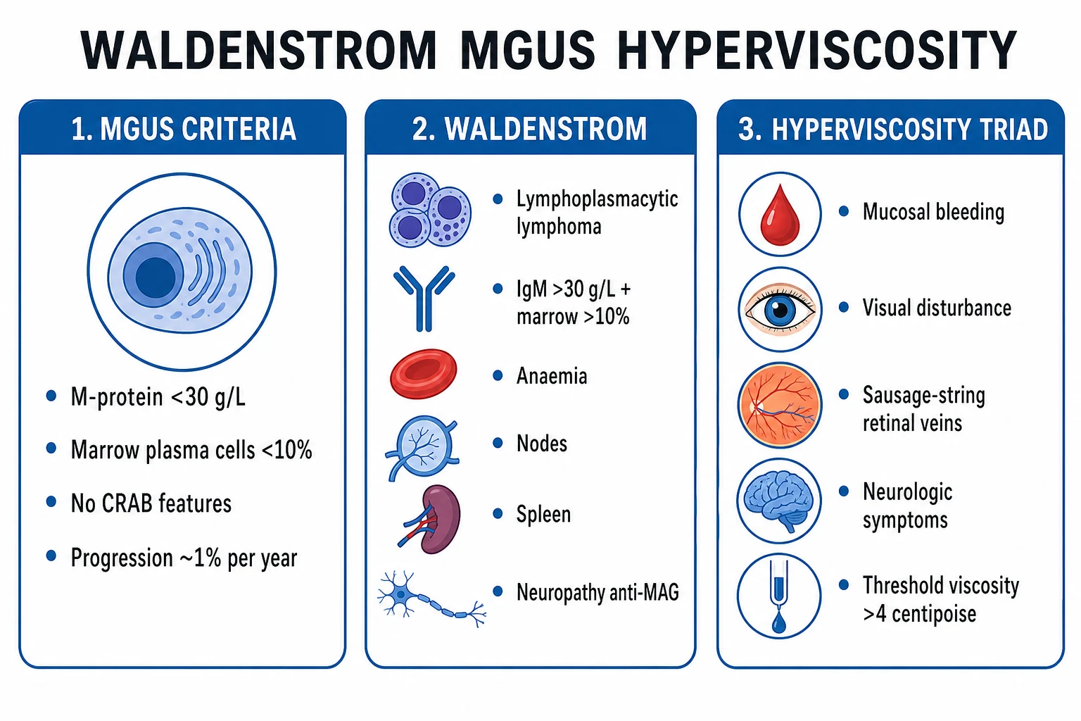
Monoclonal gammopathy of undetermined significance (MGUS) is an asymptomatic clonal plasma-cell or B-cell disorder defined by a serum monoclonal protein (M-protein) under 30 g/L, bone-marrow plasma cells under 10 percent, and absence of end-organ damage (CRAB); it is common in older adults (over 3 percent of those over 50) and carries a roughly 1 percent per year lifelong progression risk, so requires lifelong monitoring. Waldenstrom macroglobulinaemia is an indolent B-cell lymphoma (lymphoplasmacytic lymphoma) of post-germinal-centre B cells driven in over 90 percent of cases by the MYD88 L265P mutation, secreting a monoclonal IgM paraprotein. Its signature emergency is hyperviscosity syndrome — the triad of mucosal bleeding, visual change and neurological symptoms with sausage-string retinal veins on fundoscopy — because the 970-kDa pentameric IgM is largely confined to the intravascular space. Hyperviscosity is an emergency: confirm with serum viscosity and fundoscopy, treat with urgent plasmapheresis (which physically removes intravascular IgM and reverses symptoms within hours), then rituximab-based chemoimmunotherapy (BR, DRC) or a BTK inhibitor (zanubrutinib, ibrutinib) for definitive disease control.
On this page & tools
Your progress
Saved locally on this device.
Exam tags
Red flags
Overview & Definition
The IgM-secreting monoclonal gammopathies are the IgM counterpart of the IgG/IgA multiple-myeloma spectrum. They form a continuum from the benign precursor MGUS — an asymptomatic paraprotein needing only watchful waiting — through smouldering (asymptomatic) Waldenstrom to symptomatic Waldenstrom macroglobulinaemia, whose signature emergency is hyperviscosity syndrome. The pivotal clinical skill is recognising hyperviscosity — the triad of mucosal bleeding, visual change and neurological symptoms with sausage-string retinal veins on fundoscopy — and treating it with urgent plasmapheresis, which physically removes IgM and reverses symptoms within hours.[1][2][3]
Waldenstrom macroglobulinaemia (WM) is defined by the International Workshop for Waldenstrom Macroglobulinaemia (IWWM) and World Health Organization as a B-cell lymphoma of small lymphocytes, plasmacytoid lymphocytes and plasma cells (lymphoplasmacytic lymphoma, LPL) involving the bone marrow and producing a monoclonal IgM paraprotein of any concentration. When the same LPL morphology produces a non-IgM paraprotein (IgG, IgA, light chain) or no paraprotein at all, the disease is termed lymphoplasmacytic lymphoma rather than WM; conversely, an IgM paraprotein without LPL marrow infiltration is IgM MGUS (if asymptomatic) or smouldering WM (if the IgM is high or marrow infiltration is present but no end-organ damage exists).[3][7]
Waldenstrom and hyperviscosity — key numbers at a glance
Classification
Waldenstrom sits at the symptomatic end of an IgM-paraprotein spectrum distinguished by the paraprotein load, marrow infiltration, and the presence of end-organ damage.[1][3]
IgM MGUS
- Serum IgM monoclonal protein under 30 g/L
- Bone-marrow lymphoplasmacytic infiltration under 10 percent
- No end-organ damage (anaemia, hyperviscosity, lymphadenopathy, organomegaly, neuropathy, cryoglobulinaemia) all absent
- Normal free-light-chain ratio and no disease-related symptoms
- Lifelong monitoring; roughly 1.5 to 2 percent per year risk of progression (higher than non-IgM MGUS)
Smouldering (asymptomatic) WM
- IgM paraprotein over 30 g/L and/or marrow lymphoplasmacytic infiltration over 10 percent
- NO end-organ damage and no symptoms attributable to the disease
- Observe; treat only when symptomatic
- Higher risk of progression to symptomatic disease
Symptomatic WM
- IgM monoclonal protein plus lymphoplasmacytic marrow infiltration (over 10 percent)
- PLUS end-organ damage: anaemia, hyperviscosity, bulky lymphadenopathy or splenomegaly, neuropathy, cryoglobulinaemia, renal/amyloid, B-symptoms
- Requires systemic therapy
- Molecular hallmark MYD88 L265P mutation in over 90 percent
By paraprotein class causing hyperviscosity — not all hyperviscosity is Waldenstrom. The causes split into protein and cellular: protein causes are IgM (Waldenstrom — commonest cause; threshold typically over 30 to 40 g/L), IgG and IgA myeloma (only at very high levels, e.g. IgG over 60 to 70 g/L), and polyclonal hypergammaglobulinaemia (rare). Cellular causes are polycythaemia rubra vera (red-cell mass), leukaemias with hyperleukocytosis (white-cell mass — especially AML with WBC over 100 and CML in blast crisis), and sickle-cell disease (rigid sickled red cells). The IgM molecule is the only one that reliably causes hyperviscosity at modest concentrations, because of its size and near-complete intravascular confinement.[2]
Epidemiology & Risk Factors
MGUS is common. It is present in over 3 percent of people over 50 and over 5 percent of those over 70, with a slight male predominance and roughly 2 to 3 times the prevalence in Black/African populations compared with White populations. It carries an approximately 1 percent per year lifelong risk of progression to multiple myeloma, Waldenstrom macroglobulinaemia, or a related lymphoid malignancy. The risk is constant over time — a patient with stable MGUS for 30 years still has about a 1 percent risk in year 31 — which is why monitoring is lifelong.[1]
Waldenstrom macroglobulinaemia is rare — about 1,500 new cases per year in the United States, with an incidence of roughly 3 to 4 per million person-years worldwide (somewhat higher in White populations of European descent). The median age at diagnosis is about 70, with a male predominance of roughly 2:1. It accounts for 1 to 2 percent of haematological malignancies. The age-adjusted incidence has risen modestly over recent decades, partly due to improved ascertainment (wider SPEP testing) and an ageing population.[3]
Recognised risk factors: [1]
- Increasing age, male sex, and White/European ancestry are the demographic risk factors.
- Family history of a lymphoid or plasma-cell disorder confers roughly 2 to 3 times the relative risk in first-degree relatives; familial clusters account for 5 to 10 percent of cases.
- Pre-existing IgM MGUS is the precursor lesion for most WM; the risk of progression from IgM MGUS to WM is roughly 1.5 to 2 percent per year.
- Hepatitis C infection and certain autoimmune conditions (Sjogren, autoimmune cytopenias) are weakly associated with WM and other low-grade lymphomas.
- Occupational exposures (farming, wood and leather work, petroleum solvents) have been weakly linked, but no strong environmental cause is established. [1]
In India and other low- and middle-income countries, MGUS prevalence appears similar but WM is under-diagnosed because routine SPEP screening is less widespread and access to MYD88 testing, CT staging, and BTK inhibitors is limited by cost. Plasmapheresis for hyperviscosity is widely available in tertiary centres. Generic rituximab-based chemoimmunotherapy is the affordable backbone, with BTK inhibitors reserved for those who can self-pay or access patient-assistance programmes.[3]
Pathophysiology
The WM clone is a post-germinal-centre B-cell with plasmacytic differentiation — a lymphoplasmacytic cell that colonises the bone marrow (paratrabecular and nodular patterns), lymph nodes and spleen, and constitutively secretes monoclonal IgM. Its immunophenotype (surface IgM+, CD19+, CD20+, CD22+, CD25+, CD27+, FMC7+, CD5-, CD10-, CD23-, CD103-, CD138-) distinguishes it from CLL (CD5+, CD23+), mantle-cell lymphoma (CD5+, cyclin-D1/t(11;14)) and marginal-zone lymphoma. The marrow shows a mixed infiltrate of small B-lymphocytes, plasmacytoid lymphocytes and mature plasma cells, often with Dutcher bodies (intranuclear IgM inclusions) and increased mast cells.[3][7]
The molecular hallmark is the MYD88 L265P mutation, found in over 90 percent of WM cases — one of the highest frequencies of any recurrent point mutation in a B-cell lymphoma. MYD88 is an adaptor protein in the innate immune Toll/IL-1 receptor pathway; the L265P mutation (leucine to proline at position 265 in the Toll/IL-1 receptor domain) drives constitutive IRAK4–IRAK1–TRAF6 signalling, activating NF-kB transcription of survival genes (BCL2, BCL-XL) and interleukin-6 / interleukin-10 autocrine loops. The mutation is essentially absent in IgM MGUS, distinguishing precursor from established disease.[7]
The second most common mutation is CXCR4 (a WHIM-like C-terminal truncating mutation, mostly CXCR4 WHIM/RS33495 or nonsense mutations), present in roughly 30 to 40 percent of cases and often co-existing with MYD88 L265P. CXCR4 mutations drive chronic AKT/ERK signalling downstream of the CXCR4–CXCL12 axis and are associated with higher IgM levels, hyperviscosity, symptomatic disease, and a slower early response to BTK inhibitors (which nonetheless remain effective with time). The third recurrent mutation is TP53 (inactivation) in roughly 5 to 10 percent, associated with adverse outcome. MYD88 + CXCR4 mutation status is diagnostically useful (separating WM from IgM-MGUS and marginal-zone lymphoma) and therapeutically pivotal, because BTK inhibitors target the Bruton tyrosine kinase step of the B-cell-receptor signalling that the MYD88-driven clone depends on.[3][4]
Why IgM (and not IgG or IgA) causes hyperviscosity. IgM is a 970-kDa pentamer (an IgG monomer is 150 kDa), joined by a J-chain. It is mostly intravascular (about 80 percent versus about half of IgG), and its bulk raises serum viscosity non-linearly — the relationship is steep once the level exceeds roughly 30 to 40 g/L (or about 40 g/L for symptoms). The relationship between IgM and serum viscosity is roughly log-linear at high concentrations, so a doubling of IgM more than doubles viscosity. Because most of the molecule is in the circulation, plasmapheresis removes it efficiently — the basis of emergency therapy.[2]
IgM-mediated paraneoplastic phenomena (mechanisms the examiner rewards): [1]
- Type I cryoglobulinaemia — monoclonal IgM precipitates in the cold; on rewarming it re-dissolves. Deposits in skin capillaries cause purpura, acrocyanosis and livedo, arthralgia, weakness and, with renal involvement, an immune-complex membranoproliferative glomerulonephritis.
- Cold agglutinin disease (CAD) — monoclonal IgM with anti-I (or anti-i) specificity fixes complement (C4) at cool temperatures in the peripheral circulation; the membrane-attack complex lyses red cells, producing chronic haemolytic anaemia with acrocyanosis and a positive direct antiglobulin (Coombs) test for C3 (anti-C3 positive, anti-IgG negative).
- Peripheral neuropathy — IgM binds neural antigens. The commonest is myelin-associated glycoprotein (anti-MAG), producing a chronic distal demyelinating sensorimotor neuropathy with sensory ataxia and postural tremor. Less commonly the target is ganglioside (anti-GM1, anti-GD1b) or sulfatide.
- Amyloidosis (AL) — the IgM light chain (usually kappa) can deposit as amyloid, causing proteinuria, nephrotic syndrome, restrictive cardiomyopathy, hepatosplenomegaly and macroglossia, though less often than in IgG/IgA myeloma.
- Bing-Neel syndrome — direct CNS infiltration by lymphoplasmacytic cells of the brain parenchyma, meninges or cranial nerves (the blood-brain barrier normally excludes IgM, so this is a cellular, not paraprotein, phenomenon). [1]
Differential Diagnosis
A monoclonal IgM is not, by itself, Waldenstrom — the differential splits into "is this an IgM paraprotein disease?" and "is this hyperviscosity?"[1][3]
IgM MGUS
- IgM M-protein under 30 g/L
- Marrow infiltration under 10 percent
- No symptoms, no end-organ damage
- Manage by monitoring — never treat
Smouldering WM
- IgM over 30 g/L and/or marrow over 10 percent
- No end-organ damage
- Observe; treatment triggers same as symptomatic WM
Symptomatic WM
- LPL morphology + IgM paraprotein + end-organ damage
- MYD88 L265P in over 90 percent
- Treat with rituximab-based or BTK inhibitor
Other IgM-secreting B-cell lymphomas
- Marginal-zone lymphoma, CLL, mantle-cell — can all produce an IgM paraprotein
- Distinguished by immunophenotype, nodal histology and cytogenetics (t(11;14) for mantle cell)
- MYD88 L265P usually ABSENT in marginal-zone and CLL
IgG/IgA myeloma
- Plasma-cell (CD138+, CD56+) clone
- CRAB features — lytic bone lesions, cast nephropathy, hypercalcaemia
- Hyperviscosity only at very high IgG/IgA levels (over 60 to 70 g/L)
- MYD88 L265P negative
AL amyloidosis (IgM)
- Macroglossia, periorbital purpura, nephrotic-range proteinuria, restrictive cardiomyopathy
- Confirmed by Congo-red biopsy; often associated with low-grade LPL
- Treat the underlying clone
The differential of a high serum viscosity / hyperviscosity syndrome is broader than Waldenstrom and includes: [1]
- Polycythaemia rubra vera — red-cell mass; high haematocrit (over 0.55 in men, 0.50 in women); JAK2 V617F positive.
- Hyperleukocytosis in acute or chronic leukaemia — WBC over 100 in AML (with leukostasis: dyspnoea, confusion, pulmonary infiltrates, stroke) or blast crisis of CML/CLL.
- Sickle-cell disease — rigid sickled red cells; vaso-occlusive crisis.
- Very high IgG or IgA myeloma — uncommon; only at very high paraprotein loads. [1]
The cellular causes are distinguished on the blood count and film; the protein causes by electrophoresis and immunofixation. Always check both in a patient with suspected hyperviscosity.[2]
Anti-MAG neuropathy must be distinguished from chronic inflammatory demyelinating polyneuropathy (CIDP) (more rapid, predominantly motor, CSF pleocytosis, response to steroids and IVIG), diabetic distal symmetrical polyneuropathy (length-dependent, sensory, glycaemic history), and paraneoplastic neuropathies (anti-Hu in small-cell lung cancer). The clue to anti-MAG is a very slow (under 25 m/s) distal motor latency on nerve-conduction studies with high-titre anti-MAG antibody. [1]
Cryoglobulinaemic glomerulonephritis must be distinguished from other immune-complex glomerulonephritides (IgA nephropathy, post-streptococcal, lupus nephritis) by serum cryoglobulin, complement profile (low C4 with relatively normal C3 in cryoglobulinaemia), and SPEP/immunofixation showing the monoclonal IgM. [1]
Clinical & Bedside Assessment
The typical presentation of symptomatic WM is an older adult (median age 70) with insidious fatigue from anaemia, lymphadenopathy and splenomegaly, B-symptoms (fever, night sweats, weight loss), and a chronic distal sensorimotor neuropathy. Many are diagnosed incidentally on a raised ESR or a protein spike on routine bloods.[3]
The hyperviscosity triad — the emergency: [1]
HYPE — the hyperviscosity triad
HYPE
epistaxis, gum and GI bleeding from engorged friable capillaries — the commonest presenting feature
blurred vision, diplopia; fundi show sausage-string retinal veins, flame haemorrhages and papilloedema
headache, dizziness, vertigo, ataxia, somnolence, seizures (severe) — microvascular sludging in the brain
urgent plasma exchange removes intravascular IgM and reverses symptoms within hours
Mandatory bedside examination — fundoscopy. In hyperviscosity the fundus shows engorged, tortuous 'sausage-string' retinal veins (veins that alternate between dilated and constricted segments, resembling a string of sausages), retinal haemorrhages (flame and dot-blot), cotton-wool spots, and papilloedema. This finding is both supportive (confirms hyperviscosity) and a trigger for emergency plasmapheresis — examine the fundi of any patient with an IgM paraprotein and visual or neurological symptoms. The finding is sometimes called the fundus paraproteinaemicus.[2]
Cold-sensitive and paraneoplastic presentations: [1]
- Cryoglobulinaemia — cold-induced purpura (especially lower limbs, occasionally necrotic), acrocyanosis, arthralgia, weakness, and (type I with renal involvement) glomerulonephritis with haematuria and proteinuria.
- Cold agglutinin disease — chronic haemolytic anaemia with acrocyanosis of ears, nose and fingers on cold exposure, mild splenomegaly, and laboratory features of complement-mediated haemolysis (raised LDH, low haptoglobin, indirect bilirubin, reticulocytosis, positive anti-C3 direct antiglobulin test).
- Anti-MAG neuropathy — slowly progressive, distal, sensory-predominant, demyelinating neuropathy with sensory ataxia and postural tremor, often with intact motor strength initially.
- Bing-Neel syndrome — direct CNS infiltration presenting with headache, cranial-nerve palsies, seizures, or focal deficit; diagnosed on brain MRI and CSF cytology/flow cytometry showing lymphoplasmacytic cells.
- Amyloidosis (AL) — nephrotic-range proteinuria, restrictive cardiomyopathy (raised NT-proBNP, troponin), hepatomegaly, macroglossia, periorbital purpura. [1]
Atypical presentation in the elderly. Frailty, falls, delirium or isolated fatigue may dominate, with classic features subtle or absent. Maintain a low threshold to measure SPEP and IgM in any older adult with unexplained anaemia, neuropathy, raised ESR, nephrotic-range proteinuria, or chronic haemolysis. [1]
[1]Investigations
First-line bloods — full blood count and film (normocytic anaemia from marrow infiltration; rouleaux formation as the IgM stacks red cells; thrombocytopenia in advanced disease), ESR markedly elevated (often over 100 mm/h, because the paraprotein stacks red cells), CRP (variable), urea/electrolytes/creatinine (looking for amyloid or cryoglobulinaemic renal disease), calcium (typically normal — unlike myeloma, hypercalcaemia is uncommon in WM), albumin, LDH, beta-2 microglobulin (prognostic — see IPSS-WM), hepatic function, urinalysis (proteinuria suggesting amyloid or cryoglobulinaemia), and quantitative immunoglobulins (raised IgM, suppressed IgG and IgA — immune paresis).[1][3]
Paraprotein workup: [1]
- Serum protein electrophoresis (SPEP) + immunofixation — detects and types the IgM M-band (M-protein); serial measurement monitors disease burden and treatment response. Quantify IgM by nephelometry (more accurate than densitometry at high levels).
- Urine protein electrophoresis and immunofixation (Bence-Jones protein) — present in roughly 30 to 40 percent but rarely nephrotoxic in WM (unlike myeloma cast nephropathy).
- Serum free light chains (kappa/lambda ratio) — abnormal in roughly 30 to 40 percent of WM; part of MGUS risk-stratification. A kappa predominance is most common because WM kappa light chains predominate.
- Serum viscosity — measured when IgM is high or symptoms suggest hyperviscosity. Symptomatic hyperviscosity typically appears at a relative serum viscosity of about 4 (normal under 1.8; water = 1). The relationship to IgM is non-linear but, as a rule of thumb, symptoms are common when IgM is over 40 g/L and very likely when it is over 60 g/L. [1]
Bone-marrow aspirate and trephine biopsy — the diagnostic cornerstone. Lymphoplasmacytic infiltration of 10 percent or more (small B-lymphocytes, plasmacytoid lymphocytes and plasma cells, often with Dutcher bodies and increased mast cells). Immunophenotype by flow cytometry (surface IgM+, CD19+, CD20+, CD22+, CD25+, CD5-, CD10-, CD23-, CD138-). MYD88 L265P mutation testing (allele-specific PCR on marrow aspirate or peripheral blood) supports the diagnosis where morphology is ambiguous, with a sensitivity of over 95 percent. CXCR4 mutation testing is increasingly performed as it predicts early BTK-inhibitor response.[3][7]
Imaging and special tests: [1]
- Contrast-enhanced CT of neck, chest, abdomen and pelvis for lymphadenopathy and splenomegaly. A skeletal survey is NOT required (unlike myeloma — bone disease is not typical of WM; rarely, osteoederosclerosis raises POEMS in the differential).
- Cryoglobulins — sample must be drawn, transported and processed WARM (kept at 37 degrees C from the moment of draw) to avoid in-vitro precipitation; a positive test with monoclonal IgM and low serum C4 = type I cryoglobulinaemia. Many laboratories fail to do this — a falsely negative cryoglobulin is a common pitfall.
- Cold agglutinin titre and direct antiglobulin (Coombs) test — anti-C3 positive (anti-IgG negative) in CAD; cold agglutinin titre is typically over 1:64 at 4 degrees C.
- Anti-MAG antibodies and nerve-conduction studies for neuropathy — anti-MAG neuropathy shows markedly prolonged distal motor latency (often over 1.5 times the upper limit, with sensory action potentials reduced but motor conduction velocity only mildly slowed).
- Hepatitis B, C and HIV serology — before rituximab or chemo-immunotherapy (hepatitis B reactivation risk); HCV is weakly associated with WM and with mixed cryoglobulinaemia.
- Cardiac biomarkers (NT-proBNP, troponin) and echocardiogram — if amyloidosis is suspected (raised NT-proBNP/troponin with concentric LV thickening).
- Coagulation screen — IgM can interfere with fibrin polymerisation, factor VIII, von Willebrand factor and platelet function, giving a prolonged APTT or bleeding time without true factor deficiency; this does not require correction before procedures in most cases. [1]
MGUS risk-stratification — IMWG model (reproduced)
Three adverse factors — M-protein at least 15 g/L, non-IgG isotype (IgA or IgM), and an abnormal serum free-light-chain ratio (under 0.26 or over 1.65) — stratify MGUS progression risk. The model is the International Myeloma Working Group (IMWG) risk model for MGUS:[1]
- Low risk (0 factors) — about 5 percent absolute risk of progression at 20 years.
- Low-intermediate (1 factor) — about 20 percent at 20 years.
- High-intermediate (2 factors) — about 35 to 40 percent at 20 years.
- High risk (3 factors) — about 50 to 60 percent at 20 years. [1]
The same model applies to IgM MGUS (which progresses to WM, often, more often than non-IgM MGUS to myeloma) — non-IgG isotype is automatically present, so IgM MGUS sits at low-intermediate risk at minimum. [1]
IPSS-WM — International Prognostic Scoring System for Waldenstrom Macroglobulinaemia (reproduced)
The IPSS-WM (Morel et al., Blood 2009) stratifies survival in symptomatic, treated WM using five adverse factors:[5]
IPSS-WM risk groups and median survival
A revised IPSS-WM has been proposed incorporating LDH and genomic risk (TP53, MYD88/CXCR4 status), but the five-factor model remains the standard bedside stratification. [1]
Management — Resuscitation
Treat the presenting emergency first.[2]
Symptomatic hyperviscosity syndrome — URGENT PLASMAPHERESIS (therapeutic plasma exchange). Plasmapheresis physically removes the intravascular IgM pentamer; because about 80 percent of IgM is intravascular, one exchange of 1.0 to 1.5 plasma volumes typically lowers the IgM by 30 to 60 percent and reverses symptoms within hours to a day. Exchange is repeated daily or alternate-day until symptoms resolve and viscosity falls below about 4, while definitive therapy is started in parallel. The standard protocol: [1]
Therapeutic plasma exchange (plasmapheresis)
Dose
Exchange 1.0 to 1.5 plasma volumes per session, daily or alternate-day; replacement with 5 percent albumin (and FFP only if bleeding or before procedures)
Supportive cautions in hyperviscosity (these pitfalls kill patients): [1]
- Transfuse cautiously — red-cell transfusion worsens viscosity and can precipitate or aggravate hyperviscosity (and stroke or acute coronary events). Plasmapheresis first, then transfuse only as needed, slowly and after the viscosity has fallen.
- Avoid dehydration — maintains viscosity; keep the patient well hydrated with isotonic fluids (but avoid over-diuresis).
- Avoid over-diuresis and nephrotoxins (NSAIDs, contrast); manage bleeding with local measures (nasal packing, tranexamic acid mouthwash) and, if severe, after plasmapheresis.
- Avoid platelet transfusion unless profoundly thrombocytopenic — platelets add to sludging.
- Hold antiplatelet agents and anticoagulants while viscosity is high. [1]
Severe anaemia with hyperviscosity. Plasmapheresis first to reduce viscosity, then cautious transfusion of one unit at a time, slowly, with monitoring. The anaemia of WM reflects marrow infiltration and will improve with definitive therapy.[2]
Management — Definitive & Stepwise
WM is incurable but highly treatable, managed as a chronic disease. Therapy is indicated when the patient is symptomatic — never for the paraprotein level alone. The IWWM consensus indications for treatment are:[3][4]
- Hyperviscosity syndrome (the absolute indication — even after plasmapheresis).
- Symptomatic anaemia (Hb under 10 g/dL) or symptomatic cytopenia (platelets under 100 × 10⁹/L) from marrow infiltration.
- Bulky lymphadenopathy or splenomegaly (compressive symptoms or B-symptoms).
- Symptomatic neuropathy (especially anti-MAG), cryoglobulinaemia, cold agglutinin disease, amyloidosis or other paraprotein-mediated organ damage.
- B-symptoms (fever, night sweats, weight loss over 10 percent).
- Recurrent infection from immune paresis. [1]
Asymptomatic patients (smouldering WM or IgM MGUS) are NOT treated — they are observed, mirroring the watch-and-wait strategy of early CLL. [1]
First-line options — choose by age, fitness, cytopenias, IgM burden, MYD88/CXCR4 status, and need for rapid IgM reduction:[3][4][6]
Treatment algorithm for symptomatic Waldenstrom macroglobulinaemia
Symptomatic patient — confirm treatment indication (hyperviscosity, anaemia, bulky disease, neuropathy, B-symptoms, organ damage)
If hyperviscosity (IgM over 40 g/L with symptoms): URGENT plasmapheresis first; AVOID rituximab until IgM under 40 g/L (IgM flare risk)
Choose first-line regimen by fitness, age, and need for rapid control: fit patient — bendamustine-rituximab (BR); frail patient — DRC or single-agent BTK inhibitor; need for oral therapy — BTK inhibitor (zanubrutinib preferred)
Initiate therapy — monitor IgM, FBC, LFTs, infection; for BR plan 4 to 6 cycles then stop (fixed-duration); for BTK inhibitor continue until progression or intolerance
Assess response by serum IgM (IWWM-6 response criteria: major response is 50 percent or greater IgM reduction; very good partial response is 90 percent reduction)
Relapse — re-treat with non-cross-resistant regimen; consider autologous SCT in young, fit, chemosensitive relapsed disease; consider venetoclax, proteasome inhibitor, or clinical trial
Chemoimmunotherapy (fixed-duration): [1]
Bendamustine + rituximab (BR)
Dose
Bendamustine 90 mg/m² IV days 1 and 2; rituximab 375 mg/m² IV day 1; every 28 days for 4 to 6 cycles
Dexamethasone + rituximab + cyclophosphamide (DRC)
Dose
Dexamethasone 20 mg IV day 1; rituximab 375 mg/m² IV day 1; cyclophosphamide 100 mg/m² orally or IV days 1 to 5; every 21 days for 6 cycles
BTK inhibitors (continuous, oral, fixed-dose) — target the B-cell-receptor signalling the MYD88-driven clone depends on; active in both untreated and relapsed WM: [1]
Zanubrutinib (preferred BTK inhibitor)
Dose
160 mg orally twice daily (or 320 mg once daily), taken until progression or intolerance
Ibrutinib
Dose
420 mg orally once daily, until progression or intolerance
Bortezomib
Dose
1.3 to 1.6 mg/m² subcutaneously days 1, 4, 8, 11 of each 21-day cycle for 4 to 6 cycles
The IgM flare of rituximab — rituximab can trigger a transient IgM rise 4 to 8 weeks after the first dose, occasionally precipitating hyperviscosity. In a patient with a very high IgM (over 40 g/L), either delay rituximab (use bortezomib or plasmapheresis first) or use fractionated low-dose rituximab (e.g. one-quarter dose weekly) with close monitoring. The flare does not occur with bortezomib or BTK inhibitors, which is why these are preferred for rapid IgM control.[2]
Other agents and emerging therapies: [1]
- Venetoclax (BCL-2 inhibitor) — oral, active in WM (over 80 percent ORR), but caution with tumour-lysis syndrome and gradual ramp-up; useful in relapsed/refractory disease.
- Carfilzomib and daratumumab — second-line proteasome-inhibitor and anti-CD38 options; daratumumab interferes with blood-typing (CD38 on red cells — daratumumab-dithiothreitol treatment needed before typing).
- Chlorambucil — older oral alkylator; affordable in low-resource settings but less effective and leukaemogenic with prolonged use.
- Autologous stem-cell transplant — reserved for younger, fit patients with chemosensitive relapsed disease; not first-line. [1]
MGUS is NOT treated — arrange lifelong monitoring (serum electrophoresis and free-light chains, 6-monthly for the first year then annually if stable), risk-stratify by the IMWG model, and educate the patient about red flags (rising M-protein, new anaemia, bone pain, symptoms of hyperviscosity).[1]
Type I cryoglobulinaemia / cold agglutinin disease complicating WM are managed by treating the underlying disease (rituximab-based); supportive measures include cold avoidance (gloves, warm clothing), and for severe CAD, sutimlimab (anti-C1s monoclonal antibody) raises haemoglobin by blocking the classical complement pathway. [1]
Specific Subtypes & Scenarios
- Smouldering (asymptomatic) Waldenstrom — observe as for smouldering myeloma; treat only when symptomatic. Hyperviscosity, anaemia, neuropathy or bulky disease are the usual triggers. Patients with very high IgM (over 60 g/L) are at higher risk of imminent hyperviscosity and may warrant earlier therapy.
- Bing-Neel syndrome (CNS WM) — direct infiltration of brain parenchyma, meninges or CSF; presents with headache, cranial neuropathy, seizures or focal deficit. Manage with CNS-penetrant therapy: high-dose methotrexate or cytarabine, ibrutinib or zanubrutinib (which cross the blood-brain barrier and are active in Bing-Neel), and/or intrathecal therapy, plus systemic control. MRI brain and CSF flow cytometry are diagnostic.[3]
- WM-associated anti-MAG neuropathy — slowly progressive distal demyelinating neuropathy; responds variably to rituximab (often single-agent, sometimes with bendamustine). Improvement is delayed (6 to 12 months) and incomplete.
- Type I cryoglobulinaemia — cold-precipitating monoclonal IgM; treat the underlying WM. Severe skin ulceration or renal involvement warrants urgent plasmapheresis followed by rituximab-based therapy.
- Cold agglutinin disease (CAD) — IgM-mediated complement haemolytic anaemia; treat the underlying WM with rituximab-based therapy; sutimlimab (anti-C1s) is approved for refractory CAD and rapidly raises haemoglobin. Supportive: folic acid, transfusion via blood-warmer, cold avoidance, pneumatic cold suits.
- AL amyloidosis complicating WM — treat the underlying clone (often BR); cardiac and renal supportive care as for any AL amyloidosis.
Complications & Pitfalls
Disease complications: hyperviscosity syndrome (the signature emergency — stroke, retinal vein occlusion, MI, fatal haemorrhage if untreated), anaemia and cytopenias (marrow infiltration), recurrent infection (immune paresis, low IgG/IgA), cold agglutinin haemolysis and cryoglobulinaemia (renal, purpura, skin ulceration), anti-MAG neuropathy (sensory ataxia, falls), Bing-Neel CNS disease, AL amyloidosis (renal, cardiac, less common than in myeloma), and histological transformation to diffuse large B-cell lymphoma (DLBCL) (Richter-like transformation, ~5 percent of patients, poor prognosis with median survival under 2 years).[3]
Treatment complications: [1]
Clinical evidence
PMID 32731259
Key finding
Zanubrutinib non-inferior to ibrutinib for overall response in symptomatic WM, with significantly less atrial fibrillation (2 percent vs 15 percent), less hypertension, less diarrhoea and less bleeding — establishing zanubrutinib as the preferred BTK inhibitor
Practice change
Zanubrutinib is first-choice BTK inhibitor where available; ibrutinib remains an option where zanubrutinib is unaffordable
BTK-inhibitor toxicities — bleeding (platelet signalling inhibition — hold 3 to 7 days before surgery; avoid concurrent antiplatelet/anticoagulant), atrial fibrillation (more with ibrutinib than zanubrutinib), hypertension, cytopenias, infection (including fungal, encapsulated bacteria and hepatitis B reactivation — give antiviral prophylaxis), diarrhoea, musculoskeletal pain, and second primary skin cancers (sun protection, dermatology surveillance). Bendamustine — myelosuppression and infection, gonadal toxicity (avoid in those wishing to conceive), rare secondary MDS/AML. Rituximab — infusion reactions (premedicate with paracetamol, chlorphenamine, hydrocortisone), the IgM flare, hepatitis B reactivation (antiviral prophylaxis), progressive multifocal leukoencephalopathy (rare). Bortezomib — peripheral neuropathy (subcutaneous route and dose reduction mitigate), herpes zoster (aciclovir prophylaxis), thrombocytopenia.[4]
Classic diagnostic pitfalls: [1]
- Attributing anaemia, neuropathy or fatigue to "old age" and missing an IgM paraprotein — measure SPEP and IgM in any older adult with unexplained normocytic anaemia, raised ESR, chronic neuropathy, nephrotic-range proteinuria or chronic haemolysis.
- Transfusing before plasmapheresis in hyperviscosity — worsens viscosity and can precipitate stroke. Plasmapheresis first.
- Drawing the cryoglobulin sample cold — it precipitates in vitro; transport warm at 37 degrees C from draw to laboratory.
- Forgetting MYD88 L265P testing when marrow morphology is ambiguous (separates WM from marginal-zone lymphoma and IgM MGUS).
- Confusing MGUS with myeloma — MGUS has no CRAB, paraprotein under 30 g/L, and marrow under 10 percent; treat by monitoring, not chemotherapy.
- Using rituximab monotherapy in very high IgM — risks the IgM flare and hyperviscosity. Delay rituximab or plasmapheresise first.
- Misattributing a prolonged APTT to a true bleeding disorder when it is the IgM interfering with the assay in vitro. [1]
Prognosis & Disposition
WM remains incurable but treatable. Median overall survival is approximately 5 to 10 years, and is improving with BTK inhibitors and modern chemoimmunotherapy. The International Prognostic Scoring System for Waldenstrom (IPSS-WM) stratifies outcome by age, haemoglobin, platelet count, beta-2 microglobulin and monoclonal IgM (see Investigations). Adverse factors include older age (over 65), severe cytopenias (Hb at or below 11.5 g/dL, platelets at or below 100 × 10⁹/L), high beta-2 microglobulin (over 3 mg/L), high IgM (over 7 g/L), high LDH, refractory disease, 6q deletion, TP53 mutation, and transformation to DLBCL.[3][4][5]
IPSS-WM — median survival by risk group
MGUS carries a constant approximately 1 percent per year progression risk; the patient is never truly "discharged" — arrange lifelong monitoring of the M-protein and free-light chains. Histological transformation to DLBCL (a "Richter-like" event) heralds aggressive disease with a poor prognosis (median survival under 2 years) and requires aggressive combination chemotherapy (R-CHOP) ± autologous SCT in selected patients. [1]
Follow-up is by serial IgM M-protein monitoring (disease burden and response — IWWM-6 response criteria), surveillance for treatment toxicity (FBC, LFTs, immunoglobulins, infection prophylaxis), and assessment for progression or transformation (rising IgM, new B-symptoms, rising LDH, new tissue masses). [1]
Special Populations
- Elderly / frail — prefer lower-toxicity regimens (DRC, single-agent BTK inhibitor); a BTK inhibitor is oral and avoids chemotherapy myelosuppression, suiting frail patients who can swallow reliably and adhere to daily therapy. Select therapy by physiological, not chronological, age. For the very frail with low-burden disease, single-agent rituximab (with care for the IgM flare) or watchful waiting may be appropriate.
- Pregnancy — WM and symptomatic MGUS are rare in pregnancy; observe where possible. If hyperviscosity develops, plasmapheresis (safe in pregnancy) is the emergency measure. Avoid teratogenic chemotherapy (bendamustine, cyclophosphamide — both teratogenic, especially first trimester) and BTK inhibitors (animal teratogenicity). Rituximab is avoided near term (neonatal B-cell depletion for 6 months). If treatment is unavoidable, second- or third-trimester rituximab with neonatal monitoring is the safest option.
- Renal impairment / CKD — avoid nephrotoxic regimens; bendamustine and rituximab are usable with dose adjustment; manage cryoglobulinaemic glomerulonephritis by treating the underlying disease (often with plasmapheresis for severe cases).
- Hepatic impairment — bendamustine and bortezomib are hepatically metabolised; dose-adjust in severe impairment.
- Pre-existing peripheral neuropathy — avoid bortezomib (use carfilzomib or a BTK inhibitor); the underlying WM (anti-MAG) may be the cause of the neuropathy and improves with treatment.
- Prior hepatitis B — antiviral prophylaxis (entecavir or tenofovir) before rituximab or BTK inhibitor, regardless of viral load, to prevent reactivation.
- Cardiovascular disease — prefer zanubrutinib over ibrutinib if atrial fibrillation or hypertension is present (ASPEN trial); BTK inhibitors interact with CYP3A4 substrates (statins, anticoagulants).
- Community monitoring of IgM MGUS — 6-monthly SPEP and free-light chains for the first year, then annually if stable; educate on red flags (rising M-protein, new anaemia, bone pain, hyperviscosity symptoms). [1]
Evidence, Guidelines & Regional Differences
The International Workshop for Waldenstrom Macroglobulinaemia (IWWM) consensus recommendations (now in their 11th iteration) define diagnostic criteria and treatment indications: treat symptomatic disease; prefer rituximab-based chemoimmunotherapy or a BTK inhibitor for first-line. The NCCN guidelines (US) and ESMO Clinical Practice Guidelines (Europe) are largely concordant, with regional access differences driving practical variation.[3]
Landmark evidence: [1]
Clinical evidence
PMID 29856685
Key finding
Ibrutinib + rituximab significantly prolonged progression-free survival versus rituximab alone in both untreated and previously treated WM (30-month PFS 82 percent vs 28 percent), establishing ibrutinib-rituximab as first-line in many settings
Practice change
BTK inhibitor + rituximab is an evidence-based first-line option in untreated WM
Clinical evidence
PMID 22931316
Key finding
MYD88 L265P somatic mutation present in over 90 percent of WM cases and rare in other B-cell lymphomas — establishing MYD88 L265P as the molecular hallmark and diagnostic biomarker of WM
Practice change
MYD88 L265P testing is now standard in diagnostic workup; supports BTK-inhibitor dependence of the clone
- ASPEN trial — zanubrutinib vs ibrutinib in symptomatic WM (Tam et al., Blood 2020): comparable overall and major response rates, with significantly less atrial fibrillation (2 percent vs 15 percent), hypertension, diarrhoea and bleeding with zanubrutinib, supporting its use as the preferred BTK inhibitor.[4]
- INNOVATE — ibrutinib + rituximab vs rituximab alone (Dimopoulos et al., NEJM 2018): significantly improved progression-free survival in both untreated and previously treated WM.[6]
- MYD88 L265P discovery (Treon et al., NEJM 2012) — the molecular driver underpinning the BTK-inhibitor dependence of the clone (chronic active B-cell-receptor signalling through BTK).[7]
- Morel et al., Blood 2009 (IPSS-WM) — established the five-factor prognostic model that stratifies WM survival.[5]
In India and other low- and middle-income countries, rituximab-based chemoimmunotherapy (often generic BR or DRC) is the affordable backbone, with BTK inhibitors limited by cost (often out-of-pocket or via patient-assistance programmes). Plasmapheresis is widely available for hyperviscosity in tertiary centres. Access to MYD88 testing and CT staging varies; in many LMIC settings, the diagnosis is made on morphology + IgM paraprotein without molecular confirmation. Chlorambucil remains a low-cost oral option where newer agents are unaffordable.
Controversies and emerging questions: [1]
- When to treat asymptomatic disease — current consensus is watch-and-wait even with high IgM, but newer data suggest earlier BTK-inhibitor therapy may improve quality of life in some high-burden smouldering WM; trials are ongoing.
- Fixed-duration chemoimmunotherapy versus continuous BTK-inhibitor — both are first-line; choice depends on patient preference, fitness, need for treatment-free interval, and access. Fixed-duration BR offers a treatment-free interval but no maintenance; continuous BTK inhibitors offer disease control but require indefinite therapy with cumulative toxicity and cost.
- Role of venetoclax (BCL-2 inhibitor) — high response rates but concern for tumour-lysis and the need for ramp-up; useful in relapsed disease.
- MYD88/CXCR4 mutation-guided therapy — CXCR4-mutated disease responds more slowly to BTK inhibitors but does eventually respond; future trials may select therapy by genotype.
- MRD (minimal residual disease)-guided therapy — high-sensitivity MYD88 tracking may guide treatment duration in the future. [1]
Exam Pearls
[1]- MGUS criteria: under 30 g/L, under 10 percent, no CRAB. Non-IgM MGUS progresses at 1 percent per year; IgM MGUS at roughly 1.5 to 2 percent per year (often to Waldenstrom).
- MYD88 L265P in over 90 percent of WM — diagnostic and the basis of BTK-inhibitor dependence; CXCR4 mutation in 30 to 40 percent (slower early BTK response, higher IgM).
- Why IgM causes hyperviscosity but IgG rarely does: IgM is a 970-kDa pentamer and 80 percent intravascular; IgG is a 150-kDa monomer, about half extravascular. Plasmapheresis removes IgM efficiently.
- Symptomatic threshold: serum relative viscosity about 4 (water = 1; normal under 1.8); symptoms common when IgM is over 40 g/L.
- Rituximab IgM flare: in very high IgM (over 40 g/L) delay rituximab or plasmapheresise first; use bortezomib or a BTK inhibitor for rapid control.
- Hyperviscosity is broader than Waldenstrom — polycythaemia rubra vera, hyperleukocytosis (AML/CML), IgG/IgA myeloma (very high), sickle cell.
- BR dose: bendamustine 90 mg/m² days 1 and 2, rituximab 375 mg/m² day 1, every 28 days, 4 to 6 cycles.
- DRC dose: dexamethasone 20 mg, rituximab 375 mg/m², cyclophosphamide 100 mg/m² days 1 to 5, every 21 days, 6 cycles.
- BTK inhibitors: zanubrutinib 160 mg BD (preferred — ASPEN trial), ibrutinib 420 mg OD.
- Plasmapheresis: 1 to 1.5 plasma volumes daily, removes 30 to 60 percent IgM per session.
- IPSS-WM: age over 65, Hb at or below 11.5 g/dL, platelets at or below 100, beta-2 microglobulin over 3, IgM over 7 g/L — low / intermediate / high risk with median survival ~12 / ~8 / ~3.5 years.
- Bing-Neel syndrome = CNS infiltration by WM cells (not paraprotein, because IgM does not cross the BBB).
- Cold agglutinin in WM: anti-C3 positive direct antiglobulin test; sutimlimab (anti-C1s) for refractory CAD. [1]
Self-test: a 72-year-old man with blurred vision, epistaxis and ataxia — what is the next best step?
Check the fundus (sausage-string retinal veins), send SPEP/immunofixation and serum viscosity, and arrange urgent plasmapheresis — not a CT head. The triad in an older adult is hyperviscosity until proven otherwise. Do not transfuse first (worsens viscosity). After the emergency, treat the underlying WM with rituximab-based therapy (delaying rituximab if IgM over 40 g/L to avoid the IgM flare) or a BTK inhibitor.
Exam application bank (NEET-PG / INICET)
One-line answer
Monoclonal gammopathy of undetermined significance (MGUS) is an asymptomatic clonal plasma-cell or B-cell disorder defined by a serum monoclonal protein (M-protein) under 30 g/L, bone-marrow plasma cells under 10 percent, and absence of end-organ damage (CRAB); it is common in older adults (over 3 percent of those over 50) and carries a roughly 1 percent per year lifelong progression risk, so requires lifelong monitoring. Waldenstrom macroglobulinaemia is an indolent B-cell lymphoma (lymphoplasmacytic lymphoma) of post-germinal-centre B cells driven in over 90 percent of cases by the MYD88 L265P mutation, secreting a monoclonal IgM paraprotein. Its signature emergency is hyperviscosity syndrome — the triad of mucosal bleeding, visual change and neurological symptoms with sausage-string retinal veins on fundoscopy — because the 970-kDa pentameric IgM is largely confined to the intravascul [1]
Worked stems (answer without another resource)
Stem 1 — Classic presentation. Map symptoms to mechanism; name the first investigation and first treatment step with dose/route if drug therapy is standard. [1]
Stem 2 — Unstable / complicated. List red flags that force immediate resuscitation, theatre, ICU, antidote, or reperfusion — and what you do in the first 15 minutes. [1]
Stem 3 — Atypical group. Elderly, pregnancy, child, or immunocompromised: how presentation and thresholds change. [1]
Stem 4 — Differential trap. Name the three closest mimics and one discriminator for each. [1]
Stem 5 — Disposition. Who goes home with safety-netting, who is admitted, who needs HDU/ICU/theatre, and what follow-up is mandatory. [1]
Rapid viva checklist
- Definition + classification
- Pathophysiology chain
- Bedside signs / criteria
- Score with exact components (if any)
- Emergency bundle
- Definitive therapy with doses
- Complications of disease and of treatment
- Special populations
- Guideline/trial name if classic
- Three exam traps
Coverage self-check
If you cannot answer any stem above from this page alone, re-read the matching section — the page is intended to be self-sufficient for final-prof and NEET-PG/INICET questions on Waldenström Macroglobulinaemia, MGUS & Hyperviscosity Syndrome.
References
- [1]Gonsalves WI, Rajkumar SV. Monoclonal Gammopathy of Undetermined Significance Ann Intern Med, 2022.PMID 36508741
- [2]Gertz MA. Acute hyperviscosity: syndromes and management Blood, 2018.PMID 30104220
- [3]Ghafoor B, Masthan SS, Hameed M, et al. Waldenström macroglobulinemia: a review of pathogenesis, current treatment, and future prospects Ann Hematol, 2024.PMID 37414960
- [4]Tam CS, Opat S, D'Sa S, et al. A randomized phase 3 trial of zanubrutinib vs ibrutinib in symptomatic Waldenström macroglobulinemia: the ASPEN study Blood, 2020.PMID 32731259
- [5]Morel P, Duhamel A, Gobbi P, et al. International prognostic scoring system for Waldenstrom macroglobulinemia Blood, 2009.PMID 19196866
- [6]Dimopoulos MA, Tedeschi A, Trotman J, et al. Phase 3 Trial of Ibrutinib plus Rituximab in Waldenström's Macroglobulinemia N Engl J Med, 2018.PMID 29856685
- [7]Treon SP, Xu L, Yang G, et al. MYD88 L265P somatic mutation in Waldenström's macroglobulinemia N Engl J Med, 2012.PMID 22931316