Infectious Diseases · General Medicine
Candidiasis & Invasive Fungal Infection
Also known as Candidiasis · Candidaemia · Invasive candidiasis · Oral thrush · Oesophageal candidiasis · Vulvovaginal candidiasis · Candida endophthalmitis
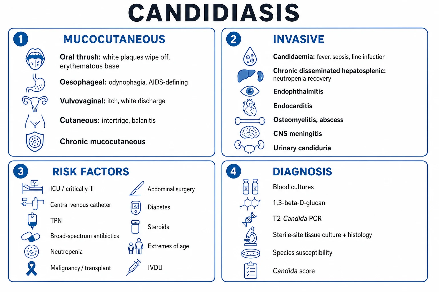
Candidiasis is infection by Candida yeasts (commonly C. albicans and increasingly non-albicans species), spanning mucocutaneous forms — oral thrush, vulvovaginal candidiasis, oesophageal (AIDS-defining), cutaneous (intertrigo, balanitis) — and invasive candidiasis (candidaemia and deep-seated infection) in the critically ill, immunocompromised, post-surgical, central-line, broad-spectrum-antibiotic, parenteral-nutrition patient. Risk factors: neutropenia, ICU/critically ill, central venous catheter, TPN, broad-spectrum antibiotics, diabetes, steroids, abdominal surgery, malignancy. Invasive candidiasis presents with fever unresponsive to antibiotics, sepsis, and may seed the eye (endophthalmitis), heart (endocarditis), liver/spleen, bone, CNS, skin. Diagnosis: blood cultures, beta-D-glucan, T2Candida/PCR, sterile-site culture/histology. Treat mucocutaneous with topical or oral azoles/nystatin; invasive with echinocandin first-line (caspofungin, micafungin, anidulafungin, rezafungin), step-down to fluconazole when stable and susceptible, remove central lines, ophthalmology exam for endophthalmitis, 2 weeks after first negative culture.
On this page & tools
Your progress
Saved locally on this device.
Exam tags
Red flags
Overview & Definition
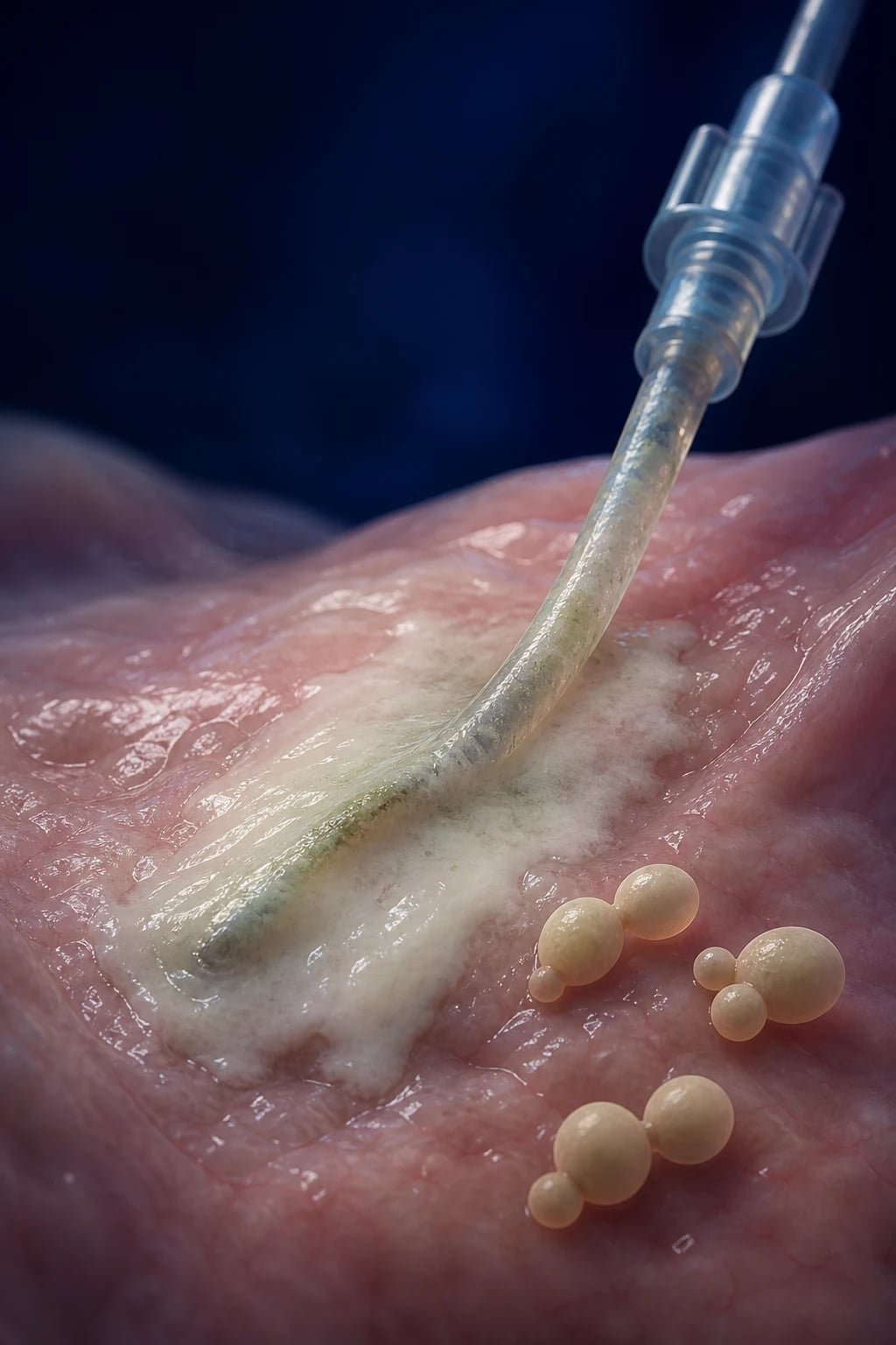
Candidiasis is infection caused by yeasts of the genus Candida — a dimorphic, asporogenous fungus that grows as budding yeast, pseudohyphae and true hyphae and is a normal commensal of the gastrointestinal tract, oropharynx, vagina and skin. The genus contains over 200 species; only a handful commonly cause human disease. The clinical spectrum is the widest of any single fungal genus, ranging from a trivial mucocutaneous complaint to a life-threatening bloodstream infection of the critically ill, and the central clinical skill is distinguishing the two realms.[1][4]
Mucocutaneous candidiasis — oral thrush, vulvovaginal candidiasis, oesophageal candidiasis, cutaneous intertrigo / balanitis / paronychia, and chronic mucocutaneous candidiasis — is common, usually benign, and often the first clue to an underlying defect in host defence (HIV, diabetes, corticosteroid/inhaled-steroid use, malignancy, broad-spectrum antibiotics).[3]
Invasive candidiasis comprises candidaemia (Candida in the bloodstream), acute disseminated candidiasis (with skin lesions, septic shock, endophthalmitis), chronic disseminated (hepatosplenic) candidiasis, and deep-seated infection of heart valves, bone, joints, meninges, peritoneum or urinary tract. It is overwhelmingly a healthcare-associated disease of the sick and immunocompromised — the ICU patient with a central line, the neutropenic haematology patient, the very-low-birth-weight neonate, the post-operative patient with GI perforation, the transplant recipient. Candida is now one of the commonest causes of nosocomial bloodstream infection worldwide and carries an attributable mortality of roughly 19 to 24%.[1][5]
The two most important recent shifts in the field drive modern management. First, the rise of non-albicans Candida — particularly C. glabrata and C. krusei (Pichia kudriavzevii), which are often or intrinsically resistant to fluconazole — has moved first-line empiric therapy for candidaemia from fluconazole to the echinocandins.[3] Second, the global emergence of Candida auris — a multidrug-resistant, environmentally persistent outbreak pathogen listed by the WHO as a critical-priority fungal threat — has made infection control, screening and susceptibility-guided therapy central to public health.[1][6]
The clinical decision is always the same: is this mucocutaneous or invasive? If invasive — who is the patient, what is the likely species, what is the source, and have I removed the line, sent the eye exam and started an echinocandin? [1]
Classification
Candidiasis is classified by clinical syndrome (the framework an examiner expects) and then by species (which determines drug choice).[3][4]
A. Mucocutaneous candidiasis [1]
- Oropharyngeal candidiasis (thrush) — white, curdy, loosely adherent plaques on buccal mucosa, tongue, palate; wipe off leaving an erythematous bleeding base; pseudomembranous (classic), atrophic (erythematous, angular cheilitis), hyperplastic (chronic, does not wipe off).
- Oesophageal candidiasis — odynophagia, dysphagia; usually coexists with oral thrush; AIDS-defining illness; also in malignancy, steroids, transplant.
- Vulvovaginal candidiasis — pruritus, dyspareunia, white curdy discharge; pH below 4.5; recurrent if 4 or more episodes per year.
- Cutaneous candidiasis — intertrigo (erythematous, macerated, satellite pustules in skin folds), diaper dermatitis, paronychia, balanitis.
- Chronic mucocutaneous candidiasis (CMC) — persistent/recurrent mucocutaneous disease from a defect in the Th17/IL-17 axis (STAT1 gain-of-function, AIRE/APECED autoantibodies against IL-17/IL-22); onset in childhood, disfiguring but rarely invasive.
- Onychomycosis / nail involvement — dystrophic, thickened nails in chronic paronychia or CMC. [1]
B. Invasive candidiasis [1]
- Candidaemia — Candida in blood cultures; may be catheter-related (line-associated) or secondary to a deep focus (abdomen, urinary tract).
- Acute disseminated candidiasis — candidaemia with sepsis and metastatic foci (skin, eye, joints, brain).
- Chronic disseminated (hepatosplenic) candidiasis — fever, RUQ pain, hepatosplenomegaly, bull's-eye lesions on imaging during recovery from prolonged neutropenia.
- Candida endophthalmitis — chorioretinal infiltrates ± vitritis; vision-threatening.
- Candida endocarditis — large friable vegetations, major embolic risk; needs surgery.
- CNS candidiasis / meningitis — usually post-neurosurgery or in neonates; shunt-associated.
- Candida osteomyelitis / septic arthritis — haematogenous or post-operative/contiguous.
- Candida peritonitis — spontaneous, post-GI-surgery leak, or continuous ambulatory peritoneal dialysis (CAPD)-associated.
- Candiduria / urinary tract candidiasis — ranges from bladder colonisation to pyelonephritis with fungal balls (bezoars) and renal candidiasis. [1]
C. By species — the layer that drives drug choice:[3][5]
C. albicans
Commonest overall (~50%)
- Forms germ tubes and chlamydospores
- Usually fluconazole-susceptible
- Causes mucocutaneous and invasive disease
- Step-down to fluconazole standard
C. glabrata
Rising; elderly, malignancy
- Often fluconazole-resistant (dose-dependent susceptible or resistant)
- Treat with echinocandin; step-down only if susceptible
- Reduced echinocandin susceptibility can emerge
- No hyphae — small budding yeast only
C. parapsilosis
Neonates, catheters, TPN
- Reduced echinocandin susceptibility — fluconazole preferred if stable
- Hand-carried by healthcare workers
- Common in neonatal ICU and TPN patients
- Forms biofilm on catheters
C. tropicalis
Neutropenia, malignancy
- Fluconazole-susceptible usually
- Tendency to tissue invasion in neutropenic patients
- Step-down to fluconazole standard
- Common in Asia
C. krusei (P. kudriavzevii)
Haematology; fluconazole exposure
- Intrinsically fluconazole-resistant
- Echinocandin then voriconazole step-down
- Mucosal and invasive disease
- Always resistance-check
C. auris
Outbreak pathogen
- Often multi-drug resistant (azole + amphotericin ± echinocandin)
- Persists on skin and environment
- Causes nosocomial outbreaks
- WHO critical-priority pathogen; screen and isolate
C. lusitaniae
Amphotericin-resistant
- The species that can be resistant to amphotericin B
- Check susceptibility before relying on amphotericin
- Rare cause of candidaemia
C. dubliniensis
HIV, oral cavity
- Closely related to C. albicans (germ tube positive)
- Recovered from oral cavity in HIV
- Can develop stable azole resistance
Epidemiology & Risk Factors
Candida is the commonest cause of invasive fungal infection in hospitalised patients and among the top three to four causes of nosocomial bloodstream infection in modern ICUs.[1][5] The incidence is dominated by C. albicans (roughly half of all isolates globally), but the proportion of non-albicans species has risen steadily — in many centres C. glabrata, C. parapsilosis and C. tropicalis together now exceed albicans. Candidaemia incidence is highest in ICU, neonatal, haematology and transplant units, and incidence rises with the intensity of medical care (lines, antibiotics, nutrition, immunosuppression).[4]
Invasive candidiasis — the numbers that examiners reward
Risk factors for invasive candidiasis (high-yield; the Candida score — see Investigations — formalises the ICU subset):[3][6]
- Central venous catheter (the single most important modifiable risk — biofilm seeds the bloodstream)
- Broad-spectrum antibiotics (suppress bacterial flora → Candida overgrowth)
- Total parenteral nutrition (TPN)
- Neutropenia (profound and prolonged; AML induction, HSCT)
- Abdominal surgery, especially GI perforation, anastomotic leak, necrotising pancreatitis (translocation)
- Prolonged ICU stay and mechanical ventilation
- Malignancy / chemotherapy / haematopoietic stem cell transplant
- Corticosteroids and other immunosuppression (calcineurin inhibitors, anti-TNF)
- Severe burns
- Haemodialysis and renal failure
- Diabetes mellitus (especially for mucocutaneous; hyperglycaemia impairs neutrophil function)
- Extreme prematurity / very-low-birth-weight neonates
- Injection drug use (rare; C. albicans endocarditis, fungal endophthalmitis) [1]
Risk factors for vulvovaginal candidiasis — antibiotics, pregnancy, uncontrolled diabetes, corticosteroids, HIV, OCP use (debated), and idiopathic recurrent disease in otherwise well women.[3]
Pathophysiology
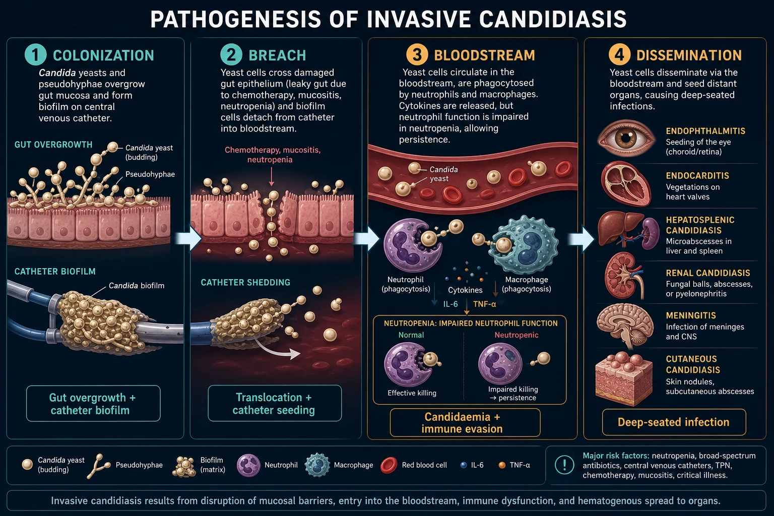
Candida is a commensal of the GI tract, oropharynx, vagina and skin in most healthy people. Disease is fundamentally opportunistic — a failure of host defences allows a commensal to become a pathogen. The two pivotal questions are how does Candida get into the bloodstream and why does it sometimes disseminate to deep organs.[1][4]
1. Two main portals into the bloodstream: [1]
- Translocation across damaged gut mucosa. Candida overgrows in the gut when broad-spectrum antibiotics suppress competing bacteria, or when chemotherapy/radiation causes mucositis and gut barrier breakdown. Yeast and pseudohyphae translocate through the damaged epithelium into the portal and then systemic circulation — the dominant mechanism in neutropenic haematology patients and after abdominal catastrophe (perforation, leak, necrotising pancreatitis).
- Biofilm on central venous catheters. Candida adheres to the polymer surface of CVCs, forms a thick extracellular biofilm with metabolically quiescent (persister) cells deep within it, and periodically seeds yeast into the bloodstream. Biofilm organisms are relatively resistant to antifungals (poor penetration, altered metabolism), which is why catheter removal is part of source control for catheter-related candidaemia. [1]
2. Virulence factors that shift Candida from commensal to invader:[5]
- Dimorphism — reversible yeast ↔ pseudohyphae ↔ hyphae transition; hyphal forms invade tissue.
- Adhesins (ALS family, Hwp1) — bind epithelium, endothelium and catheter polymers.
- Secreted aspartyl proteinases (SAP) and phospholipases — digest tissue, aid invasion.
- Phenotypic switching and biofilm formation.
- Candidalysin — a cytolytic peptide toxin (the first identified in a human fungal pathogen) that damages epithelial cells and triggers a danger-response. [1]
3. Host defence against Candida is principally neutrophil and macrophage mediated (innate immunity). Phagocytes recognise Candida through Dectin-1, DC-SIGN and Toll-like receptors (TLR2/TLR4), triggering phagocytosis and killing, and a Th17 (IL-17)-driven mucosal response that recruits neutrophils. This is why: [1]
- Neutropenia and neutrophil dysfunction (chemotherapy, leukaemia, post-transplant immunosuppression, chronic granulomatous disease) predispose to invasive, disseminated disease.
- Defects in the Th17/IL-17 axis (STAT1 gain-of-function, AIRE/APECED with autoantibodies to IL-17/IL-22, anti-IL-17 biologics) predispose to chronic mucocutaneous candidiasis rather than invasive disease. [1]
4. Dissemination to deep organs. From the bloodstream, Candida seeds any organ: the retina (endophthalmitis — white cotton-wool chorioretinal lesions, vitritis), heart valves (large friable vegetations → emboli), liver and spleen (chronic disseminated/hepatosplenic candidiasis — characteristic when neutrophils recover after prolonged neutropenia), kidneys (pyelonephritis, fungal balls), brain and meninges, bones and joints, and skin (pustular/nodular lesions). The absence of pus in the neutropenic host is itself a key exam point — impaired neutrophils mean no classic abscess, so clinical signs are blunted. [1]
5. Mechanism of chronic disseminated (hepatosplenic) candidiasis. During profound neutropenia, Candida seeds liver and spleen; lesions are subclinical because neutrophils cannot mount an inflammatory response. When the marrow recovers and neutrophils return, the immune system suddenly attacks the seeded yeast, producing fever, RUQ pain, raised alkaline phosphatase and characteristic bull's-eye lesions on imaging. This is why the diagnosis is made at the moment of marrow recovery, not during neutropenia — a classic exam pearl.[4]
6. Why antibiotics drive Candida. Broad-spectrum antibacterials eliminate the commensal bacteria that normally compete with Candida for mucosal niches and produce anti-Candida metabolites (e.g. short-chain fatty acids from anaerobes). Loss of this colonisation resistance lets Candida overgrow and translocate. [1]
Clinical Presentation
The presentation depends on whether the disease is mucocutaneous or invasive. The two are clinically distinct, although the same patient can have both (e.g. oesophageal candidiasis + systemic upset in AIDS).[3][4]
Mucocutaneous candidiasis
- Oral thrush (oropharyngeal candidiasis) — white, curdy, loosely adherent plaques on the buccal mucosa, tongue, palate, gingiva; scraped off, they leave an erythematous, bleeding base (this distinguishes thrush from leukoplakia and oral hairy leukoplakia, which do not wipe off). Variants: atrophic (erythematous, sore, under dentures), angular cheilitis (sore fissures at mouth corners), hyperplastic (chronic, firmly adherent, premalignant). Risk: infants, denture-wearers, inhaled-corticosteroid users, broad-spectrum antibiotics, diabetes, HIV, malignancy.
- Oesophageal candidiasis — odynophagia, dysphagia, retrosternal chest pain, substernal burning; frequently coexists with oral thrush but can occur without oral involvement. AIDS-defining illness (typically CD4 below 100 cells/microlitre); also in malignancy, transplant, steroids.
- Vulvovaginal candidiasis — intense pruritus, dyspareunia, dysuria, white curdy discharge; vulval erythema, oedema, excoriation, satellite lesions; vaginal pH below 4.5 (distinguishes from bacterial vaginosis and trichomoniasis). Recurrent vulvovaginal candidiasis = 4 or more episodes per year in an otherwise well woman.
- Cutaneous candidiasis (intertrigo) — erythematous, macerated, moist plaque in a skin fold (axillae, inframammary, groin, gluteal cleft, interdigital web spaces) with a scalloped advancing edge and characteristic satellite pustules. Variants: diaper dermatitis (in infants), balanitis (penis, often in uncircumcised diabetic men), paronychia (nail fold, in wet-work occupations).
- Chronic mucocutaneous candidiasis (CMC) — persistent, disfiguring, recurrent thrush, skin, nail and scalp disease from childhood; rarely invasive. Underlying Th17/IL-17 defects (STAT1, AIRE/APECED). [1]
Invasive candidiasis
- Candidaemia — persistent fever unresponsive to broad-spectrum antibiotics, chills, myalgia; clinically indistinguishable from bacterial bloodstream infection. Signs of sepsis or septic shock (tachycardia, hypotension, tachypnoea, altered mentation, oliguria, multi-organ failure). There may be no focal signs at all — a key pitfall.
- Cutaneous lesions of disseminated disease — painless or tender erythematous papules, nodules or pustules (ecthyma-gangrenosum-like) on the trunk and proximal limbs; biopsy shows yeast and pseudohyphae in dermis. A neutropenic patient with fever and new skin nodules → disseminated candidiasis (or fusariosis) until proven otherwise.
- Candida endophthalmitis — blurred vision, floaters, eye pain, photophobia, visual loss; white, cotton-wool chorioretinal lesions with overlying vitritis on fundoscopy. Found in up to a quarter of candidaemia patients on dilated fundoscopy — every candidaemia patient needs an ophthalmology exam.
- Chronic disseminated (hepatosplenic) candidiasis — persistent fever, right-upper-quadrant pain, hepatosplenomegaly, raised alkaline phosphatase in a patient recovering marrow after prolonged neutropenia; CT shows bull's-eye (target) lesions in liver and spleen.
- Candida endocarditis — fever, new murmur, embolic phenomena (stroke, mesenteric, splenic, limb); large, friable vegetations that embolise readily (often larger than bacterial vegetations).
- CNS candidiasis — meningitis/encephalitis: headache, altered mentation, seizures; usually post-neurosurgery or in neonates; may be associated with CSF shunt.
- Candida osteomyelitis / septic arthritis — back pain (vertebral), joint pain and swelling; subacute; haematogenous or post-operative.
- Candida peritonitis — fever, abdominal pain, cloudy peritoneal dialysate (in CAPD), or post-GI-surgery leak.
- Candiduria — usually asymptomatic; symptomatic disease gives dysuria, frequency, flank pain, fever; fungal balls (bezoars) in the upper tract can cause obstruction and renal candidiasis. [1]
Atypical presentations
- Neutropenic patient — no pus, no localising signs; fever alone may be the only clue (febrile neutropenia pathway). Skin lesions may be subtle.
- Neonate (VLBW) — apnoea, temperature instability, feeding intolerance, lethargy, respiratory distress; candiduria and CNS involvement common; high mortality.
- Elderly — confusion, falls, functional decline; C. glabrata over-represented; atypical fever pattern.
- Injection drug user — Candida endocarditis (classically of the tricuspid valve) and fungal endophthalmitis. [1]
Differential Diagnosis
Each candidal syndrome has its own differential; the exam favourite is mucocutaneous (what else causes a white patch, a vaginal discharge, an intertriginous rash) and the febrile ICU patient (what else causes persistent fever on antibiotics).[3][4]
White oral lesion — distinguish: [1]
- Oral hairy leukoplakia — lateral border of tongue, corrugated white, does NOT wipe off, EBV-driven, HIV marker, not painful.
- Leukoplakia — firmly adherent white plaque, does not wipe off, premalignant, biopsy mandatory.
- Lichen planus — reticulate white striae, bilateral, Wickham striae.
- Aphthous ulcers — painful round/oval ulcers.
- Oral squamous cell carcinoma — non-healing ulcer/indurated plaque, risk factors (tobacco, betel, alcohol).
- Necrotising ulcerative gingivitis (Vincent) — painful gingival ulceration with halitosis. [1]
Vaginal discharge — distinguish (pH is the discriminator): [1]
- Bacterial vaginosis — fishy, homogeneous milky discharge; pH above 4.5; clue cells on wet mount; positive whiff (KOH) test.
- Trichomoniasis — frothy yellow-green discharge; pH above 4.5; motile trichomonads on wet mount; strawberry cervix.
- Chlamydia / gonorrhoea — mucopurulent discharge, cervicitis.
- Atrophic vaginitis — thin, dry, pH above 4.5; postmenopausal. [1]
Intertriginous rash — distinguish: [1]
- Tinea (dermatophytosis) — annular, scaly, raised advancing edge, central clearing, no satellite pustules; KOH shows septate hyphae (not pseudohyphae/yeast).
- Inverse psoriasis — well-demarcated red plaques, no satellite pustules, elsewhere psoriasis.
- Erythrasma — coral-red fluorescence under Wood's lamp (Corynebacterium).
- Seborrhoeic dermatitis, contact/irritant dermatitis. [1]
Oesophageal candidiasis — distinguish: CMV oesophagitis (linear ulcers), HSV oesophagitis (vesicles/ulcers), pill oesophagitis (doxycycline, bisphosphonates — pinpoint ulcers at aortic arch), eosinophilic oesophagitis, GERD/stricture, oesophageal malignancy. [1]
Febrile ICU patient on broad-spectrum antibiotics — distinguish: [1]
- Bacterial sepsis (most common; persistent Gram-negative or resistant organism, e.g. MRSA, VRE, ESBL, carbapenemase).
- Other invasive fungal infections — invasive aspergillosis (sinus/lung, angioinvasion, halo/air-crescent sign), mucormycosis (Mucorales) (rhino-orbital-cerebral, black necrotic eschar, no septate hyphae), cryptococcosis (meningitis, pulmonary), endemic mycoses (histoplasmosis, blastomycosis, coccidioidomycosis) in endemic regions.
- Drug fever — temporal with a new drug, eosinophilia, rash.
- Underlying malignancy / tumour fever.
- Non-infective causes — PE, pancreatitis, adrenal insufficiency. [1]
Candida endophthalmitis — distinguish: bacterial endophthalmitis (post-injection/post-surgical/acute), CMV retinitis (AIDS, "pizza-pie" appearance, full-thickness retinal necrosis), Toxoplasma retinochoroiditis, Aspergillus endophthalmitis. [1]
Candiduria — distinguish: contamination, asymptomatic colonisation (most common; do not treat), catheter colonisation, true upper-UTI/pyelonephritis with fungal balls, systemic candidiasis with renal seeding (always send blood cultures if funguric). [1]
Clinical & Bedside Assessment
The bedside exam must answer three questions: is there sepsis, what is the source, and is there a metastatic focus.[3][6]
Vital signs and sepsis screen — temperature (persistent fever despite antibiotics is the cardinal sign), heart rate, blood pressure (septic shock), respiratory rate, SpO2, conscious level, urine output. Apply the qSOFA / Sepsis-6 framework: any new organ dysfunction in a febrile ICU patient is sepsis until proven otherwise. [1]
Lines and source control — daily line review (the single highest-yield bedside act): [1]
- Inspect the exit site of every central line, PICC, dialysis catheter and arterial line for erythema, tenderness, purulence, tunnel infection.
- Take peripheral blood cultures AND a culture drawn through each lumen of the central line (differential time-to-positivity more than 120 min supports catheter-related bloodstream infection).
- Plan to remove the central line in proven candidaemia as soon as the patient is stable enough — biofilm is the single biggest cause of persistent candidaemia. [1]
Look for metastatic foci: [1]
- Skin — search for papules, nodules, pustules (disseminated candidiasis); biopsy suspicious lesions.
- Eyes — dilated fundoscopy by ophthalmology for chorioretinal lesions and vitritis (endophthalmitis); this is mandatory within the first week of candidaemia and changes duration and route of therapy. If initially normal, repeat after one week.
- Heart — auscultate for a new murmur (endocarditis); echocardiography (transthoracic first; transoesophageal if endocarditis suspected or prosthetic valve).
- Abdomen — hepatosplenomegaly, RUQ tenderness (hepatosplenic), peritonitis (peritoneal source), abdominal surgical wound infection.
- Bones and joints — point tenderness, septic arthritis.
- Neurology — meningism, focal deficits, seizures (CNS disease). [1]
Mucocutaneous assessment — the scrape test: [1]
- Oral — scrape the plaque with a tongue depressor: thrush wipes off leaving an erythematous base; leukoplakia and oral hairy leukoplakia do not.
- Vagina — note discharge character and measure pH (below 4.5 = candidiasis; above 4.5 = BV/trichomonas).
- Skin — look for satellite pustules (candidal intertrigo). [1]
Bedside red flags — persistent fever despite broad-spectrum cover in a high-risk ICU patient; new skin nodules in a neutropenic patient; visual change in a patient with candidaemia; septic shock without a bacterial source; abdominal catastrophe with GI perforation; suspected C. auris in an outbreak setting. [1]
Risk-factor mnemonic for invasive candidiasis
CANDIDA
Central venous catheter — biofilm, the single biggest modifiable risk
Broad-spectrum antibiotics — suppress bacterial flora, allow overgrowth
Profound or prolonged neutropenia (AML, HSCT, chemotherapy)
Diabetes mellitus; steroids, immunosuppressants, calcineurin inhibitors
Transplant, malignancy, HIV/AIDS, anti-TNF, anti-IL-17
Abdominal surgery, GI perforation, anastomotic leak, necrotising pancreatitis (translocation), TPN
Very-low-birth-weight neonate; very elderly
Investigations
The investigation strategy depends on the suspected syndrome. Blood culture remains the gold standard for invasive candidiasis, but it is slow (typically 1 to 3 days) and only positive in roughly half of autopsy-proven disseminated cases — so non-culture diagnostics (beta-D-glucan, T2Candida, PCR) fill the gap in high-risk patients.[1][3]
Diagnosis of invasive candidiasis
- Blood cultures — gold standard; draw at least two peripheral sets plus a set through each lumen of the central line (before antifungals). Use BACTEC MycoF Lytic or lysis-centrifugation (Isolator) tubes for higher yield. Positive cultures give species ID (MALDI-TOF, chromogenic agar, germ-tube test for C. albicans) and susceptibility (CLSI/EUCAST broth microdilution; fluconazole, echinocandin, amphotericin breakpoints). Follow-up daily or alternate-day until negative.
- (1→3)-beta-D-glucan (BDG, Fungitell assay) — a pan-fungal cell-wall marker (positive in Candida, Aspergillus, Pneumocystis; NOT in Cryptococcus or Mucorales). Cut-off at least 80 pg/mL; high negative predictive value (useful rule-out). False positives: haemodialysis with cellulose membranes, IVIG/albumin/globulin, surgical gauze/sponges, severe bacteraemia, amoxicillin-clavulanate, IV amoxicillin-piperacillin. A single positive is not diagnostic — trend it and integrate clinically.
- T2Candida (T2 Magnetic Resonance) — detects C. albicans/tropicalis/parapsilosis and C. glabrata/krusei panels directly from whole blood in roughly 3 to 5 hours, no culture step required; high negative predictive value in suspected candidaemia; does not give susceptibility, so cultures must still be sent. Negative T2 + negative BDG in a moderate-risk patient argues against invasive candidiasis.
- PCR (Candida-specific or pan-fungal) — increasingly used; high NPV; combines well with BDG.
- MALDI-TOF MS — rapid species ID directly from positive blood-culture bottles within minutes (vs 24 to 48 h for traditional biochemistry).
- Antifungal susceptibility testing (AFST) — CLSI or EUCAST broth microdilution; critical for fluconazole (susceptible dose-dependent vs resistant C. glabrata) and echinocandin (emerging FKS1/FKS2 resistance). [1]
The Candida score (León / SCHOC) — ICU pre-emptive tool
Reproduced from the original León derivation — predicts the probability of invasive candidiasis in a non-neutropenic ICU patient with a new fever, to guide empirical vs pre-emptive antifungal therapy:[3][6]
| Variable | Points |
|---|---|
| Severe sepsis (per SCCM definition) | 2 |
| Colonisation at multiple sites (more than one of respiratory, urine, gastric, wound, drainage) | 1 |
| Hospitalisation prior to ICU admission | 1 |
| O — total parenteral nutrition (TPN) | 1 |
| Central venous catheter in situ | (note: original León score's components as above; some derivatives include catheter) |
A total score of 3 or more identifies patients at sufficiently high risk to justify empirical echinocandin while cultures and BDG are awaited; lower scores favour continued observation and pre-emptive (biomarker-driven) therapy. The score is not a substitute for clinical judgement and is not validated for neutropenic or transplant patients. [1]
Diagnosis of deep-seated / disseminated disease
- Ophthalmology dilated fundoscopy — within the first week of candidaemia; identifies endophthalmitis.
- Echocardiography — transthoracic (TTE); transoesophageal (TOE) if endocarditis suspected, prosthetic valve, or persistently positive cultures.
- CT abdomen (with contrast) — for hepatosplenic candidiasis: multiple hypodense lesions, classically bull's-eye (target) lesions; MRI is more sensitive and can be diagnostic.
- CT / MRI brain — for suspected CNS candidiasis; lumbar puncture with CSF culture and BDG if meningitic.
- Biopsy of skin, bone or organ lesions — histopathology with PAS and Gomori methenamine silver (GMS) stains shows budding yeast and pseudohyphae in tissue (distinguishes true invasion from colonisation); culture confirms species. [1]
Investigation of candiduria
- Urinalysis and urine culture with colony count; pyuria suggests true infection rather than colonisation.
- Rule out contamination by changing the catheter and re-culturing.
- Send blood cultures — a positive blood culture in the setting of candiduria suggests disseminated disease with renal seeding, not simple bladder colonisation.
- Imaging (renal US/CT) if upper-tract obstruction or fungal balls suspected. [1]
Mucocutaneous diagnosis
- Clinical in most cases.
- Wet mount with 10% KOH — pseudohyphae and budding yeast; rapid confirmation.
- Gram stain — Gram-positive budding yeast.
- Culture (Sabouraud dextrose, chromogenic CHROMagar Candida) — reserved for recurrent, recalcitrant or atypical disease, or to speciate.
- Vaginal pH below 4.5 (candidiasis) vs above 4.5 (BV, trichomoniasis).
- HIV test, blood glucose/HbA1c in any adult with unexplained thrush, oesophageal or recurrent mucocutaneous candidiasis. [1]
Management — Resuscitation
ABCDE first. The unstable patient with candidaemia is in septic shock and is managed with the Surviving Sepsis Campaign hour-1 bundle, adapted to the fungal cause.[3]
Airway / Breathing — oxygen to target SpO2 94–98% (or 88–92% in COPD/at risk of CO2 retention); mechanical ventilation if respiratory failure or ARDS. [1]
Circulation — two reliable IV lines (place a new peripheral set if removing the central line), balanced crystalloid 30 mL/kg for hypotension or lactate at least 2 mmol/L, noradrenaline as first-line vasopressor for fluid-refractory shock (add vasopressin if escalating; consider steroids per refractory-shock guidance). Reassess fluid responsiveness (passive leg raise, IVC, pulse-pressure variation) before further boluses. [1]
Surviving Sepsis hour-1 bundle applied to suspected fungal sepsis:[3]
- Two or more blood cultures drawn (peripheral + through each lumen of the line) before antifungal.
- Lactate.
- Serum (1→3)-beta-D-glucan and (where available) T2Candida.
- Empirical echinocandin within one hour of suspicion in the unstable or high-risk patient (do not wait for culture results).
- Urine output (catheterise), CRP, FBC, U&E, LFTs.
- Vasopressors for fluid-refractory shock. [1]
Source control (time-critical): remove or exchange the central venous catheter within 24 hours if feasible; send the tip for semi-quantitative culture (more than 15 CFU = significant catheter colonisation). Address any intra-abdominal source (drain collections, repair perforation) and infected hardware (pacemaker, prosthetic, shunt). [1]
Ophthalmology referral within the first week for dilated fundoscopy — endophthalmitis is sight-threatening and changes the duration and route of therapy. [1]
Management — Definitive & Stepwise
Drug choice is driven by syndrome, species, host and stability. The IDSA 2016 guideline underpins current practice.[3]
Candidaemia in the non-neutropenic adult
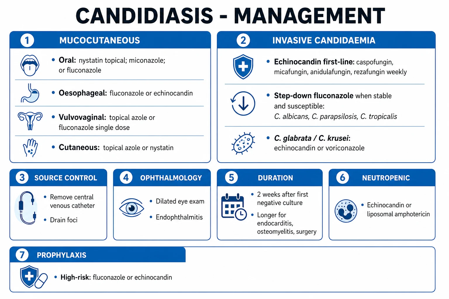
First-line: an echinocandin.[3]
- Caspofungin — 70 mg IV loading dose on day 1, then 50 mg IV once daily (increase to 70 mg/day if body weight more than 80 kg).
- Micafungin — 100 mg IV once daily (no loading dose needed).
- Anidulafungin — 200 mg IV loading dose on day 1, then 100 mg IV once daily.
- Rezafungin (long-acting, once-weekly) — 400 mg IV week 1, then 400 mg IV weekly; non-inferior to caspofungin in the ReSTORE trial; rationale is outpatient/once-weekly dosing.[2]
Step-down (oral transition) to fluconazole once all three of the following are met: clinically stable, repeat blood cultures negative, and isolate fluconazole-susceptible (typically C. albicans, C. parapsilosis, C. tropicalis):[3]
- Fluconazole 400–800 mg/day (12 mg/kg loading, then 6 mg/kg/day), oral when tolerating. [1]
Species-specific guidance:[3]
- C. glabrata — continue echinocandin; step-down only if susceptible (high-dose fluconazole 800 mg/day or voriconazole).
- C. krusei — intrinsically fluconazole-resistant; echinocandin for induction then voriconazole step-down.
- C. parapsilosis — reduced echinocandin susceptibility; if stable and isolate susceptible, fluconazole is preferred. [1]
Total duration: 2 weeks after the FIRST negative blood culture (not first day of therapy), provided the patient is clinically stable, source control achieved (line removed), and no metastatic focus (no endophthalmitis, endocarditis, etc.).[3]
Daily follow-up blood cultures (every 1–2 days) until negative to date the start of the 2-week clock. [1]
Dilated ophthalmology exam within the first week; repeat at one week if initially normal. [1]
Remove central venous catheter in catheter-associated candidaemia (within 24 hours if feasible). [1]
Neutropenic candidaemia / haematology
- An echinocandin OR liposomal amphotericin B 3–5 mg/kg/day IV as first-line.[3]
- Continue therapy through marrow recovery; switch to fluconazole, posaconazole or voriconazole step-down once neutropenia resolves and isolate is susceptible.
- Investigate for hepatosplenic candidiasis during recovery (imaging).
Candida endophthalmitis
- Systemic fluconazole (if isolate susceptible) or liposomal amphotericin B plus flucytosine.[3]
- Intravitreal amphotericin B or echinocandin for sight-threatening (vitritis/macular) lesions — ophthalmology-led.
- Treat 4–6 weeks and until ocular findings resolve; macular involvement and vitritis are sight-threatening.
Chronic disseminated (hepatosplenic) candidiasis
- Fluconazole if stable and isolate susceptible; otherwise echinocandin or liposomal amphotericin B for induction.[3]
- Step-down to fluconazole for months (often several); image to follow lesion regression.
- Consider adjunctive corticosteroids for the inflammatory syndrome (fever, pain that flares as neutrophils recover).
Candida endocarditis
- Liposomal amphotericin B plus flucytosine as initial therapy; surgical valve replacement is usually required (large friable vegetations, high embolic risk).[3]
- Chronic suppressive fluconazole post-surgery, often lifelong if prosthetic material retained.
CNS candidiasis / meningitis
- Liposomal amphotericin B plus flucytosine; remove infected CSF shunt or hardware. [1]
Candida peritonitis
- Intra-abdominal source control (drain, repair); fluconazole or echinocandin for 2 weeks after source control (longer if ongoing focus). [1]
Candiduria
- Remove or change the urinary catheter where possible; discontinue offending antibiotics.[3]
- Treat with fluconazole 200–400 mg/day for 7–14 days only if: symptomatic UTI, neutropenic, neonate/VLBW, before urologic procedure, renal transplant candidate, or evidence of upper-tract disease / fungaemia.
- Amphotericin B bladder irrigation is rarely used (toxicity, recurrence). Echinocandins do not reach therapeutic levels in urine — do not use for candiduria.
- Asymptomatic candiduria in a catheterised stable patient is colonisation — do not treat.
Candida auris
- Susceptibility-guided therapy (often resistant to fluconazole, frequently to amphotericin, sometimes to echinocandins); usually high-dose echinocandin first-line unless susceptibility dictates otherwise.
- Infection control is paramount: single-room isolation, contact precautions, dedicated equipment, terminal disinfection with a sporicidal agent (e.g. chlorine-based, hydrogen peroxide vapour), screening of roommates and high-risk contacts, and notification to public health. [1]
Mucocutaneous candidiasis
- Oral thrush — nystatin oral suspension 100 000 U/mL, 4–6 mL swish-and-swallow QID, OR topical clotrimazole troches/miconazole mucoadhesive buccal tablet 50 mg daily; oral fluconazole 100–200 mg/day for 7–14 days if moderate/severe, oesophageal involvement, or immunosuppression.
- Oesophageal candidiasis — fluconazole 100–200 mg/day for 14–21 days (IV if unable to swallow); echinocandin if fluconazole-resistant or refractory.
- Vulvovaginal candidiasis — topical azole (clotrimazole/miconazole) cream or pessary for 1–7 days, OR fluconazole 150 mg single oral dose. For recurrent disease: fluconazole 100–200 mg weekly for 6 months after induction; treat partner only if symptomatic balanitis.
- Cutaneous intertrigo — keep the fold dry, topical azole or nystatin cream for 1–2 weeks; address underlying diabetes/obesity.
- Chronic mucocutaneous candidiasis — long-term systemic azole (fluconazole/itraconazole); treat the underlying immune defect if possible. [1]
Antifungal prophylaxis (selected high-risk groups)
- Prolonged neutropenia (AML induction, MDS/AML) and haematopoietic stem cell transplant — posaconazole, fluconazole, or micafungin during the neutropenic window.[3]
- GVHD on intensive immunosuppression — posaconazole.
- High-risk VLBW neonates in high-incidence NICUs — fluconazole prophylaxis.
- Selected high-risk ICU cohorts — prophylaxis is not routine; restrict to those with very high Candida scores or BDG-driven pre-emptive strategies.
Specific Subtypes & Scenarios
- Candidaemia in the non-neutropenic ICU adult — echinocandin first-line; daily cultures until negative; line removal; eye exam; 2 weeks after first negative; consider pre-emptive therapy in high-Candida-score patients.[3]
- Neutropenic / haematology — echinocandin or liposomal amphotericin; treat through neutropenia; image for hepatosplenic disease at recovery.[3]
- Chronic disseminated (hepatosplenic) — bull's-eye lesions on CT/MRI at marrow recovery; months of azole; consider steroids for inflammatory flares.[4]
- Candida endophthalmitis — ophthalmology-led; systemic plus intravitreal therapy; 4–6 weeks.
- Candida endocarditis — amphotericin B + flucytosine + surgery; long-term suppressive fluconazole.
- Candida meningitis / CNS — post-neurosurgery, shunt or neonate; liposomal amphotericin B + flucytosine; remove shunt.
- Candida peritonitis — source control + fluconazole/echinocandin; PD-related peritonitis.
- Candiduria — distinguish colonisation (do not treat) from infection (treat defined indications); echinocandins do not reach urine.
- Neonatal invasive candidiasis — VLBW/extreme prematurity risk; amphotericin B deoxycholate (liposomal penetrates urine poorly); fluconazole in stable; remove central lines; prophylactic fluconazole in high-risk NICUs.
- Candida auris — multi-drug resistant outbreak; susceptibility-guided; strict infection control; environmental decontamination; screen contacts.
Complications & Pitfalls
Complications of invasive candidiasis: septic shock and multi-organ failure, ARDS, acute kidney injury, endophthalmitis with permanent vision loss, endocarditis (embolic stroke, mesenteric/splenic/limb infarction, heart failure), CNS involvement (meningitis, abscess, mycotic aneurysm), hepatosplenic abscesses, osteomyelitis / septic arthritis, candiduria with fungal-ball obstruction, prolonged ICU and hospital stay, and death.[1]
Persistent candidaemia despite therapy — escalate the search for:[3]
- A retained/infected central venous catheter or intravascular device (pacemaker, prosthetic, ICD).
- A sequestered focus — intra-abdominal abscess, infected thrombus (septic thrombophlebitis), endocarditis, endophthalmitis, osteomyelitis.
- A resistant organism (check susceptibility — FKS-mediated echinocandin resistance in C. glabrata; amphotericin resistance in C. lusitaniae).
- Inadequate dosing (caspofungin under-dosed in a heavy patient; fluconazole not increased for C. glabrata).
- Intravascular hardware (haemodialysis catheter, ECMOS cannula, ventriculoperitoneal shunt). [1]
- Treating mucocutaneous thrush in a healthy adult without testing for HIV/diabetes.
- Treating asymptomatic candiduria reflexively (colonisation, not infection).
- Missing endophthalmitis in a patient with candidaemia (no dilated eye exam).
- Failing to remove the central venous catheter in catheter-related candidaemia.
- Using empirical fluconazole where C. glabrata/krusei prevalent.
- Under-dosing echinocandin in a heavy patient (caspofungin should increase to 70 mg/day above 80 kg).
- Not screening for C. auris in an outbreak unit or after overseas healthcare.
- Over-treating a single positive BDG without correlation.
- Confusing oral hairy leukoplakia with thrush (it does not wipe off).
- Assuming a mucocutaneous rash is candidal when it is tinea (no satellite pustules, KOH shows septate hyphae). [1]
Prognosis & Disposition
Crude mortality of candidaemia is roughly 30 to 40%; attributable mortality 19 to 24%. Prognosis worsens with delayed appropriate antifungal therapy, septic shock, age, malignancy, breakthrough infection, C. auris (mortality up to 30–60% in outbreaks), and inadequate source control. Prognosis improves with early appropriate therapy, prompt line removal, and the absence of metastatic foci.[1][5]
Disposition: [1]
- ICU for septic shock, respiratory failure, or organ failure.
- Ward once haemodynamically stable and stepped down to oral.
- Home on oral fluconazole when afebrile, cultures negative, tolerating oral intake, source controlled, with a clear plan for follow-up cultures/imaging and an ID review. [1]
Follow-up: dilated eye exam at diagnosis and (if initially normal) repeated at one week; follow-up blood cultures until negative and to date the 2-week clock; repeat imaging for hepatosplenic or endocarditis involvement; screen for C. auris colonisation in outbreak settings. [1]
Special Populations
- Neonates (VLBW/extremely premature) — invasive candidiasis prophylaxis with fluconazole in high-incidence NICUs; amphotericin B deoxycholate preferred for treatment (liposomal formulations reach urine poorly, and renal/CNS involvement is common); remove central lines; candiduria and CNS disease are common; weight-based dosing throughout.[3]
- Pregnancy and vulvovaginal candidiasis — extremely common; treat with topical azoles for 7 days; avoid oral fluconazole (especially first trimester — associated with miscarriage and, in high doses, with birth defects).[3]
- Elderly — atypical presentation (confusion, functional decline); C. glabrata over-represented; polypharmacy and drug interactions (azoles inhibit CYP3A4 — warfarin, statins, calcineurin inhibitors); renal dose-adjust fluconazole.
- Neutropenic / haematology / transplant — empirical antifungal on persistent fever; prophylaxis (posaconazole in GVHD; fluconazole/micafungin in neutropenia); high rate of non-albicans and co-infection (Aspergillus); treat through neutropenia.
- HIV/AIDS — oesophageal candidiasis is AIDS-defining (CD4 typically below 100); fluconazole; immune restoration with ART; CMC predicts profound immunodeficiency.[3]
- Diabetes — predisposes to mucocutaneous disease (balanitis, vulvovaginitis, intertrigo); good glycaemic control; rare invasive disease without additional risk.
- Renal or hepatic impairment — dose-adjust fluconazole for CrCl; monitor voriconazole levels (hepatic metabolism, CYP2C19 polymorphism); amphotericin B nephrotoxicity (pre-hydrate, monitor K and Mg); echinocandins are largely hepatically metabolised (no renal dose adjustment, but monitor liver function).
- Injection drug use — Candida endocarditis (often tricuspid) and fungal endophthalmitis; prolonged therapy, surgery often required.
Evidence, Guidelines & Regional Differences
IDSA 2016 guideline (Pappas et al.) — the foundational framework:[3]
- Echinocandin first-line for candidaemia in non-neutropenic adults (and neutropenic patients).
- Step-down to fluconazole once stable, cultures negative, isolate susceptible.
- Remove central venous catheters early.
- 2 weeks duration after first negative culture (no metastatic focus).
- Ophthalmology exam within the first week. [1]
ESCMID / ECMM guidance (Europe) and the WHO Fungal Priority Pathogens List 2022 — the WHO list places Candida auris, C. albicans, C. tropicalis, C. parapsilosis, plus Cryptococcus neoformans, Aspergillus fumigatus and Histoplasma spp. in the critical-priority group, reflecting global mortality, rising resistance and diagnostic gaps.[1][6]
The ReSTORE trial (Thompson 2023, Lancet) — rezafungin (a long-acting, once-weekly echinocandin) 400 mg IV then 400 mg IV weekly was non-inferior to caspofungin for candidaemia and invasive candidiasis, supporting once-weekly dosing that could transform outpatient and step-down management.[2]
Pre-emptive strategies in the ICU (EMPIRICUS, León) — the Candida score (score at least 3) and beta-D-glucan-driven pre-emptive therapy aim to reduce over-treatment of low-risk ICU patients (cost, resistance, drug interactions) without missing the mortality cost of delayed therapy. The trade-off between empirical (treat all high-risk) and pre-emptive (treat on biomarker trigger) therapy remains contested in the non-neutropenic ICU.[6]
The emergence of Candida auris — first described in 2009, now a globally distributed multidrug-resistant outbreak pathogen; persisted in the environment, colonises skin, survives disinfectants; triggered WHO listing and national advisories (CDC, PHE, ICMR) on screening, contact precautions and terminal environmental disinfection.[1]
Antifungal stewardship — species identification, susceptibility testing, prompt de-escalation/step-down, source control, and defined durations reduce toxicity, cost and the emergence of resistance (especially echinocandin resistance in C. glabrata via FKS mutations). [1]
Exam Pearls
- Commonest Candida species: C. albicans; non-albicans rising (glabrata, parapsilosis, tropicalis, krusei, auris).
- Oesophageal candidiasis is AIDS-defining; oral thrush in an adult without obvious cause → test HIV.
- Endophthalmitis is found on dilated fundoscopy in up to a quarter of candidaemia patients — send every candidaemia patient to ophthalmology.
- 2 weeks of therapy after the FIRST negative blood culture (not first day of therapy).
- First-line empiric for candidaemia: echinocandin (caspofungin, micafungin, anidulafungin, rezafungin); fluconazole is the step-down, not first-line empiric.
- C. glabrata and C. krusei — fluconazole-resistant → echinocandin; C. krusei intrinsically fluconazole-resistant.
- C. parapsilosis — reduced echinocandin susceptibility → fluconazole preferred if stable; neonates, catheters, TPN, hand-carried.
- C. lusitaniae — amphotericin B-resistant.
- (1→3)-beta-D-glucan is positive in Candida, Aspergillus, Pneumocystis but NOT Cryptococcus or Mucorales.
- Remove the central line in catheter-associated candidaemia — biofilm causes persistence.
- Candida auris — multidrug-resistant outbreak pathogen; environmentally persistent; strict contact precautions, screen contacts, terminal disinfection.
- Chronic mucocutaneous candidiasis (CMC) — Th17/IL-17 pathway defect (STAT1, AIRE/APECED).
- Echinocandin mnemonic: the "-fungin" suffix — caspofungin, micafungin, anidulafungin, rezafungin; inhibit beta-1,3-glucan synthesis (cell wall).
- Candida risk-factor mnemonic: CANDIDA — Catheter, Antibiotics, Neutropenia, Diabetes/Drugs, Immunosuppression, Digestive breach (abdominal surgery/TPN), Age extremes.
- Hepatosplenic candidiasis presents at marrow RECOVERY, not during neutropenia — bull's-eye lesions on CT/MRI, raised ALP.
- Echinocandins do NOT reach the urine — wrong drug for candiduria.
- Asymptomatic candiduria = colonisation — do not treat (unless neutropenic, neonate, pre-urologic procedure, transplant candidate). [1]
Exam application bank (NEET-PG / INICET)
One-line answer
Candidiasis is infection by Candida yeasts (commonly C. albicans and increasingly non-albicans species), spanning mucocutaneous forms — oral thrush, vulvovaginal candidiasis, oesophageal (AIDS-defining), cutaneous (intertrigo, balanitis) — and invasive candidiasis (candidaemia and deep-seated infection) in the critically ill, immunocompromised, post-surgical, central-line, broad-spectrum-antibiotic, parenteral-nutrition patient. Risk factors: neutropenia, ICU/critically ill, central venous catheter, TPN, broad-spectrum antibiotics, diabetes, steroids, abdominal surgery, malignancy. Invasive candidiasis presents with fever unresponsive to antibiotics, sepsis, and may seed the eye (endophthalmitis), heart (endocarditis), liver/spleen, bone, CNS, skin. Diagnosis: blood cultures, beta-D-glucan, T2Candida/PCR, sterile-site culture/histology. Treat mucocutaneous with topical or oral azoles
Worked stems (answer without another resource)
Stem 1 — Classic presentation. Map symptoms to mechanism; name the first investigation and first treatment step with dose/route if drug therapy is standard. [1]
Stem 2 — Unstable / complicated. List red flags that force immediate resuscitation, theatre, ICU, antidote, or reperfusion — and what you do in the first 15 minutes. [1]
Stem 3 — Atypical group. Elderly, pregnancy, child, or immunocompromised: how presentation and thresholds change. [1]
Stem 4 — Differential trap. Name the three closest mimics and one discriminator for each. [1]
Stem 5 — Disposition. Who goes home with safety-netting, who is admitted, who needs HDU/ICU/theatre, and what follow-up is mandatory. [1]
Rapid viva checklist
- Definition + classification
- Pathophysiology chain
- Bedside signs / criteria
- Score with exact components (if any)
- Emergency bundle
- Definitive therapy with doses
- Complications of disease and of treatment
- Special populations
- Guideline/trial name if classic
- Three exam traps
Coverage self-check
If you cannot answer any stem above from this page alone, re-read the matching section — the page is intended to be self-sufficient for final-prof and NEET-PG/INICET questions on Candidiasis & Invasive Fungal Infection.
References
- [1]Lass-Flörl C, Kanj SS, Govender NP, et al. Invasive candidiasis Nat Rev Dis Primers, 2024.PMID 38514673
- [2]Thompson GR 3rd, Soriano A, Cornely OA, et al. Rezafungin versus caspofungin for treatment of candidaemia and invasive candidiasis (ReSTORE): a multicentre, double-blind, double-dummy, randomised phase 3 trial Lancet, 2023.PMID 36442484
- [3]Pappas PG, Kauffman CA, Andes DR, et al. Clinical Practice Guideline for the Management of Candidiasis: 2016 Update by the Infectious Diseases Society of America Clin Infect Dis, 2016.PMID 26679628
- [4]Kullberg BJ, Arendrup MC. Invasive Candidiasis N Engl J Med, 2015.PMID 26444731
- [5]Pappas PG, Lionakis MS, Arendrup MC, Ostrosky-Zeichner L, Kullberg BJ. Invasive candidiasis Nat Rev Dis Primers, 2018.PMID 29749387
- [6]Martin-Loeches I, Bassetti M, De Waele JJ, et al. Invasive candidiasis in intensive care medicine: shaping the future of diagnosis and therapy Intensive Care Med, 2025.PMID 41117944