Infectious Diseases · General Medicine
Cellulitis & Skin / Soft-Tissue Infection
Also known as Cellulitis · Erysipelas · Skin and soft-tissue infection · SSTI · Necrotising fasciitis · Cutaneous abscess
Cellulitis is an acute, spreading, non-suppurative infection of the dermis and subcutaneous tissue, usually caused by Streptococcus pyogenes or Staphylococcus aureus (including community-acquired MRSA), presenting with erythema, warmth, swelling and tenderness with poorly demarcated margins, usually on a leg and often through a portal of entry (tinea pedis, ulcer, trauma, IV site). Erysipelas is the superficial variant with raised, sharply demarcated margins. Necrotising fasciitis is the life-threatening deep-fascial variant — severe pain out of proportion, systemic toxicity, skin necrosis or bruising, bullae, crepitus, rapid spread — needing urgent surgical debridement. Management is driven by the purulent vs non-purulent and mild vs moderate vs severe axes: non-purulent cellulitis gets oral flucloxacillin (or cefalexin); purulent disease gets incision and drainage plus or minus antibiotics; MRSA cover (clindamycin, co-trimoxazole, doxycycline, vancomycin, linezolid) is added for purulent, severe, or at-risk patients. Mark the border, elevate the limb, and treat the portal of entry (tinea, lymphoedema, ulcer) to prevent recurrence.
On this page & tools
Your progress
Saved locally on this device.
Exam tags
Red flags
Overview & Definition
Skin and soft-tissue infections (SSTIs) are among the commonest reasons for antibiotic prescribing and for emergency medical admission. They span a spectrum from trivial (impetigo, a simple furuncle) to imminently life-threatening (necrotising fasciitis). The clinical skill is not usually making the diagnosis of cellulitis — that is straightforward — but rather distinguishing the common, straightforward cellulitis that can be sent home on oral antibiotics from the necrotising infection that demands immediate surgical debridement, and choosing empirical therapy that covers the right organisms (including MRSA where appropriate) without over-treating mimics.[1][2]
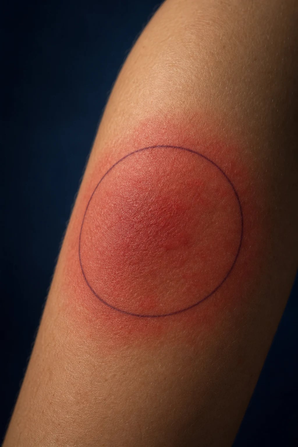
Cellulitis is an acute, spreading, non-suppurative (non-pus-forming) bacterial infection of the dermis and subcutaneous tissue, producing the classic tetrad of erythema, warmth, swelling and tenderness with poorly demarcated margins. It is almost always unilateral (a leg is the classic site) and there is usually an identifiable portal of entry through which skin flora or external bacteria enter. [1]
Erysipelas is a closely related but more superficial infection involving the upper dermis and superficial lymphatics; it produces a bright-red, shiny, indurated plaque with a sharply demarcated, raised advancing edge. Classically facial in adults and on the legs in children, it is overwhelmingly streptococcal.[1]
Nomenclature delta. UK practice (NICE CKS / NG190) treats cellulitis and erysipelas together and reserves the distinction largely for severity ("mild, moderate, severe") and for treatment route (oral vs IV). US (IDSA 2014) organises management around the purulent vs non-purulent axis and severity. Indian (ICMR/NCDC) guidance emphasises local antibiograms because of high community MRSA and Gram-negative prevalence.[1]
The two questions that decide every SSTI management decision: (1) Is it purulent? — purulence (a drainable collection) shifts the priority to incision and drainage and raises the likelihood of Staphylococcus aureus/MRSA; (2) Is it severe? — the presence of systemic signs (fever, tachycardia, hypotension, confusion, rapid spread, immunocompromise) drives the switch to IV therapy and admission. These two axes — purulent vs non-purulent, mild vs severe — are the backbone of the IDSA framework reproduced below.[1]
Classification
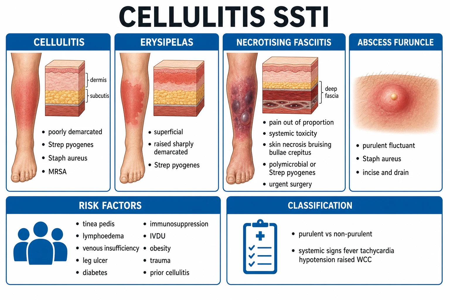
SSTIs are classified along three independent axes that each drive management. [1]
Cellulitis (non-purulent)
- Dermis + subcutis; poorly demarcated margins
- Typically Strep pyogenes and Staph aureus (incl MRSA)
- Management: anti-strep/anti-staph antibiotics; mark border; elevate
Erysipelas
- Superficial dermis + superficial lymphatics; raised, sharply demarcated border
- Overwhelmingly Strep pyogenes (GAS); bright red shiny plaque
- Often facial or lower leg; responds rapidly to penicillin
Cutaneous abscess / furuncle / carbuncle
- Purulent, fluctuant collection in dermis/subcutis
- Staph aureus (incl MRSA) predominant
- Management: incision and drainage is primary therapy; antibiotics adjunctive
Necrotising fasciitis
- Deep fascia and subcutaneous tissue necrosis
- Type I polymicrobial; Type II monomicrobial GAS
- Pain out of proportion + systemic toxicity + necrosis/bullae/crepitus; urgent surgery
Clostridial myonecrosis (gas gangrene)
- Necrosis of skeletal muscle; Clostridium perfringens alpha-toxin
- Post-trauma, post-surgery, dirty/contaminated wound
- Surgery + penicillin + clindamycin; hyperbaric O2 adjunctive
Impetigo
- Superficial epidermal infection; honey-coloured crust
- Staph aureus and Strep pyogenes
- Topical or oral anti-staph therapy
By the purulent vs non-purulent axis (the IDSA 2014 framework that drives empirical therapy): [1]
- Non-purulent (cellulitis, erysipelas without abscess) — empirical anti-streptococcal/anti-staphylococcal cover; cover MRSA only if severe or at-risk.
- Purulent (abscess, furuncle, carbuncle, purulent cellulitis) — incision and drainage first; add antibiotics if severe/systemic/immunocompromised/extensive; cover MRSA because S. aureus (often MRSA) predominates.[1]
By severity (IDSA classification): [1]
| Class | Definition | Site of care | Empirical approach |
|---|---|---|---|
| Mild | No systemic signs; no uncontrolled comorbidity | Outpatient | Oral anti-strep/staph |
| Moderate | Systemic signs (fever, tachycardia); or uncontrolled comorbidity; or failed oral therapy; or extensive | Admit (ward) | IV anti-strep/staph; add MRSA cover if indicated |
| Severe | Sepsis, shock, life-threatening infection; high-risk patient; necrotising features | Admit (often ICU) | IV broad spectrum with MRSA and toxin suppression |
Epidemiology & Risk Factors
Cellulitis is common, with an annual incidence estimated at around 200 cases per 100,000 person-years, rising steeply with age. It accounts for a substantial fraction of emergency admissions for skin infection. Most cases are managed in the community.[2]
Host risk factors — group them by mechanism: [1]
- Disrupted skin barrier (the portal of entry): tinea pedis (interdigital fissuring — the single commonest and most remediable), venous leg ulcers, diabetic foot ulcers, pressure ulcers, trauma/abrasions, burns, insect bites, IV cannulation sites, surgical wounds, body piercing, eczematous skin, injection drug use ("skin popping").
- Venous and lymphatic compromise (impaired drainage): lymphoedema (post-surgical, post-radiotherapy, filariasis, congenital), chronic venous insufficiency, post-thrombotic syndrome, saphenous vein harvest for CABG.
- Immunocompromise and metabolic disease: diabetes mellitus, chronic kidney disease, cirrhosis, HIV/neutropenia, immunosuppressive therapy, alcohol misuse, malnutrition, advancing age, obesity.
- Medications: systemic corticosteroids, biologics, chemotherapy.[1][2]
Risk factors and the organisms they favour (an examiner favourite): [1]
| Exposure / host | Organism to consider and the empirical implication |
|---|---|
| Aspirated abscess / purulent SSTI | Staphylococcus aureus (incl MRSA); I&D ± anti-MRSA |
| Dog/cat bite | Pasteurella multocida, anaerobes, Capnocytophaga; amoxicillin-clavulanate |
| Human bite | Eikenella corrodens, oral anaerobes, oral streptococci; amoxicillin-clavulanate |
| Saltwater / brackish water / raw oyster | Vibrio vulnificus (especially cirrhosis); doxycycline + third-generation cephalosporin |
| Fresh water / lake | Aeromonas hydrophila; fluoroquinolone |
| Fish tank / marine abrasion | Mycobacterium marinum (chronic); think TB-like therapy |
| Hot tub / spa folliculitis | Pseudomonas aeruginosa (usually self-limiting) |
| Diabetic foot | Polymicrobial (Gram-positive, Gram-negative, anaerobes); broad cover |
| IV drug use | S. aureus (incl MRSA), streptococci, oral anaerobes; screen for endocarditis |
| Immunocompromised / neutropenic | Add Gram-negatives, Pseudomonas, fungi; broad empiric cover |
| Animal contact / farm | Brucella, Bacillus anthracis (anthrax — painless black eschar) |
Community-acquired MRSA (CA-MRSA) is now widespread. Classic risk factors: recent antibiotic or healthcare exposure, IV drug use, contact sports, crowded living (military, prison), prior MRSA, chronic skin breaks, indwelling devices. In high-prevalence regions (much of India, parts of the US, UK pockets), empirical MRSA cover is warranted in severe or purulent SSTI.[1][1]
Recurrence is the rule, not the exception, if the predisposing factor persists: around 1 in 5 patients have a recurrence within a year, and roughly a quarter have multiple recurrences. Treating the portal of entry (tinea, lymphoedema, venous disease) and, where frequent, prophylactic penicillin, substantially reduce recurrence.[2][4]
Pathophysiology
The skin is a formidable barrier defended by the stratum corneum, the slightly acidic mantle, normal flora competing with pathogens, and intact innate and adaptive immunity. Cellulitis begins when this barrier is breached at a portal of entry, allowing bacteria to inoculate the dermis and subcutaneous tissue and overwhelm local defences.[2]
Mechanism cascade (the heart of the topic): [1]
- Portal of entry — a micro-fissure (interdigital tinea), an ulcer (venous, diabetic), trauma, an IV cannula, an insect bite, or a surgical wound. Occasionally there is no identifiable portal (idiopathic, "cryptogenic" cellulitis — classically in lymphoedema).
- Bacterial inoculation and multiplication — Streptococcus pyogenes (group A Strep, GAS) and Staphylococcus aureus (including MRSA) reach the dermis/subcutis and multiply. GAS deploys spreading enzymes — streptokinase (dissolves clots), hyaluronidase (digests the ground substance of connective tissue), DNase (degrades neutrophil extracellular traps) and streptolysins (lyse red and white cells) — that explain its rapid, extensive tissue spread. S. aureus produces coagulase (fibrin walling-off, abscess formation) and a range of cytotoxins (alpha-toxin, Panton-Valentine leukocidin in some CA-MRSA strains — linked to necrotising skin and lung infection).
- Innate immune response — tissue macrophages and keratinocytes release pro-inflammatory cytokines (IL-1 beta, TNF-alpha, IL-6) and chemokines (IL-8). These produce vasodilation (erythema, warmth), increased vascular permeability (oedema, swelling), and neutrophil and fluid recruitment into the tissue (the basis of the induration and tenderness).
- Lymphatic and systemic spread — organisms and cytokines reach lymphatics producing lymphangitis (the red linear streak) and regional lymphadenopathy; systemic cytokine spill produces fever, malaise and rigors. Bacteraemia occurs in a minority (more likely in severe disease) and seeds metastatic infection (endocarditis, osteomyelitis, septic arthritis).[2]
Why erysipelas has a sharp border while cellulitis does not: the depth of infection determines the clinical appearance. Erysipelas is confined to the upper dermis and the superficial lymphatic plexus, where the inflammatory response produces a well-circumscribed, raised, palpable edge. Cellulitis involves the deeper dermis and subcutaneous fat through which organisms spread along tissue planes without a discrete anatomic boundary, giving the poorly demarcated, advancing margin.[2]
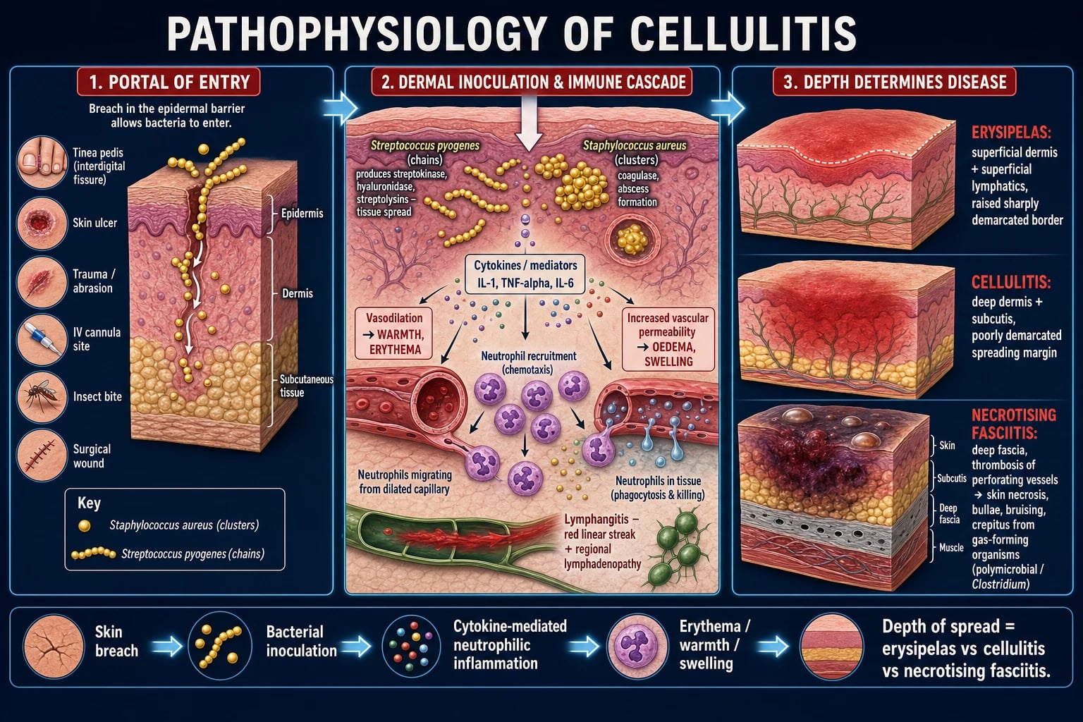
Necrotising fasciitis — the mechanism of the surgical emergency. The infection reaches the deep fascia and spreads rapidly along fascial planes. A characteristic finding is microvascular thrombosis of the perforating cutaneous vessels that traverse the fascia — infarction of these vessels produces cutaneous necrosis, haemorrhagic bullae, bruising and cutaneous anaesthesia (nerves are destroyed). The overlying skin may look deceptively normal early on, which is why pain is out of proportion to the visible skin findings. Gas in the tissues (palpable crepitus or visible on imaging) is produced by anaerobic metabolism of gas-forming organisms (Clostridium, anaerobic streptococci, some Gram-negatives).[1][3]
Toxin-mediated systemic toxicity. Staphylococcal and streptococcal exotoxins act as superantigens (TSST-1, staphylococcal enterotoxins, streptococcal pyrogenic exotoxins) that bypass normal antigen presentation and massively activate T cells, releasing cytokines (IL-1, TNF-alpha, IL-2, IFN-gamma) and producing the toxic-shock syndrome picture (fever, rash, hypotension, multi-organ failure). This is why clindamycin (a protein-synthesis inhibitor that suppresses toxin production) is added to the regimen in severe toxin-mediated disease — cell-wall antibiotics (beta-lactams, vancomycin) kill organisms but do not stop ongoing toxin release.[1]
Clinical Presentation
Classical cellulitis — a unilateral, warm, erythematous, swollen, tender area of skin with poorly demarcated margins, most often on a lower leg (the typical portal of entry is interdigital tinea pedis or a venous ulcer). There may be an orange-peel (peau d'orange) appearance, vesicles or bullae, and a spreading red lymphangitic streak tracking proximally, with tender regional lymphadenopathy (inguinal for leg lesions). Systemic upset (low-grade fever, malaise) is variable and may be absent even in significant infection.[1][2]
Erysipelas — a bright red, shiny, indurated, tender plaque with a sharply demarcated, raised advancing edge. The margin is palpable and the lesion is well-defined — the discriminator from cellulitis. Common on the face (often with a nasal vestibule portal of entry) and the lower leg. Systemic upset (fever, rigors) is often prominent and out of proportion to the visible lesion.[2]
Cutaneous abscess / furuncle / carbuncle — a tender, erythematous, fluctuant nodule with a central pustule or pointing head, often with surrounding cellulitis. A furuncle (boil) arises in a hair follicle (usually S. aureus); a carbuncle is a coalescence of furuncles draining through multiple openings, classically on the nape of the neck or back of older men. Fluctuance indicates a drainable collection. [1]
Red flags for necrotising fasciitis (the surgical emergency) — these mandate immediate surgical assessment: [1]
- Severe pain out of proportion to the visible skin findings (the cardinal early clue).
- Systemic toxicity — high fever, tachycardia, hypotension, confusion, septic shock.
- Cutaneous changes of necrosis: dusky/purple discoloration, bruising (ecchymosis), haemorrhagic bullae, skin necrosis or sloughing.
- Crepitus (gas in tissues) — palpable or audible.
- Cutaneous anaesthesia (destroyed nerves).
- Rapid spread despite antibiotics.
- Dishwater-grey or foul-smelling fluid from an involved area.[1][3]
Periorbital (preseptal) vs orbital cellulitis — a high-stakes distinction. Periorbital cellulitis is infection anterior to the orbital septum: eyelid swelling and erythema without proptosis, without painful or restricted eye movement, and with normal vision. Orbital cellulitis (post-septal) is an emergency: in addition to lid signs there is proptosis, painful or limited eye movement (ophthalmoplegia), reduced or double vision, and systemic toxicity, usually from adjacent ethmoid sinusitis. Orbital cellulitis risks cavernous sinus thrombosis, optic nerve compression and intracranial spread and needs urgent CT, IV antibiotics, and ENT/ophthalmology.[2]
Diabetic foot infection — presents over a chronic wound or ulcer; look for surrounding erythema, warmth, swelling, purulent discharge, deep tracking, exposed tendon or bone, and a foul odour. Always probe to bone at the ulcer base (a palpable gritty bone suggests osteomyelitis). Assess the vascular supply (pulses, Doppler, ABI) and sensation (neuropathy). [1]
Atypical presentations — be alert: [1]
- Elderly — blunted fever, confusion or functional decline may be the only sign; a lower threshold to investigate and admit.
- Diabetic / immunocompromised / neutropenic — may be afebrile; broader organisms; high risk of deep infection, bacteraemia and rapid progression.
- IV drug user — atypical sites (groin, forearm); MRSA; consider deep abscess, septic arthritis/osteomyelitis, and endocarditis.
- Lymphoedema — recurrent ipsilateral episodes; the limb is chronically swollen between attacks.
- Pregnancy — antibiotics must be safe in pregnancy (penicillins, cephalosporins; avoid tetracyclines).[2]
Differential Diagnosis
A red, swollen, tender leg is not always cellulitis. Bilateral red legs are particularly likely to be something else. Consider: [1]
Acute contact dermatitis
- Itch (not pain) predominates; vesicles; linear/geometric pattern
- History of allergen/irritant exposure (plant, chemical, cosmetic)
- Often bilateral; pruritic; responds to steroid withdrawal of allergen
Stasis dermatitis / venous eczema
- Bilateral; chronic venous insufficiency; medial lower leg
- Often bilateral and symmetric; haemosiderin staining (brown)
- No fever, no systemic upset; treat with compression and emollients
Lipodermatosclerosis
- Chronic, hard, bound-down subcutaneous tissue; "inverted champagne bottle" leg
- A complication of chronic venous disease; not acute infection
- Pain is chronic; no response to antibiotics
Deep vein thrombosis (DVT)
- Unilateral calf swelling/tenderness; not warm erythema typically
- Risk factors (immobility, malignancy, post-op); Wells score
- D-dimer and compression ultrasound settle it; DO NOT assume cellulitis
Acute gout
- Joint (first MTP, knee); exquisite tenderness; podagra
- History of gout; hyperuricaemia; monosodium urate crystals
- Joint aspiration — needle-shaped negatively birefringent crystals
Septic arthritis
- Joint, not skin; pain on passive movement; hot joint
- Single joint; hold the joint; aspirate before antibiotics
- Synovial fluid WBC over 50,000; urgent washout
Erythema migrans (Lyme)
- Expanding annular lesion with central clearing; tick exposure
- Endemic area; tick bite history; flu-like illness
- Serology; doxycycline
Erythema nodosum
- Tender subcutaneous nodules on shins; bilateral
- Panniculitis (streptococcal, sarcoid, drugs, IBD, pregnancy)
- No surface erythema that spreads; biopsy if needed
Insect sting / hypersensitivity
- Sudden after a sting; itchy, urticarial; bilateral if multiple
- History of sting; resolves quickly
- Antihistamines, not antibiotics
Compartment syndrome
- Pain out of proportion; tense compartment; pain on passive stretch
- After trauma/fracture; pallor, paraesthesia, pulseless late
- Surgical emergency — fasciotomy
Calciphylaxis (renal)
- Painful livedoid/necrotic skin lesions in ESRD
- High calcium-phosphate product; high mortality
- Not infection; biopsy; treat calcium-phosphate
Pseudocellulitis (any mimic)
- A common cause of unnecessary admission and antibiotic use
- Diagnose by exclusion; bilateral, chronic, or non-tender cases
- Stasis dermatitis and contact dermatitis account for most mis-labels
The "two-dermatome rule": bilateral red, swollen legs are almost never cellulitis (cellulitis is unilateral in over 90 percent of cases). Bilateral red legs point toward stasis dermatitis, contact dermatitis, lipodermatosclerosis, or a systemic cause (heart failure, nephrosis).[2]
Distinguishing cellulitis from DVT is the single most common and consequential error. The clues: cellulitis is warm with surface erythema and a sharp demarcation of the red border; DVT is calf tenderness without the bright surface erythema. When uncertain, D-dimer and compression ultrasound settle it — treating DVT as cellulitis (or vice versa) is dangerous.[2]
Distinguishing necrotising fasciitis from severe cellulitis — any of the red-flag features above (pain out of proportion, necrosis/bullae/bruising, crepitus, cutaneous anaesthesia, systemic shock) in a patient who looks unwell should trigger an immediate surgical consult. Do not be reassured by a "low" LRINEC score or "early" findings; necrotising fasciitis can present with deceptively normal-looking skin.[3]
Clinical & Bedside Assessment
Mark the leading edge. Draw a line around the perimeter of the erythema with a skin marker, dated and timed. This is the single most useful bedside manoeuvre — it allows objective tracking of spread (or response) over hours and days, and reassures both clinician and patient. Re-mark at each reassessment.[1][2]
Examine systematically: [1]
- Inspection — distribution (unilateral?), margins (demarcated vs poorly defined), colour (bright red erysipelas vs dusky/purple in necrosis), surface (vesicles, bullae, haemorrhage, necrosis, slough), portal of entry (interdigital tinea, ulcer, wound, IV site, bite), lymphangitis (red streak), lymphadenopathy.
- Palpation — warmth, induration (measure the area or circumference of the limb for monitoring), fluctuance (an abscess), crepitus (subcutaneous gas — ominous).
- Distal neurovascular — pulses, capillary refill, sensation; impaired sensation or pulse loss raises concern for deep/necrotising infection, compartment syndrome, or ischaemia.
- Lymph nodes draining the region.
- Systemic vital signs — temperature, heart rate, blood pressure, respiratory rate, oxygen saturation, conscious level. These define systemic involvement (the "severe" threshold) and trigger the sepsis pathway (qSOFA, MEWS/NEWS). [1]
The probe-to-bone test (diabetic foot): using a sterile blunt probe through the base of a clean, debrided ulcer, palpation of hard gritty bone suggests osteomyelitis (high specificity when positive; a negative test does not exclude it).[2]
The "finger test" for necrotising fasciitis (at surgical exploration): a 2 cm incision down to deep fascia; the tissue planes dissect easily with a finger, there is grey "dishwater" fluid, the fascia is friable and grey/green, and there is no bleeding — diagnostic of necrotising fasciitis. This is an operative finding, not a bedside test in unstable patients.[1]
Document the affected area, a photograph where possible, the marked border with date/time, vital signs, and a sepsis screen. A normal blood pressure does not exclude early sepsis — compensation maintains pressure until late; assess perfusion (capillary refill, lactate, urine output).[2]
Investigations
Mild, uncomplicated cellulitis usually requires no investigations — the diagnosis is clinical and empirical therapy is started immediately. Over-investigation of mild disease is a common and costly error.[1]
Indicated when severe, atypical, recurrent, immunocompromised, or failing first-line therapy: [1]
- Full blood count, CRP, U&E, LFTs, glucose — baseline; the LRINEC variables are pulled from these.
- Blood cultures — yield is low (around 5 percent) in uncomplicated cellulitis, but higher in severe disease; obtain before antibiotics when possible, never delaying therapy.
- Swab/pus culture — only useful when there is a drainable purulent collection, an open ulcer, or a weeping lesion; a routine surface swab of intact cellulitis is unhelpful (it grows contaminant skin flora). Send pus from an abscess for culture and sensitivity, especially in recurrent or MRSA-suspected disease.
- Needle aspiration/biopsy of the leading edge — occasionally performed in atypical/refractory disease or immunocompromise; low yield but can identify unusual organisms.
- Anti-streptolysin O titre (ASO/ASOT) — supports a recent streptococcal cause in recurrent disease; not needed in straightforward cellulitis.[1][2]
Imaging — choose by the question being asked: [1]
- Point-of-care ultrasound — the bedside discriminator of cellulitis from abscess. Simple cellulitis shows subcutaneous oedema and "cobblestoning" (hyperechoic strands in oedematous fat) without a discrete collection; an abscess shows a round, avascular, anechoic or heterogeneous fluid collection with posterior acoustic enhancement. This single test changes management (drainage vs antibiotics) and should be used liberally.[2]
- Plain X-ray — for gas in the soft tissues (necrotising infection, gas gangrene), foreign body, or to look for underlying osteomyelitis in the diabetic foot.
- CT or MRI — when necrotising infection is suspected (fascial fluid, gas, abscess), to define the extent of deep infection, or for diabetic foot osteomyelitis. MRI is the modality of choice for suspected necrotising fasciitis in a stable patient and for osteomyelitis. Imaging must never delay surgery in an unstable patient with a clinically obvious necrotising infection.[1]
LRINEC score — reproduced verbatim. A bedside laboratory score to risk-stratify necrotising soft-tissue infection.[3]
| Variable | 0 points | 1 point | 2 points |
|---|---|---|---|
| CRP (mg/L) | under 150 | 150 to 175 | over 176 |
| WBC (x 10^9/L) | 15 to 25 | over 25 | — |
| Haemoglobin (g/dL) | 13.5 to 16.5 | 11 to 13.4 | under 11 |
| Sodium (mmol/L) | 135 or higher | 131 to 134 | under 130 |
| Creatinine (umol/L) | 141 or less | — | over 141 |
| Glucose (mmol/L) | 10 or less | — | over 10 |
- Score 5 or less — low risk (a cellulitis that is probably not necrotising).
- Score 6 or 7 — intermediate risk.
- Score 8 or more — high risk of necrotising fasciitis. [1]
Critical caveats: the LRINEC was derived in a population of admitted patients and has a false-negative rate that rises in early disease (before biochemical derangement). A low score never overrides a clinically suspicious picture — surgery must not be delayed for a low score. Use it to raise suspicion, not to lower it.[3]
Management — Resuscitation
ABCDE first. Most mild SSTIs need no resuscitation — start empirical antibiotics and supportive care. Severe SSTI with sepsis or septic shock is managed with the Surviving Sepsis hour-1 bundle:[1]
- Oxygen to target SpO2 94 to 98 percent (88 to 92 percent if chronic CO2 retention).
- Blood cultures (and pus/tissue cultures where possible) before antibiotics, but do not delay antibiotics.
- Lactate; hourly urine output (catheterise).
- Broad-spectrum IV antibiotics within 1 hour of recognition of septic shock.
- Balanced crystalloid 30 mL/kg bolus, titrated to perfusion; noradrenaline for fluid-refractory shock.
- (UK Sepsis Six: oxygen, blood cultures, IV antibiotics, IV fluids, lactate, hourly urine output.) [1]
For suspected necrotising fasciitis — the first hour: urgent surgical exploration and debridement plus broad-spectrum IV antibiotics including clindamycin for toxin suppression. Do NOT delay surgery for imaging, fluid resuscitation, or blood pressure normalisation. Mortality roughly doubles with each 24-hour delay to surgery.[1][3]
Supportive measures that matter in all SSTIs: mark and re-mark the border, elevate the affected limb (reduces oedema and pain), analgesia (paracetamol; NSAIDs cautiously if renal function allows; opioids for severe pain), treat the portal of entry (debride ulcers, dress wounds, antifungal for tinea), and mobilise gently once improving.[2]
Management — Definitive & Stepwise
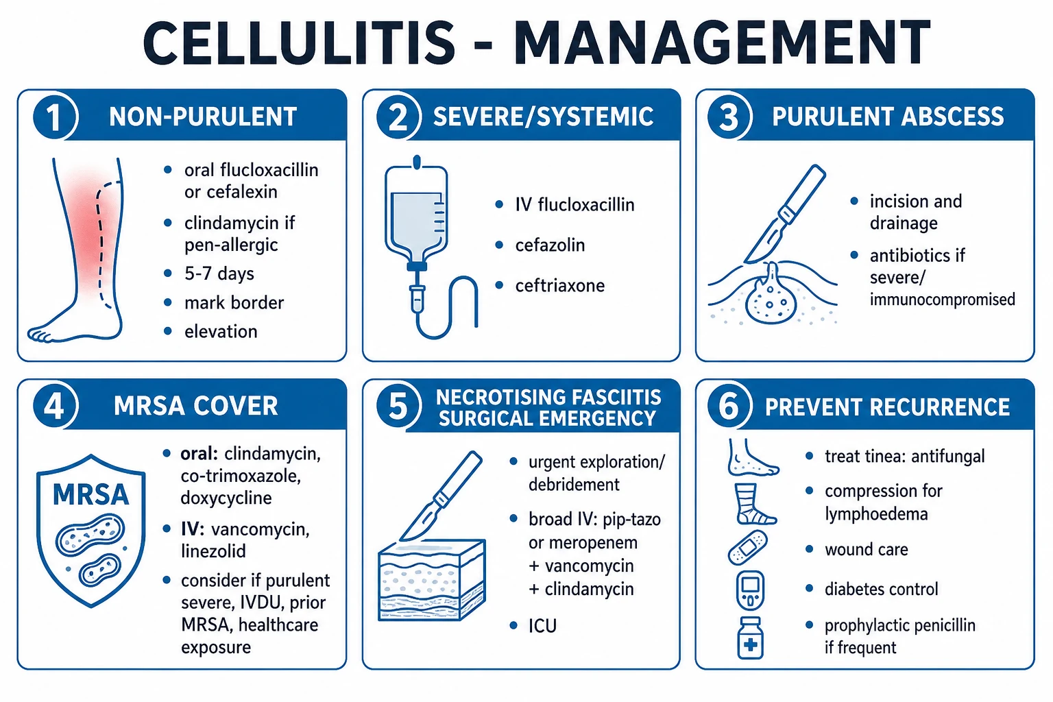
The empirical regimen is driven by the purulent vs non-purulent axis, severity, and MRSA risk.[1]
Non-purulent cellulitis / erysipelas (mild, outpatient)
- First-line: oral flucloxacillin 500 mg four times daily (QDS) for 5 to 7 days (extend if slow response). Flucloxacillin covers Strep pyogenes and methicillin-sensitive S. aureus (MSSA) — the dominant organisms.
- Penicillin allergy without anaphylaxis: oral cefalexin 500 mg QDS (a cephalosporin; cross-reactivity is low).
- Penicillin anaphylaxis: oral clindamycin 300 to 450 mg QDS, or a macrolide (azithromycin or clarithromycin) — check local streptococcal macrolide resistance.
- In pregnancy: oral cefalexin (a beta-lactam safe in pregnancy) — avoid tetracyclines, avoid sulfonamides in the first trimester and near term.
- Mark the border; elevate; treat the portal of entry; safety-net for review at 48 hours.[1][2]
Non-purulent cellulitis (moderate / severe, IV)
- IV flucloxacillin 1 to 2 g QDS (or IV cefazolin 1 to 2 g TDS, or ceftriaxone 1 to 2 g OD — useful where adherence or once-daily dosing matters, e.g. outpatient parenteral therapy).
- Penicillin allergy: IV clindamycin 600 mg QDS (severe) or a glycopeptide.
- Add MRSA cover if MRSA risk or severe disease (see below). [1]
Purulent cellulitis / abscess
- Incision and drainage is the primary therapy — the single most effective intervention. Send pus for culture and sensitivity.
- Antibiotics are adjunctive, given when there is severe surrounding cellulitis, systemic signs (fever, tachycardia), immunocompromise, extremes of age, facial/hand/location, or failure of I&D.
- Cover MRSA (purulent disease is staphylococcal, often MRSA): oral co-trimoxazole (960 mg to 1.92 g BD), doxycycline 100 mg BD, or clindamycin 300 to 450 mg QDS; IV vancomycin (target trough 15 to 20), linezolid 600 mg BD, or daptomycin.[1]
Severe SSTI, sepsis, or suspected necrotising infection
- Broad-spectrum IV: piperacillin-tazobactam 4.5 g TDS or a carbapenem (meropenem 1 g TDS) PLUS clindamycin 600 mg QDS (toxin suppression) PLUS vancomycin or linezolid for MRSA cover.
- For necrotising fasciitis, surgery is the definitive therapy — planned, aggressive debridement of all necrotic tissue with return-to-theatre every 24 to 48 hours until no further necrosis is found. IV immunoglobulin is considered in streptococcal toxic-shock-like syndrome with refractory shock. Hyperbaric oxygen is adjunctive where available, never a substitute for surgery.[1]
Specific exposure-related regimens
- Animal bite (dog/cat): oral amoxicillin-clavulanate 625 mg TDS (covers Pasteurella, anaerobes, oral streptococci) for 5 to 7 days; assess for tendon, joint and bone involvement; update tetanus; consider rabies exposure. Severe/hand bites need IV and surgical review.
- Human bite: amoxicillin-clavulanate (covers Eikenella corrodens, anaerobes); clenched-fist injuries are high-risk for septic arthritis and need urgent surgical review.
- Saltwater exposure (Vibrio vulnificus): doxycycline 100 mg BD plus ceftazidime 2 g TDS (or ceftriaxone) IV; high mortality in cirrhosis — early surgical debridement.
- Freshwater exposure (Aeromonas): ciprofloxacin (or a fluoroquinolone) plus surgical debridement.[1]
IV-to-oral switch and discharge
Switch to oral when the patient is haemodynamically stable, afebrile for 24 to 48 hours, improving clinically, and able to swallow and absorb oral medication. Discharge when clinically stable, afebrile for 24 to 48 hours, tolerating oral intake, with a safe social situation and a clear safety-net to re-present if not improving within 48 hours.[1]
Recurrent cellulitis — prevention is the cure
- Treat the portal of entry — antifungal for tinea pedis (topical allylamine/azole; oral terbinafine or itraconazole for extensive/nail disease); compression therapy for lymphoedema and chronic venous insufficiency (after arterial assessment); debridement and dressing of ulcers; keep the interdigital spaces dry.
- Prophylactic antibiotics for frequent recurrences (two or more per year): the PATCH trial showed that low-dose oral penicillin V 250 mg BD for 12 months (or benzathine penicillin intramuscular monthly, or erythromycin/clarithromycin if penicillin-allergic) reduced recurrence during the treatment period (the benefit was lost after stopping).[4]
Cellulitis — the numbers that decide
Specific Subtypes & Scenarios
- Erysipelas — overwhelmingly Strep pyogenes; treat with oral or IV penicillin (phenoxymethylpenicillin PO; benzylpenicillin IV); a bright-red, sharply demarcated plaque often on the face or lower leg with prominent systemic upset.
- Periorbital vs orbital cellulitis — periorbital (preseptal): oral/IV antibiotics targeting sinus/skin organisms; can be managed outpatient if well. Orbital (post-septal): an emergency — urgent CT of orbits/sinuses, IV antibiotics (ceftriaxone ± metronidazole, add MRSA cover if risk), and urgent ENT/ophthalmology for drainage of sinus/orbital collections.
- Diabetic foot infection — classify severity (IDSA mild/moderate/severe; IWGDF/PEDIS); probe-to-bone for osteomyelitis; send deep tissue/bone cultures; broad empiric cover (Gram-positive, Gram-negative, anaerobes); vascular assessment and revascularisation if ischaemic; offloading; multidisciplinary diabetic foot clinic.
- Necrotising fasciitis — Type I polymicrobial (anaerobes + Gram-negatives + facultative streptococci; immunocompromised, perineal/Fournier gangrene, post-surgery) vs Type II monomicrobial GAS (often healthy host, often with staphylococcal co-infection). Surgery + broad IV antibiotics (carbapenem or piperacillin-tazobactam + clindamycin + vancomycin/linezolid) + supportive ICU care.[1]
- Clostridial myonecrosis (gas gangrene) — Clostridium perfringens alpha-toxin (a phospholipase) lyses cell membranes; post-traumatic or post-surgical, dirty contaminated wounds, rapid onset, severe pain, bronze discoloration, gas, shock. Treatment: urgent surgical debridement of muscle to bleeding healthy tissue, IV penicillin G + clindamycin, supportive ICU; hyperbaric oxygen is adjunctive (where available).
- Surgical site infection (SSI) — CDC classification: superficial incisional (skin/subcutis, within 30 days), deep incisional (fascia/muscle, within 30 days, or 90 days if implant), organ/space (within 30 days, or 90 days if implant). Typical organisms S. aureus, streptococci, Gram-negatives; open the wound, culture, targeted antibiotics.[1]
- Bite and water-exposure SSTIs — see regimens above; always update tetanus and consider rabies for animal bites in endemic regions.
- IV-drug-use SSTI — cover MRSA and oral anaerobes; screen for deep abscess, septic arthritis, osteomyelitis, and endocarditis (examine for a murmur, send blood cultures, consider echocardiography).
Complications & Pitfalls
Local complications: abscess formation, progression to necrotising fasciitis or myonecrosis, lymphatic damage that worsens lymphoedema and predisposes to further recurrence, scarring and pigmentary change, spread to deep structures (osteomyelitis, septic arthritis, tenosynovitis), and in the face/orbit cavernous sinus thrombosis and intracranial spread.[2]
Systemic complications: bacteraemia and sepsis/septic shock, endocarditis, metastatic infection (osteomyelitis, septic arthritis, meningitis), post-streptococcal glomerulonephritis (after GAS), toxic shock syndrome, and acute kidney injury (from sepsis, rhabdomyolysis in necrotising disease, or nephrotoxic drugs). [1]
Classic pitfalls — each one loses marks and harms patients: [1]
- Overdiagnosing cellulitis in bilateral "red legs" (pseudocellulitis) — most often stasis dermatitis, contact dermatitis, or lipodermatosclerosis. Bilateral red legs are rarely cellulitis.
- Missing necrotising fasciitis by reassurance from "early" skin findings or a low LRINEC score — the cardinal clue is pain out of proportion.
- Failing to drain a purulent collection — antibiotics alone will not cure a drainable abscess; use bedside ultrasound to find it.
- Under-covering MRSA in purulent, severe, or at-risk disease.
- Missing a deep abscess or osteomyelitis in the diabetic foot — always probe to bone.
- Treating DVT as cellulitis (or vice versa) — the two coexist riskily; use D-dimer and ultrasound when uncertain.
- Antibiotic pitfalls: clindamycin and Clostridioides difficile (and inducible resistance in staphylococci); vancomycin nephrotoxicity (monitor troughs, especially with concurrent piperacillin-tazobactam); linezolid and thrombocytopenia/myelosuppression (over 2 weeks), peripheral and optic neuropathy, and serotonin syndrome with SSRIs/MAOIs; fluoroquinolones and tendon/AESD and QT prolongation; tetracyclines avoid in pregnancy and children under 12. [1]
Prognosis & Disposition
Outpatient management is appropriate for mild SSTI with no systemic signs, no uncontrolled comorbidity, a reliable patient with follow-up, and ability to take oral therapy. Expect improvement within 48 to 72 hours of appropriate therapy — at reassessment the border should be stable or shrinking, erythema fading, warmth and induration reducing. If not improving by 48 to 72 hours, reassess: wrong diagnosis (pseudocellulitis), wrong organism (MRSA, atypical), unrecognised abscess needing drainage, deep infection (osteomyelitis), or an immunocompromised host.[1][2]
Admit (ward) for systemic signs (fever, tachycardia, hypotension), uncontrolled comorbidity (diabetes, CKD, immunosuppression), failed oral therapy, extensive or rapidly spreading infection, hand/face/perineal involvement, suspected deep infection, or an unsafe social situation. [1]
Admit (ICU) for sepsis or septic shock, necrotising infection, or the immunocompromised with rapid progression. [1]
Necrotising fasciitis carries a mortality of 20 to 40 percent, rising steeply with surgical delay — each 24-hour delay roughly doubles mortality. Diabetic foot infection complicated by osteomyelitis has a guarded prognosis and a high recurrence/amputation risk without multidisciplinary care. [1]
Special Populations
- Children — common organisms Strep pyogenes and S. aureus (including CA-MRSA); weight-based flucloxacillin (12.5 mg/kg QDS, max 500 mg per dose) or cefalexin (10 mg/kg TDS); periorbital cellulitis is common and must be distinguished from orbital cellulitis; a low threshold for admission in young infants.
- Pregnancy — use penicillins and cephalosporins (safe); avoid tetracyclines (fetal bone/tooth), sulfonamides (first trimester, near term), and be cautious with clindamycin near term (rare); flucloxacillin and cefalexin are first-line.
- Elderly — atypical/blunted presentation (no fever, confusion, falls); lower threshold to admit; watch for sepsis and aspiration of necrotic tissue.
- Immunocompromised / neutropenic — broader organisms (Gram-negatives, Pseudomonas, fungi); broad empiric IV cover and a low threshold to image and biopsy.
- Diabetic — diabetic foot; assess for osteomyelitis and ischaemia; multidisciplinary care; offloading.
- IV drug user — cover MRSA and anaerobes; screen for endocarditis and deep abscess.
- Lymphoedema / chronic venous insufficiency — recurrent cellulitis; compression (after arterial assessment) and prophylactic penicillin where recurrences are frequent.
- Renal impairment — dose-adjust flucloxacillin (rarely, in severe CKD), cefalexin, vancomycin (by trough/level), co-trimoxazole; clindamycin needs no renal adjustment; linezolid needs no adjustment.[1][2]
Evidence, Guidelines & Regional Differences
Regional empirical differences: [1]
- US (IDSA 2014) — the purulent vs non-purulent framework. Non-purulent mild: anti-strep/staph oral (cephalexin, dicloxacillin, clindamycin); severe or purulent at-risk: add MRSA cover (vancomycin/linezolid/daptomycin IV; clindamycin/co-trimoxazole/doxycycline oral). Necrotising infection: surgery + broad IV antibiotics with clindamycin.
- UK (NICE CKS / NG190) — oral flucloxacillin 500 mg QDS first-line for uncomplicated cellulitis; cefalexin in penicillin allergy and in pregnancy; clindamycin 300 mg QDS for severe infection or first-line in some trusts; mark the border, 5 to 7 days extendable; IV for moderate/severe (flucloxacillin, ceftriaxone, clindamycin).
- India (ICMR/NCDC) — high community MRSA and Gram-negative prevalence; empirical severe SSTI often requires vancomycin + Gram-negative cover (piperacillin-tazobactam or carbapenem); filariasis is a major cause of lymphoedema driving recurrent lower-limb cellulitis in endemic states; always apply the local antibiogram.[1]
Landmark evidence and controversies: [1]
- PATCH trial (Thomas 2013, NEJM) — low-dose oral penicillin V for 12 months reduced the recurrence of leg cellulitis during treatment (hazard ratio around 0.5); benefit was lost after stopping, and prophylaxis is recommended only for patients with recurrent disease (two or more episodes per year) alongside treating the portal of entry.[4]
- LRINEC score — a useful bedside risk-raiser for necrotising fasciitis, but false-negatives in early disease mean it must never delay surgery in a clinically suspicious case.[3]
- Bedside ultrasound — clearly improves the discrimination of abscess from cellulitis and changes management; under-used.
- Corticosteroids (prednisolone) as adjunct in severe cellulitis (the PATCH-II-era studies) — modest evidence of faster resolution in non-purulent cellulitis in some patients, controversial and not routine; consider only in selected non-diabetic, non-immunocompromised patients with marked inflammation.
- Short vs standard duration — evidence supports short courses (5 to 7 days) for uncomplicated cellulitis that is improving; longer courses (10 to 14 days) for severe, slow-resolving, or complicated disease.[1]
Exam Pearls
Necrotising fasciitis red flags — call surgery NOW
PAIN-CUT
severe pain beyond visible skin findings — the cardinal early clue
destroyed cutaneous nerves — paradoxical numbness over a "painful" area
woody hard subcutaneous tissue, crossing fascial planes, advancing hourly
dusky purple skin, haemorrhagic bullae, sloughing, skin gangrene
palpable or radiographic gas — anaerobes producing tissue gas
fever, tachycardia, hypotension, confusion, septic shock
add clindamycin — it stops toxin synthesis that cell-wall antibiotics cannot
Recurrent leg cellulitis — treat the cause
LITE-FOOT
compression therapy; the limb that swells, recurs
the commonest remediable portal — antifungal, keep dry
the entry point for streptococci — treat the maceration
dress, debride, emolliate the broken skin
podiatry, offloading, footwear
compression stockings after arterial check
weight loss reduces lymphatic pressure
prophylactic penicillin V (PATCH trial)
Exam application bank (NEET-PG / INICET)
One-line answer
Cellulitis is an acute, spreading, non-suppurative infection of the dermis and subcutaneous tissue, usually caused by Streptococcus pyogenes or Staphylococcus aureus (including community-acquired MRSA), presenting with erythema, warmth, swelling and tenderness with poorly demarcated margins, usually on a leg and often through a portal of entry (tinea pedis, ulcer, trauma, IV site). Erysipelas is the superficial variant with raised, sharply demarcated margins. Necrotising fasciitis is the life-threatening deep-fascial variant — severe pain out of proportion, systemic toxicity, skin necrosis or bruising, bullae, crepitus, rapid spread — needing urgent surgical debridement. Management is driven by the purulent vs non-purulent and mild vs moderate vs severe axes: non-purulent cellulitis gets oral flucloxacillin (or cefalexin); purulent disease gets incision and drainage plus or minus antibio
Worked stems (answer without another resource)
Stem 1 — Classic presentation. Map symptoms to mechanism; name the first investigation and first treatment step with dose/route if drug therapy is standard. [1]
Stem 2 — Unstable / complicated. List red flags that force immediate resuscitation, theatre, ICU, antidote, or reperfusion — and what you do in the first 15 minutes. [1]
Stem 3 — Atypical group. Elderly, pregnancy, child, or immunocompromised: how presentation and thresholds change. [1]
Stem 4 — Differential trap. Name the three closest mimics and one discriminator for each. [1]
Stem 5 — Disposition. Who goes home with safety-netting, who is admitted, who needs HDU/ICU/theatre, and what follow-up is mandatory. [1]
Rapid viva checklist
- Definition + classification
- Pathophysiology chain
- Bedside signs / criteria
- Score with exact components (if any)
- Emergency bundle
- Definitive therapy with doses
- Complications of disease and of treatment
- Special populations
- Guideline/trial name if classic
- Three exam traps
Coverage self-check
If you cannot answer any stem above from this page alone, re-read the matching section — the page is intended to be self-sufficient for final-prof and NEET-PG/INICET questions on Cellulitis & Skin / Soft-Tissue Infection.
References
- [1]Stevens DL, Bisno AL, Chambers HF, et al. Practice guidelines for the diagnosis and management of skin and soft tissue infections: 2014 update by the infectious diseases society of America Clin Infect Dis, 2014.PMID 24947530
- [2]Rrapi R, Chand S, Kroshinsky D. Cellulitis: A Review of Pathogenesis, Diagnosis, and Management Med Clin North Am, 2021.PMID 34059247
- [3]Wong CH, Khin LW, Heng KS, Tan KC, Low CO. The LRINEC (Laboratory Risk Indicator for Necrotizing Fasciitis) score: a tool for distinguishing necrotizing fasciitis from other soft tissue infections Crit Care Med, 2004.PMID 15241098
- [4]Thomas KS, Crook AM, Nunn AJ, et al. Penicillin to prevent recurrent leg cellulitis N Engl J Med, 2013.PMID 23635049