Infectious Diseases · Infectious Diseases
COVID-19 (SARS-CoV-2)
Also known as COVID-19 · Coronavirus disease 2019 · SARS-CoV-2 infection · 2019-nCoV
COVID-19 is the disease caused by the betacoronavirus SARS-CoV-2, declared a pandemic by the WHO in March 2020. The S1 subunit of the spike protein binds the ACE2 receptor (high density in type-II pneumocytes, nasal goblet cells, enterocytes, renal tubules, myocardium) and S2 is primed by TMPRSS2 and furin before membrane fusion. Spectrum runs from asymptomatic through mild upper-respiratory illness, pneumonia with hypoxaemia, ARDS and multi-organ failure. Hallmarks: anosmia/ageusia, silent hypoxaemia, bilateral peripheral ground-glass opacities on CT, and a lymphopenia/raised CRP/ferritin/D-dimer/IL-6 profile. Severe disease is driven by a cytokine storm with endothelialitis and microthrombi. Dexamethasone 6 mg OD reduces mortality in patients on oxygen (RECOVERY); remdesivir shortens recovery; nirmatrelvir-ritonavir (Paxlovid) prevents hospitalisation in high-risk outpatients; tocilizumab/baricitinib in rapidly progressing severe disease; mandatory VTE prophylaxis with LMWH. Vaccines (mRNA: Pfizer BNT162b2, Moderna mRNA-1273; viral-vector: ChAdOx1 Covishield/AstraZeneca, Ad26.COV2.S Janssen; inactivated: Covaxin BBV152, CoronaVac; protein subunit: Novavax) are the single most effective prevention and reduce severe disease by more than 90%.
On this page & tools
Your progress
Saved locally on this device.
Exam tags
Red flags
Overview & Definition
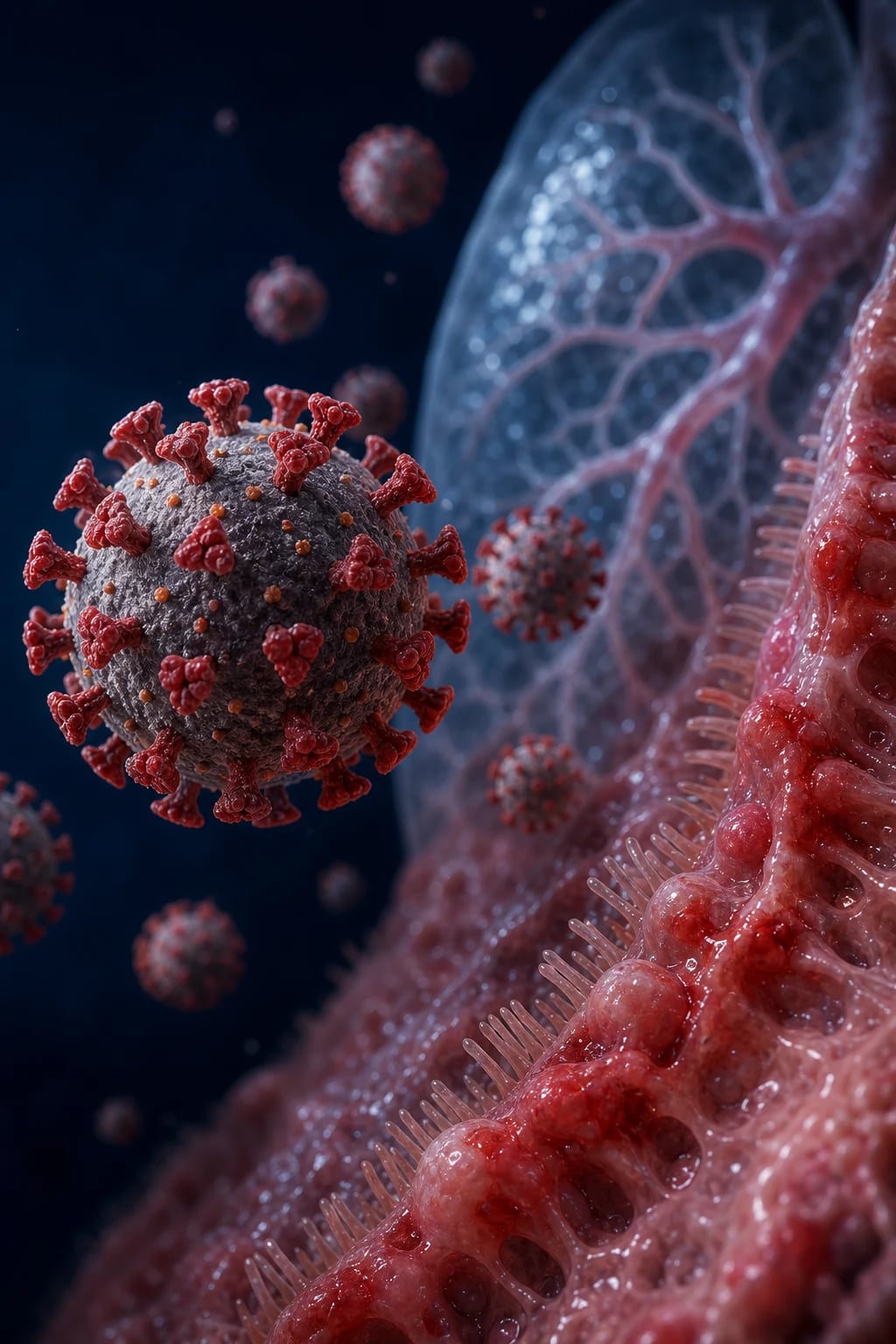
Coronavirus disease 2019 (COVID-19) is the clinical illness caused by severe acute respiratory syndrome coronavirus 2 (SARS-CoV-2), an enveloped, positive-sense, single-stranded RNA betacoronavirus of the subgenus Sarbecovirus that emerged in Wuhan, China in late 2019 and was declared a global pandemic by the WHO on 11 March 2020.[1]
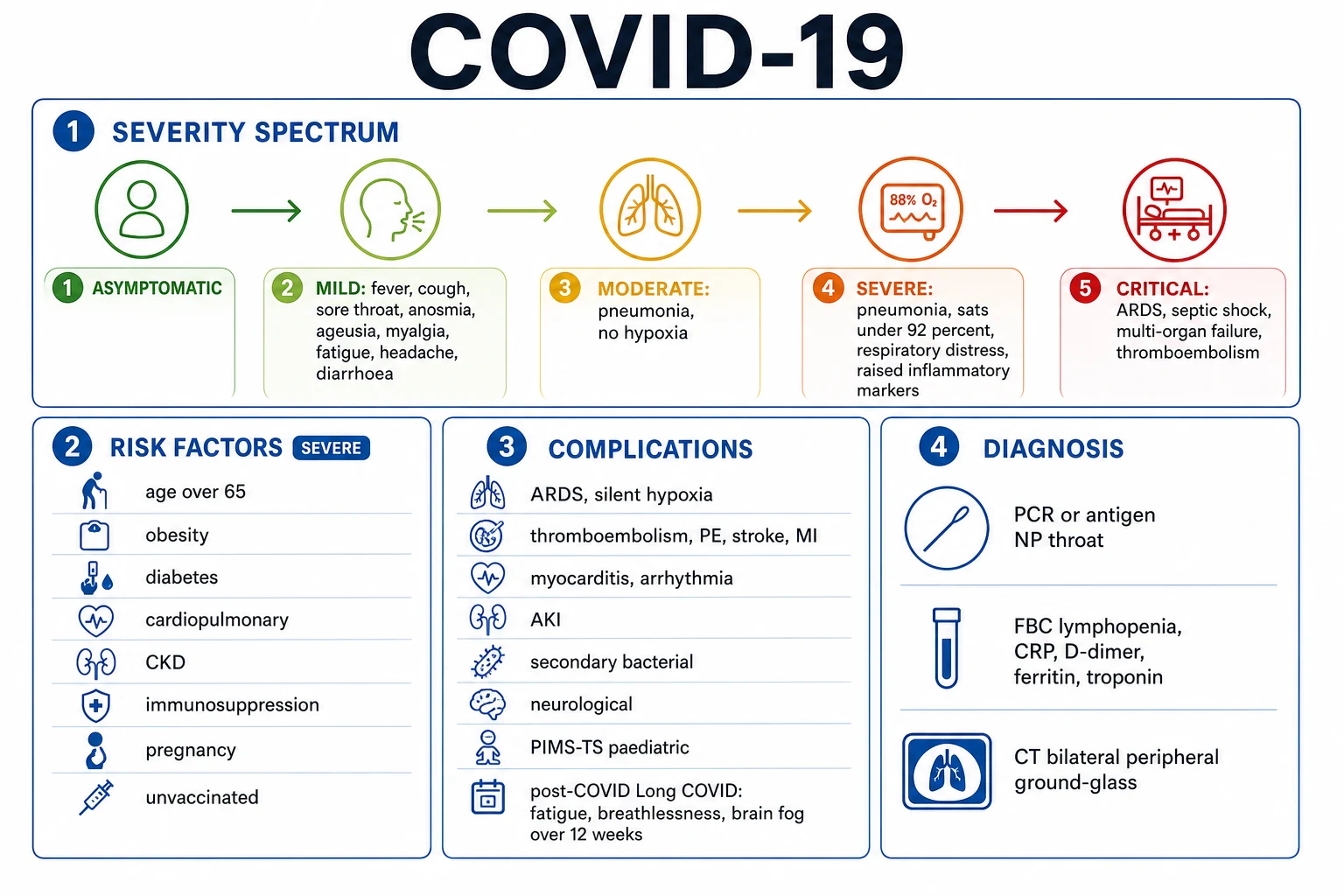
The clinical spectrum is exceptionally wide: [1]
- Asymptomatic / pre-symptomatic infection — viral RNA detectable without symptoms; an important driver of transmission.[8]
- Mild illness — upper-respiratory or influenza-like symptoms (fever, cough, sore throat, fatigue, myalgia, anosmia), no hypoxaemia, no radiographic pneumonia.
- Moderate illness — clinical or radiographic pneumonia with SpO2 at least 92% on room air.
- Severe illness — pneumonia with SpO2 below 92% on room air, RR over 30, or lung infiltrates over 50% of lung fields.
- Critical illness — ARDS, sepsis/septic shock, or multi-organ failure requiring intensive care.[1][2]
Two post-infectious syndromes are integral to the definition: [1]
- Multisystem inflammatory syndrome (MIS) — MIS-C in children and MIS-A in adults, a hyperinflammatory illness with persistent fever, multi-organ involvement and shock, occurring 2 to 6 weeks after SARS-CoV-2 infection.
- Post-COVID condition ('long COVID') — WHO definition: symptoms (commonly fatigue, breathlessness, cognitive dysfunction 'brain fog', anosmia) that develop within 3 months of onset, last at least 2 months, and are not explained by an alternative diagnosis. [1]
The clinical skill in COVID-19 is not the diagnosis (clinical plus PCR/antigen) but recognising severity and the trajectory of deterioration, choosing the right tier of therapy (outpatient antiviral, inpatient steroid ± antiviral ± immunomodulator, ICU ventilatory support), preventing venous thromboembolism, and anticipating the post-COVID sequelae. [1]
Classification
By clinical severity (the classification that drives therapy — adapted from WHO and NIH):[1]
| Severity | Definition | Site of care |
|---|---|---|
| Asymptomatic | Positive test, no symptoms | Isolate; treat comorbidity |
| Mild | Symptomatic, no pneumonia, SpO2 at least 92% room air | Home; supportive; consider antiviral if high risk |
| Moderate | Clinical/radiographic pneumonia, SpO2 at least 92% room air | Hospital; oxygen if needed |
| Severe | SpO2 under 92% room air, RR over 30, or infiltrates over 50% | Hospital; oxygen, dexamethasone, remdesivir ± immunomodulator |
| Critical | ARDS, sepsis/septic shock, multi-organ failure | ICU; ventilation, organ support |
By viral lineage — variants of concern (VOC) (an exam favourite): [1]
Ancestral Wuhan
- Original 2019 strain
- R0 ~2.5 to 3.5
- D614G mutation became dominant mid-2020
Alpha (B.1.1.7)
- UK, late 2020
- ~50% more transmissible
- Modest increase in severity
Beta (B.1.351)
- South Africa
- Significant immune evasion (K417N, E484K)
- Reduced vaccine efficacy against mild disease
Gamma (P.1)
- Brazil (Manaus)
- Immune evasion (K417T, E484K, N501Y)
- Reinfection outbreaks
Delta (B.1.617.2)
- India, 2021 — drove the devastating 'second wave'
- More transmissible and more severe
- Reduced (but preserved) vaccine efficacy against severe disease
Omicron (B.1.1.529)
- Southern Africa, late 2021; many sublineages (BA.x, XBB, JN.1, KP.x)
- Markedly higher transmissibility and immune evasion
- Generally less severe (lower-tropism upper airway), but huge case counts
By ARDS phenotype (an advanced concept): Type-L ('Low') — low elastance, low V/Q homogeneity, low lung weight (early COVID-19, often surprisingly compliant); Type-H ('High') — high elastance, high D-dimer, typical dense ARDS (later/severe). The distinction influences PEEP strategy — high PEEP may worsen Type-L by overdistension. [1]
Epidemiology & Risk Factors
COVID-19 caused over 770 million confirmed cases and over 7 million reported deaths globally by 2024 (true excess-mortality estimates are 2 to 3 times higher). The pandemic progressed in successive waves driven by variants and population immunity, with Omicron sublineages dominating from late 2021 onwards.[1][1]
Modes of transmission: [1]
- Respiratory droplet (greater than 5 micrometres, short range under 1 m) — the classical route.
- Aerosol (smaller droplet nuclei, under 5 micrometres, can travel further and linger) — now recognised as the dominant mode, particularly in closed, poorly ventilated, crowded indoor spaces — the basis for airborne-precaution PPE and ventilation.
- Contact/fomite (contaminated surfaces then mucous membranes) — minor route.
- Rare — vertical transmission (possible but uncommon), blood-borne, zoonotic origin. [1]
Key epidemiological parameters: [1]
- Incubation period — mean 5 days (range 2 to 14); Omicron is shorter (~3 to 4 days).
- Infectious period — from ~2 days before symptom onset (peak infectiousness) to ~7 to 10 days after; severely immunocompromised may shed for weeks.
- R0 (ancestral) — ~2.5 to 3.5; Omicron effective reproduction number much higher (~7 to 10 in susceptible populations). [1]
COVID-19 — key numbers
Risk factors for severe COVID-19 and death (the consistent global signal):[2]
- Age 65 years and over — by far the strongest risk factor; mortality rises exponentially with age.
- Comorbidities — cardiovascular disease, diabetes mellitus, chronic obstructive pulmonary disease, asthma, chronic kidney disease (especially dialysis), chronic liver disease, cerebrovascular disease.
- Obesity (BMI 30 and over) — an independent risk factor that became prominent in COVID-19 (mechanical, metabolic, and pro-inflammatory mechanisms).
- Immunocompromise — solid-organ or haematopoietic stem-cell transplant, chemotherapy, advanced HIV (CD4 under 200), inherited immunodeficiency, high-dose corticosteroids.
- Pregnancy — increased risk of ICU admission, mechanical ventilation and preterm birth.
- Smoking — current smoking.
- Socioeconomic deprivation and ethnic minority status — driven by structural factors (multigenerational households, occupational exposure, healthcare access). [1]
Risk factors for transmission (the 3 C's): Crowded places, Close-contact settings, Confined and enclosed spaces. [1]
Pathophysiology
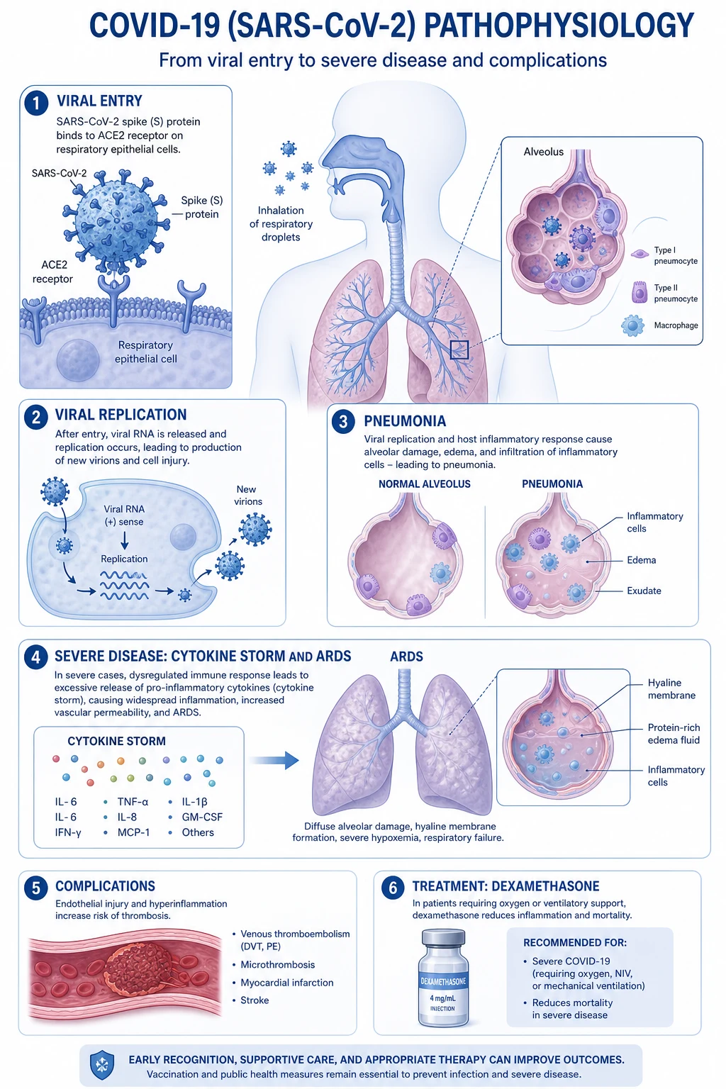
The pathophysiology of COVID-19 is best understood as a triphasic process: viral entry and replication, then dysregulated host inflammation, then endothelial-thrombotic injury.[1][2]
1. The virion and viral entry. SARS-CoV-2 is an enveloped, positive-sense single-stranded RNA virus with four structural proteins — spike (S), envelope (E), membrane (M), and nucleocapsid (N) — and a ~30 kb genome. The S protein is cleaved into two functional subunits: [1]
- S1 contains the receptor-binding domain (RBD) that binds the angiotensin-converting enzyme 2 (ACE2) receptor — densely expressed on type-II pneumocytes, nasal goblet and ciliated cells, bronchial epithelium, enterocytes, renal tubular cells, cardiomyocytes, vascular endothelium, and pancreatic islet cells (explaining the multi-organ signature).
- S2 mediates membrane fusion after priming by the transmembrane serine protease TMPRSS2 (and furin, which pre-cleaves S at the polybasic S1/S2 site — a feature absent from the original SARS-CoV that increases transmissibility). [1]
Binding → conformational change → fusion → viral RNA released into the cytoplasm → translation of ORF1a/1b polyproteins → RNA-dependent RNA polymerase (RdRp) → replication-transcription → new virions → exocytosis. (RdRp is the target of remdesivir.) [1]
2. The immune response — and why it sometimes fails. Productive innate immunity (early type-I and type-III interferon) usually aborts infection. Severe COVID-19 is characterised by a delayed, blunted interferon response combined with a dysregulated, exuberant inflammatory response: macrophage activation, IL-6, TNF-α, IL-1β, IL-12, MCP-1, complement (C5a) activation, and lymphocyte apoptosis (the characteristic lymphopenia). This is the 'cytokine storm' phenotype, the target of tocilizumab (IL-6 receptor blockade), baricitinib (JAK1/2 inhibition) and corticosteroids.[9]
3. Endothelialitis, microthrombi, and the hypercoagulable state. ACE2 on vascular endothelium becomes a viral target; endothelial injury + complement activation + cytokines produce diffuse endothelialitis, lymphocytic endothelialitis, and in-situ microthrombi (especially pulmonary). Combined with immobilisation and critical illness, this produces a striking prothrombotic state: D-dimer is typically markedly raised, and the rate of deep-vein thrombosis and pulmonary embolism in hospitalised COVID-19 is several-fold higher than in matched non-COVID inpatients. This is the rationale for mandatory pharmacological VTE prophylaxis in every hospitalised patient.[2]
4. The renin-angiotensin-system twist. ACE2 normally converts Ang-II to Ang-(1-7), which is vasodilatory, anti-fibrotic and anti-inflammatory. Viral binding and internalisation downregulates ACE2, leaving unopposed Ang-II — vasoconstriction, inflammation, oxidative stress, fibrosis — a mechanism that contributes to cardiac, renal and vascular injury. [1]
5. Pulmonary phase — why hypoxaemia. Alveolar macrophage cytokine release → neutrophil and proteinaceous exudate → diffuse alveolar damage with hyaline membranes (early exudative phase; later organising/proliferative and, in survivors, fibrosis). The radiological correlate is the bilateral peripheral ground-glass opacity. Hypoxaemia results from ventilation-perfusion mismatch, intrapulmonary shunt, and loss of hypoxic pulmonary vasoconstriction. The paradoxical preserved lung compliance in early COVID-19 ('Type-L') with severe hypoxaemia explains 'silent/happy hypoxaemia' — patients may walk and talk with SpO2 in the 70s. [1]
6. Anosmia — mechanism. SARS-CoV-2 infects the sustentacular (supporting) cells of the olfactory epithelium (which express ACE2/TMPRSS2), not the olfactory neurons themselves — disrupts support of olfactory receptors → anosmia. Usually recovers; mechanism of persistent anosmia involves olfactory-epithelium regeneration. [1]
Clinical Presentation
Incubation 2 to 14 days (mean ~5; Omicron shorter). The presentation depends on age, comorbidity, vaccination status and viral lineage. [1]
Typical symptomatic (mild-to-moderate) COVID-19 — influenza-like illness: [1]
- Fever (often high, with rigors) — though afebrile in 10 to 20% and in the elderly.
- Cough — usually dry initially; may become productive later.
- Sore throat, coryza, headache, myalgia, fatigue.
- Anosmia and/or ageusia — a discriminating feature present in 40 to 60% of symptomatic patients (more pronounced with ancestral and Alpha strains), often preceding respiratory symptoms.[1]
- Diarrhoea and other GI symptoms in ~10 to 20%.
Severe COVID-19 — pneumonia and ARDS: [1]
- Dyspnoea (often the discriminant that prompts admission), tachypnoea (RR over 30), accessory-muscle use.
- Hypoxaemia (SpO2 under 92% on room air) — classically 'silent/happy hypoxaemia' (cyanosis, low SpO2, minimal breathlessness — explained by preserved lung compliance, low dyspnoea threshold set by hypoxia).
- Auscultation may be surprisingly quiet despite impressive CT changes; bilateral fine crackles develop later.
- Rapidly progressive bilateral pneumonia over 24 to 72 hours; cytokine-release phase around days 7 to 10 (often after initial improvement). [1]
Critical COVID-19 — ARDS, septic shock, multi-organ failure: [1]
- ARDS (Berlin definition — see Investigations).
- Septic shock — vasopressor-requiring hypotension with elevated lactate.
- Acute kidney injury — often requiring renal replacement therapy.
- Cardiac — myocardial injury (raised troponin), myocarditis, new arrhythmia (including atrial fibrillation), Takotsubo-like cardiomyopathy, acute coronary syndrome.
- Neurological — encephalopathy (very common in ICU), stroke (large-vessel and microvascular, hypercoagulable), Guillain-Barré syndrome, seizures.
- Thrombotic — pulmonary embolism, deep-vein thrombosis, MI, ischaemic stroke, catheter-related and unusual-site thromboses.
- Dermatological — chilblain-like acral lesions ('COVID toes'), urticarial, maculopapular, livedoid rash. [1]
Atypical presentations (examiner-favourite): [1]
- Elderly — delirium, falls, reduced mobility, anorexia, loss of usual function, drowsiness; fever and cough may be absent; lower threshold to test, image and admit.
- Diabetic — may present in diabetic ketoacidosis as the first manifestation of COVID-19; glycaemic control often destabilised.
- Pregnant — risk of severe pneumonia and preterm birth; may present with reduced fetal movements.
- Immunocompromised — silently progressive hypoxaemia, prolonged shedding, atypical or sparse symptoms; high risk of variant emergence under prolonged replication.
- Children — predominantly mild or asymptomatic; present with croup-like, bronchiolitis-like illness; Omicron produces more upper-airway disease (laryngotracheobronchitis). MIS-C (see Specific Subtypes) is the principal severe paediatric phenotype. [1]
Multisystem inflammatory syndrome in children (MIS-C) — persistent fever (3 or more days) plus multi-organ involvement (rash, conjunctivitis, mucosal involvement, abdominal pain, vomiting/diarrhoea, myocarditis with shock, coronary-artery aneurysm, AKI, hepatitis, neurological features) 2 to 6 weeks after SARS-CoV-2 infection, often with serological evidence of recent infection (PCR may be negative). It overlaps with but differs from Kawasaki disease (older children, more GI, more cardiac, prominent shock, thrombocytopenia). [1]
Differential Diagnosis
A febrile illness with respiratory and/or radiographic findings in the COVID era is not always COVID-19. Distinguish:[1]
- Influenza pneumonia — similar dry cough, fever, myalgia, malaise; more abrupt onset; seasonal epidemiology; rapid antigen/PCR positive for influenza; vaccination history. CT pattern often more central, lobular, peribronchovascular ground-glass.
- Bacterial community-acquired pneumonia (typical — S. pneumoniae) — abrupt, productive purulent/rust-coloured sputum, lobar consolidation on CXR, neutrophilia rather than lymphopenia, rapid response to beta-lactam.
- Atypical bacterial pneumonia (Mycoplasma, Chlamydophila, Legionella) — insidious, dry cough, prominent systemic features, patchy interstitial CXR. Legionella — GI symptoms, confusion, hyponatraemia, raised LFTs, urinary antigen.
- Other viral pneumonias (RSV, adenovirus, hMPV, parainfluenza) — distinguish by multiplex respiratory PCR; more common in children/immunocompromised.
- Pulmonary embolism — sudden dyspnoea, pleuritic pain, syncope; risk factors; CXR often normal; D-dimer raised (also in COVID-19 — confusing); CT pulmonary angiography is definitive. Both can coexist in COVID-19.
- Tuberculosis — subacute/chronic, night sweats, weight loss, upper-lobe cavitation, positive sputum AFB/NAAT.
- Pneumocystis jirovecii pneumonia (HIV/immunocompromised) — subacute dyspnoea, perihilar ground-glass, pneumothorax, raised LDH, low CD4.
- Cardiogenic pulmonary oedema — bilateral alveolar infiltrates, cardiomegaly, raised BNP/NT-proBNP, echocardiographic systolic/diastolic dysfunction; history of heart failure.
- Acute exacerbation of COPD/asthma — usually triggered, wheeze prominent, less fever/CRP, response to bronchodilator/steroid.
- Hypersensitivity pneumonitis / organising pneumonia / drug-induced pneumonitis — exposure/drug history, eosinophilia.
- MIS-C differentials: Kawasaki disease, staphylococcal toxic-shock syndrome, streptococcal toxic-shock, bacterial sepsis, juvenile-onset systemic lupus or other autoimmune flare, haemophagocytic lymphohistiocytosis (HLH). [1]
Always consider co-infections — bacterial superinfection (pneumococcal, staphylococcal especially post-influenza) and other respiratory viruses. Send respiratory virus PCR panel in moderate-to-severe disease. [1]
Clinical & Bedside Assessment
Vital signs drive severity and disposition. In every suspected case: [1]
- SpO2 on room air (the single most important bedside number) — measure on room air for at least 5 minutes; record response to supplemental oxygen. Target 92 to 96% in most adults, 88 to 92% in COPD/type-2 respiratory failure.
- Respiratory rate — the most sensitive single sign of lower-respiratory compromise; RR over 30 is a severe-disease criterion.
- Work of breathing — accessory-muscle use, intercostal recession, inability to speak full sentences.
- Blood pressure, heart rate, temperature, capillary refill, conscious level (GCS / NEWS2).
- Hydration, urine output (AKI is common in severe disease). [1]
Detecting silent hypoxaemia (a clinical pitfall): [1]
- 6-minute walk test (6MWT) in ambulatory patients — a drop in SpO2 by 3 percentage points or more (or to under 92%) is abnormal and warrants admission.
- 1-minute sit-to-stand test — useful bedside surrogate. [1]
Awake proning — the bedside manoeuvre that improves oxygenation by recruiting dorsal-lung segments and improving V/Q matching (reduces shunt, redistributes perfusion, improves diaphragmatic mechanics): [1]
- Indication — awake, cooperative patient with severe hypoxaemia (SpO2 under 92% despite supplemental oxygen) without need for immediate intubation.
- Protocol — prone 2 hours at a time, 3 to 4 sessions daily, building to 12 to 16 hours/day; rotate between prone and lateral positions.
- Contraindications — spinal instability, recent abdominal or thoracic surgery, 3rd-trimester pregnancy, haemodynamic instability, severe obesity that precludes safe turning, agitation, raised intracranial pressure. [1]
Chest auscultation — often deceptively quiet early in the course despite impressive imaging; bilateral fine basal crackles and reduced breath sounds develop as exudate accumulates. A silent hemithorax with dullness suggests effusion, collapse, or pneumothorax (a recognised COVID-19 complication). [1]
Daily trajectory assessment — every hospitalised patient should have a disease-severity trajectory documented: improving, static, or worsening. Worsening trajectory with rising CRP/ferritin/D-dimer and falling lymphocyte count is the trigger to consider tocilizumab or baricitinib and ICU escalation. [1]
Investigations
Diagnostic tests (confirming SARS-CoV-2 infection): [1]
- Reverse-transcription polymerase chain reaction (RT-PCR) — gold standard. Detects viral RNA in a nasopharyngeal (or combined naso/oropharyngeal) swab, sputum, BAL. Sensitivity highest days 3 to 7; sensitivity falls after day 10 but the patient may remain PCR-positive (non-viable RNA) for weeks. Reported as a cycle-threshold (Ct) — a low Ct (under 25) indicates high viral load; a Ct over 35 typically corresponds to non-infectious residual RNA.
- Rapid antigen test (RAT) — detects viral nucleocapsid antigen; faster and cheaper; lower sensitivity early in disease and in asymptomatics; useful for triage and community surveillance. False-negatives occur with low viral load.
- Nucleic acid amplification tests (NAAT), CRISPR-based, and molecular point-of-care tests — varying speed/accuracy; the standard is RT-PCR.
- Serology (anti-S, anti-N antibodies) — confirms prior infection; anti-N distinguishes natural infection from vaccination (vaccines target S only); not useful for acute diagnosis (IgM unreliable).
- Whole-genome sequencing / variant surveillance — for VOC identification. [1]
Chest imaging: [1]
- Chest X-ray — often normal early; classic finding is bilateral, peripheral, mid- and lower-zone opacities (ground-glass then consolidation). Useful for severity and follow-up.
- Chest CT — the most sensitive imaging; classic findings:[5]
- Bilateral, peripheral (subpleural), posterior-basal ground-glass opacities (GGOs) — the hallmark.
- Crazy-paving (GGO with interlobular septal thickening) and consolidation.
- Vascular enlargement, reverse-halo (atoll) sign, fibrotic bands in organising phase.
- Define ground-glass opacity — hazy increased lung attenuation without obscuration of underlying bronchovascular markings (distinguished from consolidation, which obscures them).
- Lung ultrasound — bedside; demonstrates B-lines (comet-tail artefacts) in a multifocal, patchy, often spared pattern; useful in resource-limited or infection-control settings.
Laboratory profile of severe COVID-19 (high-yield — reproduces the inflammatory phenotype):[2]
| Test | Finding | Prognostic implication |
|---|---|---|
| Full blood count | Lymphopenia (under 1.0 × 10⁹/L) | Severity and mortality |
| Neutrophil/lymphocyte ratio over 3 | Adverse | |
| Mild thrombocytopenia; later reactive thrombocytosis | — | |
| CRP | Raised (often 50 to 200 mg/L) | Trajectory marker; ↑ = worsening |
| Ferritin | Markedly raised (often 1000+) | Macrophage activation |
| D-dimer | Markedly raised | Coagulopathy, VTE, mortality |
| LDH | Raised | Tissue injury; mortality marker |
| IL-6 | Raised (where available) | Target of tocilizumab; trajectory marker |
| Troponin / NT-proBNP | Raised | Cardiac injury; adverse |
| ALT/AST | Mild transaminitis | Hepatic involvement |
| Creatinine / urea | Raised | AKI; adverse |
| Creatine kinase | Raised | Myositis/rhabdomyolysis |
| Coagulation | Prolonged PT/aPTT (mild), low fibrinogen (in severe DIC-like) | Hypercoagulable; bleeding rare |
Severity scores: [1]
-
NEWS2 (National Early Warning Score 2) — 7 physiological parameters (RR, SpO2, supplemental oxygen, temperature, systolic BP, heart rate, AVPU/GCS); aggregate 5+ = urgent review, 7+ = emergency. Widely used for triage and deterioration detection. [1]
-
ARDS — Berlin definition (reproduce verbatim — exam favourite):
- Timing — within 1 week of a known clinical insult or new/worsening respiratory symptoms.
- Imaging — bilateral opacities not fully explained by effusions, lobar/lung collapse, or nodules.
- Origin of oedema — respiratory failure not fully explained by cardiac failure or fluid overload (objective assessment, e.g. echocardiography, if no risk factor).
- Hypoxaemia (with PEEP or CPAP at least 5 cmH2O):
- Mild — PaO2/FiO2 (P/F ratio) between 200 and 300 (≤300 with the original definition).
- Moderate — P/F between 100 and 200.
- Severe — P/F under 100. [1]
-
For MIS-C — echocardiography (coronary aneurysm, ventricular dysfunction, pericardial effusion), troponin and NT-proBNP, ferritin, CRP, coagulation (PT, aPTT, fibrinogen, D-dimer), IL-6, SARS-CoV-2 serology (often positive even when PCR negative). [1]
-
VTE work-up — D-dimer (raised in COVID-19, so use trajectory and pre-test probability), CT pulmonary angiography for PE (look for subsegmental and saddle emboli), compression Doppler of lower limbs, echocardiography (RV strain pattern). [1]
Management — Resuscitation
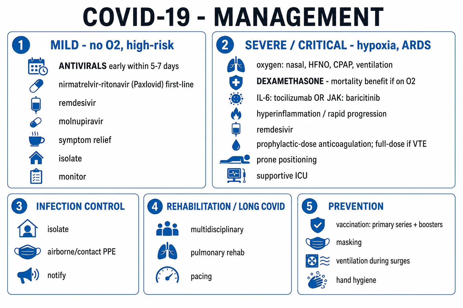
ABCDE approach. Isolate immediately (airborne/droplet precautions) and notify infection control. [1]
Oxygen therapy — the cornerstone of supportive care. Target SpO2 92 to 96% in most adults (or 88 to 92% in COPD/type-2 respiratory failure risk).[1]
Escalation ladder (apply stepwise, guided by SpO2 and work of breathing): [1]
- Nasal cannula (1 to 5 L/min).
- Simple face mask (5 to 10 L/min) or non-rebreather mask (15 L/min) for immediate resuscitation.
- High-flow nasal oxygen (HFNO) — up to 60 L/min, FiO2 up to 100%; humidified; well-tolerated; reduces work of breathing. (Closely monitor — 'HFNO failure' = early intubation, not delayed.)
- Continuous positive airway pressure (CPAP) / non-invasive ventilation (NIV) — useful in COVID-19 with hypoxaemic respiratory failure (some benefit; risk of aerosol generation — use with close-fitting mask and viral filter).
- Invasive mechanical ventilation — when HFNO/NIV fails, with worsening respiratory failure, hypoxaemia despite maximal non-invasive support, altered consciousness, or haemodynamic instability. Intubate early rather than as a crash can't-intubate-can't-oxygenate situation.
- Extracorporeal membrane oxygenation (ECMO) — veno-venous ECMO for refractory severe ARDS in expert centres. [1]
Awake proning at the bedside (see Clinical Assessment) — start in patients with SpO2 under 92% on oxygen, who can cooperate. [1]
Septic shock bundle (Surviving Sepsis hour-1) — applies to COVID-19 with shock: two large-bore IV cannulae, blood cultures, lactate, broad-spectrum antibiotics within 1 hour, balanced crystalloid 30 mL/kg, noradrenaline for fluid-refractory shock; reassess fluid responsiveness before further boluses. [1]
Concurrent therapy at resuscitation stage — start VTE prophylaxis (LMWH) on admission in all non-bleeding patients (see Management — Definitive). [1]
End-of-life / goals-of-care discussion in patients with advanced frailty or irreversible multi-organ failure — comfort care and palliative measures appropriate. [1]
Management — Definitive & Stepwise
Therapy is stratified by disease severity, and time-from-symptom-onset is critical (early = antiviral; late/inflammatory = immunomodulatory).[3][1]
Mild disease (home, SpO2 at least 92%) — supportive care (rest, hydration, antipyretic — paracetamol 1 g QDS). Isolate (5 days from symptom onset with negative RAT, or symptom/time-based — see Prognosis). Antibiotics are NOT routinely indicated. Consider outpatient antiviral in high-risk patients (see below). [1]
High-risk mild/moderate outpatient (within 5 to 7 days of symptom onset) — antiviral therapy to prevent progression: [1]
- Nirmatrelvir-ritonavir (Paxlovid) — 300 mg nirmatrelvir + 100 mg ritonavir PO twice daily for 5 days (reduce to nirmatrelvir 150 mg/ritonavir 100 mg BD if eGFR 30 to 60 mL/min; contraindicated if eGFR under 30 or severe hepatic impairment). Mechanism: nirmatrelvir inhibits the SARS-CoV-2 3CL protease; ritonavir boosts nirmatrelvir by CYP3A inhibition. EPIC-HR: 89% reduction in hospitalisation/death in unvaccinated high-risk adults.[7] Major caveat: many CYP3A drug-drug interactions (statins, antiarrhythmics, anticoagulants, calcineurin inhibitors, many antiretrovirals) — review medication list; adjust or withhold interacting drugs for the 5-day course.
- Remdesivir (outpatient, IV) — for those who cannot take Paxlovid: 200 mg IV day 1, then 100 mg IV OD on days 2 and 3 (3-day course).
- Molnupiravir — 800 mg PO twice daily for 5 days; less effective than Paxlovid (~30% reduction); avoid in pregnancy and in those of childbearing potential not using contraception.
Moderate-to-severe disease (hospitalised, on oxygen) — combination therapy: [1]
-
Dexamethasone 6 mg PO/IV once daily for up to 10 days — the single most important mortality-reducing therapy in severe/critical COVID-19.[3]
- RECOVERY trial (PMID 32678530): reduced 28-day mortality — from 41% to 28% in invasively ventilated patients; from 26% to 21% in patients on oxygen; no benefit (trend to harm) in patients not requiring oxygen. Hence: give ONLY to patients on supplemental oxygen or ventilatory support.
- Mechanism: broad suppression of the dysregulated host inflammatory response (cytokine storm, endothelialitis).
- Side-effects to monitor: hyperglycaemia (often marked, may need insulin), secondary infection, neuromyopathy, psychiatric disturbance, GI bleeding.
- In pregnant patients with severe COVID-19, dexamethasone 6 mg is preferred over prednisolone (also benefits fetal lung maturity); avoid dexamethasone for fetal lung maturation dosing (use betamethasone/dexamethasone as per obstetric indication).
-
Remdesivir (inpatient, IV) — 200 mg IV loading dose on day 1, then 100 mg IV OD for 4 days (total 5 days); extend to 10 days in severely immunocompromised with prolonged replication.[4]
- ACTT-1 trial (PMID 32445440): shortened median time to recovery from 15 to 10 days in hospitalised hypoxic patients.
- Mechanism: nucleotide analogue inhibitor of SARS-CoV-2 RNA-dependent RNA polymerase (RdRp).
- Best benefit in early (first 10 days) disease with hypoxaemia; little benefit in late ARDS/multi-organ failure or those already intubated/extubated.
- Monitoring: transamitis (reduce dose or stop if ALT over 5 to 10 times upper limit of normal); renal adjustment.
-
IL-6 inhibition — tocilizumab — 8 mg/kg IV (maximum 800 mg) as a single dose; may repeat once after 8 to 12 hours if no clinical response.[6]
- Indication — severe/critical disease with hypoxaemia on oxygen and a rising inflammatory marker profile (CRP, ferritin) with rapid progression, particularly within 24 hours of new high-flow oxygen or ventilation. The REMAP-CAP tocilizumab evidence showed mortality and organ-support benefit in this group.
- Mechanism: monoclonal antibody to the IL-6 receptor.
- Caveats: increased risk of secondary bacterial/fungal infection; do not combine routinely with baricitinib.
-
JAK inhibition — baricitinib — 4 mg PO OD for up to 14 days (renal dose adjustment); alternative to tocilizumab in selected severe/critical patients. Mechanism: JAK1/2 inhibitor, blocks intracellular signalling downstream of multiple cytokines. (Tofacitinib is an alternative.) [1]
-
Corticosteroid + remdesivir combination is appropriate for the patient with new-onset hypoxaemia on oxygen. [1]
-
Convalescent plasma, hydroxychloroquine, lopinavir-ritonavir, ivermectin, azithromycin — NOT recommended; high-quality trials showed no benefit (and harm in some — HCQ caused arrhythmia and excess mortality; lopinavir-ritonavir no benefit and GI side-effects). [1]
-
Monoclonal antibodies (casirivimab-imdevimab REGEN-COV, sotrovimab, regdanvimab, bamlanivimab-etesevimab) — for early outpatient treatment in high-risk patients; early-generation mAbs lost neutralising activity against Omicron sublineages, so their current role is limited; check current local neutralisation data. [1]
Venous thromboembolism prophylaxis (mandatory in all hospitalised non-bleeding patients): [1]
- Enoxaparin 40 mg subcutaneous once daily (or dalteparin 5000 IU SC OD); renal dose reduction to enoxaparin 20 mg SC OD if eGFR under 30; unfractionated heparin 5000 IU SC TDS as alternative in severe renal impairment.
- Therapeutic-dose anticoagulation — NOT routinely in critically ill ICU patients (REMAP-CAP/ATTACC/ACTIV-4a: therapeutic-dose anticoagulation was harmful in critically ill patients); consider therapeutic-dose LMWH in moderately ill (non-ICU) hospitalised patients with high D-dimer (these trials showed benefit).
- Therapeutic anticoagulation is, of course, mandatory for confirmed VTE, PE or acute coronary syndrome. [1]
Ventilator strategy for COVID-19 ARDS (lung-protective ventilation): [1]
- Low tidal volume 6 mL/kg predicted body weight; plateau pressure under 30 cmH2O; driving pressure under 15 cmH2O.
- PEEP titration — individualised; high PEEP may worsen Type-L physiology.
- Prone positioning 16 hours per day in moderate-to-severe ARDS (P/F under 150) — improves oxygenation and reduces mortality.
- Neuromuscular blockade (cisatracurium) if severe asynchrony or P/F under 150 — short course.
- Inhaled pulmonary vasodilators (nitric oxide, epoprostenol) — rescue oxygenation, no mortality benefit.
- VV-ECMO — refractory severe ARDS in expert centre. [1]
Step-down and discharge criteria — clinically stable, afebrile for 24 to 48 hours, SpO2 over 92% on room air for 24 hours, no organ-support requirement, stable comorbidity, safe social situation. Thromboprophylaxis is usually continued for 2 to 6 weeks post-discharge (post-hospital thromboprophylaxis) in high-risk patients. [1]
Specific Subtypes & Scenarios
MIS-C (and MIS-A) — persistent fever plus multi-organ involvement 2 to 6 weeks after SARS-CoV-2 infection; managed in hospital with: [1]
- Supportive ICU for shock (fluids, vasoactive agents), respiratory support.
- IV immunoglobulin (IVIG) 2 g/kg single infusion (over 10 to 12 hours; max 100 g), and methylprednisolone 1 to 2 mg/kg/day (pulse methylprednisolone 10 to 30 mg/kg/day in severe shock).
- Aspirin (antiplatelet dose, 3 to 5 mg/kg/day, max 75 mg) for coronary-artery aneurysm prophylaxis (after Kawasaki protocols); therapeutic anticoagulation if large aneurysm or thrombus.
- Serial echocardiography for coronary aneurysms and cardiac function.
- Refractory cases — anakinra (IL-1 receptor antagonist), infliximab. [1]
COVID-19 in pregnancy — increased risk of ICU admission, mechanical ventilation and preterm birth, but no convincing increase in vertical transmission. Dexamethasone 6 mg is safe in pregnancy and is the corticosteroid of choice for severe maternal COVID-19 (with the caveat of avoiding dexamethasone as routine antenatal fetal-lung-maturation dosing if used for COVID). Remdesivir — not contraindicated; benefit-risk favours use in hypoxaemic pregnant women. Tocilizumab can be used in life-threatening disease (limited data, no clear teratogenicity). mRNA vaccines are safe and recommended in pregnancy and lactation; vaccination reduces maternal severe disease and the risk of preterm birth. Timing of delivery is obstetric (not driven by COVID-19 unless maternal compromise). [1]
COVID-19 in the immunocompromised — prolonged viral shedding (weeks to months, especially transplant recipients on rituximab, anti-CD20, calcineurin inhibitors), higher risk of severe disease and death, and a substrate for the emergence of variants with resistance (e.g. SARS-CoV-2 with escape mutations during prolonged replication in immunocompromised hosts). Strategies: prophylactic monoclonal antibodies (where active against current variants, e.g. tixagevimab-cilgavimab Evusheld — now limited use against Omicron), longer courses of remdesivir (10 days), and early combination antiviral-immunomodulator therapy. Reduction of immunosuppression where possible (e.g. transient reduction of calcineurin inhibitor, holding rituximab). [1]
Long COVID (post-COVID condition) — WHO definition (≥3 months from onset, ≥2 months duration, not explained by alternative diagnosis). Common manifestations: fatigue (most common, often disabling), breathlessness, cognitive dysfunction ('brain fog' — attention, executive, memory), persistent anosmia/ageusia, post-exertional malaise, autonomic dysfunction (POTS-like postural tachycardia), mental-health sequelae (depression, anxiety, PTSD), persistent cough, sleep disturbance. Management is symptom-focused and rehabilitative: pacing for fatigue and post-exertional malaise (avoid aggressive exercise reconditioning which can worsen), pulmonary rehabilitation, cognitive rehabilitation, treatable organ-specific disease (cardiac — myocarditis; respiratory — organising pneumonia/fibrosis — trial corticosteroid; thrombotic — anticoagulation; endocrine — new-onset diabetes), mental-health support. No single biomarker or treatment; multidisciplinary post-COVID clinics. [1]
Paediatric COVID-19 — predominantly mild or asymptomatic; Omicron era produces more upper-airway disease (croup, bronchiolitis-like). Vaccination is recommended from 6 months of age. Watch for MIS-C 2 to 6 weeks after infection. Multisystem inflammatory syndrome is the principal paediatric emergency. [1]
Asymptomatic infection — isolate per local guidance (5 days + mask for 5 days post-CDC, or test-based), do not require antiviral unless high-risk; check for progression. [1]
Complications & Pitfalls
Pulmonary: ARDS, secondary bacterial pneumonia (S. pneumoniae, Staph aureus, Gram-negatives — high index of suspicion with new fever, purulent sputum, leucocytosis after initial improvement), pulmonary fibrosis (organising pneumonia, post-ARDS fibrosis — some respond to corticosteroid), pneumothorax / pneumomediastinum (barotrauma in ventilation; also spontaneous in severe COVID), pulmonary embolism, pleural effusion (uncommon — usually reactive). [1]
Cardiac: myocarditis (raised troponin, heart failure, arrhythmia; severe mimics acute coronary syndrome), pericarditis, new arrhythmias (atrial fibrillation, ventricular), Takotsubo (stress) cardiomyopathy, acute coronary syndrome / MI (in-situ thrombosis of a ruptured plaque or type-2 MI from hypoxaemia/sepsis), heart failure exacerbation. [1]
Thromboembolic: DVT, PE, MI, ischaemic stroke, mesenteric and unusual-site thromboses, catheter-related thrombosis; coagulopathy resembling DIC but predominantly thrombotic (high D-dimer, often normal/mildly abnormal PT, aPTT, low fibrinogen only in severe cases). [1]
Neurological: encephalopathy (common in ICU), ischaemic stroke (large-vessel, hypercoagulable), intracerebral haemorrhage (rare, anticoagulation-related), seizures, Guillain-Barré syndrome, acute transverse myelitis, anosmia/ageusia (may persist), critical-illness polyneuropathy/myopathy. [1]
Renal: AKI (multifactorial — sepsis, cytokine, thrombotic microangiopathy, nephrotoxic drugs; often requires RRT), proteinuria and haematuria. [1]
Hepatic/GI: transaminitis (mild), chostatic disease in severe/critical, bowel ischaemia (mesenteric thrombosis), pancreatitis (rare), new-onset diabetes post-COVID. [1]
Endocrine: new-onset diabetes, adrenal insufficiency (in critical illness or adrenal haemorrhage), subacute thyroiditis. [1]
Dermatological: chilblain-like 'COVID toes', urticarial, maculopapular, livedoid, vesicular eruptions. [1]
MIS-C / MIS-A — see Specific Subtypes. [1]
Long COVID — see Specific Subtypes. [1]
Classic pitfalls in management: [1]
- Missing silent hypoxaemia — always measure SpO2 on room air; walking tests to unmask.
- Late intubation — recognise HFNO/NIV failure early (rising RR, rising work of breathing, falling SpO2); do not delay.
- Not giving dexamethasone to oxygen-requiring patients (or, conversely, giving it to patients NOT on oxygen — harm, no benefit).
- Not giving VTE prophylaxis in hospitalised patients.
- Over-using antibiotics in viral illness; reserve for documented/suspected secondary bacterial infection.
- Using disproven therapies (hydroxychloroquine, lopinavir-ritonavir, ivermectin, convalescent plasma in late disease).
- Inappropriate oxygen targeting in COPD (target 88 to 92%, not 100% — risk of CO2 retention).
- Missing secondary bacterial infection in the deteriorating ventilated patient — repeat cultures, procalcitonin, bronchoscopy if needed.
- Missing PE in the deteriorating COVID-19 inpatient — high index of suspicion; CTPA.
- Neglecting proning in awake and ventilated patients with severe ARDS.
- Forgetting post-discharge VTE prophylaxis and post-COVID rehabilitation. [1]
Prognosis & Disposition
Overall case-fatality rate — varied 10-fold by country, age and healthcare capacity; symptomatic CFR ~5% globally (true infection-fatality rate much lower due to undiagnosed asymptomatic infection, ~0.5 to 1% overall). Mortality rises exponentially with age — under 0.1% in under-50s to over 15% in over-80s; comorbidity multiplies risk.[2]
Poor prognostic markers: older age, comorbidity (cardiac, diabetic, renal, respiratory, obesity, immunocompromise), SpO2 below 92% on room air, raised D-dimer, ferritin, CRP, LDH, troponin, IL-6, lymphocyte count under 0.8 × 10⁹/L, high neutrophil/lymphocyte ratio, low albumin, AKI, multilobar infiltrates, low PaO2/FiO2 ratio, vaccination non-receipt. [1]
Disposition: [1]
- Home — mild disease, SpO2 at least 94% on room air, no red flags; safety-net advice on return (worsening dyspnoea, chest pain, confusion, haemoptysis).
- Hospital admission — moderate/severe disease (SpO2 under 92% on room air, RR over 30, clinical/radiographic pneumonia, comorbidity decompensation, frailty/elderly, social concerns).
- ICU — severe/critical disease (ARDS with P/F under 200, septic shock, need for mechanical ventilation, multi-organ failure). [1]
Discharge criteria: clinically improving, afebrile for 24 to 48 hours, SpO2 over 92% on room air for 24 hours, no oxygen requirement, tolerating oral intake, stable comorbidity, safe social situation. Consider post-discharge LMWH for 2 to 6 weeks in high-VTE-risk patients (post-hospital thromboprophylaxis). [1]
Ending isolation — modern symptom/time-based criteria (CDC, WHO): at least 5 days from symptom onset with 24 hours fever-free and improving symptoms (CDC), then mask for 5 more days; for severe/immunocompromised, longer (up to 20 days). Test-based strategy (two negative RATs 24 hours apart) is an alternative. For asymptomatic positives: 5 days from positive test. Healthcare workers and immunocompromised have stricter protocols. [1]
Special Populations
-
Elderly — atypical/blunted presentation (delirium, falls, anorexia, afebrile), higher mortality, higher risk of delirium, frailty, secondary bacterial infection, aspiration, and anticoagulation-related bleeding. Lower threshold to admit; protect against delirium; preserve function; consider advance care planning. [1]
-
Pregnancy — increased risk of ICU admission, mechanical ventilation and preterm birth; mRNA vaccines safe and recommended in pregnancy/lactation; dexamethasone and remdesivir are safe; tocilizumab in life-threatening disease; manage with obstetric and intensive-care input; delivery is obstetric (not COVID-driven unless maternal compromise). [1]
-
Immunocompromised (transplant, chemotherapy, advanced HIV with low CD4, rituximab, calcineurin inhibitors) — prolonged viral shedding, higher mortality, risk of variant emergence, lower vaccine response; consider prophylactic mAbs (where active), additional vaccine doses, longer antiviral courses, reduction of immunosuppression where possible. [1]
-
Children — predominantly mild; Omicron upper-airway phenotype (croup-like); MIS-C 2 to 6 weeks post-infection; vaccination from 6 months; weight-based dosing for remdesivir, nirmatrelvir-ritonavir (Paediatric EPIC), tocilizumab. [1]
-
CKD and dialysis — high-risk; renal dose adjustments of remdesivir, nirmatrelvir, enoxaparin (unfractionated heparin if eGFR under 30); adjust fluid balance in dialysis; protect vascular access. [1]
-
Obesity (BMI 30+) — independent risk factor for severe disease; prone-positioning can be technically challenging; weight-based dosing of LMWH and tocilizumab; lower threshold for ICU. [1]
-
The COVID-19 vaccines (high-yield): [1]
- mRNA platform (Pfizer BNT162b2, Moderna mRNA-1273) — lipid-nanoparticle-encapsulated mRNA encoding the spike protein; ~95% efficacy against symptomatic COVID-19 (BNT162b2, Polack et al, NEJM 2020)[10]; adverse events include reactogenicity, lymphadenopathy, anaphylaxis (rare), and mRNA-vaccine myocarditis — particularly in young males (under-30) after the second dose, usually mild and self-limiting.
- Viral vector (ChAdOx1 Covishield/AstraZeneca, Ad26.COV2.S J&J/Janssen, Sputnik V) — recombinant adenovirus encoding spike; lower reactogenicity than mRNA; vaccine-induced thrombosis and thrombocytopenia (VITT/TTS) — a rare but serious autoimmune-HIT-like syndrome (anti-PF4 antibodies, thrombosis at unusual sites — cerebral venous sinus, splanchnic; thrombocytopenia; onset 5 to 30 days post-vaccination). Treat with non-heparin anticoagulant (argatroban, danaparoid, fondaparinux, DOAC), IVIG, avoid heparin and platelet transfusion. Most countries restricted viral-vector vaccines to older adults or switched to mRNA.
- Inactivated (Covaxin BBV152, CoronaVac Sinovac) — whole inactivated virion + alum/adjuvant; lower efficacy against symptomatic disease but good protection against severe disease; very low rates of serious adverse events.
- Protein subunit (Novavax Nuvaxovid, Corbevax) — recombinant spike + adjuvant.
- Heterologous prime-boost (mixing platforms for primary and booster) — improves immunogenicity and is widely practised.
Evidence, Guidelines & Regional Differences
Landmark trials (reproduce the result and its implication): [1]
- RECOVERY (PMID 32678530) — open-label UK platform trial; dexamethasone 6 mg OD for 10 days reduced 28-day mortality in patients on invasive mechanical ventilation (41% to 28%) and in patients on oxygen (26% to 21%); no benefit in those not on oxygen. Single most practice-changing trial of the pandemic — transformed dexamethasone into the standard of care worldwide.[3]
- ACTT-1 (PMID 32445440) — double-blind US trial; remdesivir shortened median time to recovery from 15 to 10 days in hospitalised patients with lower-tract infection and hypoxaemia.[4]
- REMAP-CAP tocilizumab/baricitinib — both reduced mortality and organ-support-free days in severe/critical COVID-19 within 24 hours of new high-flow oxygen/ventilation with rising inflammatory markers.[6]
- EPIC-HR (PMID 35172054) — nirmatrelvir-ritonavir reduced hospitalisation/death by 89% in unvaccinated high-risk symptomatic outpatients within 5 days of symptom onset.[7]
- Polack et al BNT162b2 (PMID 33301246) — 95% efficacy against symptomatic COVID-19 in the pivotal phase-3 trial.[10]
- RECOVERY/WHO/SOLIDARITY hydroxychloroquine and lopinavir-ritonavir arms — no benefit; abandoned.
- REMAP-CAP/ATTACC/ACTIV-4a therapeutic-dose anticoagulation — harmful in critically ill ICU patients; modest benefit in moderately ill (non-ICU) hospitalised patients with high D-dimer.
- Molnupiravir MOVe-OUT — ~30% reduction in hospitalisation/death; less than Paxlovid.
- Disproven therapies (high-yield) — hydroxychloroquine (no benefit, arrhythmia/mortality risk), lopinavir-ritonavir (no benefit), ivermectin (no benefit in well-designed trials — TOGETHER, COVID-OUT), azithromycin (no benefit), convalescent plasma in late disease (no benefit; modest benefit in early high-risk immunocompromised).
Regional differences (high-yield): [1]
- WHO Living Guideline (2023) — recommends dexamethasone (or equivalent) for severe/critical COVID-19; remdesivir for high-risk moderate; tocilizumab or baricitinib for severe/critical with rapid progression; nirmatrelvir-ritonavir for high-risk non-severe; recommends against hydroxychloroquine, lopinavir-ritonavir, ivermectin, convalescent plasma.[1]
- NIH (US) COVID-19 Treatment Guidelines — most detailed stratified guidance; preferential nirmatrelvir-ritonavir for high-risk outpatients; dexamethasone + remdesivir ± baricitinib/tocilizumab for hospitalised; therapeutic anticoagulation for moderately ill with high D-dimer.
- NICE (UK) NG191 / NG188 — dexamethasone-based; tocilizumab and sarilumab for severe/critical; remdesivir for selected; sotrovimab/mAbs in selected immunocompromised.
- ICMR/AIIMS India[1] — dexamethasone for severe; remdesivir for early moderate (within 10 days); tocilizumab for severe with rapid progression; LMWH prophylaxis for all hospitalised; convalescent plasma (now restricted); Ivermectin NOT recommended in current Indian guidance.
- Vaccination programmes differ by platform mix (see Special Populations) and by booster strategy.
Controversies — the role of ivermectin (extensively hyped, no high-quality benefit), early use of corticosteroids (harmful in non-hypoxaemic disease — must reserve for oxygen-requiring patients), prophylactic anticoagulation dose (prophylactic vs intermediate vs therapeutic in non-ICU — current consensus: prophylactic with therapeutic for selected high D-dimer), timing of intubation (early vs late — modern practice favours early rather than waiting for crash), and mixing vaccine platforms (heterologous schedules increasingly evidence-supported). [1]
Exam Pearls
SEVEN high-yield COVID-19 pearls — remember C-O-V-I-D-19
COVID19
Severe disease = IL-6/TNF/IL-1 macrophage activation; treat with dexamethasone 6 mg + tocilizumab or baricitinib.
Dexamethasone ONLY if on oxygen (RECOVERY); harm if not on oxygen.
mRNA (Pfizer, Moderna); viral-vector (Covishield/AZ, J&J); inactivated (Covaxin). Viral-vector: VITT; mRNA: myocarditis in young males.
Lymphopenia, raised CRP/ferritin/D-dimer/LDH/IL-6; bilateral peripheral ground-glass on CT; RT-PCR nasopharyngeal swab gold standard.
Ct under 25 = high viral load; Ct over 35 = non-infectious residual RNA. Anti-N antibodies distinguish natural infection from vaccination.
S1 binds ACE2; S2 primed by TMPRSS2 and furin (target of camostat, nafamostat; remdesivir targets RdRp).
Worsening around days 7 to 10 marks the inflammatory phase — trigger for immunomodulation.
- SARS-CoV-2 binds ACE2 via S1; S2 primed by TMPRSS2 and furin. Remdesivir targets RNA-dependent RNA polymerase.
- Ground-glass opacities on CT are bilateral, peripheral, posterior-basal — the imaging signature.
- Lymphopenia, raised CRP/ferritin/D-dimer/LDH/IL-6 — the severe-disease lab profile.
- Dexamethasone 6 mg OD for patients on oxygen — the single mortality-reducing therapy (RECOVERY). Harm if not on oxygen.
- Awake proning improves oxygenation; ventilated proning 16 h/day reduces ARDS mortality.
- Thromboprophylaxis with LMWH is mandatory in every hospitalised non-bleeding patient.
- Anosmia/ageusia is a discriminating feature — sustentacular cells of olfactory epithelium (not neurons).
- Silent/happy hypoxaemia — preserved lung compliance, low dyspnoea threshold; walking tests to unmask.
- Variants of concern: Alpha, Beta, Gamma, Delta, Omicron (Delta drove India's 2021 second wave).
- MIS-C — fever + multi-organ involvement 2 to 6 weeks post-COVID; treat with IVIG 2 g/kg + methylprednisolone + aspirin.
- VITT — autoimmune-HIT-like after viral-vector vaccines; treat with non-heparin anticoagulant + IVIG.
- Disproven therapies: hydroxychloroquine, lopinavir-ritonavir, ivermectin, convalescent plasma in late disease, azithromycin.
- Long COVID (post-COVID condition) — symptoms ≥3 months from onset, lasting ≥2 months, not otherwise explained.
- ARDS Berlin criteria — within 1 week; bilateral opacities; not cardiac; P/F with PEEP ≥5: mild 200-300, moderate 100-200, severe under 100.
- Paxlovid (nirmatrelvir-ritonavir) — within 5 days of symptoms; many CYP3A drug interactions; renally/hepatically dose-adjusted. [1]
Exam application bank (NEET-PG / INICET)
One-line answer
COVID-19 is the disease caused by the betacoronavirus SARS-CoV-2, declared a pandemic by the WHO in March 2020. The S1 subunit of the spike protein binds the ACE2 receptor (high density in type-II pneumocytes, nasal goblet cells, enterocytes, renal tubules, myocardium) and S2 is primed by TMPRSS2 and furin before membrane fusion. Spectrum runs from asymptomatic through mild upper-respiratory illness, pneumonia with hypoxaemia, ARDS and multi-organ failure. Hallmarks: anosmia/ageusia, silent hypoxaemia, bilateral peripheral ground-glass opacities on CT, and a lymphopenia/raised CRP/ferritin/D-dimer/IL-6 profile. Severe disease is driven by a cytokine storm with endothelialitis and microthrombi. Dexamethasone 6 mg OD reduces mortality in patients on oxygen (RECOVERY); remdesivir shortens recovery; nirmatrelvir-ritonavir (Paxlovid) prevents hospitalisation in high-risk outpatients; tocilizu [1]
Worked stems (answer without another resource)
Stem 1 — Classic presentation. Map symptoms to mechanism; name the first investigation and first treatment step with dose/route if drug therapy is standard. [1]
Stem 2 — Unstable / complicated. List red flags that force immediate resuscitation, theatre, ICU, antidote, or reperfusion — and what you do in the first 15 minutes. [1]
Stem 3 — Atypical group. Elderly, pregnancy, child, or immunocompromised: how presentation and thresholds change. [1]
Stem 4 — Differential trap. Name the three closest mimics and one discriminator for each. [1]
Stem 5 — Disposition. Who goes home with safety-netting, who is admitted, who needs HDU/ICU/theatre, and what follow-up is mandatory. [1]
Rapid viva checklist
- Definition + classification
- Pathophysiology chain
- Bedside signs / criteria
- Score with exact components (if any)
- Emergency bundle
- Definitive therapy with doses
- Complications of disease and of treatment
- Special populations
- Guideline/trial name if classic
- Three exam traps
Coverage self-check
If you cannot answer any stem above from this page alone, re-read the matching section — the page is intended to be self-sufficient for final-prof and NEET-PG/INICET questions on COVID-19 (SARS-CoV-2).
References
- [1]Huang C, Wang Y, Li X, et al. Clinical features of patients infected with 2019 novel coronavirus in Wuhan, China Lancet, 2020.PMID 31986264
- [2]Zhou F, Yu T, Du R, et al. Clinical course and risk factors for mortality of adult inpatients with COVID-19 in Wuhan, China: a retrospective cohort study Lancet, 2020.PMID 32171076
- [3]RECOVERY Collaborative Group. Dexamethasone in Hospitalized Patients with Covid-19 N Engl J Med, 2021.PMID 32678530
- [4]Beigel JH, Tomashek KM, Dodd LE, et al. Remdesivir for the Treatment of Covid-19 - Final Report N Engl J Med, 2020.PMID 32445440
- [5]Shi H, Han X, Jiang N, et al. Radiological findings from 81 patients with COVID-19 pneumonia in Wuhan, China: a descriptive study Lancet Infect Dis, 2020.PMID 32105637
- [6]Rosas IO, Brau N, Waters M, et al. Tocilizumab in Hospitalized Patients with Severe Covid-19 Pneumonia N Engl J Med, 2021.PMID 33631066
- [7]Hammond J, Leister-Tebbe H, Gardner A, et al. Oral Nirmatrelvir for High-Risk, Nonhospitalized Adults with Covid-19 N Engl J Med, 2022.PMID 35172054
- [8]Han D, Li R, Han Y, et al. COVID-19: Insight into the asymptomatic SARS-COV-2 infection and transmission Int J Biol Sci, 2020.PMID 33061797
- [9]Burrage DR, Koushesh S, Sofat N. Immunomodulatory Drugs in the Management of SARS-CoV-2 Front Immunol, 2020.PMID 32903555
- [10]Polack FP, Thomas SJ, Kitchin N, et al. Safety and Efficacy of the BNT162b2 mRNA Covid-19 Vaccine N Engl J Med, 2020.PMID 33301246