Infectious Diseases · General Medicine
Dengue & Dengue Haemorrhagic Fever
Also known as Dengue fever · Break-bone fever · Dengue haemorrhagic fever · DHF · Dengue shock syndrome · DSS · Dandy fever
Dengue is an acute mosquito-borne flaviviral illness caused by four antigenically distinct serotypes (DENV-1 to DENV-4), transmitted mainly by the day-biting Aedes aegypti mosquito (also A. albopictus). It is the most important arboviral disease of humans, endemic across the tropics with ~100 million symptomatic cases a year. The clinical spectrum runs from undifferentiated fever through classic dengue fever (DF) to dengue haemorrhagic fever (DHF) and dengue shock syndrome (DSS) — the latter driven by plasma leakage, thrombocytopenia and bleeding. A secondary infection with a different serotype is markedly more severe through antibody-dependent enhancement (ADE). The illness has three phases — febrile (2-7 days), critical (24-48 h around defervescence) when leakage peaks and shock supervenes, and recovery. Diagnosis rests on NS1 antigen (days 1-5) and IgM ELISA (after day 5). There is no specific antiviral: management is fluid titration — oral fluids and paracetamol in mild disease (AVOID NSAIDs and aspirin); isotonic crystalloid for the leaking patient; bolus resuscitation for shock. Treated DSS mortality is under 1%; untreated, 10-20%. Dengvaxia (CYD-TDV) and Qdenga (TAK-003) vaccines now exist.
On this page & tools
Your progress
Saved locally on this device.
Exam tags
Red flags
Overview & Definition
Dengue is an acute systemic viral infection caused by any of four antigenically distinct dengue virus serotypes (DENV-1, DENV-2, DENV-3 and DENV-4), transmitted to humans through the bite of infected Aedes mosquitoes — predominantly Aedes aegypti (the principal urban vector) and, less commonly, Aedes albopictus. The virus is a positive-sense single-stranded RNA virus of the genus Flavivirus, family Flaviviridae — a sibling of yellow fever, Japanese encephalitis, Zika and West Nile viruses.[1]
Dengue is the most important arboviral (arthropod-borne) disease of humans and a major tropical public-health problem. The clinical spectrum is wide and ranges from a mild self-limiting febrile illness through classic dengue fever (DF) to the life-threatening dengue haemorrhagic fever (DHF) with plasma leakage, thrombocytopenia and bleeding tendency, and its most severe expression, dengue shock syndrome (DSS), characterised by hypovolaemic shock from massive plasma leakage.[3][1]
The clinical skill in dengue is not making the diagnosis (clinical plus NS1/IgM does that) but anticipating the critical phase — the 24-48 hour window around defervescence when the leaking patient can crash into shock — and titrating fluid to traverse it safely: too little and the patient shocks; too much and the patient floods into pulmonary oedema. A second recognition is that a secondary infection with a heterologous serotype is far more dangerous than a primary one (antibody-dependent enhancement), and never give NSAIDs, aspirin or intramuscular injections to a suspected dengue patient. [1]
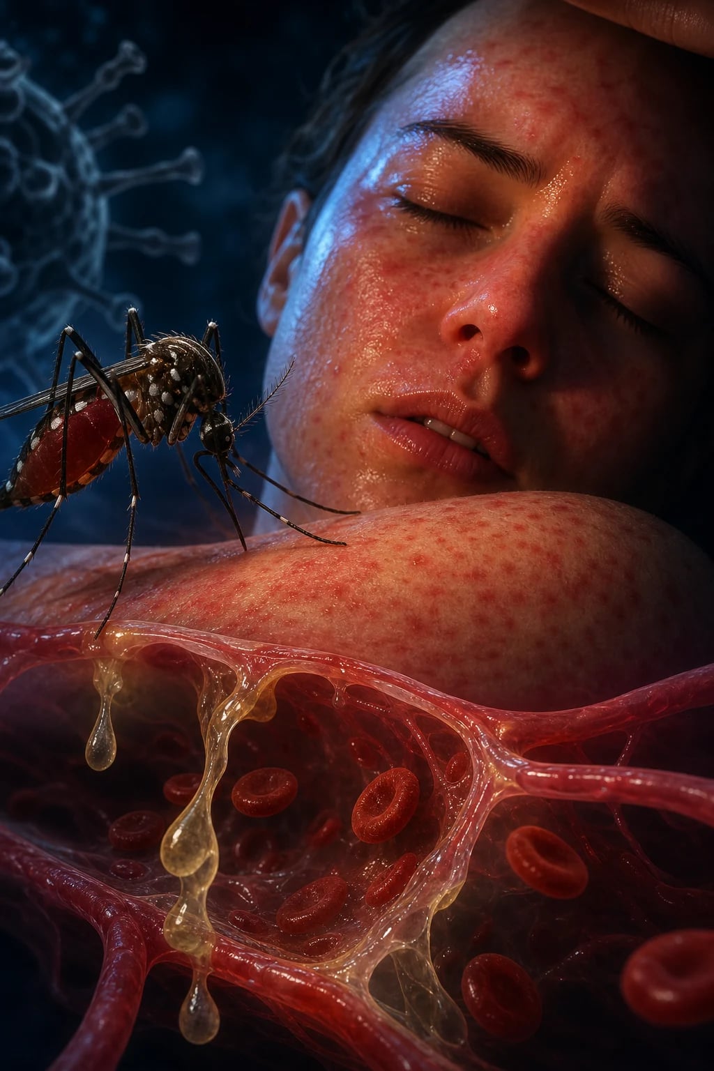
A note on naming and history: 'dengue' is thought to derive from the Swahili ka-dinga pepo (a sudden cramp-like seizure, attributed to evil spirits); the Spanish adapted dinga to dengue ('fastidious' or 'careful' — describing the gait of sufferers with severe bone pain). It is also called break-bone fever and dandy fever. The biphasic (saddle-back) fever pattern is classical but not invariable. [1]
Classification
Two classification systems coexist in dengue, both examinable. The WHO 1997 scheme (traditional, still taught and used in India and many exams) divides disease into DF, DHF (grades I-IV) and DSS; the WHO 2009 scheme (revised, level-of-severity-based, used for triage and management) divides it into dengue (with or without warning signs) and severe dengue. Knowing both is essential. [1]
WHO 1997 classification — DF, DHF and DSS (still the Indian exam standard)[1]
Dengue fever (DF) — acute febrile illness with two or more of: headache, retro-orbital pain, myalgia, arthralgia, rash, haemorrhagic manifestation (petechiae, positive tourniquet test), leucopenia — in the absence of plasma leakage. Usually self-limiting. [1]
Dengue haemorrhagic fever (DHF) — requires all four of:
- Fever (acute, high, 2-7 days).
- Haemorrhagic tendency — positive tourniquet test, OR spontaneous petechiae/purpura/ecchymoses, OR mucosal/GI bleeding, OR haematemesis/melaena.
- Thrombocytopenia — platelet count under 100 × 10⁹/L.
- Plasma leakage — haematocrit rise of 20% or more above baseline (or population mean for age/sex), OR a 20% or greater fall in haematocrit after fluids, OR clinical evidence of leakage (ascites, pleural effusion — classically right-sided, hypoproteinaemia). [1]
DHF is graded I-IV by the presence and severity of shock: [1]
| Grade | Definition |
|---|---|
| I | Fever + non-specific symptoms, positive tourniquet test only |
| II | Grade I + spontaneous bleeding (skin, gums, nose, GI) |
| III | Circulatory failure — weak, rapid pulse, narrow pulse pressure (under 20 mmHg), hypotension, cold/clammy skin, restlessness |
| IV | Profound shock — pulse and blood pressure undetectable |
Grades III and IV = dengue shock syndrome (DSS). The grading is high-yield.[1]
WHO 2009 classification — for triage and management[1][1]
The 2009 revision simplified classification into two clinical levels to aid triage and decision-making: [1]
- Dengue (without warning signs) — fever (usually 38°C or above, 2-7 days) plus two of: nausea/vomiting, rash, aches/pains, tourniquet test positive, leucopenia, AND no warning signs and no plasma leakage/organ impairment.
- Dengue with warning signs — fever as above plus any warning sign (see Clinical Presentation): abdominal pain/persistent vomiting, fluid accumulation, mucosal bleeding, lethargy, liver enlargement over 2 cm, rising haematocrit with falling platelets.
- Severe dengue — defined by any one of: (i) plasma leakage leading to shock (DSS) or fluid accumulation with respiratory distress; (ii) severe bleeding (evaluated by clinician); (iii) severe organ impairment (liver — AST/ALT over 1000; CNS — impaired consciousness; heart; kidney; other). [1]
The 2009 scheme is preferred at the bedside and by WHO 2012; many national programmes (including India's NVBDCP) still report using 1997 categories. Learn both. [1]
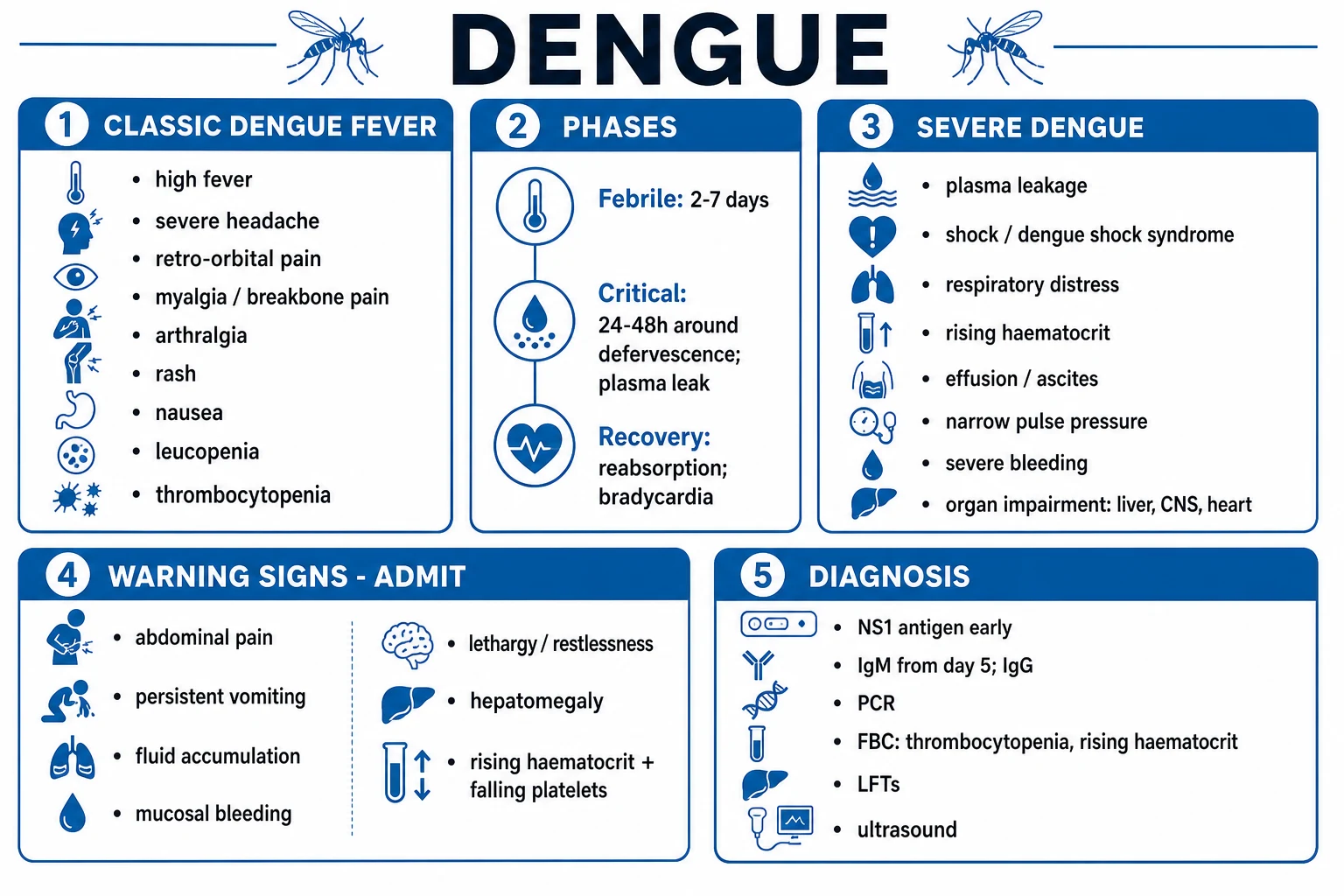
Classic dengue fever (DF)
Self-limiting, no leakage
- High fever 2-7 days, retro-orbital headache, myalgia, 'break-bone' pain, rash
- Thrombocytopenia common but NO haemoconcentration / plasma leakage
- Positive tourniquet test and scattered petechiae may occur
- Recovers spontaneously; management is symptomatic (oral fluids, paracetamol)
Dengue haemorrhagic fever (DHF)
Plasma leakage + thrombocytopenia + bleeding tendency
- All 4 WHO 1997 criteria: fever + bleeding tendency + platelets under 100k + plasma leakage (Hct rises 20% or effusion/ascites)
- Critical phase = 24-48 h around defervescence, when leakage peaks
- Right-sided pleural effusion, ascites, gallbladder wall thickening on ultrasound
- Needs IV isotonic crystalloid titrated to Hct and urine output (WHO Group B)
Dengue shock syndrome (DSS)
DHF grades III-IV — hypovolaemic shock from leakage
- Narrow pulse pressure under 20 mmHg (Grade III); undetectable BP (Grade IV)
- Cold clammy skin, prolonged capillary refill, tachycardia, oliguria
- Immediate isotonic crystalloid bolus 10-20 mL/kg; ICU (WHO Group C)
- Untreated mortality 10-20%; treated under 1%
Expanded dengue syndrome
Atypical severe organ involvement
- Severe hepatitis (AST/ALT over 1000), acute liver failure
- Encephalitis / encephalopathy, Guillain-Barre, transverse myelitis
- Myocarditis, acute kidney injury, ARDS, haemophagocytic lymphohistiocytosis (HLH)
- Managed with organ support; steroids controversial for HLH
Epidemiology & Risk Factors
Dengue is a disease of the tropics and subtropics, expanding its geographic reach with urbanisation, climate change, global travel and the spread of Aedes aegypti. WHO estimates approximately 390 million infections a year, of which ~96 million are clinically apparent, and roughly 3.9 billion people in 129 countries are at risk. The heaviest burden is in South and Southeast Asia (India, Indonesia, Vietnam, Philippines, Thailand, Bangladesh, Sri Lanka), the Americas (Brazil, Mexico, the Caribbean), the Western Pacific and sub-Saharan Africa.[1]
Dengue — key numbers
Vector biology and transmission (heavily examinable): [1]
- Aedes aegypti is a small dark mosquito with white lyre-shaped markings on the thorax and white bands on the legs. It is day-biting (peaks early morning and late afternoon), peri-domestic, anthropophilic (prefers human blood), and breeds in small collections of clean standing water — flowerpots, discarded tyres, coconut shells, water-storage containers, roof gutters, AC trays. It is a nervous, low-flying mosquito that bites mainly below the knee.[1]
- Aedes albopictus (the Asian tiger mosquito) is a secondary vector, more rural/sylvatic and cold-tolerant, and has spread into temperate regions (southern Europe, North America).
- Transmission cycle: human → mosquito → human. There is no animal reservoir for the urban cycle (a sylvatic monkey-mosquito cycle exists in Southeast Asia but does not sustain urban transmission). The mosquito acquires infection by biting a viraemic human (viraemia lasts roughly 2-12 days after symptom onset, peaking around day 3); after an extrinsic incubation period of 8-12 days, the mosquito is infectious for the rest of its life. Transovarial transmission in the mosquito sustains the virus between epidemics.
- Vertical (maternal-fetal) transmission can occur, particularly in primary maternal infection in late pregnancy. Blood transfusion, organ transplant and needle-stick transmission have been reported but are rare.
Risk factors for severe dengue (DHF/DSS):[1][2]
- Secondary (heterologous) infection — the single most important host risk factor; ADE doubles to quintuples the risk of severe disease.
- Young age — infants (born to dengue-immune mothers) and children aged 5-15 years have the highest DHF rates in hyperendemic regions.
- Viral serotype / genotype — DENV-2 and DENV-3, particularly Asian genotypes, are more virulent than DENV-1 or DENV-4.
- Host genetics — HLA class I and II alleles, polymorphisms in DC-SIGN, Fc-gamma receptor, vitamin D receptor and tumour necrosis factor genes modify severity.
- Female sex, high body mass index, comorbidity (diabetes, hypertension, asthma, sickle-cell trait, chronic kidney disease), pregnancy, infants under 1 year with maternally derived antibody.
- Climate — the monsoon season (June-November in India) drives case surges through vector-breeding expansion. [1]
Endemicity patterns:
- Hyperendemic regions (Southeast Asia, the Caribbean, parts of South America) — all four serotypes co-circulate; secondary infections and DHF are common.
- India — all four serotypes circulate; DENV-1 and DENV-2 predominate in most outbreaks. Reported cases have multiplied roughly 25-fold over the last two decades; dengue is now endemic to all states and union territories with annual monsoon-driven outbreaks.[1]
Pathophysiology
Dengue pathogenesis has three intertwined arms: viral entry and replication, the host immune response (cytokine storm), and the antibody-dependent enhancement phenomenon that explains why second infections are dangerous. The end-result — capillary endothelial dysfunction with plasma leakage — defines DHF/DSS. [1]
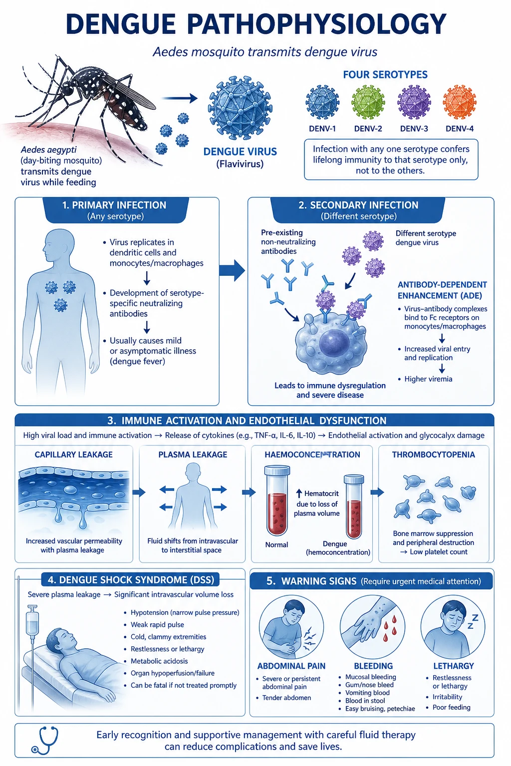
The cascade in molecular detail:[1][2][3]
- Inoculation. An infective A. aegypti female probes the dermis and inoculates virus-laden saliva with anticoagulant into the capillary plexus.
- Skin replication and primary viraemia. Dermal Langerhans cells and dendritic cells express DC-SIGN (CD209), the principal attachment receptor. The virus replicates; infected cells mature and migrate to draining lymph nodes, then spill into the blood as primary viraemia peaking at roughly day 2-4 of fever. Other receptors include heparan sulphate, mannose receptor and the phosphatidylserine receptors TIM/TAM (which 'eat-me' signal-recognise apoptotic-cell-mimicking virions).
- Systemic dissemination. Virus seeds monocytes, macrophages, liver Kupffer cells and hepatocytes, splenic and bone-marrow macrophages. Hepatocyte infection drives transaminitis (AST higher than ALT). Bone-marrow infection causes transient megakaryocyte suppression, contributing to thrombocytopenia.
- Antibody-dependent enhancement (ADE) — the central concept. Infection with one serotype induces lifelong serotype-specific neutralising antibody and transient cross-reactive, non-neutralising antibody against other serotypes. When, months to years later, the person is infected with a different serotype, the cross-reactive IgG binds the virion but fails to neutralise it; the virus-IgG complex is taken up by Fc-gamma receptors on monocytes/macrophages, which internalise and replicate virus far more efficiently than naive cells. The result is a 10- to 100-fold higher viral load, a heavier cytokine storm and a markedly greater risk of DHF/DSS. This is why secondary dengue is more severe than primary dengue, why infants born to dengue-immune mothers develop severe primary dengue (maternal antibody wanes into the enhancing range at 6-9 months), and why the first Dengvaxia dose can harm seronegative vaccinees (vaccine behaves like a silent primary infection, setting up ADE on later wild-type infection).[2][4]
- Cytokine storm. Infected and activated monocytes/macrophages release TNF-α, IL-1β, IL-6, IL-8, IL-10, IL-12, IL-18, IFN-γ, MCP-1, RANTES and VEGF; complement activation releases C3a and C5a; mast cells release tryptase, chymase and TNF-α. The combined effect is capillary endothelial dysfunction: gaps open between endothelial cells, albumin and plasma leak into the interstitium and serous cavities (peritoneum, pleura — usually right-sided because the right hemidiaphragm is lower and the lymphatic drainage favours the right pleural space). Plasma leakage, not haemorrhage, is the dominant lesion of DHF/DSS.[2]
- Thrombocytopenia and bleeding. Multifactorial: bone-marrow megakaryocyte suppression, peripheral immune destruction (anti-platelet antibodies, complement), platelet activation and consumption, and splenic sequestration. Platelet dysfunction (impaired aggregation) plus mild disseminated intravascular coagulation (prolonged APTT, low fibrinogen, raised D-dimer) and hepatic synthetic compromise produce the haemorrhagic tendency. Severe bleeding is more often a late (recovery-phase) event driven by prolonged shock and DIC than the cause of shock itself.
- The critical phase. Around the time of defervescence (day 3-7), the leaking patient reaches the critical phase, lasting 24-48 hours. Haematocrit climbs as plasma leaves the vasculature; if leakage is severe and unchecked, hypovolaemic shock (DSS) supervenes. The same cytokine milieu can rarely drive organ impairment (hepatitis, encephalopathy, myocarditis, AKI, ARDS) — expanded dengue syndrome.
- Recovery. After 48-72 hours, the endothelium recovers, leaked fluid is reabsorbed (which can cause transient haemodilution and pulmonary oedema if IV fluids were over-given), a diuresis appears, the platelet count recovers, and the characteristic convalescent rash (confluent petechiae with round white islands of normal skin — 'isles of white in a sea of red') appears, often with bradycardia and pruritus.[3]
Clinical Presentation
Dengue is a phasic illness. Recognising which phase the patient is in — and where in that phase the danger lies — is the central bedside skill. [1]
Febrile phase (days 1-7)
High fever, viraemia, symptoms
- Abrupt-onset high fever (39-40°C) often with a saddle-back/biphasic pattern
- Bifrontal and retro-orbital headache; severe myalgia and arthralgia ('break-bone fever')
- Nausea, vomiting, anorexia; flushed face, injected conjunctivae and pharynx
- Maculopapular or petechial rash; mild bleeding (epistaxis, gum, menorrhagia); positive tourniquet test
- NS1 antigen and PCR are positive (viraemia)
Critical phase (24-48 h around defervescence)
Plasma leakage peaks; shock risk
- Fever drops — but the patient looks WORSE, not better (the paradox)
- Abdominal pain, persistent vomiting, lethargy, restlessness (warning signs)
- Rising haematocrit (plasma leak) with rapidly falling platelets
- Pleural effusion (right-sided), ascites, gallbladder wall thickening on ultrasound
- Narrow pulse pressure under 20 mmHg = DSS; needs immediate fluid bolus
Recovery phase (48-72 h)
Reabsorption, convalescence
- General wellbeing, appetite and urine output improve; diuresis
- Bradycardia (recovery-phase bradycardia); pruritus
- Platelet count recovers; haematocrit falls back toward baseline
- Convalescent rash: confluent petechiae with round white islands of sparing
- Beware fluid overload from continuing IV fluids into this phase
Febrile phase (days 1-7)
The illness begins abruptly with high fever (often 39-40°C), accompanied by two or more of the following (the WHO clinical case definition):[1]
- Headache — typically bifrontal.
- Retro-orbital pain — pain behind the eyes, worsened by eye movement; a classical feature.
- Myalgia and arthralgia — severe, often described as 'break-bone' pain; back pain and muscle pain are prominent.
- Rash — occurs in up to 50-80%. The early rash is flushing/erythema of the face, neck and chest ('dengue facies'); a maculopapular or morbilliform rash may then appear on the trunk and extensor surfaces, sometimes islands of sparing. A petechial eruption and a positive tourniquet test indicate the bleeding tendency.
- Nausea, vomiting, anorexia, altered taste.
- Mild bleeding — petechiae, easy bruising, epistaxis, gum bleeding, menorrhagia, haematuria; melaena/haematemesis signal more severe disease.
- Injected conjunctivae and pharyngeal injection, cervical lymphadenopathy, periorbital oedema, and a relative bradycardia may be noted.
- Hepatomegaly — present in many children; tender hepatomegaly suggests progression. [1]
Fever typically lasts 2-7 days. A biphasic/saddle-back course is classical (fever settling on day 3-5, recurring) but is not universally present.[1]
Critical phase — defervescence and the warning signs
The transition to the critical phase occurs at defervescence (day 3-7), when the fever drops. The paradoxical sign of severe dengue is that the patient deteriorates as the fever settles: this is the leakage window. The WHO warning signs (2009) indicate impending severe disease and mandate admission with IV fluid therapy:[1][1]
WHO dengue warning signs
WARNING
Often periumbilical or right-upper-quadrant; a key warning sign
Vomiting is the single most reliable warning sign in children
Behavioural change, drowsiness — impending encephalopathy/shock
Pulse pressure under 20 mmHg = DSS (Grade III)
Rising Hct + falling platelets = plasma leakage in progress
Gum, nose, GI, haematuria, menorrhagia
Ultrasound and bedside exam findings of leakage
Clinical shock (DSS) — once plasma loss is severe enough, the patient develops cold clammy skin, prolonged capillary refill, weak rapid pulse, narrow pulse pressure (under 20 mmHg), hypotension (a late sign), oliguria, restlessness progressing to lethargy and impaired consciousness. Grade IV = undetectable pulse and BP. Hypotension is a late, pre-arrest sign in dengue — assess perfusion and pulse pressure, not pressure alone.[1]
Recovery phase (48-72 hours)
With endothelial recovery, leaked plasma is reabsorbed, appetite returns, urine output rises (diuresis), the platelet count recovers and haematocrit falls. A convalescent rash (confluent petechiae with round white islands of normal skin) is characteristic, often with generalised pruritus and sinus bradycardia. Beware continued IV fluids — the patient who was leaking is now reabsorbing, and fluid overload with pulmonary oedema is an iatrogenic danger. [1]
Atypical presentations (deliberately tested)
- Infants and young children — non-specific febrile illness, febrile seizures, refusal of feeds, vomiting; severe primary dengue in infants of immune mothers (maternal antibody wanes into the enhancing range at 6-9 months).
- Elderly — blunted fever, comorbidity-driven severity (diabetes, hypertension, CKD), higher rates of severe bleeding, AKI, encephalopathy and mortality; shock may be masked by beta-blocker or diuretic therapy.
- Pregnant women — higher risk of severe dengue, miscarriage, preterm labour, fetal distress and vertical transmission; close fetal monitoring is essential.
- Diabetics, hypertensives, those with sickle-cell trait, asthma, CKD, obesity — more severe course.
- Primary infection in adults may present with severe hepatitis, encephalitis, myocarditis or AKI without classical warning signs (expanded dengue syndrome).
- "Feeling better on day 1 but worse on day 5" — the patient who improves then deteriorates is in the critical phase; do not discharge on fever pattern alone.[2]
Differential Diagnosis
Dengue sits at the centre of the acute undifferentiated febrile illness of the tropics — a syndrome shared with malaria, chikungunya, leptospirosis, typhoid, scrub typhus, influenza and a host of others. Always exclude malaria first (a film or antigen test is rapid and changes management), and exclude bacteraemia / meningococcaemia in the toxic patient. [1]
Falciparum malaria
- Abrupt high fever with rigors, periodicity; splenomegaly, jaundice, anaemia
- Travel to malaria zone; thick/thin film or malaria antigen
- Thrombocytopenia common — does NOT exclude dengue; the two can coexist
- Exclude FIRST: a missed falciparum can kill in hours
Chikungunya
- Same Aedes vector; fever with SEVERE symmetrical polyarthralgia out of proportion (small joints, debilitating, may persist months)
- Maculopapular rash, lymphadenopathy; thrombocytopenia milder than dengue
- IgM anti-CHIK; clinically the arthritis is the give-away
- Often co-epidemic with dengue in monsoon
Zika virus
- Milder fever, conjunctivitis, maculopapular rash, arthralgia
- Risk in pregnancy (microcephaly, congenital Zika syndrome) and Guillain-Barre
- RT-PCR (early) or IgM; same Aedes vector
- Mild, self-limiting in non-pregnant adults
Leptospirosis
- Conjunctival suffusion, severe myalgia (calves), jaundice, renal failure (Weil disease)
- Contaminated water or farm/animal exposure; doxycycline/penicillin
- Thrombocytopenia + AKI + jaundice triad
- Differentiate by exposure history and conjunctival suffusion
Enteric (typhoid) fever
- Step-ladder rising fever, relative bradycardia, rose spots, splenomegaly, coated tongue
- Constipation then 'pea-soup' diarrhoea; no myalgia/rash of dengue
- Blood culture gold standard; Widal unreliable alone
- NS1/IgM negative in typhoid
Influenza / viral respiratory illness
- Abrupt fever, cough, sore throat, myalgia, headache
- Respiratory symptoms dominate; leucopenia can mimic dengue
- PCR/antigen on nasopharyngeal swab
- Thrombocytopenia mild or absent; rash uncommon
Scrub typhus / rickettsial fever
- Eschar at bite site, regional lymphadenopathy, maculopapular rash
- Rural/agricultural exposure; doxycycline response often dramatic
- Weil-Felix or IgM IFA
- Thrombocytopenia and transaminitis overlap with dengue
Meningococcaemia / bacterial sepsis
- Rapidly progressive petechial/purpuric rash with shock
- High fever, toxic appearance; DIC and multi-organ failure
- Blood cultures; urgent IV antibiotics (meningococcus can kill in hours)
- Any petechial rash with shock: treat as sepsis until proven otherwise
Measles
- Prodrome of fever, coryza, cough, conjunctivitis (3 Cs); Koplik spots
- Maculopapular rash spreads head-to-toe; unvaccinated child
- Thrombocytopenia unusual; leucopenia can occur
- IgM anti-measles
Other / non-infectious
- Acute leukaemia / aplastic anaemia: pancytopenia with blast cells
- ITP / TTP / HUS: isolated low platelets or microangiopathy
- Drug reaction with eosinophilia (DRESS); Kawasaki disease (children)
- Viral haemorrhagic fevers (yellow fever, hantavirus, Lassa, Ebola) if exposure fits
The exam trap: the two diseases that look most like dengue are malaria and chikungunya, and all three are mosquito-borne. A returned traveller with fever, headache, myalgia and thrombocytopenia should have malaria excluded first (film/antigen), then dengue (NS1/IgM) and chikungunya (IgM) tested. The two can coexist — a mixed infection is more severe. [1]
Clinical & Bedside Assessment
The bedside assessment in suspected dengue rests on vital signs and perfusion (especially pulse pressure), a focused fluid-balance and abdominal exam, and the tourniquet test — none of which requires a laboratory. [1]
History. Establish the tempo (abrupt fever, day of illness — central to interpretation of NS1/IgM), residence/travel to a dengue-endemic area, monsoon/exposure to Aedes breeding sites, prior dengue infection (raises ADE risk), comorbidity (diabetes, hypertension, CKD, asthma, sickle-cell, pregnancy, immunosuppression), and any NSAID/aspirin/anticoagulant use (bleeding risk). Ask specifically about warning signs: abdominal pain, persistent vomiting, bleeding, lethargy, restlessness, oliguria, dizziness on standing.[1]
Vital signs and perfusion — every 1-4 hours in the critical phase:
- Temperature — dropping fever with worsening patient = critical phase.
- Pulse rate and pulse pressure — pulse pressure under 20 mmHg (e.g. 100/80) defines DSS Grade III; undetectable BP = Grade IV.
- Blood pressure — a late sign; shock is present before hypotension in dengue.
- Capillary refill time, peripheral temperature, urine output — markers of perfusion.
- Respiratory rate — rising RR may signal pleural effusion, pulmonary oedema, or metabolic acidosis of shock. [1]
Tourniquet test (Hess test). Inflate a sphygmomanometer cuff to a pressure midway between systolic and diastolic, maintain for 5 minutes, then count petechiae that appear distal to the cuff in a 2.5 × 2.5 cm square on the volar forearm. Ten or more petechiae = positive. It is positive in roughly one-third to half of DHF cases, and supports (but does not confirm) the diagnosis — specificity is low (any thrombocytopenic or vasculitic state can be positive).[1]
Abdomen. Look for hepatomegaly (often tender, with mild transaminitis), right-upper-quadrant tenderness, ascites (shifting dullness, fluid thrill) and bowel-bladder pattern — abdominal pain is a warning sign. Gallbladder wall thickening is best seen on ultrasound but tender hepatomegaly and a positive Murphy sign (without stones) point to it. [1]
Skin. Examine for petechiae, ecchymoses, purpura, rash, mucosal bleeding (gingival, nasal, conjunctival), and bleeding from venepuncture sites (a marker of significant coagulopathy). [1]
Other systems. Assess for pleural effusion (dullness, reduced breath sounds, typically right-sided), encephalopathy (altered consciousness, irritability, seizures — rare but serious), dehydration, and shock (cool peripheries, weak pulse, prolonged refill, oliguria). [1]
Monitoring. In the admitted patient during the critical phase, monitor hourly pulse, BP, pulse pressure, capillary refill, urine output; 4-6 hourly haematocrit and platelets; daily ultrasound for effusion/ascites if available. Rising haematocrit with falling platelets is the bedside signature of plasma leakage. [1]
Investigations
There is no single perfect test. The choice depends on the day of illness, because viraemia, antigenaemia and antibody responses follow a predictable time course. [1]
Diagnostic tests (virology/serology)
NS1 antigen ELISA/RDT
Days 1-5 (best in first 72 h)
- Detects viral non-structural protein 1 in blood
- Sensitivity 70-90% in first 5 days of fever; falls after day 5
- High specificity (~95-100%) — a positive NS1 within 5 days confirms dengue
- Detects ALL four serotypes; doesn't distinguish them
IgM anti-dengue ELISA
After day 5
- IgM appears from day 4-5, peaks at week 2, persists ~2-3 months
- Sensitivity 60-90% after day 5; before day 5 it is unreliable
- A positive IgM with high clinical suspicion = recent dengue
- Cross-reacts with other flaviviruses (yellow fever vaccine, JE, Zika)
IgG anti-dengue ELISA
Primary vs secondary
- IgG appears by day 7-10 in primary; SECONDARY shows HIGH IgG from day 2-3
- High IgG early + IgM/IgG ratio under 1.2 = secondary infection
- A four-fold rise in paired sera confirms infection (gold standard but slow)
- Persists for life — single positive does not prove current infection
RT-PCR for DENV RNA
Days 1-5; serotyping
- Detects viral RNA; very high specificity (~100%)
- Sensitivity high only in first 5 days (during viraemia)
- Allows SEROTYPING (DENV-1 to 4) — useful for surveillance
- Costly; mainly reference-lab use
Viral isolation / culture
Research / surveillance
- Mosquito cell line (C6/36) or mammalian (Vero) culture
- Sensitive in first 5 days; slow (1-2 weeks)
- Not used clinically; gold standard for serotyping in research
- Replaced by RT-PCR for routine typing
Combined NS1 + IgM/IgG
Highest single-sample yield
- NS1 (early) + IgM (late) covers most of the illness
- Recommended single-sample approach in many labs
- Improves diagnostic yield across the febrile-to-convalescent window
- A point-of-care combined RDT is widely used in India
Timing summary (heavily examinable):[1][1]
| Day of fever | Best test |
|---|---|
| Days 1-5 | NS1 antigen (sensitivity 70-90%) or RT-PCR (gold standard if available) |
| Days 5-10 | IgM ELISA (rising sensitivity as NS1 falls) |
| After day 10 | IgM (still positive) or paired IgG titre (four-fold rise) |
A practical rule: in the first week, send NS1 + FBC; in the second week, send IgM + FBC. Combine NS1 + IgM when the timing is uncertain. [1]
Supportive bloods and imaging
- Full blood count — thrombocytopenia (platelet count under 100 × 10⁹/L; often under 50 in DHF, sometimes under 20) is the cardinal finding. Leucopenia (WCC under 5 × 10⁹/L) with a relative lymphocytosis and atypical lymphocytes is common early. Rising haematocrit by 20% above baseline (or population mean) indicates plasma leakage and is the WHO 1997 criterion for DHF.
- Coagulation — prolonged APTT, low fibrinogen, raised D-dimer (mild DIC) in severe disease; PT often normal or mildly prolonged.
- LFTs — AST and ALT raised (AST more than ALT, the reverse of viral hepatitis), sometimes above 1000 IU/L in severe hepatitis; low albumin reflects leakage.
- U&E, creatinine — acute kidney injury in severe disease; hyponatraemia common (SIADH + third-spacing).
- Lactate — raised in shock.
- Blood glucose — hypoglycaemia in children and severe disease.
- Chest X-ray — right-sided pleural effusion is the radiological hallmark of plasma leakage; bilateral in severe cases. Look for it in the erect PA film.
- Ultrasound abdomen/thorax — gallbladder wall thickening (over 3-5 mm) is highly suggestive of dengue; ascites, right pleural effusion, peri-renal fluid. May be the earliest sign of leakage, preceding haemoconcentration.[3]
- Blood cultures, malaria film — to exclude bacterial sepsis and malaria (always exclude malaria first in any tropical fever).[3]
Criteria reproduced verbatim — WHO 1997 DHF criteria
A diagnosis of DHF requires all four of the following:[1]
- Fever — acute, high, biphasic, lasting 2-7 days.
- Haemorrhagic manifestations — at least one of: positive tourniquet test, petechiae, ecchymoses/purpura, epistaxis, gum bleeding, haematemesis, melaena, haematuria; plus a positive tourniquet test alone counts if other features are absent.
- Thrombocytopenia — platelet count under 100 × 10⁹/L.
- Plasma leakage — haematocrit rise of 20% or more above baseline (or above population mean for age/sex), OR a 20% or greater fall in haematocrit after fluid therapy, OR clinical/biochemical evidence of leakage (ascites, pleural effusion, hypoproteinaemia or hypoalbuminaemia). [1]
DHF grading (and the threshold for DSS):[1]
- Grade I: fever + non-specific symptoms, the ONLY haemorrhagic manifestation being a positive tourniquet test.
- Grade II: grade I signs plus spontaneous bleeding (skin, mucosa, GI tract).
- Grade III: circulatory failure — weak, rapid pulse, narrow pulse pressure (under 20 mmHg), hypotension, cold/clammy skin, restlessness.
- Grade IV: profound shock — pulse and blood pressure undetectable. [1]
Grades III and IV = dengue shock syndrome (DSS). The pulse pressure threshold of 20 mmHg is the single most tested number in dengue. [1]
WHO 2009 severity criteria (severe dengue)
Severe dengue is defined by any one of:[1][1]
- Severe plasma leakage leading to shock (DSS) or fluid accumulation with respiratory distress.
- Severe bleeding (as evaluated by a clinician) — e.g. haematemesis, melaena, intracranial bleed.
- Severe organ impairment: hepatic (AST/ALT over 1000 IU/L), CNS (impaired consciousness), cardiac (myocarditis), renal (AKI), or other (e.g. ARDS). [1]
Management — Resuscitation
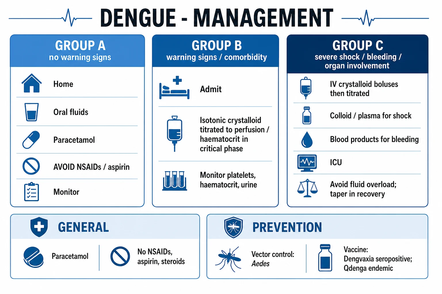
Dengue has no specific antiviral therapy. The entire treatment is fluid titration — the central life-saving intervention, and the one that distinguishes treated (mortality under 1%) from untreated (10-20%) DSS. [1]
ABCDE. Secure the airway, give oxygen to maintain SpO₂ at 94-98%, establish IV access, and identify the WHO triage group (A, B or C).[1][1]
Surviving sepsis parallel. In suspected/established DSS, take blood cultures, send lactate, and begin fluid resuscitation immediately — but not with broad-spectrum antibiotics as routine (they are reserved for proven or strongly suspected bacterial co-infection, which is more common in severe dengue than historically appreciated).[1]
Fluid resuscitation in shock (WHO Group C): [1]
- Isotonic crystalloid is first-line — 0.9% sodium chloride or Ringer's lactate / Hartmann's solution (Ringer's lactate is preferred by WHO 2012).
- Bolus 10-20 mL/kg over 1 hour (faster — over 15-30 minutes — in profound Grade IV shock), then reassess: pulse pressure, capillary refill, urine output, conscious level, haematocrit.
- Repeat bolus (10-20 mL/kg) if shock persists, up to 60 mL/kg in the first few hours.
- Switch to colloid (e.g. 6% dextran 70, hydroxyethyl starch, or 4-5% human albumin) for refractory shock or if haematocrit fails to fall after crystalloid. Colloids are reserved for the most unstable.
- Maintenance fluid as isotonic crystalloid once perfusion is restored; carefully titrate the rate down as the patient stabilises to avoid fluid overload.
- Transfuse for major bleeding (whole blood or packed red cells; fresh frozen plasma and platelets as guided by coagulation and bleeding severity). Blood is the volume expander of choice in major haemorrhage — crystalloid alone cannot replace lost red cell mass.
- Avoid prophylactic platelet transfusions for low platelet counts alone (see Definitive Management).[1]
Antipyretics and analgesia. Paracetamol 10-15 mg/kg/dose every 4-6 hours (max 60 mg/kg/day in children, 4 g/day in adults) is the ONLY recommended antipyretic-analgesic. Strictly AVOID NSAIDs (ibuprofen, diclofenac, aspirin, naproxen, mefenamic acid) and intramuscular injections — they worsen bleeding risk. Tepid sponging for refractory fever.[1][1]
Treat hypoglycaemia, correct electrolytes, and monitor closely — hourly observations in the critical phase.[1]
Management — Definitive & Stepwise
WHO 2009/2012 organises management by triage group (A, B, C), decided at presentation by the presence of warning signs and severity. The principle: fluid volume and route are titrated to the phase of illness and degree of leakage. [1]
WHO Group A — outpatient management (mild, no warning signs)[1]
- Encourage oral fluids — water, ORS, fruit juice, soup; aim for normal urine output (≥1 mL/kg/h).
- Paracetamol 10-15 mg/kg/dose every 4-6 hours for fever/pain. No NSAIDs, no aspirin, no IM injections.
- Daily review for warning signs, platelet and haematocrit trend.
- Safety-net: return immediately if any warning sign appears — abdominal pain, persistent vomiting, bleeding, lethargy, restlessness, no urine for 4-6 hours, cold hands/feet, giddiness on standing. [1]
WHO Group B — ward management (warning signs, plasma leakage without shock)[1][1]
- Admit. Establish IV access, monitor 4-6 hourly in the febrile phase and 1-2 hourly in the critical phase.
- Isotonic crystalloid — 0.9% saline, Ringer's lactate or Hartmann's — at a maintenance-plus-leakage rate. A practical starting regimen is 5-7 mL/kg/hour for the first 2-4 hours, then 3-5 mL/kg/hour, then taper to 2-3 mL/kg/hour as haematocrit stabilises. Titrate to clinical status, urine output (0.5-1.5 mL/kg/h) and haematocrit (target: stable or falling without shock).
- Reduce the rate as soon as the haematocrit falls and the patient stabilises — to avoid the fluid overload that causes pulmonary oedema in the recovery phase.
- Oxygen if SpO₂ under 94%; transfuse for major bleeding (not for low platelets alone).
- Daily FBC, LFTs, U&E; ultrasound for effusion/ascites. [1]
WHO Group C — ICU management (severe dengue, DSS)[1][1]
Compensated shock (Grade III)
Narrow pulse pressure under 20 mmHg
- Isotonic crystalloid 10-20 mL/kg over 1 h; reassess
- If improving: continue crystalloid at 5-10 mL/kg/h, titrate down
- If not improving: repeat bolus, then consider colloid
- Target: pulse pressure over 20, capillary refill under 2 s, urine output over 0.5 mL/kg/h
Hypotensive shock (Grade IV)
Undetectable BP/pulse
- Colloid 10-20 mL/kg FAST (over 15-30 min); 6% dextran, HES or 4-5% albumin
- Reassess; repeat if needed; switch to crystalloid maintenance once stable
- Match fluid type to haematocrit: high Hct = colloid; falling Hct with shock = occult bleed → blood
- ICU monitoring; treat fluid overload aggressively with furosemide once stable
Major bleeding
GI, intracranial, massive
- Transfuse WHOLE BLOOD or packed red cells — crystalloid/colloid cannot replace RBCs
- Platelet transfusion (1 adult dose = ~4-6 units) ONLY if active bleeding with thrombocytopenia
- Fresh frozen plasma if prolonged APTT/DIC; cryoprecipitate if low fibrinogen
- Look for and treat occult GI bleed; stop NSAIDs/aspirin; protect airway if haematemesis
Fluid overload
Iatrogenic, common pitfall
- Pulmonary oedema, pleural effusion, respiratory distress, gallop
- Reduce/stop IV fluids immediately
- IV furosemide 20-40 mg (1 mg/kg in children) once stable
- Oxygen; consider non-invasive ventilation; rare intubation
Severe organ impairment
Expanded dengue syndrome
- Severe hepatitis/failure: hepatology, lactulose, N-acetylcysteine
- Encephalopathy: treat seizures, control ICP; exclude other causes
- Myocarditis: inotropes, careful fluids; ARDS: lung-protective ventilation
- AKI: nephrology; renal replacement therapy if needed
Platelet transfusion — when (and when NOT) to give
Prophylactic platelet transfusion for thrombocytopenia alone is NOT recommended. It does not prevent bleeding, can cause transfusion reactions and fluid overload, and delays natural recovery. Indications for platelet transfusion (and to manage an active bleed):[1][1]
- Active major bleeding (haematemesis, melaena, intracranial, massive epistaxis, post-partum) — give 1 adult therapeutic dose of platelets (~4-6 units), repeated as needed.
- Platelet count under 10 × 10⁹/L — high spontaneous bleed risk; transfuse.
- Platelet count 10-20 × 10⁹/L with risk factors for bleeding (fever, agitation, comorbidity, concomitant anticoagulation, severe liver disease, impending surgery) — transfuse.
- Platelet count under 20-30 × 10⁹/L in a patient with shock or ongoing leakage — transfuse.
- Above 20-30 × 10⁹/L without bleeding — do NOT transfuse; observe. [1]
Drugs to AVOID (the high-yield contraindication list)
- NSAIDs (ibuprofen, diclofenac, mefenamic acid, naproxen, aspirin) — platelet inhibition + gastric irritation → severe bleeding. The single most tested 'do not' in dengue.
- Intramuscular injections — risk of haematoma; route all medication IV or oral.
- Corticosteroids — not recommended routinely; some studies of high-dose methylprednisolone in DSS showed no mortality benefit. May be considered for haemophagocytic lymphohistiocytosis or refractory shock on a case-by-case basis.
- Prophylactic platelet transfusions (above).
- Carbaspirin calcium-containing 'aspirin-combination' antipyretics — read labels in India. [1]
In India (NVBDCP / ICMR), the monsoon-driven epidemic season dominates caseload; NVBDCP recommends NS1 + IgM combination RDT at the periphery, paracetamol only, strict avoidance of NSAIDs/aspirin, no platelet transfusion without a clear indication, and isotonic crystalloid as the fluid of choice for leakage. Vector control rests on source reduction (remove stagnant water), larviciding, adulticiding (thermal fogging during outbreaks), and personal protection (full-sleeved clothing, repellents). A live-attenuated tetravalent vaccine is under Indian development.[1]
Discharge criteria
Discharge when the patient is clinically well, afebrile for 24-48 hours without antipyretics, haemodynamically stable, passing urine normally, platelet count recovering, haematocrit stable/baseline, tolerating oral fluids, no respiratory distress from pleural effusion, with no warning signs. Counsel on the recovery-phase rash, pruritus and bradycardia. [1]
Specific Subtypes & Scenarios
- Primary vs secondary dengue — secondary infections (high IgG early, IgM/IgG ratio under 1.2, or a documented prior episode) carry 2-5x the risk of DHF/DSS through ADE. The clinical course is more rapid, leakage more profound, and the critical phase more dangerous.[2]
- Dengue haemorrhagic fever (DHF) — WHO 1997 grade I-IV; managed as WHO 2009 group B/C. Right-sided pleural effusion, gallbladder wall thickening on ultrasound and rising haematocrit with falling platelets are the bedside signature.
- Dengue shock syndrome (DSS) — grades III-IV; immediate isotonic crystalloid bolus 10-20 mL/kg; colloid for refractory shock; ICU.
- Expanded dengue syndrome — atypical severe organ involvement (hepatitis/failure, encephalopathy/encephalitis, myocarditis, AKI, ARDS, haemophagocytic lymphohistiocytosis, Guillain-Barre, transverse myelitis, retinitis). Managed with organ-specific support; IVIG or steroids may be tried for HLH on specialist advice.
- Dengue in pregnancy — higher risk of severe dengue, miscarriage, preterm labour, low birth weight, fetal distress, vertical transmission and peripartum haemorrhage. Close fetal monitoring; platelet transfusion before caesarean if indicated; coordinate obstetrics and infectious-disease teams.[2]
- Dengue in infants — non-specific febrile illness; infants of dengue-immune mothers can develop severe primary dengue at 6-9 months as maternally derived antibody falls into the enhancing range. Lower threshold to admit.
- Dengue with comorbidity — diabetes, hypertension, CKD, asthma, sickle-cell trait, obesity, immunosuppression, anticoagulation; higher severity and mortality.
- Co-infections — dengue with malaria, leptospirosis, chikungunya, typhoid, scrub typhus, COVID-19 or bacterial sepsis produce more severe disease; always look for and treat a co-pathogen if the course is atypical or worsening.
- Post-dengue fatigue syndrome — weeks to months of fatigue, depression and cognitive symptoms after recovery; supportive management.[2]
Complications & Pitfalls
Plasma leakage-related: DSS (hypovolaemic shock), pleural effusion (often right-sided, occasionally bilateral), ascites, hypoproteinaemia, electrolyte disturbance (hyponatraemia, hypokalaemia). [1]
Bleeding: petechiae, purpura, ecchymoses, epistaxis, gum bleeding, menorrhagia, haematuria, haematemesis, melaena (GI bleed from gastric mucosal microvascular injury and stress ulceration), intracranial haemorrhage (rare but devastating, especially with concomitant NSAIDs), retroperitoneal bleed, haemothorax. [1]
Organ impairment (expanded dengue syndrome):
- Hepatic — transaminitis (AST over ALT), jaundice, acute liver failure (rare).
- Neurological — encephalopathy (metabolic, often reversible), encephalitis (rare, virus directly isolated from CSF), seizures, Guillain-Barre syndrome, transverse myelitis, hypoglycaemic brain injury in children.
- Cardiac — myocarditis, arrhythmias, bradycardia (also a recovery-phase sign).
- Renal — acute kidney injury (shock, rhabdomyolysis, HLH).
- Pulmonary — ARDS, pulmonary haemorrhage, pneumonitis.
- Haematological — disseminated intravascular coagulation, haemophagocytic lymphohistiocytosis (HLH). [1]
Iatrogenic complications:
- Fluid overload — the single most common iatrogenic complication; pulmonary oedema, pleural effusion, respiratory distress, especially when fluids continue into the recovery phase. Treat with furosemide once stable.
- Transfusion-transmitted infection, allergic reaction, TRALI from unnecessary blood products.
- Aspirin in children — risk of Reye syndrome as well as bleeding.
- NSAID-induced renal injury, GI bleed. [1]
Classic pitfalls:
- Treating 'fever that has settled' as recovery — the dropping fever is the start of the critical phase, not the end of illness. The patient who 'feels worse as the fever goes' is leaking.
- Using haemoglobin instead of haematocrit to monitor leakage — haematocrit is the marker; haemoglobin is unaffected by plasma leakage.
- Giving NSAIDs/aspirin/IM injections — the most preventable cause of severe bleeding.
- Transfusing platelets for a low count alone — no benefit, real harm.
- Over-resuscitating with crystalloid — flooding into pulmonary oedema.
- Missing a co-pathogen — malaria, leptospirosis, bacterial sepsis are common and lethal if untreated.
- Treating a single Widal or malaria slide as definitive exclusion — repeat films; mixed infections occur. [1]
Prognosis & Disposition
Treated uncomplicated dengue fever: case fatality under 0.1%; full recovery in 7-10 days. [1]
DHF without shock: mortality under 0.2% with appropriate fluid therapy. [1]
DSS: mortality 10-20% untreated, reduced to under 1% with prompt fluid resuscitation — the entire mortality difference is fluid therapy.[1]
Severe dengue with major bleeding, expanded dengue syndrome, or in the elderly/comorbid/pregnant: mortality higher; hepatic failure, ARDS, intracranial haemorrhage carry the worst outcomes.[2]
Predictors of poor outcome: secondary infection, age extremes, comorbidity (diabetes, CKD, hypertension, obesity), pregnancy, late presentation, delayed fluid resuscitation, NSAID/aspirin use, major bleeding, organ impairment.[2]
Disposition:
- Outpatient (Group A): clinically well, no warning signs, tolerating oral fluids, platelet trend monitored, safety-net given.
- Ward (Group B): any warning sign, plasma leakage, comorbidity, pregnancy, infants, social reasons.
- ICU (Group C): DSS, severe bleeding, severe organ impairment, refractory shock, requiring vasoactive support, ventilation or renal replacement. [1]
Follow-up is short — most patients recover fully within 1-2 weeks. Counsel on post-dengue fatigue (may last weeks), pruritus and convalescent rash, and mosquito avoidance for the febrile week (the patient is infectious to mosquitoes). [1]
Special Populations
- Children — non-specific febrile illness, febrile seizures, refusal of feeds, vomiting; higher DHF/DSS rates in hyperendemic regions; hypoglycaemia is common and dangerous; weight-based fluid and paracetamol dosing; paracetamol only — never aspirin (Reye syndrome); close monitoring of capillary refill and urine output. Tourniquet test and gallbladder wall thickening are particularly useful in children.[1]
- Pregnancy — higher risk of severe dengue, miscarriage, preterm labour, fetal distress, low birth weight, vertical transmission, peripartum haemorrhage; close fetal monitoring; platelet transfusion before surgical delivery; coordinate obstetrics and infectious disease. Vertical transmission causes neonatal thrombocytopenia, fever and rash.[2]
- Elderly — blunted fever, comorbidity-driven severity, higher rates of severe bleeding, AKI, encephalopathy and mortality; shock may be masked by beta-blockers; lower threshold to admit and to use ICU.
- Diabetes, hypertension, CKD, asthma, sickle-cell trait, obesity, immunosuppression — more severe disease; adjust fluid and drug doses; monitor glucose and renal function closely.
- Anticoagulated / antiplatelet patients — temporarily stop warfarin/DOAC/aspirin/clopidogrel during acute dengue; bridge with heparin only if thrombotic risk is very high and after haematology advice; vitamin K and FFP if INR prolonged.
- Healthcare workers and laboratory staff — standard precautions; viraemic blood is infectious by needle-stick.[1]
Evidence, Guidelines & Regional Differences
The WHO 1997 vs 2009 classification debate
The WHO 1997 scheme (DF / DHF I-IV / DSS) is highly specific but cumbersome; many cases fall into indeterminate categories. The WHO 2009 revision (dengue without/with warning signs; severe dengue) is simpler and better suited to triage but is less specific for severe dengue in non-Asian populations. Both are used in parallel — many national programmes (including India's NVBDCP) report 1997 categories, while bedside and trial work increasingly uses 2009. Know both for the exam.[1]
Landmark evidence
- Halstead 1970s (the ADE hypothesis) — observational data showing secondary dengue infection was over-represented in DHF/DSS led to the antibody-dependent enhancement concept, now the central pathophysiological paradigm.[1]
- DENV seroprevalence studies — confirm that secondary infection raises DHF risk 2-5x, especially in infants of immune mothers and in sequential serotype-2 outbreaks.
- Dengue Antiviral (balapiravir, celgosivir) trials — no licensed antiviral has yet demonstrated clinical benefit; fluid therapy remains the only specific intervention.
- Sanofi Dengvaxia (CYD-TDV) post-marketing (2017) — in seronegative vaccinees under 9 years, the vaccine (behaving like a silent primary infection) was associated with increased hospitalisation and severe dengue on later wild-type infection through ADE — leading to a WHO (2018) recommendation that Dengvaxia be given only to seropositive individuals.[4]
- WHO 2023 prequalification of TAK-003 (Qdenga, Takeda) — a live-attenuated tetravalent vaccine with broader protection, including in seronegative recipients; recommended by WHO (2023) in children aged 6-16 years in high-burden settings.
- Steroids in DSS — randomised trials of high-dose methylprednisolone showed no mortality benefit; not recommended routinely.[2]
Vaccines (heavily examinable)
- Dengvaxia (CYD-TDV, Sanofi) — first licensed dengue vaccine (live attenuated tetravalent chimeric yellow-fever backbone). Three doses at 0, 6, 12 months. WHO 2018: only for seropositive individuals aged 9-45 years in high-burden settings — seronegative recipients have increased severe dengue risk.[4]
- Qdenga (TAK-003, Takeda) — live attenuated tetravalent (DENV-2 backbone). Two doses at 0 and 3 months. WHO 2023 prequalified; recommended for children aged 6-16 in high-burden settings. Active in seronegative recipients — addresses Dengvaxia's limitation.
- Other candidates — TAK-003, TV003/TV005 (NIH/LNI/Merck), WRAIR pipelines; Indian indigenous live-attenuated tetravalent vaccine in development.[1]
Regional deltas in management
- India (NVBDCP/ICMR) — NS1+IgM combination RDT at periphery, paracetamol only, no NSAIDs/aspirin/IM, no prophylactic platelets, isotonic crystalloid for leakage; vector control via source reduction, larviciding, fogging; indigenous vaccine in development.[1]
- WHO TDR (2012 Handbook) — emphasises fluid titration by triage group, oral fluids in Group A, isotonic crystalloid in Group B, crystalloid-first bolus with colloid for refractory shock in Group C.[1]
- Southeast Asia (Singapore, Thailand, Vietnam) — mature dengue programmes with Wolbachia-infected Aedes aegypti release (a novel biological control: Wolbachia-carrying mosquitoes have reduced DENV replication; the World Mosquito Programme has shown large reductions in dengue incidence in field trials).
- Americas (Brazil, PAHO) — high DENV-2 and DENV-3 burden; integrated vector management; Dengvaxia/Qdenga rollout in Brazil.
- UK (UKHSA) — notifiable; NS1 then IgM; mandatory malaria exclusion; mosquito-avoidance advice for travellers.[1]
Exam Pearls
Dengue — the core mnemonic
DENGUE
White lyre on thorax; breeds in CLEAN standing water; peri-domestic; bites below the knee
Capillary dysfunction → Hct rises 20%, effusions, ascites; THE lesion of DHF/DSS (not haemorrhage)
Then IgM (after day 5); thrombocytopenia + haemoconcentration = DHF
Pulse pressure under 20 mmHg = Grade III; undetectable BP = Grade IV
The single most tested number; check at every assessment
Paracetamol ONLY; the single most preventable cause of severe bleeding
Dengue warning signs
ABCD-WARN
Often periumbilical or RUQ — a key warning sign
Spontaneous bleeding; haematemesis/melaena is severe
Patient gets WORSE as fever settles — do not discharge on fever pattern alone
Behavioural change heralds shock/encephalopathy
The most reliable warning sign in children
Plasma leakage in progress; start IV crystalloid
Pleural effusion (right-sided), ascites, gallbladder wall thickening
DSS Grade III — immediate fluid bolus
- "Break-bone fever" = dengue (severe myalgia/arthralgia).[1]
- Aedes aegypti is day-biting (early morning + late afternoon), breeds in clean standing water, has a white lyre on the thorax and white-banded legs, and bites below the knee.
- Plasma leakage, not haemorrhage, is the dominant lesion of DHF/DSS. Monitor haematocrit (not haemoglobin).[2]
- NS1 antigen = first 5 days; IgM = after day 5. Combine NS1 + IgM when timing is uncertain.
- Critical phase = defervescence (day 3-7), lasting 24-48 hours — the patient gets worse as the fever settles. Never discharge on fever pattern alone.
- Pulse pressure under 20 mmHg = DSS Grade III; undetectable BP = Grade IV. Pulse pressure is the most tested number in dengue.[1]
- NSAIDs, aspirin and IM injections are CONTRAINDICATED. Use paracetamol only.[1]
- Right-sided pleural effusion and gallbladder wall thickening on ultrasound are characteristic findings of plasma leakage.[3]
- Convalescent rash — confluent petechiae with round white islands of sparing ("isles of white in a sea of red"); plus bradycardia and pruritus.
- Tourniquet test — cuff at midpoint SBP/DBP for 5 min, ≥10 petechiae in 2.5 cm square = positive; positive in ~one-third to half of DHF.[1]
- No prophylactic platelet transfusion — transfuse only for active bleeding, platelets under 10, or 10-20 with risk factors.[1]
- Secondary infection is worse (ADE) — non-neutralising antibody-virus complex taken up via Fc receptors into monocytes; viral load 10-100x higher. Also explains severe primary dengue in infants of immune mothers at 6-9 months, and the Dengvaxia seronegative-risk issue.[2][4]
- Dengue vaccine: Dengvaxia (CYD-TDV) — seropositive only, 9-45 years; Qdenga (TAK-003) — WHO 2023, also active in seronegative.[4]
- Beware fluid overload — pulmonary oedema in the recovery phase; treat with furosemide once stable.[1]
- Treated DSS mortality under 1%; untreated 10-20%. Fluid therapy is the entire difference.[1]
Exam application bank (NEET-PG / INICET)
One-line answer
Dengue is an acute mosquito-borne flaviviral illness caused by four antigenically distinct serotypes (DENV-1 to DENV-4), transmitted mainly by the day-biting Aedes aegypti mosquito (also A. albopictus). It is the most important arboviral disease of humans, endemic across the tropics with ~100 million symptomatic cases a year. The clinical spectrum runs from undifferentiated fever through classic dengue fever (DF) to dengue haemorrhagic fever (DHF) and dengue shock syndrome (DSS) — the latter driven by plasma leakage, thrombocytopenia and bleeding. A secondary infection with a different serotype is markedly more severe through antibody-dependent enhancement (ADE). The illness has three phases — febrile (2-7 days), critical (24-48 h around defervescence) when leakage peaks and shock supervenes, and recovery. Diagnosis rests on NS1 antigen (days 1-5) and IgM ELISA (after day 5). There is
Worked stems (answer without another resource)
Stem 1 — Classic presentation. Map symptoms to mechanism; name the first investigation and first treatment step with dose/route if drug therapy is standard. [1]
Stem 2 — Unstable / complicated. List red flags that force immediate resuscitation, theatre, ICU, antidote, or reperfusion — and what you do in the first 15 minutes. [1]
Stem 3 — Atypical group. Elderly, pregnancy, child, or immunocompromised: how presentation and thresholds change. [1]
Stem 4 — Differential trap. Name the three closest mimics and one discriminator for each. [1]
Stem 5 — Disposition. Who goes home with safety-netting, who is admitted, who needs HDU/ICU/theatre, and what follow-up is mandatory. [1]
Rapid viva checklist
- Definition + classification
- Pathophysiology chain
- Bedside signs / criteria
- Score with exact components (if any)
- Emergency bundle
- Definitive therapy with doses
- Complications of disease and of treatment
- Special populations
- Guideline/trial name if classic
- Three exam traps
Coverage self-check
If you cannot answer any stem above from this page alone, re-read the matching section — the page is intended to be self-sufficient for final-prof and NEET-PG/INICET questions on Dengue & Dengue Haemorrhagic Fever.
References
- [1]Guzman MG, Gubler DJ, Izquierdo A, Martinez E, Halstead SB. Dengue infection Nat Rev Dis Primers, 2016.PMID 27534439
- [2]Wang WH, Urbina AN, Chang MR, et al. Dengue hemorrhagic fever - A systemic literature review of current perspectives on pathogenesis, prevention and control J Microbiol Immunol Infect, 2020.PMID 32265181
- [3]Simmons CP, Farrar JJ, Nguyen vV, Wills B. Lateral root initiation: one step at a time New Phytol, 2012.PMID 22403823
- [4]World Health Organization. Safety and Outcome of Endovascular Treatment for Minor Ischemic Stroke: Results From the Multicenter Clinical Registry of Endovascular Treatment of Acute Ischemic Stroke in the Netherlands J Stroke Cerebrovasc Dis, 2019.PMID 30527790