Infectious Diseases · General Medicine
Intestinal & Tissue Helminth Infections (STH, Tapeworms, Schistosomiasis, Strongyloides, Filariasis)
Also known as Helminth infection · Soil-transmitted helminth · STH · Ascariasis · Hookworm · Whipworm · Tapeworm · Schistosomiasis · Strongyloidiasis · Neurocysticercosis · Hydatid disease · Filariasis
Helminth infections are parasitic worm infestations caused by nematodes (roundworms), cestodes (tapeworms), and trematodes (flukes), infecting approximately 1.5 billion people worldwide and dominating the tropical infectious-disease burden alongside malaria and tuberculosis. The soil-transmitted helminths (STH) - Ascaris lumbricoides, hookworm (Necator americanus / Ancylostoma duodenale), and Trichuris trichiura - are acquired by egg ingestion or larval skin penetration from contaminated soil; tapeworms (Taenia solium/saginata) by undercooked meat; schistosomes by freshwater cercariae; filarial worms by insect vectors. Many infections are asymptomatic or pauci-symptomatic, but heavy burdens cause iron-deficiency anaemia (hookworm), growth and cognitive impairment in children (all STH), intestinal obstruction and biliary migration (Ascaris), Loeffler eosinophilic pneumonitis (larval migration), neurocysticercosis (Taenia solium eggs), portal hypertension and bladder cancer (schistosomiasis), elephantiasis (lymphatic filariasis), and fatal hyperinfection (Strongyloides in the immunosuppressed). The unifying laboratory signature is peripheral eosinophilia, and the unifying diagnostic test is stool microscopy for ova, cysts, and parasites (OCP) with species-specific serology or antigen tests for tissue stages. Treatment is with benzimidazoles (albendazole/mebendazole) for STH, praziquantel for cestodes and trematodes, ivermectin for Strongyloides and onchocerciasis, and diethylcarbamazine for lymphatic filariasis. Prevention rests on sanitation, safe water, footwear, thorough cooking of meat and fish, and mass drug administration (MDA) to at-risk children in endemic regions.
On this page & tools
Your progress
Saved locally on this device.
Exam tags
Red flags
Overview & Definition
Helminths (from the Greek helmins, worm) are multicellular, macroscopic parasitic worms that colonise the human host for years. Unlike protozoa, they do not multiply within the definitive human host — the worm burden reflects the inoculum (the number of eggs or larvae acquired), and intensity of infection drives disease. The three phyla of medical importance are:[1]
- Nematodes (roundworms) — elongated, cylindrical, unsegmented worms with a complete digestive tract. The major human species are the intestinal nematodes (Ascaris lumbricoides, hookworm, Trichuris trichiura, Enterobius vermicularis, Strongyloides stercoralis) and the tissue nematodes (filarial worms: Wuchereria bancrofti, Brugia malayi, Onchocerca volvulus, Loa loa; also Trichinella spiralis, Dracunculus medinensis, and the zoonotic larva migrans).
- Cestodes (tapeworms) — segmented, ribbon-like, hermaphroditic flatworms without a digestive tract (nutrients absorbed through the tegument). The human-relevant species are Taenia solium (pork) and Taenia saginata (beef), Diphyllobothrium latum (fish), Hymenolepis nana (dwarf tapeworm), and Echinococcus granulosus/multilocularis (hydatid disease).
- Trematodes (flukes) — non-segmented, leaf-shaped flatworms. The human species are the blood flukes (Schistosoma species — the most clinically important trematodes), the liver flukes (Fasciola hepatica, Clonorchis sinensis, Opisthorchis species), the lung fluke (Paragonimus westermani), and the intestinal flukes (Fasciolopsis buski, Heterophyes). [1]
The clinical skill rests on four reflexes: (1) recognising the syndrome of unexplained eosinophilia and linking it to a travel, dietary, or environmental exposure; (2) choosing the right diagnostic sample (stool for most, urine for Schistosoma haematobium, sellotape for Enterobius, serology/antigen for tissue stages); (3) matching the right species-specific drug (benzimidazoles, praziquantel, ivermectin, or diethylcarbamazine — they are not interchangeable); and (4) screening for Strongyloides before any immunosuppression, because untreated strongyloidiasis causes fatal hyperinfection.[9]
A note on terminology: soil-transmitted helminths (STH) refers specifically to the three major intestinal nematodes whose eggs require a soil-maturation phase — Ascaris lumbricoides, hookworm, and Trichuris trichiura. Enterobius and Strongyloides are intestinal nematodes but are not classed as STH (Enterobius eggs are immediately infective and do not need soil; Strongyloides larvae can complete the cycle entirely within the host). The WHO and the public-health literature use STH in this narrow sense. [1]
Classification
Helminths are classified along three axes: phylum (aetiology), route of acquisition, and primary site of pathology (intestinal vs tissue). The classification below is the practical framework examiners use. [1]
Intestinal nematodes (STH)
Faecal-oral or skin penetration; gut-dwelling adults
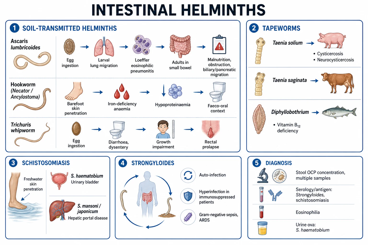
- **Ascaris lumbricoides** - egg ingestion; larval hepatopulmonary migration (Loeffler); adult in jejunum; obstruction, biliary migration
- **Hookworm (Necator americanus, Ancylostoma duodenale)** - filariform larval skin penetration; adult in duodenum; iron-deficiency anaemia, hypoproteinaemia
- **Trichuris trichiura (whipworm)** - egg ingestion; adult embedded in caecum/colon; dysentery, rectal prolapse (children)
- **Enterobius vermicularis (threadworm/pinworm)** - egg ingestion (retroinfection); perianal pruritus; sellotape test
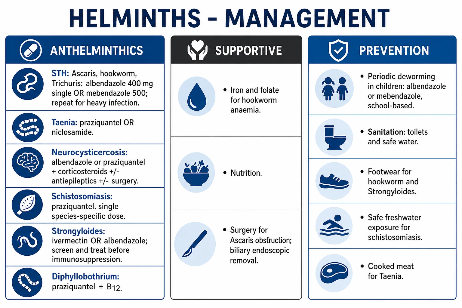
- Treat: albendazole 400 mg or mebendazole 500 mg single dose (mebendazole 100 mg BD x3 d for Trichuris)
Strongyloides stercoralis
Skin penetration + AUTO-INFECTION; the dangerous one
- Filariform larvae penetrate skin; migrate via lung; adult in duodenum
- **Auto-infection**: rhabditiform larvae in gut moult to filariform, penetrate perianal skin or bowel mucosa
- Allows **decades of carriage**; precipitates **hyperinfection in immunosuppression** (corticosteroids, HTLV-1, transplant)
- Hyperinfection: Gram-negative sepsis, ARDS, meningitis; mortality 60-85 percent
- Diagnosis: stool OCP (low sensitivity), **agar plate culture**, serology. Treat: **ivermectin** (NOT albendazole first-line)
Cestodes (tapeworms)
Larval cysts in meat or eggs causing tissue disease
- **Taenia solium (pork)** - cysticerci in undercooked pork cause intestinal taeniasis; eggs cause **cysticercosis/neurocysticercosis**
- **Taenia saginata (beef)** - cysticerci in undercooked beef; intestinal taeniasis only (no human cysticercosis)
- **Diphyllobothrium latum (fish)** - longest tapeworm (up to 10 m); causes **B12 deficiency/megaloblastic anaemia**
- **Echinococcus granulosus** - dog tapeworm; eggs cause **hydatid cysts** (liver, lung); anaphylaxis on rupture
- Treat: **praziquantel** (or niclosamide) for adult tapeworms; **albendazole** for cysticercosis/hydatid
Trematodes (flukes)
Freshwater/snail cycle; tissue-dwelling
- **Schistosoma mansoni/japonicum** - bowel; lateral-spined eggs in stool; periportal fibrosis, portal hypertension
- **Schistosoma haematobium** - bladder; terminal-spined eggs in urine; haematuria, **squamous cell carcinoma of bladder**
- **Fasciola hepatica** - sheep liver fluke; raw watercress; biliary obstruction
- **Clonorchis/Opisthorchis** - raw fish; biliary; **cholangiocarcinoma** (IARC Group 1 carcinogen)
- **Paragonimus westermani** - raw crab/crayfish; lung fluke; mimics TB
- Treat: **praziquantel** (all trematodes); triclabendazole for Fasciola
Tissue nematodes (filaria)
Vector-borne; lymphatic/dermal/ocular disease
- **Wuchereria bancrofti / Brugia malayi** - mosquito; lymphatic filariasis (elephantiasis, hydrocoele)
- **Onchocerca volvulus** - blackfly (Simulium); onchocerciasis ('river blindness'); subcutaneous nodules, ocular lesions
- **Loa loa** - Chrysops fly; Calabar swellings; **subconjunctival migration** ('eye worm')
- Diagnosis: **nocturnal microfilariaemia** (Wuchereria/Brugia - midnight blood); skin snip (Onchocerca); antibody/antigen
- Treat: **diethylcarbamazine (DEC)** for Wuchereria/Brugia/Loa; **ivermectin** for Onchococcus (NEVER DEC - Mazzotti reaction); doxycycline kills Wolbachia
Epidemiology & Risk Factors
Helminths are among the commonest infections of humans. The WHO estimates that approximately 1.5 billion people (nearly one in four of the world's population) are infected with STH alone, with the greatest burden in sub-Saharan Africa, the Indian subcontinent, China, Southeast Asia, and Latin America. Schistosomiasis affects approximately 240 million people, and lymphatic filariasis around 50 million, with over 880 million at risk in the tropics.[1]
The single most important determinant of helminth endemicity is poverty — specifically the combination of warm, moist climate; poor sanitation; use of untreated human faeces (night soil) as fertiliser; and lack of access to safe water and footwear. Worm eggs and larvae require warm (25 to 35 degrees C), moist, well-aerated soil to mature, which is why STH and schistosomiasis cluster in the tropics and disappear in temperate, arid regions with reticulated sewerage.[1]
Host and environmental risk factors, and the worms they favour: [1]
| Risk factor / exposure | Worm(s) to consider |
|---|---|
| Residence/travel in tropical, resource-poor region | All STH, schistosomiasis, filariasis |
| Walking barefoot on soil / faecally contaminated ground | Hookworm, Strongyloides (skin penetration) |
| Use of untreated night soil / open defecation | Ascaris, Trichuris (egg ingestion) |
| Consumption of undercooked pork | Taenia solium (taeniasis and cysticercosis) |
| Consumption of undercooked beef | Taenia saginata (taeniasis) |
| Consumption of raw freshwater fish | Diphyllobothrium latum, Clonorchis, Opisthorchis |
| Consumption of raw crab/crayfish | Paragonimus westermani (lung fluke) |
| Consumption of raw watercress | Fasciola hepatica (sheep liver fluke) |
| Freshwater swimming/wading in Africa, Middle East, China, Philippines | Schistosoma species (cercarial penetration) |
| Sheep- and dog-rearing rural communities | Echinococcus granulosus (hydatid) |
| Children in endemic areas | All STH, Enterobius (intense exposure, poor hygiene) |
| Immunosuppression (corticosteroids, HTLV-1, transplant, haematological malignancy) | Strongyloides hyperinfection |
| Pregnancy with hookworm in endemic region | Iron-deficiency anaemia, low birthweight |
Pathophysiology
The clinical and laboratory features of every helminth infection derive from where the worm lives, what it feeds on, and how the host immune system responds to it. The unifying immunological signature is the Th2 cytokine response — driven by IL-4, IL-5, IL-9, IL-13 and IL-31 — which drives eosinophilopoiesis (via IL-5), IgE class-switching, goblet-cell hyperplasia, mucin secretion, smooth-muscle hypercontractility, and macrophage alternative activation. This Th2 axis is the host's principal defence against worms (it expels them by mucous trapping, weep-and-sweep, and mechanical grip-loosening), which is why eosinophilia and high IgE are the laboratory hallmarks of tissue helminth infection, and why Th2-deficient states (corticosteroids, HTLV-1) tip the balance toward the parasite.[9]
The Ascaris lumbricoides life cycle — the paradigm of hepatopulmonary migration
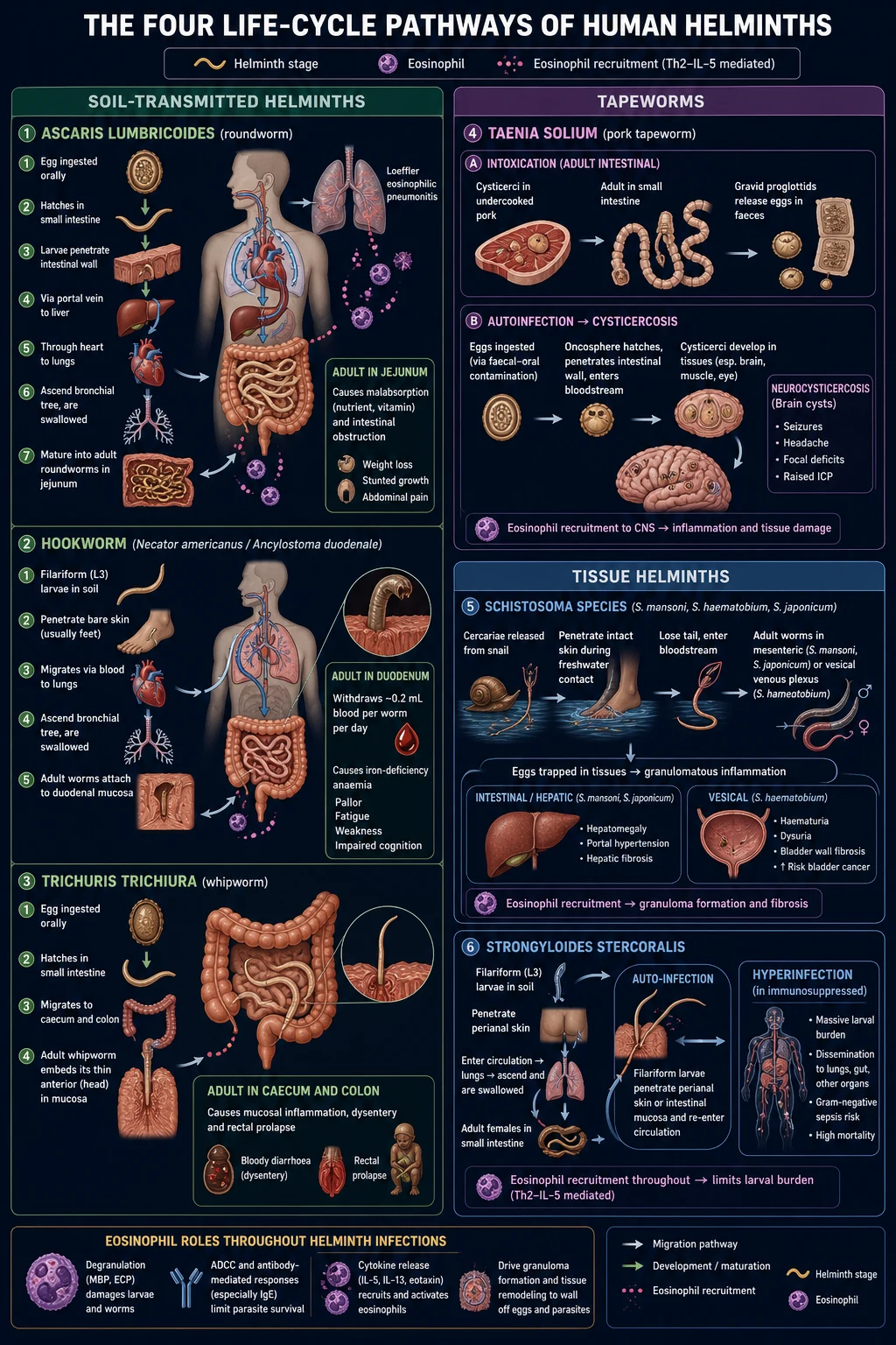
- Egg ingestion. Fertilised eggs (rounded, thick-shelled, mammillated brown coat) are swallowed in soil-contaminated food or water. They require 2 to 6 weeks of soil maturation to become embryonated and infective — so fresh faecal-oral transit does not transmit.
- Small-intestinal hatching and mucosal penetration. Eggs hatch in the jejunum, releasing rhabditiform larvae that penetrate the intestinal wall and enter the portal venous system.
- Hepatic and pulmonary migration. Larvae reach the liver (4 to 7 days), then pass through the right heart to the lungs (days 7 to 14), where they lodge in alveolar capillaries, moult twice, and mature into third- and fourth-stage larvae (about 1 to 2 mm long). The host mounts a brisk eosinophilic inflammatory response (Loeffler syndrome) — cough, wheeze, transient pulmonary infiltrates, and marked peripheral eosinophilia.
- Airway ascent and swallowing. Larvae ascend the bronchial tree to the glottis (day 14), are swallowed, and return to the small intestine, where they mature into adult roundworms (15 to 35 cm) in the jejunum. Adults do not attach — they live free in the lumen, consuming semi-digested food.
- Egg laying and excretion. Adult females begin laying eggs at 8 to 12 weeks after infection. Eggs appear in stool. Adult worms live 1 to 2 years. [1]
The pathological consequences of Ascaris are mechanical: a heavy burden (hundreds of worms) can ball up and obstruct the small bowel (especially the ileum, in children), and an adult worm can migrate into the bile duct, pancreatic duct, or appendix, causing biliary colic, ascending cholangitis, acute pancreatitis, or appendicitis. The migrating larval phase causes the Loeffler syndrome.[1]
The hookworm life cycle — the paradigm of blood loss
- Skin penetration. Filariform (third-stage) larvae in soil penetrate exposed skin (usually the feet), often producing a transient pruritic 'ground itch' at the entry site.
- Venous migration to lungs. Larvae enter subcutaneous venules, travel to the right heart and lungs, rupture into alveoli (day 3 to 5), ascend the bronchial tree, and are swallowed. This migration also causes a mild Loeffler-like pneumonitis.
- Duodenal attachment and blood feeding. Adult worms (8 to 13 mm) attach to the duodenal and proximal jejunal mucosa with cutting plates or teeth, secrete anticoagulants (e.g. ancylostomatin), and ingest blood and mucosal tissue. Each Necator withdraws approximately 0.03 mL/day and each Ancylostoma approximately 0.2 mL/day; a heavy burden (dozens of worms) drives iron-deficiency anaemia and hypoproteinaemia within months.
- Egg excretion. Females lay eggs (60 x 40 micrometres) in the bowel lumen; eggs appear in stool within 4 to 8 weeks. Ancylostoma duodenale can additionally transmit via transmammary (breast milk) and oral routes, causing neonatal infection.[1]
The Strongyloides stercoralis cycle — the paradigm of auto-infection
Strongyloides is unique among human helminths in that it can complete its entire life cycle within a single human host (homogonic cycle), through auto-infection. This is the single most important fact about the parasite, because it explains both decades of asymptomatic carriage (in a person who left an endemic region years ago) and catastrophic hyperinfection when cell-mediated immunity is suppressed.[9]
- Skin penetration and migration. As for hookworm: filariform larvae penetrate skin, migrate to lungs, are swallowed, and adult parasitic females (2 to 3 mm) embed in the duodenal and jejunal mucosa, where they reproduce parthenogenetically (no male required).
- Egg laying and rhabditiform larvae. Females lay eggs that hatch within the mucosa to release rhabditiform larvae which pass out in stool. In the external environment, rhabditiform larvae can either develop into free-living adult worms (the heterogonic cycle, unique among human nematodes) or moult into infective filariform larvae.
- Auto-infection. Some rhabditiform larvae within the bowel lumen moult into filariform larvae before being excreted, then penetrate the perianal skin (external auto-infection) or the colonic mucosa (internal auto-infection), re-migrating through the lungs to re-establish infection. This loop, repeating every 2 weeks, sustains infection for the life of the host.
- Hyperinfection. When cell-mediated (Th2) immunity is impaired — classically by corticosteroids (which also directly stimulate larval moult via ecdysteroid-like receptors), HTLV-1, haematological malignancy, solid-organ transplantation, or malnutrition — the auto-infection cycle accelerates enormously, producing thousands of migrating larvae. The larvae carry enteric bacteria (notably Escherichia coli, Klebsiella, Enterococcus, and Bacteroides) on their cuticle and disrupt the bowel wall, producing Gram-negative bacteraemia, meningitis, and pneumonia, often with ARDS, ileus, and shock. Mortality is 60 to 85 percent despite treatment. Notably, HIV is NOT a major risk factor for Strongyloides hyperinfection — the parasite relies on Th2, which HIV (a Th1/CD4-pathogen) relatively spares. [1]
The Schistosoma cycle — the paradigm of egg-driven granulomatous disease
- Cercarial penetration. Cercariae released by infected freshwater snails (the intermediate host) penetrate the skin during swimming, bathing, or wading. They shed their forked tail, become schistosomula, and migrate via venous circulation through the lungs to the liver, where they mature and pair.
- Adult worm pairing and venous migration. Mature male and female worms pair and migrate against portal venous flow to their species-specific final niche: S. mansoni and S. japonicum to the inferior mesenteric and superior mesenteric venules (bowel); S. haematobium to the vesical and pelvic venous plexus (bladder). Adult worms live 3 to 10 years (occasionally much longer), sheathed in a host-derived tegument that hides them from immunity.
- Egg deposition — the pathogenic stage. The female lays eggs (each species has a characteristic spine: S. mansoni = lateral, S. haematobium = terminal, S. japonicum = small lateral). To exit the host, eggs must traverse the bowel or bladder wall, a process that only a minority achieve — the rest are swept by the portal/systemic circulation to the liver (S. mansoni/japonicum), lungs, brain, spinal cord, or genitourinary tract, where they elicit a granulomatous Th2 response around the egg.
- Granulomatous disease. The egg-antigen-driven granuloma is the pathological hallmark of schistosomiasis. In the liver, periportal ('clay-pipestem') Symmers fibrosis develops over years, producing presinusoidal portal hypertension (splenomegaly, varices, hypersplenism) with preserved hepatocellular function (normal synthetic function — distinguishes schistosomiasis from cirrhosis). In the bladder, chronic S. haematobium egg deposition causes haematuria, bladder-wall calcification, obstruction, and, over decades, squamous cell carcinoma of the bladder (IARC Group 1 carcinogen).
- Acute schistosomiasis (Katayama fever). Two to six weeks after a heavy primary exposure, a serum-sickness-like immune-complex reaction produces fever, rigors, urticaria, myalgia, arthralgia, cough, diarrhoea, hepatosplenomegaly, and marked eosinophilia — Katayama fever. It is most severe with S. japonicum and can be fatal.[3]
The Taenia solium cycle — the paradigm of cysticercosis
Humans are the definitive host (adult tapeworm) for both T. solium (pork) and T. saginata (beef), and the intermediate host (larval cysticercus) for T. solium alone. This dual role is the source of the most important exam distinction: eating undercooked pork causes taeniasis (the adult worm in the gut); ingesting T. solium eggs causes cysticercosis (larval cysts in brain, muscle, and eye).[4]
- Taeniasis (adult worm). Humans eat undercooked pork containing cysticerci. The cysticercus evaginates in the small intestine, attaches to the jejunal mucosa by its scolex (with four suckers and a double ring of hooks — T. solium; hooks absent in T. saginata), and matures into an adult tapeworm of 2 to 8 metres in 2 to 3 months. The adult is usually single and asymptomatic; gravid proglottids detach and pass in stool or migrate actively per rectum/per anus.
- Cysticercosis (larval cysts). T. solium eggs (indistinguishable from T. saginata eggs morphologically) ingested in faecally contaminated food or water, or by auto-infection (a taeniasis carrier swallowing their own eggs via hand-to-mouth), release oncospheres that penetrate the intestinal wall, enter the bloodstream, and lodge in skeletal muscle, subcutaneous tissue, brain, and eye, where they develop into cysticerci — fluid-filled bladders of about 5 to 20 mm containing an invaginated scolex.
- Neurocysticercosis (NCC). Cysticerci in the brain parenchyma, subarachnoid space, ventricles, spinal cord, or eye provoke disease when they degenerate and the host immune system recognises them — releasing antigen and eliciting inflammation, oedema, and seizures. Live (viable) cysticerci evade immunity and are often silent for years; calcified cysts are inactive but remain epileptogenic foci.[4]
Clinical Presentation
Most helminth infections are asymptomatic or pauci-symptomatic at low intensity; clinical disease is a function of worm burden, larval migration, tissue deposition of eggs, and host immune response. The syndromes below are organised by worm. [1]
Soil-transmitted helminths (Ascaris, hookworm, Trichuris)
Ascaris lumbricoides. Light infection: asymptomatic. Heavy infection produces three syndromes: (1) the migrating-larval phase (Loeffler syndrome) — at days 7 to 14, dry cough, wheeze, dyspnoea, low-grade fever, and transient pulmonary infiltrates with marked eosinophilia (resolves in 1 to 2 weeks); (2) established intestinal infection — non-specific abdominal pain, anorexia, nausea, occasionally malabsorption and failure to thrive in children; and (3) mechanical complications — acute small-bowel obstruction (a heavy burden balls up in the ileum, classically in a child with a palpable abdominal mass of worms), biliary migration (biliary colic, obstructive jaundice, ascending cholangitis), pancreatic duct migration (acute pancreatitis), and appendicitis. A worm may be vomited or passed per rectum, which is often the presenting complaint.[1]
Hookworm. The classical presentation is insidious iron-deficiency anaemia in an endemic or returned-travel patient — pallor, fatigue, exertional dyspnoea, koilonychia, angular cheilitis, pica (geophagia), and in children, growth and cognitive impairment. Hypoalbuminaemia produces dependent oedema. At skin penetration, 'ground itch' (pruritic erythematous papule at the entry site) may be recalled. The migrating-larval phase can produce a mild Loeffler-like pneumonitis.[1]
Trichuris trichiura. Light infection: asymptomatic. Heavy infection (especially in children) produces the Trichuris dysentery syndrome (TDS) — chronic mucoid, bloody diarrhoee, tenesmus, abdominal pain, rectal prolapse, growth failure, and anaemia, mimicking inflammatory bowel disease. The 'Trichuris trichiura dysentery' picture is a classic exam vignette: a child in an endemic region with chronic bloody diarrhoea and a prolapsed rectum on straining. [1]
Strongyloides stercoralis
Chronic strongyloidiasis (decades of auto-infection) is usually mild and non-specific — epigastric pain, intermittent diarrhoea alternating with constipation, nausea, urticaria, and 'larva currens' (a rapidly migrating, serpiginous, pruritic urticarial weal, typically perianal, buttock or trunk, that moves several centimetres per hour — pathognomonic and distinct from cutaneous larva migrans which moves only millimetres per day). Irritable-bowel-like symptoms are common.[9]
Hyperinfection / disseminated disease (in the immunosuppressed) is a medical emergency: Gram-negative sepsis (often with E. coli, Klebsiella, Enterococcus, Bacteroides — the larvae carry them from the gut), pneumonia and ARDS, meningitis, ileus and abdominal distension, shock, and Gram-negative bacteraemia of gut origin without an obvious source. The clue is astonishing peripheral eosinophilia may be ABSENT in the immunosuppressed (paradoxically), but larvae are found in sputum, bronchoalveolar lavage, and stool in large numbers. Suspect it in any patient with HTLV-1, recent corticosteroids, transplant, or haematological malignancy who develops unexplained Gram-negative sepsis.[9]
Enterobius vermicularis (threadworm / pinworm)
The classical presentation is nocturnal perianal pruritus in a child (the gravid female migrates out at night to lay eggs on the perianal skin), with restless sleep, irritability, and secondary excoriation or vulvovaginitis. Whole-household infection is the rule. Eosinophilia is absent (no tissue migration). [1]
Tapeworms (Taenia, Diphyllobothrium)
Intestinal taeniasis (adult worm) is usually asymptomatic or mild — vague abdominal discomfort, nausea, weight loss, and the distressing passage of motile proglottids per anus (the proglottid may crawl out onto underclothes — a pathognomonic complaint). The key clinical importance of T. solium taeniasis is that the carrier is at risk of auto-infection with cysticercosis and is the source of cysticercosis for household contacts. [1]
Neurocysticercosis (NCC). The presentation depends on cyst location, number, and viability: (1) parenchymal NCC — new-onset seizures (the commonest cause of adult-onset epilepsy in endemic regions), headache, focal deficits; (2) subarachnoid NCC — chronic meningitis, basal arachnoiditis, hydrocephalus, vasculitis and stroke; (3) intraventricular NCC — obstructive hydrocephalus with episodic intracranial-pressure spikes (Brun syndrome — positional vertigo and drop attacks); (4) spinal NCC — radicular pain, myelopathy; (5) ocular NCC — visual loss from intraocular cysts. Subcutaneous cysticerci present as firm, painless nodules over muscle.[4]
Diphyllobothrium latum (fish tapeworm) is usually asymptomatic but can cause vitamin B12 deficiency (the worm competes for dietary B12 in the terminal ileum) — megaloblastic anaemia, glossitis, neuropathy, and rarely subacute combined degeneration of the cord. [1]
Schistosomiasis
- Acute schistosomiasis (Katayama fever). Two to six weeks after heavy primary freshwater exposure (often a returning traveller who swam in Lake Malawi or the Nile): abrupt-onset fever, rigors, headache, myalgia, arthralgia, dry cough, abdominal pain, diarrhoea, and a generalised urticarial rash, with hepatosplenomegaly and marked eosinophilia. Severe S. japonicum Katayama can be fatal.
- Chronic intestinal/hepatic schistosomiasis (S. mansoni/japonicum). Years after infection: abdominal pain, diarrhoea (± blood/mucus), hepatosplenomegaly, progressing to periportal fibrosis (Symmers), presinusoidal portal hypertension — splenomegaly, varices, ascites, hypersplenism (pancytopenia) — with preserved hepatic synthetic function (no encephalopathy, normal albumin/coagulation — the key distinction from decompensated cirrhosis). Death is from variceal haemorrhage.
- Urogenital schistosomiasis (S. haematobium). Terminal (end-of-stream) haematuria is the classical presentation, with dysuria, frequency, suprapubic pain, and, on examination, bladder-wall nodularity, sandy patches, and hydro-ureter/hydronephrosis from chronic granulomatous inflammation and fibrosis. Chronic disease leads to squamous cell carcinoma of the bladder (presents years later), infertility, and in women, genital lesions (worms in the cervix/vagina — 'female genital schistosomiasis').
- Neuroschistosomiasis. Ectopic egg deposition in the spinal cord (acute transverse myelitis, especially S. mansoni) or brain (seizures, focal deficit).[3]
Lymphatic filariasis (Wuchereria, Brugia)
- Acute adenolymphangitis (ADLA). Recurrent episodes of fever, tender lymphadenitis, and lymphangitis (classically inguinal), often preceded by a 'filarial fever', lasting days. Repeated episodes scar and obstruct lymphatics.
- Chronic lymphatic disease. Lymphoedema of the limbs (usually lower, asymmetric) progressing to elephantiasis — massive, warty, weeping limb swelling with recurrent bacterial cellulitis (the skin fissures and is superinfected with streptococci). Hydrocoele (the commonest chronic manifestation in men), chylocele, chyluria (milky white urine from lymphatico-urinary fistula), and genital elephantiasis.
- Tropical pulmonary eosinophilia (TPE). A hypersensitivity reaction to microfilariae trapped in the lungs — nocturnal cough, wheeze, dyspnoea, marked eosinophilia (over 3 x10^9/L), and miliary pulmonary infiltrates; microfilariae are absent from blood (they are trapped/dying in the lungs).[7]
Onchocerciasis (river blindness) and loiasis
- Onchocerciasis. Subcutaneous nodules (onchocercomata) over bony prominences (iliac crest, greater trochanter, skull), pruritic papular dermatitis ('lizard skin', 'leopard skin' from depigmentation and atrophy), and ocular disease (punctate and sclerosing keratitis, anterior uveitis, chorioretinitis, optic atrophy) progressing to blindness. Microfilariae are in the skin and eye, not blood.
- Loiasis (African eye worm). Calabar swellings (transient 5 to 10 cm subcutaneous angio-oedematous swellings, often on the limbs) and the dramatic migration of an adult worm across the conjunctiva ('eye worm') — alarming but usually benign. [1]
Hydatid disease (Echinococcus granulosus)
Often asymptomatic for years until the cyst grows large enough to cause mass effect. The liver (right lobe) is the commonest site (~65 percent), then the lung (~25 percent). Hepatic hydatid presents as a painless right-upper-quadrant mass, occasionally with biliary obstruction (jaundice, cholangitis). Pulmonary hydatid may cause cough, haemoptysis, or chest pain. Rupture (spontaneous, traumatic, or iatrogenic) releases antigenic fluid and causes acute anaphylaxis (hypotension, urticaria, bronchospasm) and secondary dissemination.[6]
Atypical presentations (deliberately tested)
- Immunosuppressed patient with Gram-negative sepsis, ARDS, or meningitis — Strongyloides hyperinfection; larvae in sputum/stool; ivermectin urgently.
- Returned traveller from Africa with terminal haematuria — S. haematobium; urine for ova.
- Adult-onset seizures in a pork-endemic region — neurocysticercosis; CT/MRI brain.
- Chronic bloody diarrhoea and rectal prolapse in a child — Trichuris dysentery syndrome.
- Painless RUQ mass with a folded membrane on ultrasound — hydatid cyst.
- Iron-deficiency anaemia with eosinophilia in a returned traveller / barefoot child — hookworm. [1]
Differential Diagnosis
Because helminths cause diverse syndromes (eosinophilia, anaemia, diarrhoea, seizures, portal hypertension), the differential is syndrome-driven. [1]
Eosinophilia (worm vs other)
- **Helminth (tissue)** - migration, schistosomiasis, filariasis, strongyloidiasis, fascioliasis; travel/diet/exposure; stool OCP + serology
- **Drug hypersensitivity** - timing with new drug, rash, eosinophilia, AKI (DRESS)
- **Atopy/asthma/eczema** - history, IgE raised, no organ dysfunction
- **Adrenal insufficiency** - fatigue, hypotension, hyperkalaemia, hyponatraemia; check cortisol
- **Hypereosinophilic syndrome** - eosinophils over 1.5 x10^9/L for over 6 months, end-organ damage, no other cause
- **Neoplasia** - T-cell lymphoma, mastocytosis, CML, solid tumours
Iron-deficiency anaemia
- **Hookworm** - eosinophilia, tropical/returned, positive stool OCP
- **GI blood loss** - peptic ulcer, malignancy (especially older), IBD, angiodysplasia
- **Menorrhagia** - premenopausal women; ferritin low, eosinophils normal
- **Malabsorption** - coeliac disease (anti-tTG), Helicobacter, gastric surgery
- **Dietary deficiency** - poor intake, vegetarian, infant weaning
- **Chronic disease** - normal/high ferritin, underlying inflammation
Adult-onset seizures (ring lesion)
- **Neurocysticercosis** - pork-endemic, scolex 'hole-with-dot', calcified lesions
- **Cerebral toxoplasmosis** - HIV/CD4 under 100, ring-enhancing, multiple, basal ganglia
- **Tuberculoma** - TB contact, basal exudates, positron-emitting rim
- **Brain abscess** - fever, source (sinus, ear, endocarditis), thin-walled ring
- **Metastasis** - known primary, multiple lesions at grey-white junction
- **Glioblastoma** - thick irregular ring, necrotic centre, white-matter spread
Acute febrile traveller (Katayama)
- **Acute schistosomiasis (Katayama)** - freshwater exposure, eosinophilia, urticaria
- **Malaria** - abrupt fever, periodicity, thick/thin film; co-endemic
- **Enteric fever** - step-ladder fever, rose spots, blood culture
- **Dengue** - retro-orbital pain, thrombocytopenia, saddle-back fever
- **Leptospirosis** - conjunctival suffusion, myalgia, jaundice, AKI
- **Rickettsial (scrub typhus)** - eschar, regional lymphadenopathy, doxycycline response
Chronic bloody diarrhoea (Trichuris)
- **Trichuris dysentery syndrome** - child, endemic, rectal prolapse, stool OCP
- **Inflammatory bowel disease** - extraintestinal features, ASCA/p-ANCA, colonoscopy
- **Amoebic dysentery** - Entamoeba histolytica, flask-shaped ulcers, liver abscess
- **Giardiasis** - foul stools, bloating, no blood (usually)
- **Balantidium coli** - pigs, bloody diarrhoea, ciliate on stool
- **Intestinal TB** - caecal mass, ascites, GeneXpert
Hepatic cyst (hydatid vs other)
- **Hydatid (Echinococcus)** - sheep/dog, daughter cysts, 'water-lily', hydatid sand
- **Simple liver cyst** - anechoic, thin wall, no septations, asymptomatic
- **Amoebic liver abscess** - travel, fever, RUQ pain, 'anchovy sauce' pus
- **Pyogenic liver abscess** - fever, biliary source, polymicrobial
- **Congenital (Caroli, choledochal)** - biliary communication, recurrent cholangitis
- **Hepatocellular carcinoma** - cirrhosis, arterial enhancement, washout, raised AFP
The exam trap: cutaneous larva migrans (CLM) and larva currens are easily confused. CLM is zoonotic (Ancylostoma braziliense from dogs/cats), produces a slow (1 to 2 cm/day) serpiginous pruritic track usually on the feet/buttocks, and is treated with albendazole or ivermectin (or topical tiabendazole). Larva currens is Strongyloides, moves much faster (5 to 10 cm/hour, typically perianal/gluteal), and requires systemic ivermectin. [1]
Clinical & Bedside Assessment
The bedside assessment is syndrome-driven and depends on the suspected worm, but every assessment should include a structured exposure history, growth assessment in children, and a search for the anaemia, eosinophilia, and organ-specific signs that betray tissue disease. [1]
History. Establish (1) residence or travel in a tropical endemic region (sub-Saharan Africa, India, Southeast Asia, Latin America, China); (2) the specific exposure — barefoot walking, open defecation, untreated water, consumption of undercooked pork, beef, freshwater fish, crab, or raw watercress, and freshwater swimming/wading (ask specifically about Lake Malawi, the Nile, Lake Victoria, the Mekong, and Philippines freshwater); (3) occupational exposure — sheep-farming and dogs (hydatid), abattoir work (Taenia), rice farming (filariasis, schistosomiasis); (4) the symptom tempo and pattern — perianal pruritus at night (Enterobius), terminal haematuria (S. haematobium), new seizures (NCC), chronic bloody diarrhoea and rectal prolapse (Trichuris), milky urine (chyluria in filariasis), a worm passed per mouth/rectum (Ascaris), Calabar swellings and an eye worm (Loa loa); and (5) immunosuppression — corticosteroids, HTLV-1, transplant, haematological malignancy (the trigger to screen for Strongyloides). [1]
Examination. Look for pallor (anaemia), koilonychia, angular cheilitis (hookworm iron deficiency); growth parameters — weight, height, mid-upper-arm circumference — in children (STH growth impairment); abdomen — hepatosplenomegaly (schistosomiasis, katayama, capillaria), mass (Ascaris bolus, hydatid cyst), tenderness (acute pancreatitis/cholangitis from Ascaris migration); skin — urticaria (acute helminthiasis), larva currens (Strongyloides), creeping eruption (CLM), subcutaneous nodules (onchocercomata, cysticerci, hydatid, Loa), 'leopard/lizard skin' (onchocerciasis); lymphatics — lymphoedema/elephantiasis of limbs and genitalia, hydrocoele (filaria); eyes — subconjunctival worm (Loa), chorioretinitis (onchocerciasis), intraocular cyst (cysticercosis); neurology — focal deficit, seizures, signs of raised intracranial pressure (NCC), acute transverse myelitis (neuroschistosomiasis); respiratory — Loeffler pneumonitis (wheeze, crackles, transient infiltrates), tropical pulmonary eosinophilia (TPE). Rectal examination for fresh blood/mucus (Trichuris dysentery, schistosomiasis) and rectal prolapse (Trichuris in children). [1]
Monitoring. In severe disease — Ascaris obstruction, Strongyloides hyperinfection, Katayama fever — serial observations (temperature, heart rate, blood pressure, respiratory rate, oxygen saturation, GCS), hourly urine output, and serial full blood count (haemoglobin, eosinophil trend) and organ function. [1]
Investigations
The diagnostic strategy is (a) parasitological (find the worm, eggs, or larvae), (b) immunological (antibody, antigen), (c) haematological/biochemical (eosinophilia, anaemia), and (d) imaging (for tissue disease). The choice of sample is species-specific and is the single most important practical decision. [1]
Parasitological diagnosis — stool, urine, and sellotape
Stool microscopy for ova, cysts, and parasites (OCP) is the cornerstone. Eggs of Ascaris, hookworm, Trichuris, Taenia, Schistosoma mansoni/japonicum, Fasciola, Clonorchis, and Fasciolopsis are shed in stool. Single-sample sensitivity is only 50 to 70 percent in light infection; three samples on alternate days (concentration technique — formol-ether or zinc-sulphate flotation) raise sensitivity to over 90 percent. The Kato-Katz thick smear quantifies eggs per gram (epg) of faeces, used in mass-drug-administration programmes and to grade intensity (heavy = Ascaris over 50,000 epg; hookworm over 4000 epg; Trichuris over 10,000 epg).[1]
Stool OCP (ova, cysts, parasites)
First-line for most intestinal worms
- Detects eggs of Ascaris, hookworm, Trichuris, Taenia, S. mansoni/japonicum, Fasciola
- Three samples on alternate days + concentration technique raise sensitivity to over 90 percent
- **Kato-Katz thick smear** quantifies eggs per gram for MDA programmes
- Sensitivity LOW in light infection, low-burden carriers, and the immunosuppressed
Strongyloides stool
The 'sneaky' worm - special techniques needed
- Rhabditiform larvae (not eggs) in stool - single OCP sensitivity only 30 percent
- **Agar plate culture** - sensitivity 90 percent+, the diagnostic gold standard
- Multiple samples (4 to 7) increase yield substantially
- Paired with serology (ELISA) which is sensitive but cannot distinguish current from past
- **Filariform larvae in sputum/BAL in hyperinfection** - pathognomonic
Enterobius sellotape test
Stool OCP is often NEGATIVE
- Clear adhesive tape pressed against perianal skin first thing in the morning (before bathing)
- Examined under microscope for characteristically asymmetrical eggs
- Sensitivity about 50 percent single, over 90 percent with 3 samples
- Whole household should be tested
Schistosoma - urine vs stool
Species-specific sample is critical
- **S. haematobium** - terminal-spined eggs in URINE (midday terminal sample, filtration)
- **S. mansoni / japonicum** - lateral-spined eggs in STOOL
- **S. japonicum** eggs are smaller and more numerous; also rectal snip
- Serology (FAL) confirms exposure but cannot distinguish current from past
Filarial - blood and skin
Time-of-day and tissue matter
- **Wuchereria / Brugia** - microfilariae in blood drawn at MIDNIGHT (nocturnal periodicity); membrane filtration
- **Onchocerca** - microfilariae in SKIN SNIP (not blood); nodules may yield adults
- **Loa loa** - microfilariae in daytime blood (diurnal periodicity)
- **Antigen (Og4C3, ICT BinaxNOW)** - detects W. bancrofti antigen at any time, replaces midnight blood
- PCR where available
Immunological (serology/antigen)
Strongyloides antibody (ELISA) is the screening test of choice in returned travellers and before immunosuppression — sensitivity about 85 to 95 percent; cross-reactivity with other filariae/Ascaris limits specificity; titres fall slowly after cure (so cannot reliably confirm cure). Schistosoma serology (FAST-ELISA, Falcon Assay Screening Test) is the test of choice for returned travellers (sensitive but genus-specific; cannot distinguish species — eggs in stool/urine confirm active disease). Cysticercosis antibody/antigen (EITB immunoblot, sensitivity about 98 percent for multiple lesions) supports neurocysticercosis. Echinococcus antibody (IHA, immunoblot, Arc 5 antigen) supports hydatid diagnosis but may be negative in intact liver cysts. Filarial antigen (ICT/Og4C3) detects W. bancrofti without regard to nocturnal periodicity.[9]
Haematology and biochemistry
- Eosinophilia (over 0.5 x10^9/L) is the screening red flag for tissue helminthiasis — marked (over 3 x10^9/L) in Katayama, fascioliasis, TPE, and the migrating-larval phase; absent in established intraluminal gut infection and in Strongyloides hyperinfection in the immunosuppressed.
- Iron studies (low ferritin, low iron, high TIBC) confirm hookworm-related iron-deficiency anaemia.
- B12 and folate — megaloblastic picture in Diphyllobothrium.
- Liver function — preserved in schistosomal portal hypertension (normal synthetic function); cholestatic/obstructive in Ascaris biliary migration and Fasciola.
- Urinalysis — haematuria (macroscopic terminal, or dipstick micro) in S. haematobium.
- IgE — markedly elevated in tissue helminthiasis and TPE. [1]
Imaging
- Chest X-ray — transient pulmonary infiltrates (Loeffler), miliary pattern (TPE), 'water-lily' sign (collapsed hydatid cyst membrane floating in fluid), pleural effusion.
- Abdominal ultrasound — hydatid cyst (WHO-INN classification: CE1 active simple cyst with hydatid sand; CE2 active multivesicular; CE3a/CE3b transitional with detached membrane; CE4 inactive heterogeneous; CE5 inactive with calcified wall), periportal fibrosis (Symmers) and hepatosplenomegaly in schistosomiasis, Ascaris bolus (target sign, intra-biliary worms).
- CT / MRI brain — neurocysticercosis: vesicular (live cyst, CSF-density fluid, scolex — 'hole-with-dot' or 'starry sky'), colloidal vesicular (degenerating, ring-enhancing, oedema), granular nodular (shrinking, calcifying), calcified nodular (dense, inactive but epileptogenic). Hydrocephalus and subarachnoid cysts in extraparenchymal NCC.
- Endoscopic retrograde cholangiopancreatography (ERCP) — diagnostic and therapeutic for biliary Ascaris and biliary Fasciola. [1]
Special: the Strongyloides screen before immunosuppression
Every patient about to start corticosteroids (especially high-dose or prolonged), transplantation immunosuppression, biologicals, or haematological-malignancy chemotherapy, and every patient with HTLV-1, who has lived in or travelled to an endemic region, should be screened with Strongyloides serology plus stool (with agar plate culture where available) and treated with ivermectin 200 micrograms/kg single dose (two doses 24 hours apart) before immunosuppression. This is among the most important and most overlooked reflexes in clinical medicine.[9][10]
Management — Resuscitation
Most helminth infections are outpatient, oral-therapy conditions, but several scenarios are emergencies. [1]
Ascaris intestinal obstruction. A child (or adult) with a heavy Ascaris burden and signs of obstruction requires nasogastric decompression, IV fluids (balanced crystalloid), analgesia, and correction of electrolytes. Piperazine citrate 75 mg/kg (max 3.5 g) orally or via NG tube (paralyses the worms, allowing them to be passed) or albendazole 400 mg once obstruction is resolving. Surgery (laparotomy with manual de-bulking, with or without resection) is reserved for refractory obstruction, peritonitis, perforation, or ischaemia — never for uncomplicated obstruction. For biliary/pancreatic migration, ERCP with worm extraction is first-line.[1]
Strongyloides hyperinfection. This is a critical-care emergency: ICU admission, broad-spectrum empiric antibiotics covering gut-origin Gram-negatives and anaerobes (e.g. piperacillin-tazobactam or meropenem, adding vancomycin if line/soft-tissue sepsis is possible), fluid resuscitation and vasopressors for shock, mechanical ventilation for ARDS, and urgent ivermectin 200 micrograms/kg orally daily (subcutaneous ivermectin if ileus or absorption is impaired) for 7 to 14 days or until negative stool/sputum. Reduce or hold immunosuppression where possible. Treat until parasitologically clear — relapse is common. Mortality remains 60 to 85 percent.[9]
Ruptured hydatid cyst / anaphylaxis. Adrenaline 0.5 mg IM (adult), oxygen, IV fluids, hydrocortisone 200 mg IV and chlorphenamine 10 mg IV, plus surgical consultation for source control. Commence albendazole to prevent secondary dissemination. [1]
Severe hookworm anaemia in pregnancy. Transfuse packed red cells if haemoglobin under 7 g/dL or symptomatic; oral iron and folate; albendazole 400 mg single dose in the second or third trimester (avoid first trimester). [1]
Status epilepticus due to neurocysticercosis. IV lorazepam 4 mg (repeat once) or diazepam 10 mg, followed by IV levetiracetam 60 mg/kg or fosphenytoin 20 mg/kg, plus IV dexamethasone 10 mg to reduce perilesional oedema, and definitive cysticidal therapy once controlled. Hydrocephalus requires neurosurgical shunt/ventriculostomy before cysticidal drugs.[4]
Katayama fever. Corticosteroids (prednisolone 20 to 40 mg daily for 5 to 7 days, or IV hydrocortisone in severe disease) to suppress the immune-complex reaction, followed by praziquantel once the acute reaction settles (giving praziquantel too early can worsen symptoms by increasing antigen release). [1]
Management — Definitive & Stepwise
The choice of antihelminthic is species-specific and not interchangeable. The benzimidazoles (albendazole, mebendazole) are first-line for STH; praziquantel for cestodes and trematodes; ivermectin for Strongyloides and onchocerciasis; diethylcarbamazine (DEC) for lymphatic filariasis and loiasis (with care). [1]
Anthelmintic formulary — drug, dose, route, rationale
Albendazole
Benzimidazole - STH, cysticercosis, hydatid
- **STH (Ascaris, hookworm, Enterobius)**: 400 mg single oral dose (adults and over 2 y)
- **Trichuris**: 400 mg daily for 3 days (single-dose cure rate lower)
- **Neurocysticercosis**: 15 mg/kg/day in divided doses for 8-30 days (with steroids)
- **Hydatid**: 10-15 mg/kg/day for 1-6 months (cycles of 28 days with 14-day gaps)
- **Avoid in first trimester of pregnancy** and children under 12 months (teratogenic)
- **Monitor LFTs and CBC** on prolonged courses; causa leucopenia, transaminitis, alopecia
Mebendazole
Benzimidazole - alternative for STH
- **STH**: 500 mg single oral dose (WHO MDA regimen)
- **Trichuris and hookworm**: 100 mg BD for 3 days (better than single dose)
- **Enterobius**: 100 mg single dose, repeat after 2 weeks; treat whole household
- **Poor systemic absorption** - excellent for intraluminal gut worms
- **Avoid in pregnancy** first trimester
Ivermectin
Avermectin - Strongyloides, onchocerciasis, CLM
- **Strongyloides**: 200 micrograms/kg orally daily for 1-2 days (FIRST-LINE)
- **Strongyloides hyperinfection**: 200 micrograms/kg daily for 7-14 days or until stool/sputum negative; SC if ileus
- **Onchocerciasis**: 150 micrograms/kg single dose, repeat every 6-12 months (kills microfilariae)
- **Cutaneous larva migrans**: 200 micrograms/kg single dose (or 2 doses)
- **Caution in Loa loa** - ivermectin can cause fatal encephalopathy if microfilaraemia over 30,000/mL
- **NEVER in pregnancy** or children under 15 kg
Praziquantel
Pyrazinoisoquinoline - tapeworms, schistosomes, flukes
- **Taenia, Diphyllobothrium, Hymenolepis**: 5-10 mg/kg single oral dose
- **S. mansoni/haematobium**: 40 mg/kg single dose (or 20 mg/kg BD)
- **S. japonicum/mekongi**: 60 mg/kg in 3 divided doses
- **Neurocysticercosis**: 50-100 mg/kg/day in 3 divided doses for 15 days (alternative to albendazole)
- **Fasciola** is RESISTANT - use **triclabendazole 10 mg/kg single or 2 doses**
- Side-effects: nausea, headache, dizziness, abdominal pain; rare CNS effects
- **Cysticidal therapy accelerates radiographic resolution and seizure control in NCC** (Garcia NEJM 2004)
Diethylcarbamazine (DEC)
Piperazine - lymphatic filariasis, loiasis
- **Wuchereria / Brugia**: 6 mg/kg orally in divided doses for 12 days (or 6 mg/kg single-dose MDA with albendazole)
- **Loa loa**: 6-9 mg/kg/day for 21 days (assess microfilaraemia first; gradual escalation if heavy)
- **Tropical pulmonary eosinophilia**: 6 mg/kg/day for 21 days
- **CONTRAINDICATED in Onchocerca** - severe Mazzotti reaction (systemic collapse, exacerbation of ocular/skin disease)
- **Side-effects**: Mazzotti-like reaction in filariasis (fever, rash, lymphadenitis, hypotension)
Niclosamide
Salicylanilide - tapeworms
- **Taenia saginata/solium, Diphyllobothrium, Hymenolepis**: 2 g (4 x 500 mg tablets) chewed single dose on empty stomach
- **Poorly absorbed** - acts on adult worm in gut lumen
- **Laxative often given 2 h after** to expel the degenerated worm (prevent egg release and cysticercosis in T. solium)
- Less effective than praziquantel; not useful for cysticercosis
Doxycycline
Tetracycline - Wolbachia endosymbiont
- **Lymphatic filariasis and onchocerciasis**: 100-200 mg/day for 4-6 weeks
- **Kills Wolbachia** (intracellular endosymbiont of filarial worms) - sterilises adult worms (macrofilaricidal)
- Reduces lymphatic inflammation, microfilaraemia, and disease progression
- Adjunct, not replacement for DEC/ivermectin; long course is a logistical challenge in MDA
Neurocysticercosis — the management algorithm
Management of NCC is sub-type specific and depends on cyst location, viability, and number:[4][5]
- Antiepileptics for all patients with seizures (start a standard agent — levetiracetam, phenytoin, or carbamazepine). Seizures are the presenting feature in 70 to 90 percent of parenchymal NCC.
- Corticosteroids (prednisolone 1 mg/kg/day or dexamethasone 0.1 mg/kg/day) before and during cysticidal therapy to reduce the inflammatory response to dying cysts (which worsens oedema, seizures, and intracranial pressure).
- Cysticidal therapy for viable (vesicular/colloidal) parenchymal cysts: albendazole 15 mg/kg/day for 8 to 30 days (preferred — better penetration, fewer doses) or praziquantel 50 to 100 mg/kg/day for 15 days. Combination albendazole + praziquantel is superior to either alone for multiple cysts. The Garcia 2004 NEJM trial showed cysticidal therapy reduces seizure frequency and accelerates cyst resolution.[5]
- Surgery / shunt for intraventricular, subarachnoid (giant), spinal, or intraocular NCC, and for hydrocephalus — neuroendoscopic removal, ventriculoperitoneal shunt, or ocular surgery. Cysticidal drugs are contraindicated in intraocular NCC (irreversible blindness from intraocular inflammation).
- Calcified (inactive) NCC: cysticidal therapy is not indicated — manage with antiepileptics alone (calcified lesions remain epileptogenic).
Public-health (mass drug administration) therapy
The WHO preventive chemotherapy strategy targets at-risk populations rather than individuals: [1]
- STH — single-dose albendazole 400 mg (or mebendazole 500 mg) annually or twice yearly to all preschool and school-age children in endemic regions (prevalence over 20 percent); once-yearly if 20 to 50 percent, twice-yearly if over 50 percent.
- Schistosomiasis — praziquantel 40 mg/kg annually to school-age children and at-risk adults in endemic regions.
- Lymphatic filariasis — annual albendazole + DEC (or albendazole + ivermectin in onchocerciasis-coendemic areas) for 5 to 7 years (the lifespan of the adult worm).
- Strongyloides — the 2024 WHO guideline recommends ivermectin-based preventive chemotherapy (albendazole + ivermectin co-formulation) in endemic preschool and school-age children.[10][1]
Specific Subtypes & Scenarios
- Neurocysticercosis — covered above; the clinical forms (parenchymal, subarachnoid, intraventricular, spinal, ocular) determine management; seizures are the dominant presentation; the 'starry sky' or 'hole-with-dot' scolex is the radiological signature.
- Strongyloides hyperinfection — covered above; the cardinal reflex is to screen before immunosuppression.
- Acute schistosomiasis (Katayama fever) — corticosteroids first, then praziquantel; watch for severe S. japonicum.
- Cutaneous larva migrans — zoonotic dog/cat hookworm (Ancylostoma braziliense); serpiginous pruritic track; treat with albendazole 400 mg daily for 3 days or ivermectin 200 micrograms/kg single dose; topical tiabendazole for early/localised disease.
- Hydatid disease — covered above; WHO-INN CE1 to CE5 classification drives PAIR vs surgery vs conservative; never spill a cyst (anaphylaxis, secondary dissemination); peri-operative albendazole cover is standard.
- Lymphatic filariasis — covered above; morbidity management (WHO grades I to III lymphoedema) is hygiene, skin care, limb elevation, compression bandaging, exercise, and treatment of acute attacks; hydrocoele surgery; doxycycline to kill Wolbachia reduces lymphatic inflammation.
- Diphyllobothrium latum — fish tapeworm; up to 10 m long; treat with praziquantel 10 mg/kg single dose; correct B12 deficiency (parenteral B12) — megaloblastic anaemia resolves.
- Enterobius vermicularis — threadworm; treat the whole household with mebendazole 100 mg single dose (repeat after 2 weeks); morning shower, wash bedding/nightclothes, trim nails, discourage scratching.
- Dracunculus medinensis (Guinea worm) — on the verge of eradication; emerges through the skin (usually the foot); extract by slow winding on a stick over days to weeks; no drug is effective; metronidazole may ease extraction.
- Trichinella spiralis — undercooked pork/bear; larvae encyst in muscle; fever, periorbital oedema, myalgia, eosinophilia; treat with albendazole 400 mg BD for 8 to 14 days plus corticosteroids for severe systemic disease.
- Fasciola hepatica — sheep liver fluke from raw watercress; acute phase (fever, RUQ pain, eosinophilia, migratory subcapsular liver lesions on imaging), then chronic biliary phase; treat with triclabendazole 10 mg/kg single dose (NOT praziquantel — resistant). [1]
Complications & Pitfalls
Ascaris
- **Small-bowel obstruction** (heavy burden balls up in ileum; children)
- **Biliary migration** - cholangitis, biliary colic, obstructive jaundice
- **Pancreatic duct migration** - acute pancreatitis
- **Appendicitis**; **peritonitis** from perforation
- **Loeffler pneumonitis** during larval migration
- **Malnutrition, growth and cognitive impairment** in children
Schistosomiasis
- **Periportal (Symmers) fibrosis and portal hypertension** - varices, splenomegaly, hypersplenism
- **Variceal haemorrhage** (the dominant cause of death in chronic intestinal disease)
- **Squamous cell carcinoma of bladder** (S. haematobium, after decades)
- **Pulmonary hypertension** (Schistosoma is the world's commonest cause)
- **Neuroschistosomiasis** - acute transverse myelitis (S. mansoni), cerebral lesions
- **Infertility and genital disease** (female genital schistosomiasis)
- **Hydronephrosis, chronic kidney disease** from urinary tract obstruction
Neurocysticercosis
- **Epilepsy** - the commonest cause of adult-onset epilepsy in endemic regions
- **Hydrocephalus** (intraventricular or subarachnoid NCC) - raised ICP
- **Stroke** (subarachnoid NCC vasculitis)
- **Chronic headache, cognitive decline**
- **Ocular involvement** - blindness (uveitis, retinal detachment)
- **Spinal cord compression** - myelopathy
Strongyloides
- **Hyperinfection** - Gram-negative sepsis, ARDS, meningitis, ileus, shock
- Mortality **60-85 percent** despite treatment
- **Gram-negative bacteraemia without an obvious source** - the cardinal clue
- **Eosinophilia may be ABSENT** in the immunosuppressed (paradox)
- **Larvae in sputum, BAL, urine, and CSF** in disseminated disease
Hydatid
- **Cyst rupture - acute anaphylaxis** (the surgical and radiological emergency)
- **Secondary dissemination** of daughter cysts after spillage
- **Biliary communication** - cholangitis, obstructive jaundice
- **Secondary bacterial infection** of the cyst (abscess)
- **Recurrence** after surgery (2 to 25 percent) - long albendazole courses
The cardinal pitfalls in helminth diagnosis and management: [1]
- Failure to screen for Strongyloides before immunosuppression — the most important and most preventable fatal error in helminthology. Screen any patient from (or who has lived in) an endemic region with serology before corticosteroids, transplant, biologicals, or chemotherapy.[9]
- Treating onchocerciasis with DEC — causes the severe Mazzotti reaction (hypotension, pruritus, fever, exacerbation of ocular and skin lesions, occasionally fatal). Use ivermectin. DEC is reserved for lymphatic filariasis and loiasis.
- Treating Loa loa with ivermectin without checking microfilaraemia — causes fatal encephalopathy if Loa microfilaraemia is over 30,000/mL. Assess with a daytime blood film first.
- Aspirating a hydatid cyst without albendazole cover — risks anaphylaxis and spillage. Always cover with albendazole for at least 4 days before and 1 to 3 months after PAIR or surgery.
- Over-reliance on a single stool sample — sensitivity 50 to 70 percent in light infection. Repeat with concentration, three samples on alternate days.
- Assuming eosinophilia must be present — it is absent in established intraluminal gut infection (Ascaris, Enterobius, intraluminal taeniasis) and paradoxically absent in Strongyloides hyperinfection in the immunosuppressed.
- Confusing taeniasis (gut adult worm) with cysticercosis (tissue larval cysts) — both are caused by T. solium; eating undercooked pork causes taeniasis, ingesting eggs (or auto-infection) causes cysticercosis. A taeniasis carrier must be treated to prevent auto-infection and household spread.
- Using praziquantel for Fasciola — Fasciola hepatica is resistant; use triclabendazole.
Prognosis & Disposition
Most treated helminth infections have an excellent prognosis — cure rates for uncomplicated STH with benzimidazoles are over 95 percent, and most tissue infections respond to single-dose or short-course therapy. The major exceptions and high-mortality scenarios are: [1]
- Strongyloides hyperinfection — mortality 60 to 85 percent despite ivermectin and ICU support; survivors need prolonged treatment until stool/sputum are repeatedly negative.
- Neurocysticercosis with hydrocephalus or subarachnoid disease — significant morbidity; calcified parenchymal lesions remain epileptogenic and may require lifelong antiepileptics.
- Advanced schistosomal periportal fibrosis with portal hypertension — irreversible; risk of variceal haemorrhage is the dominant determinant of survival. Early disease is reversible with praziquantel.
- Ruptured hydatid cyst with anaphylaxis — surgical and ICU emergency; recurrence after surgery 2 to 25 percent.
- Bladder squamous cell carcinoma complicating chronic S. haematobium — presents years to decades after infection; prognosis is that of bladder cancer. [1]
Disposition is usually outpatient with oral therapy and follow-up stool/serology at 2 to 4 weeks. Admit for Ascaris obstruction/migration, Strongyloides hyperinfection, ruptured hydatid, Katayama fever, neurocysticercosis with raised ICP or status epilepticus, and severe anaemia (especially in pregnancy). All patients with tissue disease need repeat stool/serology to confirm cure and re-treatment if eggs persist at 2 to 4 weeks. [1]
Special Populations
- Children in endemic areas — the highest-intensity, highest-morbidity group; growth faltering, anaemia, and cognitive impairment are proportional to worm burden. School-based mass deworming (albendazole 400 mg or mebendazole 500 mg, annually or biannually) is among the most cost-effective public-health interventions known — it improves haemoglobin, growth, school attendance, and cognition.[2]
- Pregnancy — albendazole and mebendazole are avoided in the first trimester (animal teratogenicity). The WHO permits single-dose benzimidazole in the second and third trimester where hookworm anaemia is prevalent, with clear net benefit. Praziquantel is safe in pregnancy (used in MDA including pregnant women). Ivermectin is avoided in pregnancy. Treat severe anaemia with iron, folate, and transfusion (if Hb under 7 g/dL).
- Returned traveller / unexplained eosinophilia — take a structured travel, food, water, and freshwater-exposure history; request stool OCP (three samples with concentration) plus Strongyloides serology, schistosoma serology, and filarial serology/antigen as guided by exposure. Treat by species. The magnitude of eosinophilia guides urgency but never the absence of a single normal count.
- Before immunosuppression (corticosteroids, transplantation, biologicals, chemotherapy, radiation) and in HTLV-1 — screen with Strongyloides serology and treat with ivermectin 200 micrograms/kg (2 doses) BEFORE immunosuppression. This single reflex prevents most fatal hyperinfections.[9][10]
- Patients with HIV — notably NOT a major risk for Strongyloides hyperinfection (paradoxical, because the parasite relies on Th2 which HIV relatively spares). HIV does, however, increase susceptibility to crusted scabies, disseminated Strongyloides in advanced disease, and other opportunistic infections — always exclude co-pathogens.
- Coendemic Loa loa and onchocerciasis — assess Loa microfilaraemia with a daytime blood film before ivermectin; high counts (over 30,000/mL) risk fatal encephalopathy. Use albendazole or apheresis first.
Evidence, Guidelines & Regional Differences
Key guideline changes and landmark evidence: [1]
- WHO 2030 Neglected Tropical Disease Road Map — sets elimination of STH, schistosomiasis, and lymphatic filariasis as public-health problems (intensity of infection below the threshold of morbidity, not eradication) by 2030, through preventive chemotherapy, sanitation, and behaviour change.[1]
- WHO 2024 strongyloidiasis guideline — first WHO recommendation for ivermectin-based preventive chemotherapy (albendazole + ivermectin co-formulation) for preschool and school-age children in endemic regions, recognising strongyloidiasis as a discrete and neglected public-health problem.[10]
- Garcia 2004 NEJM trial — randomised placebo-controlled trial showing albendazole reduces seizures and accelerates cyst resolution in neurocysticercosis; established cysticidal therapy as standard of care for viable parenchymal NCC.[5]
- Taylor 2005 Lancet / Hoerauf — doxycycline kills Wolbachia, the obligate intracellular endosymbiont of filarial worms, sterilising adult worms (macrofilaricidal effect) and reducing lymphatic inflammation — a paradigm-shifting addition to filariasis therapy.[8]
- WHO-INN hydatid classification (CE1 to CE5) — guides whether to use PAIR, surgery, or 'watch and wait' for cystic echinococcosis, replacing the older Gharbi classification.
- The Wolbachia story is one of the great translational triumphs of tropical-medicine research — targeting a bacterial endosymbiont to treat a worm infection.
Regional empirical differences: [1]
- US (CDC) — recommends albendazole/mebendazole for STH, praziquantel for cestodes and schistosomiasis, ivermectin for Strongyloides and onchocerciasis, DEC for lymphatic filariasis; CDC Parasitology Diagnostic Service provides expert reference identification of stool and tissue specimens.
- UK (NICE / UKHSA / BSAC) — refugee/asylum-seeker screening protocols; travel-associated eosinophilia workup; specialist tropical-medicine centres (Hospital for Tropical Diseases, Liverpool).
- India (ICMR / NCVBDCP / NTEP) — National Deworming Day (albendazole); Lymphatic Filariasis Elimination (DEC + albendazole + ivermectin MDA); focal schistosomiasis surveillance.
- WHO global — preventive chemotherapy with single-dose regimens to at-risk populations is the cornerstone; the Albendazole + Ivermectin co-formulation is being scaled up for combined STH and Strongyloides control.[10]
Exam Pearls
Helminth antihelminthics — the drug-species pairings you MUST know
WORMS
**Albendazole 400 mg** or **mebendazole 500 mg** single dose (mebendazole 100 mg BD x 3 d for Trichuris)
**Ivermectin** - 150 micrograms/kg for Onchocerca, 200 micrograms/kg x 1-2 d for Strongyloides (NEVER DEC in onchocerciasis - Mazzotti reaction)
**Albendazole** - 15 mg/kg/day for NCC, 10-15 mg/kg/day x months for hydatid
**Praziquantel** - 5-10 mg/kg for tapeworm, 40 mg/kg for S. mansoni/haematobium, 60 mg/kg for S. japonicum
**Screen serology and treat with ivermectin BEFORE immunosuppression** - prevents fatal hyperinfection (the cardinal reflex)
Ascaris life cycle — IILLAS
IILLAS
Fertilised mammillated eggs in soil-contaminated food/water (need 2-6 weeks soil maturation to infect)
Larvae hatch in jejunum and penetrate the mucosa into the portal venous system
Larvae reach the liver (days 4-7); may cause transient hepatitis
Via right heart to the lungs (days 7-14); mature in alveoli; **Loeffler eosinophilic pneumonitis**
Up the bronchial tree to the glottis
Return to jejunum, mature to adults (15-35 cm); eggs in stool at 8-12 weeks
Schistosoma species — egg spine and site
SHBL
Blood flukes; cercariae penetrate skin in freshwater
Terminal-spined egg in URINE; bladder; squamous cell carcinoma
S. mansoni: LATERAL-spined egg in STOOL; bowel; periportal fibrosis. S. japonicum: small lateral, bowel + brain
Symmers 'clay-pipestem' periportal fibrosis -> PRESINUSOIDAL portal hypertension (preserved synthetic function)
Exam application bank (NEET-PG / INICET)
One-line answer
Helminth infections are parasitic worm infestations caused by nematodes (roundworms), cestodes (tapeworms), and trematodes (flukes), infecting approximately 1.5 billion people worldwide and dominating the tropical infectious-disease burden alongside malaria and tuberculosis. The soil-transmitted helminths (STH) - Ascaris lumbricoides, hookworm (Necator americanus / Ancylostoma duodenale), and Trichuris trichiura - are acquired by egg ingestion or larval skin penetration from contaminated soil; tapeworms (Taenia solium/saginata) by undercooked meat; schistosomes by freshwater cercariae; filarial worms by insect vectors. Many infections are asymptomatic or pauci-symptomatic, but heavy burdens cause iron-deficiency anaemia (hookworm), growth and cognitive impairment in children (all STH), intestinal obstruction and biliary migration (Ascaris), Loeffler eosinophilic pneumonitis (larval migra
Worked stems (answer without another resource)
Stem 1 — Classic presentation. Map symptoms to mechanism; name the first investigation and first treatment step with dose/route if drug therapy is standard. [1]
Stem 2 — Unstable / complicated. List red flags that force immediate resuscitation, theatre, ICU, antidote, or reperfusion — and what you do in the first 15 minutes. [1]
Stem 3 — Atypical group. Elderly, pregnancy, child, or immunocompromised: how presentation and thresholds change. [1]
Stem 4 — Differential trap. Name the three closest mimics and one discriminator for each. [1]
Stem 5 — Disposition. Who goes home with safety-netting, who is admitted, who needs HDU/ICU/theatre, and what follow-up is mandatory. [1]
Rapid viva checklist
- Definition + classification
- Pathophysiology chain
- Bedside signs / criteria
- Score with exact components (if any)
- Emergency bundle
- Definitive therapy with doses
- Complications of disease and of treatment
- Special populations
- Guideline/trial name if classic
- Three exam traps
Coverage self-check
If you cannot answer any stem above from this page alone, re-read the matching section — the page is intended to be self-sufficient for final-prof and NEET-PG/INICET questions on Intestinal & Tissue Helminth Infections (STH, Tapeworms, Schistosomiasis, Strongyloides, Filariasis).
References
- [1]Jourdan PM, Lamberton PHL, Fenwick A, et al. Soil-transmitted helminth infections Lancet, 2018.PMID 28882382
- [2]Yap P, Utzinger J, Hattendorf J, et al. Influence of nutrition on infection and re-infection with soil-transmitted helminths: a systematic review Parasit Vectors, 2014.PMID 24885622
- [3]Colley DG, Bustinduy AL, Secor WE, et al. Human schistosomiasis Lancet, 2014.PMID 24698483
- [4]Garcia HH, Nash TE, Del Brutto OH. Clinical symptoms, diagnosis, and treatment of neurocysticercosis Lancet Neurol, 2014.PMID 25453460
- [5]Garcia HH, Pretell EJ, Gilman RH, et al. A trial of antiparasitic treatment to reduce the rate of seizures due to cerebral cysticercosis N Engl J Med, 2004.PMID 14724304
- [6]McManus DP, Zhang W, Li J, et al. Echinococcosis Lancet, 2003.PMID 14575976
- [7]Taylor MJ, Hoerauf A, Bockarie M. Lymphatic filariasis and onchocerciasis Lancet, 2010.PMID 20739055
- [8]Taylor MJ, Makunde WH, McGarry HF, et al. Macrofilaricidal activity after doxycycline treatment of Wuchereria bancrofti: a double-blind, randomised placebo-controlled trial Lancet, 2005.PMID 15964448
- [9]Buonfrate D, Bisoffi Z, Bonfanti C, et al. Human strongyloidiasis: complexities and pathways forward Clin Microbiol Rev, 2023.PMID 37937980
- [10]Lo NC, Bisoffi Z, Huppatz C, et al. Review of the WHO guideline on preventive chemotherapy for public health control of strongyloidiasis Lancet Infect Dis, 2025.PMID 39481419