Infectious Diseases · General Medicine
Infectious Mononucleosis (Epstein-Barr Virus)
Also known as Infectious mononucleosis · Mononucleosis · Glandular fever · Epstein-Barr virus · EBV infection · Kissing disease
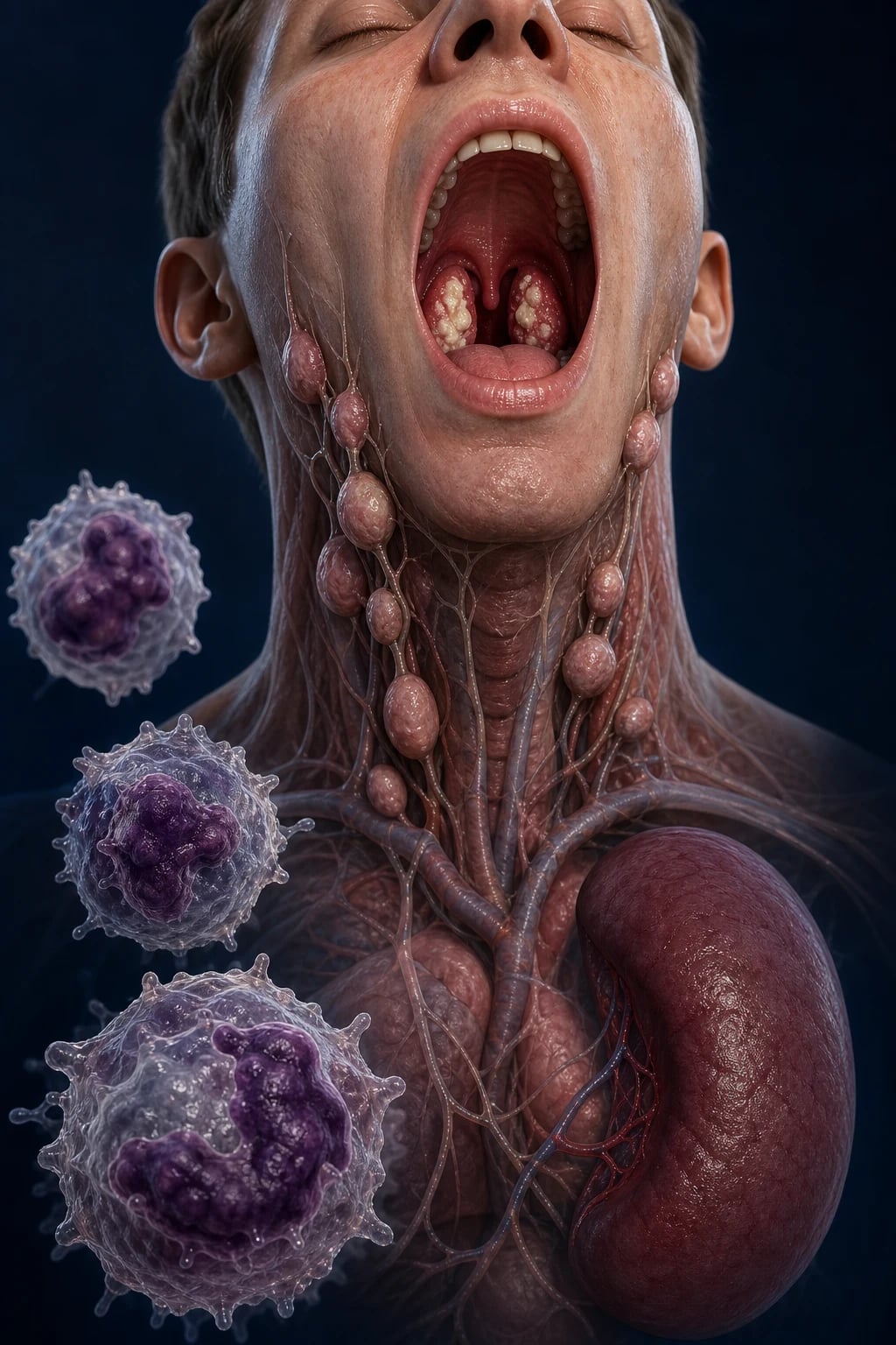
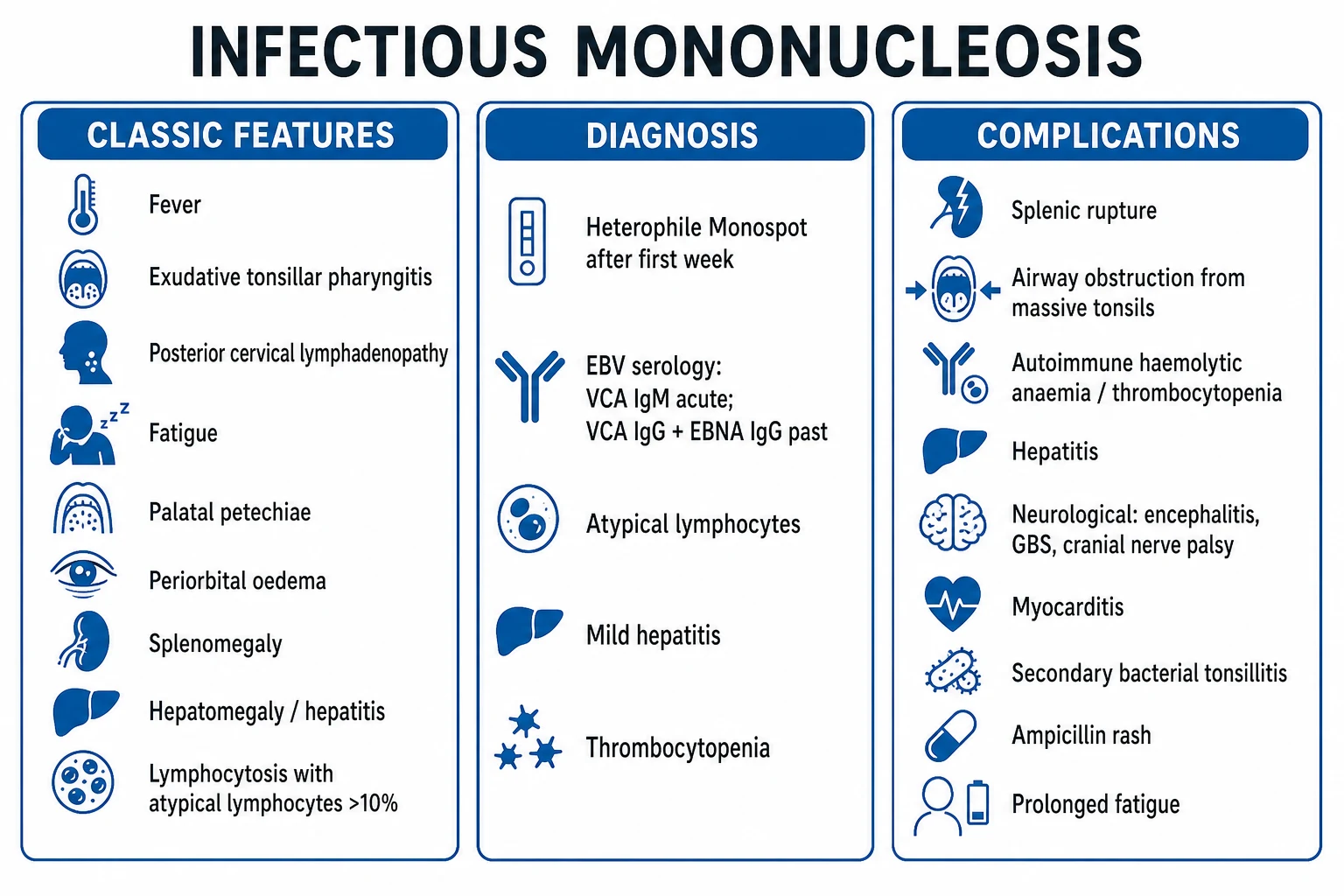
Infectious mononucleosis is the acute, self-limiting clinical syndrome of primary Epstein-Barr virus (EBV, human herpesvirus 4) infection, transmitted mainly by saliva ('kissing disease') and classically affecting adolescents and young adults. The classic triad is fever, exudative tonsillar pharyngitis and (especially posterior cervical) lymphadenopathy, with prolonged fatigue, palatal petechiae, splenomegaly, hepatitis and atypical lymphocytes on the blood film. EBV enters B lymphocytes via the CD21 (CR2) receptor; the atypical lymphocytes are reactive CD8+ cytotoxic T cells (Downey cells). Diagnosis is by heterophile antibody (Monospot) plus EBV-specific serology (VCA IgM = acute). Management is supportive (rest, analgesia); AVOID ampicillin/amoxicillin (diffuse rash), alcohol (hepatitis), and contact sport for at least 3 to 4 weeks (splenic rupture). Corticosteroids are reserved for impending airway obstruction, severe cytopenia/haemolysis or neurology. Key complications: splenic rupture, airway obstruction, autoimmune haemolytic anaemia/thrombocytopenia, neurological disease, and EBV-driven malignancy (Burkitt, Hodgkin, nasopharyngeal carcinoma, PTLD).
On this page & tools
Your progress
Saved locally on this device.
Exam tags
Red flags
Overview & Definition
Infectious mononucleosis (IM; "glandular fever") is the acute clinical syndrome produced by primary Epstein-Barr virus (EBV, human herpesvirus 4, HHV-4) infection, characterised by the triad of fever, exudative tonsillar pharyngitis and generalised (especially posterior cervical) lymphadenopathy, accompanied by prolonged fatigue, splenomegaly, mild hepatitis, and a reactive lymphocytosis with atypical lymphocytes on the blood film.[1][2]
The disease matters to the practising doctor less for its (usually self-limiting) course than for four specific pitfalls that examiners return to again and again: [1]
- The ampicillin/amoxicillin rash — diffuse, non-allergic, occurs in 80 to 100 percent of IM patients given these drugs; often mislabelled as penicillin allergy.[6]
- Splenic rupture — rare (around 0.1 to 0.5 percent) but potentially fatal; mandates activity restriction for at least 3 to 4 weeks.[7]
- Prolonged fatigue — symptoms may persist weeks to months; a small minority develop a chronic-fatigue-like syndrome.
- EBV oncogenicity — EBV drives endemic Burkitt lymphoma, Hodgkin lymphoma, nasopharyngeal carcinoma, EBV-associated gastric cancer, post-transplant lymphoproliferative disease (PTLD) and oral hairy leukoplakia in HIV; recent evidence links EBV to multiple sclerosis.[9][11]
EBV is the archetype of acute viral syndromes in young adults: the clinical task is to recognise the syndrome, confirm with Monospot and EBV serology, exclude mimics (group-A strep, CMV, toxoplasmosis, HIV seroconversion, acute hepatitis, leukaemia), and counsel the patient on drug avoidance and activity restriction while remaining alert to the rare but serious complications.[1][4]
Classification
EBV infection is classified by the clinical phenotype of primary infection and by its long-term consequences:[1][10]
- Asymptomatic / subclinical primary infection — the global norm; the vast majority of infections in early childhood go unnoticed.
- Symptomatic infectious mononucleosis — the classic adolescent triad; the exam archetype.
- EBV hepatitis — predominant transaminitis ± jaundice.
- EBV neurological disease — meningoencephalitis, Guillain-Barre syndrome, transverse myelitis, Bell palsy, cerebellitis.
- EBV haematological disease — autoimmune haemolytic anaemia, immune thrombocytopenia, aplastic anaemia, disseminated intravascular coagulation, haemophagocytic lymphohistiocytosis (HLH).
- Chronic active EBV disease (CAEBD) — persistent IM-like illness with high EBV load, organ damage, poor prognosis (especially East Asia).[8]
- EBV in the immunocompromised — post-transplant lymphoproliferative disease (PTLD); EBV-HLH in X-linked lymphoproliferative disease (Duncan syndrome).
- EBV-associated malignancies — endemic Burkitt lymphoma; Hodgkin lymphoma (nodular sclerosis and mixed cellularity); nasopharyngeal carcinoma; EBV-positive gastric cancer; extranodal NK/T-cell lymphoma.[9]
By aetiology (the "mononucleosis syndromes") — the term mononucleosis refers to the absolute lymphocytosis with atypical lymphocytes, not to EBV alone:[3]
- EBV mononucleosis — exudative pharyngitis + marked cervical adenopathy; heterophile positive.
- CMV mononucleosis — older patients; less pharyngitis/adenopathy, more fever and fatigue; heterophile negative.
- Toxoplasma mononucleosis — cervical nodes without pharyngitis; cat/meat exposure.
- HIV seroconversion illness — rash, mucosal ulcers, sexual/contact risk. [1]
Epidemiology & Risk Factors
EBV is ubiquitous: over 90 percent of the world's adults are seropositive, harbouring EBV in memory B cells for life with intermittent asymptomatic salivary shedding.[1][10]
- Age and geography shape presentation. In low- and middle-income countries (including India), most children are infected by age 3 to 5, usually asymptomatically — florid IM is uncommon. In high-income settings, primary infection is delayed to adolescence and young adulthood (15 to 24 years), where the risk of symptomatic IM is far higher (around 30 to 70 percent of primary infections in this age group).[1]
- Transmission. Spread is by intimate salivary contact ("kissing disease"), and less efficiently by shared utensils, toys and blood/transplant. The virus is shed in saliva for months after primary infection and intermittently for life. Incubation is 4 to 6 weeks.
- Risk factors: intimate oral contact in young adults, crowded living conditions, immunosuppression (solid-organ or haematopoietic stem-cell transplant recipients, HIV), and genetic susceptibility — notably X-linked lymphoproliferative disease (XLP, Duncan syndrome, SAP/SH2D1A mutation), in which primary EBV infection triggers fulminant, often fatal EBV-HLH.[10]
EBV by the numbers
Pathophysiology
EBV is a gamma-1 herpesvirus (Lymphocryptovirus): enveloped, double-stranded DNA, that has co-evolved to establish lifelong latent infection of B lymphocytes.[1][3]
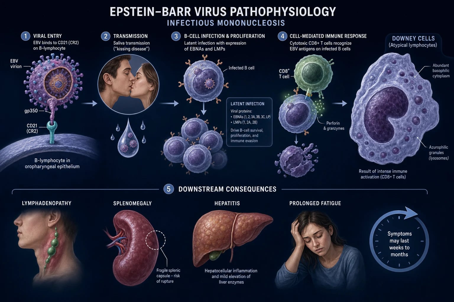
Entry. The viral envelope glycoprotein gp350/220 binds the CD21 (CR2) complement receptor on oropharyngeal epithelial cells and mature B lymphocytes. EBV initially replicates in the oropharyngeal (especially nasopharyngeal and tonsillar) epithelium, then infects B cells that trafficked through the mucosa. The viral genome circularises within the B-cell nucleus as an episome and establishes latency, expressing a restricted set of latent proteins: EBNA-1, EBNA-2, EBNA-3A/B/C, EBNA-LP, LMP-1 and LMP-2.[3][9]
Polyclonal B-cell activation. EBNA-2 and LMP-1 mimic constitutively active CD40 and BCR signalling, driving polyclonal B-cell proliferation — the basis of the lymphocytosis, the lymphadenopathy/splenomegaly, and (over a lifetime) EBV's contribution to B-cell lymphoid malignancy.[9]
The cellular immune response is the disease. The clinical illness of IM is not the cytopathic effect of EBV — it is the vigorous cytotoxic CD8+ T-cell response directed at EBV-infected B cells. The atypical lymphocytes on the blood film (the Downey cells) are these reactive, antigen-experienced CD8+ T cells — large cells with abundant basophilic cytoplasm that scallops around neighbouring erythrocytes. This is why the lymphocytosis of IM is reactive, not clonal — do not mistake florid atypical lymphocytosis for leukaemia.[1][3]
Organ consequences follow infiltration of lymphoid tissues by reactive T cells and infected B cells: [1]
- Lymph nodes and tonsils — reactive hyperplasia (cervical, generalised).
- Spleen — splenic congestion, sinusoidal and red-pulp infiltration by reactive lymphocytes, capsular distension and oedema. This is the pathological basis of splenic rupture: the spleen is enlarged, soft and friable, and rupture risk peaks at weeks 2 to 3 of illness. Even trivial trauma (and rarely no trauma at all) can rupture an IM spleen.[7]
- Liver — portal and sinusoidal lymphocytic infiltrate produces the mild hepatitis (transaminitis) seen in around 90 percent of patients; jaundice is uncommon (around 5 percent).
- Bone marrow — suppressed megakaryopoiesis and immune-mediated destruction contribute to mild thrombocytopenia (around 50 percent); rare severe autoimmune haemolysis (Coombs-positive with anti-i specificity), aplastic anaemia, or DIC.
- Nervous system — neural, meningeal and perivascular lymphocytic infiltrates underlie the EBV neurological complications (meningoencephalitis, Guillain-Barre, Bell palsy, transverse myelitis).
- Fatigue — the mechanism is poorly defined but probably cytokine-mediated (interferon-gamma, IL-6, TNF-alpha), reflecting a sustained immune activation.
Latency and oncogenesis. EBV persists for life in a small pool of circulating memory B cells as a silent latent episome, periodically reactivating in the oropharynx. EBV's latent genes (especially EBNA-2, LMP-1) are B-cell growth-transforming; when latency is dysregulated (immunosuppression, malaria, specific host genetics, c-myc translocation), EBV drives malignant B-cell and epithelial tumours.[9]
EBV is the master of latency programmes, switching between distinct gene-expression states to suit its niche — a recurring viva topic because these programmes map directly onto EBV's tumour associations:[9]
- Lytic (productive) replication — in oropharyngeal epithelium, EBV expresses the full cascade of immediate-early (BZLF1/Zebra, BRLF1), early (DNA polymerase, thymidine kinase) and late (structural capsid) proteins, assembling infectious virions shed into saliva. This is the phase that aciclovir targets (inhibiting the viral DNA polymerase) — which is precisely why aciclovir only suppresses shedding and does not treat established latency.
- Latency Type III (growth programme) — the pattern of fresh acute IM in vivo and of lymphoblastoid cell lines in vitro: expression of all EBNAs plus LMP-1 and LMP-2, driving polyclonal B-cell proliferation. This is the programme the CD8+ T-cell response is fighting in symptomatic IM.
- Latency Type II — EBNA-1 only plus LMP-1/LMP-2; the pattern of Hodgkin lymphoma and nasopharyngeal carcinoma, where LMP-1 provides the survival/proliferation signal.
- Latency Type I — EBNA-1 alone; the pattern of endemic Burkitt lymphoma, where the c-myc translocation (not LMP-1) is the oncogenic driver.
- Latency Type 0 — silent carriage in resting memory B cells with no latent protein expression; the lifelong reservoir. [1]
Understanding these programmes explains why EBV-driven tumours differ in immunogenicity and behaviour: latency I/II tumours are relatively "invisible" to the EBV-specific T-cell response, whereas the latency III programme of PTLD (under iatrogenic immunosuppression) is exquisitely sensitive to reduction of immunosuppression and rituximab.[9]
Clinical Presentation
The presentation of IM is dominated by age and immune competence.[1][2]
Prodrome (3 to 5 days): malaise, fatigue, anorexia, headache, myalgia, chills — easily mistaken for a nonspecific viral illness. [1]
Established IM (classical): [1]
- Fever — typically 38 to 40 degrees C, often peaking in the late afternoon/evening; settles within 1 to 2 weeks.
- Tonsillar pharyngitis — exudative; white, grey-white or yellow-green membrane on enlarged, erythematous tonsils, often with palatal petechiae (nearly pathognomonic). Dysphagia and odynophagia may be severe.
- Lymphadenopathy — tender, symmetric; posterior cervical chain larger than anterior, but generalised (cervical, axillary, epitrochlear, inguinal). Epitrochlear nodes are a useful pointer to a viral (rather than streptococcal) cause.
- Splenomegaly — palpable in around 50 percent of patients, usually peaking in the second to third week; the capsule is stretched and vulnerable.
- Hepatomegaly and hepatitis — transaminitis in around 90 percent (usually mild); frank jaundice in around 5 percent.
- Cutaneous — the maculopapular ampicillin/amoxicillin rash (see below); an EBV-related morbilliform or urticarial rash occurs in around 5 percent of untreated patients; periorbital oedema (Hoagland sign) in up to a third.
- Fatigue — often the most disabling symptom; the patient is exhausted by trivial activity. Fatigue may persist for weeks to months. [1]
The temporal course is itself diagnostic — knowing the expected arc helps the clinician distinguish a normal protracted recovery from a complication, and frames the return-to-play counselling:[2]
The natural history of infectious mononucleosis
Named signs of IM (high-yield for viva): [1]
Named signs of infectious mononucleosis
Palatal petechiae (Hall sign)
- Petechiae on the soft palate, near the junction with the hard palate
- Nearly pathognomonic for IM in the right clinical context
- Persist for several days to a week
Hoagland sign
- Periorbital (especially upper-eyelid) oedema
- Early morning; transient
- Attributed to lymphatic obstruction from cervical adenopathy
Ampicillin rash
- Diffuse, maculopapular, non-pruritic or mildly pruritic
- Occurs in 80-100 percent given ampicillin/amoxicillin (also some cefprozil)
- NOT a true penicillin allergy - mechanism is EBV-driven polyclonal B-cell activation
Atypical presentation — elderly. In older adults IM often presents with prolonged fever, jaundice and little lymphadenopathy, mimicking lymphoma, hepatitis, or an occult malignancy — heterophile may be weaker; send EBV serology.[10]
Immunocompromised / XLP. Primary EBV in X-linked lymphoproliferative disease (Duncan syndrome, SAP/SH2D1A mutation), post-transplant immunosuppression, or congenital immunodeficiency produces fulminant EBV-associated haemophagocytic lymphohistiocytosis (EBV-HLH): fever, hepatosplenomegaly, pancytopenia, ferritin often over 10,000 micrograms/L, hypertriglyceridaemia, hypofibrinogenaemia, haemophagocytosis on marrow, high EBV viral load. Mortality is high without specific therapy.[8]
Chronic active EBV disease (CAEBD). Persistent IM-like symptoms, organ involvement (hydroa vacciniforme-like skin lesions, myocarditis, lymphoid interstitial pneumonitis), persistent high EBV DNA, and progression to EBV-positive T/NK-cell lymphoproliferative disease. Curative treatment is haematopoietic stem-cell transplantation.[8]
Differential Diagnosis
A young adult with fever + sore throat + adenopathy is not always EBV. Distinguish:[1][5]
Differential diagnosis of the mononucleosis syndrome
Group-A streptococcal pharyngitis
- Abrupt onset, anterior cervical adenopathy, no splenomegaly, no atypical lymphocytes, no hepatomegaly
- Centor/McIsaac criteria; rapid strep test or throat swab/ASO
- Treat with phenoxymethylpenicillin (penicillin V) or cephalexin - NEVER ampicillin/amoxicillin when IM possible
CMV mononucleosis
- Older patients; less pharyngitis and lymphadenopathy; more fever, profound fatigue
- Heterophile NEGATIVE; CMV IgM positive; CMV DNA PCR if immunocompromised
- Course similar; supportive management
Toxoplasmosis
- Painless cervical lymphadenopathy WITHOUT pharyngitis or splenomegaly
- Cat contact / undercooked meat exposure; Toxoplasma IgM
- Heterophile negative; usually self-limiting in immunocompetent
HIV seroconversion illness
- Sexual/blood-borne exposure; mucosal ulcers, rash, diarrhoea
- HIV RNA (viral load) positive before antibodies; 4th-gen Ag-Ab
- Critical not to miss - check HIV in any mononucleosis-like illness
Acute viral hepatitis (A, B, E)
- Predominant jaundice and transaminitis; less pharyngitis/adenopathy
- HAV IgM, HBsAg/anti-HBc IgM, HEV
- Usually a prodrome then jaundice
Acute leukaemia / lymphoma
- Persistent cytopenias, weight loss, night sweats; nodes hard/fixed or supraclavicular
- Blood film - blast cells; flow cytometry; lymph node biopsy
- When 'atypical lymphocytes' are atypical, persistent or accompanied by cytopenias
Always specifically consider CMV, HIV seroconversion, and group-A strep in any apparent mononucleosis — these are the three most commonly missed. [1]
Clinical & Bedside Assessment
The diagnosis of IM is clinical, supported by Monospot and EBV serology; the bedside assessment must also screen for the complications that change management.[1][4]
- Vital signs and airway: fever pattern, hydration, and — critically — stridor, drooling, muffled 'hot-potato' voice, dyspnoea or intercostal recession indicating impending airway obstruction from massive tonsillar/adenoidal hypertrophy (a surgical emergency).
- Throat: exudate, palatal petechiae, exclude peritonsillar abscess.
- Lymph nodes: size, tenderness, distribution (cervical, epitrochlear, axillary, inguinal); hard/fixed or supraclavicular nodes suggest malignancy.
- Abdomen: examine gently for splenomegaly and hepatomegaly — do not palpate the spleen vigorously (rupture risk); assess for peritoneal signs / LUQ tenderness / shoulder-tip referred pain (Kehr sign) that would suggest splenic rupture.
- Skin: ampicillin rash, jaundice, periorbital oedema.
- Neurology: if any meningism, cranial-nerve deficit, weakness or altered consciousness — assess for EBV neurology (meningoencephalitis, Guillain-Barre, Bell palsy, transverse myelitis).
- Severity markers to escalate: airway compromise; splenic rupture (LUQ pain, falling Hb, shock); severe thrombocytopenia with bleeding; severe haemolysis (falling Hb, jaundice); neurological signs; HLH picture (high fever, hepatosplenomegaly, cytopenias, high ferritin) in immunocompromised. [1]
Investigations
First-line (the IM work-up):[1][4][5]
- Full blood count and film: lymphocytosis (greater than 50 percent or absolute lymphocytosis greater than 4.5 x 10⁹/L) with atypical lymphocytes over 10 percent of lymphocytes — the classic Downey cells. Historically described in three forms: Downey type I (small, round, hyperchromatic), type II (the commonest — large cells with abundant basophilic cytoplasm that scallops around neighbouring erythrocytes, the cell most undergraduates picture), and type III (large, plasmacytoid, deeply basophilic with a perinuclear hof). All three are reactive CD8+ cytotoxic T cells, not leukaemic blasts. Mild thrombocytopenia in around 50 percent; haemoglobin usually normal unless autoimmune haemolysis develops.
- Heterophile antibody test (Monospot / Paul-Bunnell-Davidsohn): detects IgM antibodies that agglutinate horse, sheep or bovine erythrocytes. The classical differential absorption distinguishes true heterophile from Forssman antibody: heterophile is absorbed by beef (ox) cells but NOT by guinea-pig kidney, while Forssman antibody is absorbed by guinea-pig kidney. Sensitivity around 85 percent, specificity around 97 percent in adults. Becomes positive in the first week, peaks at 2 to 3 weeks, may persist for up to a year. Negative in children under 4 years (who do not mount a heterophile response — only around 30 percent positive under age 12) and in the first few days of illness — recheck at week 2 or send EBV-specific serology. The heterophile test is negative in CMV, toxoplasmosis and HIV seroconversion, which is why a negative Monospot in a compatible illness demands those alternatives be sought.
- EBV-specific serology (definitive) — the pattern that determines disease phase (see table below).
- Liver function tests: AST/ALT typically 2 to 3 times upper limit (mild hepatitis in around 90 percent); bilirubin mildly raised; LDH raised (reflecting lymphocyte turnover); gamma-GT often mildly elevated. Coagulation usually normal; a rising INR suggests severe hepatitis or fulminant failure and mandates urgent hepatology input.
- Cold agglutinins — low-titre anti-i autoantibody detectable in around 50 to 70 percent during the second week; when titre is high it produces cold autoimmune haemolytic anaemia (complement-mediated, Coombs-positive with anti-C3). This is the mechanism of the haemolytic complication and the basis of the Donath-Landsteiner-type picture occasionally seen.
- EBV DNA PCR (quantitative): reserved for immunocompromised patients, CAEBD, suspected CNS disease (CSF), and PTLD monitoring — not for routine immunocompetent IM, where serology suffices. Whole-blood PCR is more sensitive than plasma in PTLD surveillance.
- EBV antibody kinetics (high-yield for exams): VCA IgM appears first (peaks at 2-3 weeks, fades by 3-6 months); VCA IgG appears early and persists for life; EA (early antigen, diffuse component) IgG appears in acute infection and fades over months; EBNA IgG appears late — only after 3 to 4 weeks — and persists for life. This late EBNA is the key discriminator: a patient with VCA IgM positive + EBNA IgG negative has acute IM, while VCA IgG positive + EBNA IgG positive = past infection. [1]
EBV-specific serology - how to interpret the pattern
Never infected (susceptible)
- VCA IgM negative, VCA IgG negative, EBNA IgG negative
- Patient is at risk of primary EBV in future
Acute / primary infection
- VCA IgM POSITIVE, VCA IgG positive (rising), EBNA IgG NEGATIVE
- The pattern that confirms acute IM - EBNA does not appear for 3-4 weeks
Recent / convalescent (past 3-12 months)
- VCA IgM may be trace-positive, VCA IgG positive, EBNA IgG becoming positive
- EA (early antigen) IgG often raised
Past infection (immune)
- VCA IgG POSITIVE, EBNA IgG POSITIVE, VCA IgM NEGATIVE
- Lifelong immunity - the resting pattern of >90 percent of adults
Reactivation / immunocompromised
- EA IgG raised, VCA IgG high titre, EBNA IgG variable
- Use EBV DNA PCR; clinical correlation required
Diagnostic algorithm — a stepwise approach that matches test to clinical context and avoids over-testing or mislabelling:[4][5]
Diagnostic approach to suspected infectious mononucleosis
Clinical suspicion
Adolescent/young adult with fever + exudative tonsillitis + posterior cervical lymphadenopathy + fatigue. Apply Centor/McIsaac to assess streptococcal overlap.
First-line tests
FBC and film (lymphocytosis + atypical lymphocytes >10%), LFTs (mild hepatitis), CRP, and heterophile (Monospot) in those over 12 years. Throat swab/rapid strep for co-infection.
Monospot positive
Confirms EBV IM in the right clinical context. No further serology needed in immunocompetent adults. Counsel on drug avoidance and activity restriction.
Monospot negative - recheck or escalate
If early illness (under 1 week), retest at week 2. If under 12 years, immunocompromised, or atypical presentation, send EBV-specific serology (VCA IgM, VCA IgG, EBNA IgG).
EBV serology negative
Actively exclude the mimics: CMV IgM, Toxoplasma IgM, HIV 4th-gen Ag-Ab + RNA, hepatitis serology, and blood film for leukaemia if cytopenias persist.
Complication screen if unwell
Abdominal ultrasound for spleen size/rupture (LUQ pain); Coombs and reticulocytes if falling Hb; CSF analysis + EBV PCR if neurological signs; ferritin/triglycerides/fibrinogen if HLH suspected.
Imaging. Abdominal ultrasound to measure spleen size and to screen for rupture (free intraperitoneal fluid) when LUQ pain is present. CT abdomen is more sensitive for rupture and characterises splenic laceration. Chest X-ray is unnecessary unless a complication (effusion, pneumonitis) is suspected. [1]
Testing for strep co-infection: use Centor / McIsaac criteria plus rapid strep or throat swab. Streptococcal co-infection occurs in around 5 to 30 percent of IM, but does not justify ampicillin/amoxicillin — use phenoxymethylpenicillin or cephalexin.[5]
Do NOT routinely send atypical-lymphocyte counts as a leukaemia work-up unless cells are bizarre, persistent, or accompanied by significant cytopenias — then blood film, flow cytometry and marrow. [1]
Management — Resuscitation
Most IM is ambulatory and self-limiting; resuscitation is reserved for rare, life-threatening complications.[1][7]
Airway obstruction (massive tonsillar/adenoidal hypertrophy, drooling, stridor):
- Urgent ENT and anaesthetic involvement.
- Sit upright, humidified oxygen, nebulised adrenaline if stridor.
- IV dexamethasone 0.6 mg/kg (max 10 mg) once, then oral prednisolone for a short course.
- Be prepared to secure the airway (nasopharyngeal airway, intubation by an expert, rarely tracheostomy). The inflamed, friable tissue makes this difficult. [1]
Splenic rupture (LUQ pain, shoulder-tip/Kehr sign, falling Hb, signs of shock):
- ABC, two large-bore IV cannulae, crossmatch, IV crystalloid resuscitation, treat for haemorrhagic shock.
- Urgent focused ultrasound / FAST or CT abdomen.
- Urgent surgical referral; many stable ruptures are managed non-operatively (observation, bed rest, serial haematocrit) — unstable or continuing bleed -> splenectomy / splenic artery embolisation. [1]
Autoimmune haemolytic anaemia (severe Coombs-positive with anti-i antibody, falling Hb):
- Transfuse if symptomatic; IV corticosteroids (prednisolone 1 mg/kg/day). [1]
Severe thrombocytopenia with bleeding:
- IVIG (1 g/kg/day for 1 to 2 days) and/or corticosteroids; platelet transfusion only if actively bleeding. [1]
EBV-HLH (XLP, immunocompromised): treat per the HLH-2004 protocol (etoposide, dexamethasone, ciclosporin), with EBV-targeted rituximab for B-cell disease; refer urgently to a haematology/immunology centre.[8]
Management — Definitive & Stepwise
The mainstay of treatment for uncomplicated IM is supportive care and patient counselling:[1][4][5]
1. Supportive care (the mainstay): [1]
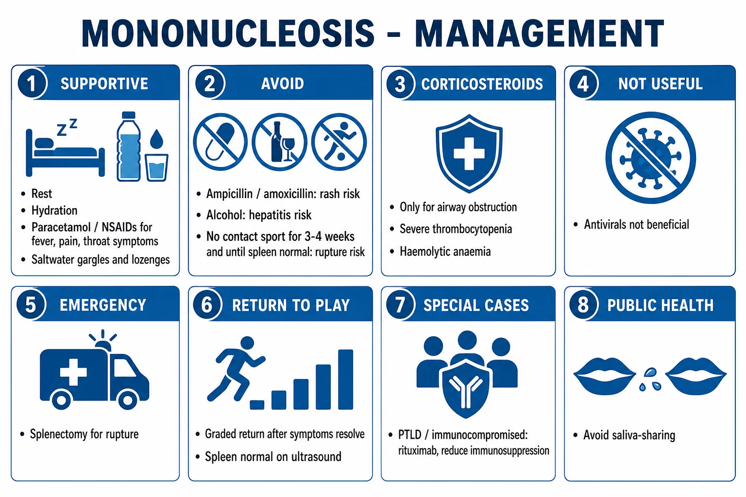
- Rest as tolerated — strict bed rest is not required unless severe; pace activity to symptoms.
- Adequate hydration and a nutritious diet.
- Antipyretic / analgesic: paracetamol 1 g orally every 6 hours (adult; max 4 g/day), or ibuprofen 400 mg every 8 hours for fever, throat pain and headache. Avoid codeine/opioids if airway compromise is possible.
- Salt-water gargles and throat lozenges for odynophagia. [1]
2. Drugs to AVOID: [1]
- AVOID ampicillin, amoxicillin, and (where possible) cefprozil — they trigger the diffuse maculopapular rash in 80 to 100 percent of IM patients.[6] If a beta-lactam is needed for proven streptococcal co-infection, use phenoxymethylpenicillin (penicillin V) 500 mg PO QID for 10 days, or cephalexin, or azithromycin if penicillin-allergic.
- AVOID alcohol during the hepatitis phase.
- AVOID contact sports, strenuous exercise, heavy lifting, and abdominal trauma for a minimum of 3 to 4 weeks from onset (some guidelines 4 weeks) — until the patient is asymptomatic, spleen non-palpable / normal on ultrasound, and bloods normal.[7]
3. Drugs that are NOT routinely indicated: [1]
- Corticosteroids are not recommended for symptom control in uncomplicated IM (Cochrane 2015 — no consistent benefit; theoretical risk of secondary bacterial infection and rebound). Reserve steroids for: impending airway obstruction; severe thrombocytopenia with bleeding; autoimmune haemolytic anaemia; severe neurological complications; HIV-associated EBV.[4] Typical regimen: prednisolone 0.5 to 1 mg/kg/day (max 60 mg) for 5 to 10 days, tapered.
- Antivirals (aciclovir, valaciclovir, ganciclovir) reduce oropharyngeal EBV shedding but do not alter the clinical course of uncomplicated IM — not recommended.[4]
4. Activity restriction and return-to-play:[7]
- 3 weeks minimum off all contact sport / vigorous exertion in all patients (including those without palpable splenomegaly — the spleen may be enlarged and friable without being clinically palpable).
- 4 weeks for competitive athletes; some sports-medicine guidance requires an ultrasound at 3 to 4 weeks confirming the spleen has returned to normal before return.
- Graded return to activity once asymptomatic, spleen normal, bloods normal. [1]
5. Patient counselling: explain the disease course (2 to 3 weeks acute; fatigue may persist weeks to months), drug avoidance, activity restriction, and the warning signs that mandate return — LUQ or shoulder-tip pain, dyspnoea, bleeding, severe headache, weakness, confusion. [1]
Specific Subtypes & Scenarios
- Classic adolescent IM — the archetype; managed supportively with the counselling above.
- Childhood EBV — usually asymptomatic or mild; heterophile often negative — rely on EBV serology (VCA IgM). Manage supportively.
- CMV mononucleosis — the commonest mimic; older patients, less pharyngitis/adenopathy, heterophile negative, CMV IgM positive. Manage supportively.
- EBV hepatitis — predominant transaminitis ± jaundice; usually self-limiting; supportive.
- EBV neurological disease — meningoencephalitis (CSF lymphocytic pleocytosis, EBV DNA PCR positive), Guillain-Barre syndrome (ascending paralysis — support/IVIG), Bell palsy, transverse myelitis, cerebellitis. Steroids considered for severe disease; supportive + neurology input.
- EBV haematological disease — autoimmune haemolytic anaemia (anti-i antibody, Coombs positive; transfuse ± steroids), immune thrombocytopenia (steroids/IVIG), rare aplastic anaemia, DIC, EBV-HLH.
- Chronic active EBV disease (CAEBD) — persistent IM-like illness, high EBV load, organ damage (skin, marrow, heart, lung); haematopoietic stem-cell transplantation is the only curative therapy.[8]
- PTLD in transplant recipients — driven by EBV in B cells under immunosuppression; managed by reducing immunosuppression, rituximab (anti-CD20) for CD20-positive disease, and immunochemotherapy (R-CHOP) for overt lymphoma. Monitor EBV DNA viral load post-transplant.[9]
- EBV-associated malignancies (lifetime risk): endemic (African) Burkitt lymphoma (EBV in over 95 percent; t(8;14) c-myc translocation; jaw mass in children), Hodgkin lymphoma (EBV in around 40 percent overall, higher in mixed-cellularity and HIV), nasopharyngeal carcinoma (almost 100 percent EBV; South Chinese / Cantonese predisposition), EBV-positive gastric cancer (around 10 percent of gastric adenocarcinomas), extranodal NK/T-cell lymphoma (nasal type), and oral hairy leukoplakia (HIV, on the lateral tongue).[9]
Complications & Pitfalls
Haematological: autoimmune haemolytic anaemia (Coombs-positive, anti-i; 1 to 3 percent), immune thrombocytopenia, rare aplastic anaemia, DIC, EBV-HLH (especially XLP/immunocompromised).[1]
Splenic: splenic rupture (around 0.1 to 0.5 percent; peaks weeks 2 to 3; LUQ pain, Kehr sign, falling Hb; some cases atraumatic).[7]
Airway: impending airway obstruction from massive tonsillar/adenoidal hypertrophy (rare, but a surgical emergency; more common in children and young adults). [1]
Hepatic: mild hepatitis in around 90 percent; jaundice in around 5 percent; rare fulminant hepatic failure; pancreatitis uncommon. [1]
Neurological (1 to 5 percent): meningoencephalitis, Guillain-Barre syndrome, transverse myelitis, Bell palsy, optic neuritis, cerebellitis. [1]
Cardiac (rare): myocarditis, pericarditis, rarely heart block. [1]
Renal: interstitial nephritis, microscopic haematuria. [1]
Pulmonary: interstitial pneumonitis, pleural effusion. [1]
Classic pitfalls:
- Mislabelling the ampicillin rash as true penicillin allergy — most patients tolerate penicillins later.[6]
- Missing splenic rupture in a patient with LUQ/shoulder pain sent home with "just glandular fever".
- Over-treating uncomplicated IM with steroids or antivirals.
- Failing to consider CMV, HIV seroconversion, toxoplasmosis in a heterophile-negative illness.
- Missing EBV-HLH/PTLD in the immunocompromised patient with fever and cytopenias.
- Prematurely diagnosing leukaemia on atypical lymphocytosis — IM is reactive, not clonal.
Prognosis & Disposition
- Acute illness (fever, pharyngitis) settles in 1 to 2 weeks; lymphadenopathy and splenomegaly over 3 to 6 weeks.
- Fatigue persists weeks to months in around 10 to 20 percent; a small minority meet criteria for chronic fatigue syndrome-like illness; reassurance and graded activity are key.
- EBV infection is lifelong — intermittent reactivation and salivary shedding occur; the oncogenic potential persists for life.
- Overall mortality is very low — death is from splenic rupture, airway obstruction, neurological disease, HLH, or underlying immunodeficiency (XLP). [1]
Disposition. Almost all patients are managed as outpatients with supportive care and counselling. Admit for: [1]
- Airway compromise (ENT/anaesthetic/HDU).
- Splenic rupture (surgical).
- Severe cytopenias / haemolysis / bleeding (haematology).
- Neurological complications (neurology, monitor for GBS respiratory involvement).
- Severe hepatitis / dehydration.
- Immunocompromise / HLH / PTLD (specialist team).
- Social concerns (inability to manage at home, returning student). [1]
Special Populations
- Athletes and the military. Serial ultrasound of the spleen; NO contact sport for a minimum of 3 to 4 weeks (competitive athletes often 4 weeks), and graded return-to-play only once asymptomatic with a normal spleen and normal bloods. Document the counselling — splenic rupture is a leading cause of avoidable death in this group.[7]
- Pregnancy. Primary EBV is rare in pregnancy (most women are already seropositive); where it occurs, the disease is self-limiting and no proven teratogenicity has been shown — CMV, not EBV, is the herpesvirus that causes congenital harm. Manage supportively; avoid ampicillin/amoxicillin and contact sport.
- Immunocompromised / transplant. Monitor EBV DNA viral load; reduce immunosuppression at the first sign of PTLD; rituximab for CD20-positive PTLD; R-CHOP for overt lymphoma. Primary EBV can be fulminant.[9]
- X-linked lymphoproliferative disease (Duncan syndrome, SAP/SH2D1A mutation). Primary EBV triggers fulminant EBV-HLH with high mortality; refer urgently for HLH-2004 therapy and consider haematopoietic stem-cell transplantation.[8]
- Children. Usually mild or asymptomatic; heterophile negative — send EBV serology; supportive care.
- Elderly. Atypical — prolonged fever, jaundice, less adenopathy; send EBV serology and exclude malignancy/HIV.
Evidence, Guidelines & Regional Differences
- Cochrane 2015 (Reitman et al., Cochrane Database Syst Rev). Antivirals (aciclovir, valaciclovir) and corticosteroids do not improve symptom control in uncomplicated IM; steroids are not recommended routinely.[4]
- Systematic review of splenic rupture (Bartlett et al., Injury 2016). Rupture is rare but peaks at week 2; many cases are managed non-operatively; case fatality is low with prompt recognition.[7]
- Updated CAEBV guidelines (Kawada et al., Int J Hematol 2023). Haematopoietic stem-cell transplantation is the only curative therapy for CAEBD; EBV DNA load thresholds and rituximab/cytotoxic chemo bridge to transplant.[8]
- EBV and multiple sclerosis (Soldan & Lieberman, Nat Rev Microbiol 2023). A large US military cohort (Bjornevik 2022, NEJM) showed that EBV seroconversion precedes essentially all MS, establishing EBV as the single strongest known risk factor for MS.[11]
- EBV and cancer (Farrell, Annu Rev Pathol 2019). EBV contributes to around 1.5 percent of the global cancer burden through Burkitt, Hodgkin, NPC and EBV+ gastric carcinoma.[9]
- Regional note (India). Most Indian children are EBV-seropositive before adolescence, so florid IM is uncommon; a young adult with apparent IM should prompt exclusion of CMV, HIV seroconversion, dengue, enteric fever and leptospirosis — all of which mimic the syndrome in the Indian context. Use EBV-specific serology (VCA IgM) rather than relying on Monospot in young children, in whom heterophile is often negative.[1]
Cochrane 2015: Corticosteroids and antivirals in uncomplicated IM
PMID 25822555
Systematic review of 7 RCTs (corticosteroids, 750 patients) + antiviral trials
Population: Adolescents/adults with confirmed or suspected uncomplicated EBV mononucleosis
Key finding
No consistent benefit of corticosteroids on duration or severity of symptoms at 1-2 weeks; Modest transient reduction in throat pain at day 2-4 only (not sustained); Antivirals reduce oropharyngeal EBV shedding but do not alter clinical recovery or symptom duration; No evidence of harm, but theoretical risk of secondary bacterial infection and corticosteroid side-effects
Exam Pearls
[1]SPLAAT — the complications of mononucleosis
Exam application bank (NEET-PG / INICET)
One-line answer
Infectious mononucleosis is the acute, self-limiting clinical syndrome of primary Epstein-Barr virus (EBV, human herpesvirus 4) infection, transmitted mainly by saliva ('kissing disease') and classically affecting adolescents and young adults. The classic triad is fever, exudative tonsillar pharyngitis and (especially posterior cervical) lymphadenopathy, with prolonged fatigue, palatal petechiae, splenomegaly, hepatitis and atypical lymphocytes on the blood film. EBV enters B lymphocytes via the CD21 (CR2) receptor; the atypical lymphocytes are reactive CD8+ cytotoxic T cells (Downey cells). Diagnosis is by heterophile antibody (Monospot) plus EBV-specific serology (VCA IgM = acute). Management is supportive (rest, analgesia); AVOID ampicillin/amoxicillin (diffuse rash), alcohol (hepatitis), and contact sport for at least 3 to 4 weeks (splenic rupture). Corticosteroids are reserved for i
Worked stems (answer without another resource)
Stem 1 — Classic presentation. Map symptoms to mechanism; name the first investigation and first treatment step with dose/route if drug therapy is standard. [1]
Stem 2 — Unstable / complicated. List red flags that force immediate resuscitation, theatre, ICU, antidote, or reperfusion — and what you do in the first 15 minutes. [1]
Stem 3 — Atypical group. Elderly, pregnancy, child, or immunocompromised: how presentation and thresholds change. [1]
Stem 4 — Differential trap. Name the three closest mimics and one discriminator for each. [1]
Stem 5 — Disposition. Who goes home with safety-netting, who is admitted, who needs HDU/ICU/theatre, and what follow-up is mandatory. [1]
Rapid viva checklist
- Definition + classification
- Pathophysiology chain
- Bedside signs / criteria
- Score with exact components (if any)
- Emergency bundle
- Definitive therapy with doses
- Complications of disease and of treatment
- Special populations
- Guideline/trial name if classic
- Three exam traps
Coverage self-check
If you cannot answer any stem above from this page alone, re-read the matching section — the page is intended to be self-sufficient for final-prof and NEET-PG/INICET questions on Infectious Mononucleosis (Epstein-Barr Virus).
References
- [1]Dunmire SK, Hogquist KA, Balfour HH. Infectious Mononucleosis Curr Top Microbiol Immunol, 2015.PMID 26424648
- [2]Leung AKC, Lam JM, Barankin B, et al. Infectious Mononucleosis: An Updated Review Curr Pediatr Rev, 2024.PMID 37526456
- [3]Luzuriaga K, Sullivan JL. Infectious mononucleosis N Engl J Med, 2010.PMID 20505178
- [4]Sylvester JE, Buchanan BK, Silva TW. Infectious Mononucleosis: Rapid Evidence Review Am Fam Physician, 2023.PMID 36689975
- [5]Womack J, Jimenez M. Common questions about infectious mononucleosis Am Fam Physician, 2015.PMID 25822555
- [6]Thompson DF, Ramos CL. Antibiotic-Induced Rash in Patients With Infectious Mononucleosis Ann Pharmacother, 2017.PMID 27620494
- [7]Bartlett A, Williams R, Hilton M. Splenic rupture in infectious mononucleosis: A systematic review of published case reports Injury, 2016.PMID 26563483
- [8]Kawada JI, Ito Y, Ohshima K, et al. Updated guidelines for chronic active Epstein-Barr virus disease Int J Hematol, 2023.PMID 37728704
- [9]Farrell PJ. Epstein-Barr Virus and Cancer Annu Rev Pathol, 2019.PMID 30125149
- [10]Fugl A, Andersen CL. Epstein-Barr virus and its association with disease - a review of relevance to general practice BMC Fam Pract, 2019.PMID 31088382
- [11]Soldan SS, Lieberman PM. Epstein-Barr virus and multiple sclerosis Nat Rev Microbiol, 2023.PMID 35931816