Infectious Diseases · General Medicine
Intra-Abdominal Infection & Peritonitis
Also known as Peritonitis · Intra-abdominal infection · Spontaneous bacterial peritonitis · SBP · Secondary peritonitis · Tertiary peritonitis · Intra-abdominal abscess
Intra-abdominal infection (IAI) and peritonitis are time-critical emergencies that span two very different diseases. Primary peritonitis — spontaneous bacterial peritonitis (SBP) — is infection of cirrhotic ascites without an intra-abdominal source, presenting subtly (fever, abdominal pain, hepatic encephalopathy, renal failure) and treated medically with cefotaxime/ceftriaxone plus IV albumin (the ascitic neutrophil count over 250 cells per mm³ is diagnostic). Secondary peritonitis is polymicrobial contamination of the peritoneum from a perforated or translocating hollow viscus (perforated peptic ulcer, appendicitis, diverticulitis, ischaemic bowel, post-operative leak, trauma) and is a surgical emergency: resuscitation, broad-spectrum antibiotics covering enteric Gram-negatives and anaerobes, and urgent source control. Tertiary peritonitis is persistent or recurrent infection in the critically ill, often with multi-drug-resistant organisms (Pseudomonas, Enterococcus, Candida). Presents with abdominal pain, peritonism (rigidity, guarding, rebound tenderness), fever, sepsis and ileus. Diagnose with diagnostic ascitic tap (PMN over 250), CT abdomen with contrast and blood cultures. Treat with antibiotics plus source control (surgery or radiological drainage); SBP additionally receives albumin to prevent hepatorenal syndrome.
On this page & tools
Your progress
Saved locally on this device.
Exam tags
Red flags
Overview & Definition
Peritonitis is inflammation of the parietal and visceral peritoneum, the serous membrane that lines the abdominal cavity and invests its organs (the total peritoneal surface area is about 1.7 m² in an adult, comparable to the skin).[5] Intra-abdominal infection (IAI) extends the term to include infection of the abdominal viscera and/or the peritoneal space, with or without abscess formation. The two overlap and are managed by the same principles — antibiotics plus source control plus resuscitation — but the aetiology, organisms, definitive treatment and prognosis differ sharply between the major subtypes.[4][5]
The clinical art in peritonitis is NOT making the diagnosis (acute generalised peritonitis is unmistakable at the bedside) but in distinguishing the three mechanistic classes, because that distinction drives everything that follows:[1][5]
- A cirrhotic with ascites and fever or encephalopathy needs a diagnostic tap and cefotaxime plus albumin (SBP — a medical peritonitis; surgery will not help).
- A previously well patient with sudden severe peritonism and sepsis needs CT, broad-spectrum antibiotics and urgent surgery/source control (secondary peritonitis from a perforated viscus).
- A critically ill ICU patient with persistent fever, leucocytosis and organ dysfunction despite prior source control has tertiary peritonitis with MDR organisms and needs broadened empiric cover and reoperation. [1]
In all three, the unifying principles are early antibiotics, source control and resuscitation; in secondary peritonitis specifically, delay in source control is fatal. The two biggest process levers in mortality reduction are (1) diagnostic paracentesis in every cirrhotic with ascites (catches SBP while still treatable) and (2) early source control in secondary peritonitis within hours of recognition.[1][6]
Classification
Peritonitis is classified by mechanism (the axis that decides treatment), then by extent, duration and route.[1][5]
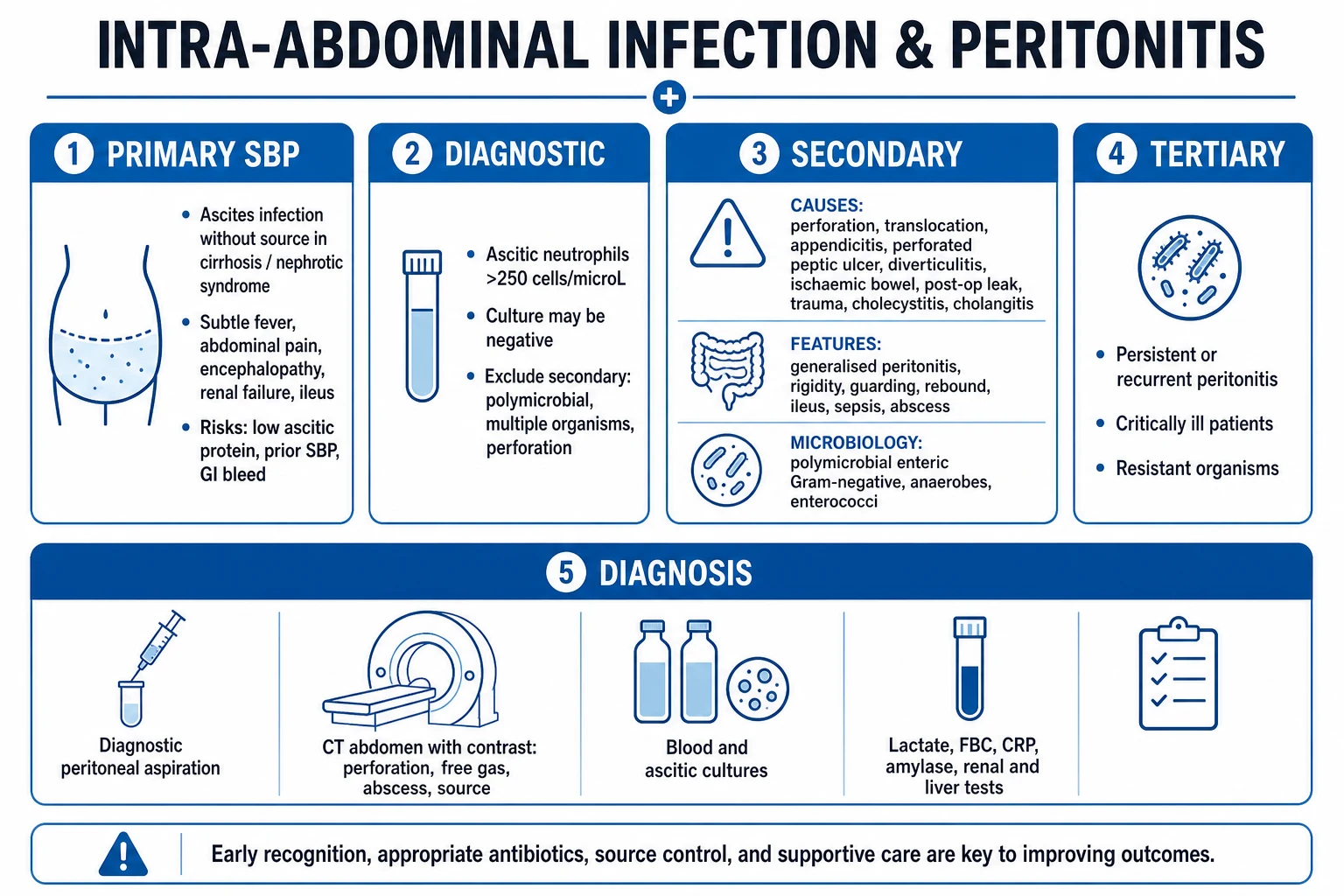
By mechanism — the classification that decides treatment
PRIMARY (SBP)
- Infection of ascites WITHOUT an intra-abdominal source
- Setting: cirrhosis with ascites, nephrotic syndrome, CLD
- Monomicrobial (one organism on culture)
- Organisms: E. coli, Klebsiella, S. pneumoniae; anaerobes are RARE
- Ascitic PMN over 250 cells/mm³
- Treat medically: cefotaxime/ceftriaxone + albumin; NO surgery
SECONDARY
- Perforation or translocation of a hollow viscus INTO the peritoneum
- Sources: perforated peptic ulcer, appendicitis, diverticulitis, ischaemic bowel, post-op leak, trauma, cholecystitis, anastomotic leak
- Polymicrobial: Gram-negatives + anaerobes (Bacteroides fragilis) + Enterococcus
- Ascitic fluid: PMN very high, glucose under 50 mg/dL, LDH high, polymicrobial
- Treat: broad-spectrum antibiotics + URGENT source control (surgery/drainage)
TERTIARY
- Persistent or recurrent peritonitis over 48 h despite adequate source control
- Critically ill ICU patients, post-operative, immunosuppressed
- MDR organisms: Pseudomonas aeruginosa, Enterococcus (VRE), Candida, MRSA, Acinetobacter
- Often low-grade, persistent fever, organ dysfunction
- Treat: re-operation, broad empiric cover incl. antifungal, source control
By extent
- Generalised (diffuse) peritonitis — contamination spread throughout the peritoneal cavity; the classic acute abdomen with rigidity, guarding and rebound. Surgical emergency.
- Localised peritonitis — inflammation walled off around a source (e.g. RIF in appendicitis, RUQ in cholecystitis); may progress to abscess if not drained. [1]
By duration and special forms
- Acute (hours–days) — the common surgical pattern.
- Chronic / granulomatous — tuberculous peritonitis, fungal peritonitis; presents insidiously with fever, ascites, weight loss.
- Chemical peritonitis — sterile inflammation from bile, gastric acid, pancreatic juice, blood or urine released into the peritoneum (e.g. early perforated peptic ulcer, post-ERCP); becomes secondary bacterial within hours as bacteria translocate.
- Peritoneal dialysis-associated peritonitis — special entity in CAPD/PD patients; touch preparation and effluent cell count; intraperitoneal antibiotics (see Specific Subtypes).[5]
Variants of ascitic infection in cirrhosis
In cirrhosis, the AASLD defines three related entities, distinguished by ascitic PMN count and culture:[1]
- SBP — ascitic PMN over 250 cells/mm³ with a positive culture (monomicrobial, no surgical source).
- Culture-negative neutrocytic ascites (CNNA) — ascitic PMN over 250 cells/mm³ with negative culture, in a symptomatic patient; treated identically to SBP.
- Bacterascites — positive culture with a normal ascitic PMN (under 250 cells/mm³); treat if symptomatic or repeat the tap.
- Secondary bacterial peritonitis — polymicrobial ascites (with anaerobes) OR a very high PMN with low glucose (under 50 mg/dL), high LDH, high protein (over 1 g/dL), high amylase or CEA (over 5) — implies a perforation; search for the source with CT.[1]
Epidemiology & Risk Factors
Peritonitis / IAI — key numbers
Spontaneous bacterial peritonitis complicates cirrhotic ascites in roughly 10–30% of cirrhotics at some point; the one-year mortality after an episode is about 30–50%, and recurrence is up to 70% within one year without secondary prophylaxis.[1] Risk factors for SBP are the conditions that combine low ascitic defence (low opsonic activity) with bacterial over-translocation:[1]
- Low ascitic total protein (under 1 g/dL) — the single most powerful risk factor; reflects low complement and opsonic activity.
- Advanced liver disease (high Child-Pugh / MELD score, bilirubin over 2.5 mg/dL).
- Platelet count under 116 x 10⁹/L (a marker of severe portal hypertension).
- Prior SBP (recurrence rate up to 70% at one year).
- Acute gastrointestinal bleeding (variceal) — bacterial translocation and bacteraemia.
- Urinary tract infection and invasive procedures (IV cannulation, endoscopy, catheterisation).
- High serum creatinine (over 1 mg/dL) and hyponatraemia (under 130 mEq/L). [1]
Secondary peritonitis is the commonest surgical abdominal emergency. Causes by age: appendicitis dominates in the young; perforated peptic ulcer, perforated diverticulitis and mesenteric ischaemia in the elderly. Post-operative anastomotic leak is an increasingly important cause in the surgical ICU.[2][5]
Tertiary peritonitis arises in the critically ill: prolonged ICU stay, prior broad-spectrum antibiotics, immunosuppression, post-operative state, multi-organ failure. The organisms are MDR by selection.[5]
Pathophysiology
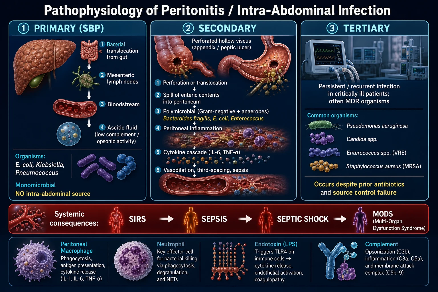
Primary peritonitis (SBP) — bacterial translocation
The hallmark is infection of ascitic fluid without an intra-abdominal surgical source. The pathogenesis has four steps:[1]
- Bacterial translocation — disruption of the intestinal mucosal barrier (cirrhosis-induced increased intestinal permeability, bacterial overgrowth from reduced motility) allows enteric bacteria to reach mesenteric lymph nodes, then the bloodstream.
- Impaired reticuloendothelial (Kupffer cell) function in cirrhosis — bacteria that reach the portal/systemic circulation are not cleared.
- Ascitic fluid with low defence — cirrhotic ascites has low complement (C3) and low opsonic activity, which scales directly with ascitic total protein; protein under 1 g/dL is essentially defenceless.
- Bacterial seeding of ascites — the most defenceless compartment in the body becomes infected. The result is a monomicrobial infection (because only one organism succeeds in establishing), most often E. coli, Klebsiella pneumoniae or Streptococcus pneumoniae. Anaerobes are rare in SBP (they do not translocate easily); their presence should prompt a search for secondary peritonitis.[1]
Secondary peritonitis — polymicrobial contamination
The peritoneum is flooded by enteric contents from a perforated or translocating viscus. The cascade is:[5]
- Contamination — gastric juice, bile, small bowel contents, faecal matter or blood enters the peritoneum. Initial inflammation may be chemical (sterile) but becomes bacterial within hours.
- Mesothelial and peritoneal macrophage activation — release of IL-1, IL-6, TNF-alpha, leukotrienes and complement activation.
- Neutrophil influx into the peritoneal cavity (ascitic PMN rises sharply).
- Cytokine amplification — local cytokines spill into the systemic circulation producing SIRS, then sepsis, septic shock and multi-organ dysfunction syndrome (MODS).
- Splanchnic vasodilation, capillary leak and third-spacing — large volumes of fluid sequester into the peritoneum; hypovolaemia, abdominal compartment syndrome and shock follow.
- Localisation / walled-off infection — omentum and adjacent loops wall off contamination, forming an intra-abdominal abscess if source control fails. [1]
The microbial load is polymicrobial: any perforation distal to the ligament of Treitz yields Gram-negative aerobes (E. coli, Klebsiella, Proteus), anaerobes (Bacteroides fragilis — the dominant anaerobe), and Enterococcus. Pseudomonas emerges with healthcare exposure.[4]
Tertiary peritonitis — failure of source control
In the critically ill, prior broad-spectrum antibiotics and impaired immunity select for low-virulence but multi-drug-resistant organisms — Pseudomonas aeruginosa, Enterococcus (including VRE), Candida species, MRSA, Acinetobacter, Stenotrophomonas. The peritoneum is unable to clear even modest contamination; biofilm on foreign material and necrotic tissue sustain infection. Clinical signs are often blunted in the ICU patient (sedation, analgesia, mechanical ventilation mask the peritonism).[5]
The cascade to sepsis, shock and organ failure
All three subtypes converge on the SIRS → sepsis → septic shock → MODS pathway through the cytokine storm, endotoxin (LPS)-mediated nitric oxide release and mitochondrial dysfunction. Tissue hypoperfusion manifests as lactataemia, oliguria, altered mental status and coagulopathy (DIC). The peritoneum itself contributes a large third space that worsens hypovolaemia. In cirrhosis, SBP is the classic precipitant of hepatorenal syndrome (HRS-AKI): the systemic inflammatory response causes splanchnic vasodilation, effective arterial underfilling, activation of the renin-angiotensin-aldosterone system and sympathetic nervous system, and intense renal vasoconstriction — the rationale for IV albumin, which expands the effective arterial volume and breaks this loop.[3][8]
Clinical Presentation
Generalised (secondary) peritonitis — the classic acute abdomen
The patient lies still, motionless, with shallow breathing because any movement worsens the pain. Key features:[5]
- Severe, constant, poorly localised abdominal pain that is worse on movement, coughing and deep inspiration. Pain may be referred to the shoulder from diaphragmatic irritation.
- Peritonism — tenderness, guarding (voluntary then involuntary), rigidity (board-like abdomen) and rebound tenderness.
- Absent or markedly reduced bowel sounds (paralytic ileus).
- Fever, tachycardia, tachypnoea, then hypotension, oliguria and altered mental status as sepsis progresses.
- Signs of the underlying source — epigastric guarding and free gas (perforated peptic ulcer); RIF signs (appendicitis); LIF signs (diverticulitis); RUQ (cholecystitis); distension and obstipation (obstruction/ischaemia). [1]
Spontaneous bacterial peritonitis — the subtle presentation in cirrhosis
SBP may present overtly (fever, abdominal pain) but is frequently subtle or asymptomatic. A diagnostic tap is therefore mandatory in any cirrhotic with ascites who has any of the following:[1]
- Abdominal pain or tenderness.
- Fever (over 37.8°C) or rigors.
- New or worsening hepatic encephalopathy (confusion, drowsiness, asterixis).
- Worsening renal function (rising creatinine, falling urine output).
- GI bleeding, paralytic ileus, diarrhoea or worsening ascites.
- Routine tap on admission in any cirrhotic with ascites hospitalised for any reason. [1]
A high index of suspicion is essential because up to a third of SBP cases have no abdominal symptoms. [1]
Atypical presentations
Elderly
- Confusion may be the only sign
- Absence of fever is common
- Mild or poorly localised pain
- Perforation often from diverticulitis or ischaemic bowel
Diabetic
- Blunted inflammatory response (neuropathy)
- Pain may be mild despite advanced peritonitis
- Higher risk of emphysematous complications
Immunocompromised / neutropenic
- Signs markedly blunted (no neutrophils to mount peritonism)
- Atypical organisms: Pseudomonas, Candida, moulds
- Low threshold for CT and broad empiric therapy
Pregnant
- Appendicitis is the commonest surgical emergency
- Appendix shifts superiorly and laterally with uterine enlargement
- Physiological leucocytosis of pregnancy confounds CBC
- Laparoscopic appendectomy is safe in any trimester
Post-operative
- Anastomotic leak typically day 5–7
- Pain/ileus may be attributed to surgery
- Unexplained tachycardia, fever or oliguria = suspect leak
- CT with water-soluble contrast
Localising signs by source
- Right upper quadrant — perforated peptic ulcer, acute cholecystitis, cholangitis.
- Right iliac fossa — appendicitis (most common surgical cause in the young), Meckel diverticulitis, right-sided colonic pathology.
- Left iliac fossa — perforated diverticulitis (most common in the elderly), sigmoid pathology.
- Pelvic — pelvic abscess: diarrhoea, urinary frequency, tenesmus, a tender boggy anterior mass on digital rectal examination.
- Diffuse — perforated peptic ulcer (free gas throughout), ischaemic bowel, post-operative leak, primary peritonitis. [1]
Differential Diagnosis
The differential of the acute abdomen with peritonism is broad. The discriminating features below separate the commonest mimics.[2][5]
Perforated peptic ulcer
- Sudden onset, severe epigastric pain
- History of peptic ulcer / NSAID use
- Free gas under diaphragm on erect CXR
- Board-like rigidity, 'vanished liver dullness'
Acute pancreatitis
- Epigastric pain radiating to back
- Lipase over 3x ULN
- No free gas (sterile inflammation)
- CT: enlarged pancreas, stranding, necrosis
Acute appendicitis
- Periumbilical → RIF migration
- Anorexia, nausea, low-grade fever
- McBurney point tenderness, Rovsing sign
- Alvarado score high
Mesenteric ischaemia
- Pain out of proportion to examination
- Metabolic acidosis, raised lactate
- History of AF / vascular disease
- CT angiography is diagnostic
Ruptured AAA
- Elderly male, back/flank pain, collapse
- Pulsatile abdominal mass
- Hypotension, haemorrhagic shock
- Straight to theatre if unstable — no CT
Ruptured ectopic pregnancy
- Young woman, amenorrhoea, PV bleeding
- Shoulder-tip pain, shock
- Positive beta-hCG, empty uterus on US
- Surgical emergency
Diabetic ketoacidosis
- Acute abdomen, vomiting, dehydration
- Hyperglycaemia, ketones, acidosis
- Treat DKA — pain resolves; avoid laparotomy
- Beware pseudoperitonitis
Lower-lobe pneumonia
- Abdominal pain referred from diaphragmatic pleura
- Cough, dyspnoea, productive sputum
- Consolidation on CXR
- Especially in children
Distinguishing SBP from secondary peritonitis in ascitic fluid (the most examinable tap-level distinction):[1]
| Fluid feature | SBP (primary) | Secondary |
|---|---|---|
| PMN | over 250 cells/mm³ | very high (often over 5000) |
| Culture | monomicrobial (single organism) | polymicrobial (incl. anaerobes) |
| Glucose | normal (over 50 mg/dL) | under 50 mg/dL |
| LDH | normal | high (over serum) |
| Total protein | variable | over 1 g/dL |
| CEA | normal | over 5 ng/mL |
| Amylase | normal | high (gut origin) |
Clinical & Bedside Assessment
General examination
- Vital signs and qSOFA — assess severity early. qSOFA: 2 or more = high risk of poor outcome (respiratory rate 22 or more per minute, altered mentation (GCS under 15), systolic blood pressure 100 mmHg or less).
- General: posture (lying still), facial expression (anxious, in pain), hydration, signs of chronic liver disease (palmar erythema, spider naevi, jaundice, ascites, caput) if SBP is suspected. [1]
Abdominal examination — the peritonitic signs
- Inspection: motionless abdomen; distension (ileus, ascites, third-spacing); visible peristalsis suggests obstruction, not peritonitis.
- Palpation:
- Tenderness — localised or generalised.
- Guarding — voluntary (the patient tenses up) vs involuntary (reflex muscle spasm) — involuntary guarding implies true peritoneal inflammation.
- Rigidity — board-like abdomen; a sign of established generalised peritonitis (perforated ulcer).
- Rebound tenderness — pain on sudden release of deep palpation; tests peritoneal irritation.
- Percussion: shifting dullness and fluid thrill (ascites); lost liver dullness (free intraperitoneal gas from perforated viscus).
- Auscultation: absent or markedly reduced bowel sounds (paralytic ileus); high-pitched tinkles suggest obstruction.
- Special manoeuvres (source-specific):
- Murphy sign — inspiratory arrest on RUQ palpation (acute cholecystitis).
- McBurney point tenderness (one-third between ASIS and umbilicus) — appendicitis.
- Rovsing sign — LIF palpation causes RIF pain (peritoneal irritation).
- Psoas sign — pain on extending the right hip (retrocaecal appendix).
- Obturator sign — pain on internal rotation of the flexed right hip (pelvic appendix).
- Digital rectal examination: a tender, boggy, anterior mass suggests a pelvic abscess; essential in any patient with pelvic symptoms.
- Hernial orifices and genitalia — exclude strangulated hernia (a cause of small bowel obstruction and peritonitis) and testicular torsion (referred abdominal pain). [1]
Diagnostic peritoneal aspiration / lavage
- Diagnostic peritoneal aspiration (DPA) — a one-off tap of ascitic fluid for cell count, Gram stain, culture and biochemistry. Diagnostic paracentesis is done in EVERY cirrhotic with ascites on admission and at any decompensation.[1]
- Four-quadrant tap — aspirating from all four abdominal quadrants in suspected haemoperitoneum (trauma) or when ascites is minimal.
- Diagnostic peritoneal lavage (DPL) — historically for occult intra-abdominal injury in trauma; largely replaced by FAST ultrasound and CT.
Measurement of intra-abdominal pressure
At the bedside, bladder pressure (transvesical) approximates intra-abdominal pressure (IAP). Normal 5–7 mmHg. Intra-abdominal hypertension (IAH) is a sustained IAP over 12 mmHg. Abdominal compartment syndrome (ACS) is IAP over 20 mmHg with new organ dysfunction (oliguria, high airway pressures, reduced cardiac output).[5]
Investigations
First-line bloods and bedside tests
- Full blood count — leucocytosis with neutrophilia (may be normal or low in immunocompromised/elderly); thrombocytopenia in cirrhosis.
- CRP — usually markedly raised.
- Urea and electrolytes, creatinine — AKI is common; baseline renal function in cirrhosis (HRS risk).
- Liver function tests, coagulation, albumin — gauge liver disease severity; INR guides paracentesis risk.
- Serum lactate — marker of tissue hypoperfusion and severity (over 2 mmol/L is concerning; over 4 mmol/L with hypotension is septic shock).
- Amylase / lipase — exclude pancreatitis (lipase is more specific).
- Blood cultures (two sets) — before antibiotics; bacteraemia in roughly 50% of SBP.
- Arterial or venous blood gas — metabolic acidosis, raised lactate, hypoxaemia.
- Urinalysis and culture — exclude UTI as a source.
- ECG — exclude MI (inferior MI can present as upper abdominal pain).
- Chest X-ray erect — free gas under the diaphragm is diagnostic of a perforated viscus (sensitivity roughly 70%; a lateral decubitus film improves sensitivity). Also excludes lower-lobe pneumonia.
- Point-of-care ultrasound (FAST / bedside) — ascites, intra-abdominal free fluid, AAA, biliary pathology. [1]
Diagnostic ascitic tap (paracentesis) — the cornerstone for SBP
Performed in the left iliac fossa (two finger-breadths cephalad and two medial to the anterior superior iliac spine), with a sterile technique and local anaesthetic; superficial infection and bleeding are the main risks; bowel injury is rare. Coagulopathy is not a contraindication in cirrhosis (INR up to 1.5–2.0 is acceptable).[1] Send for:
- Cell count and differential — PMN count over 250 cells/mm³ (0.25 x 10⁹/L) = SBP. Treat empirically before culture returns.[1]
- Gram stain — usually negative in SBP (low bacterial load).
- Culture — inoculate blood culture bottles directly at the bedside (yield rises from about 50% to 80%).[1]
- Total protein, albumin — calculate serum-to-ascites albumin gradient (SAAG): SAAG over 1.1 g/dL = portal hypertension (cirrhosis, heart failure).
- Glucose, LDH, amylase, CEA — to distinguish secondary peritonitis (low glucose, high LDH, polymicrobial, high amylase/CEA).
CT abdomen with intravenous contrast
The definitive imaging for secondary peritonitis. Look for: free intraperitoneal gas, free fluid, fat stranding, bowel wall thickening, pneumatosis intestinalis (ischaemia), abscess, mesenteric vascular occlusion, and the underlying source (perforation site, inflamed appendix, diverticulum, ischaemic segment). In the haemodynamically stable patient with suspected secondary peritonitis, CT before theatre shortens operative time and may redirect management (e.g. percutaneous drainage of a walled-off abscess instead of surgery).[5]
Severity scores (reproduced verbatim)
qSOFA (use at the bedside for suspected sepsis — 2 or more predicts poor outcome):[5]
- Respiratory rate 22 per minute or more.
- Altered mentation (GCS under 15).
- Systolic blood pressure 100 mmHg or less. [1]
Sepsis-3 definitions — sepsis is life-threatening organ dysfunction caused by a dysregulated host response to infection (an acute increase in SOFA score of 2 or more points); septic shock is sepsis with circulatory and cellular/metabolic abnormalities sufficient to increase mortality — clinically, hypotension requiring vasopressors to maintain MAP 65 mmHg or more AND serum lactate over 2 mmol/L despite adequate fluid resuscitation. [1]
AASLD diagnostic criteria for SBP:[1]
- Ascitic PMN count over 250 cells/mm³ (0.25 x 10⁹/L) (regardless of culture result) — diagnostic; treat empirically.
- Ascitic culture (positive in monomicrobial form) supports but is not required. [1]
Management — Resuscitation
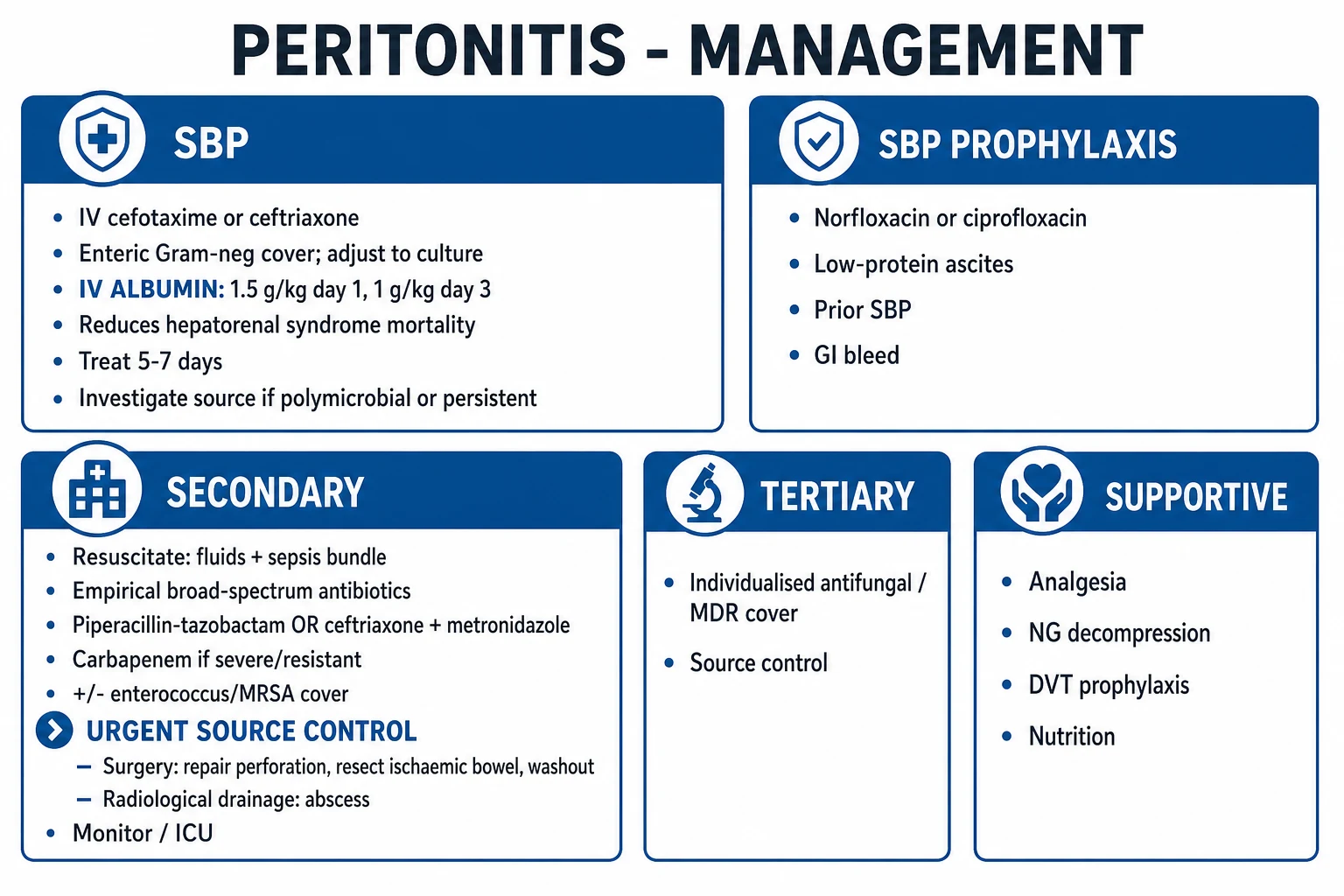
The hour-1 priorities follow the ABCDE and the Sepsis-6 / Surviving Sepsis hour-1 bundle.[5]
Airway, breathing, oxygen
- Airway — secure if comatose or vomiting (aspiration risk).
- Oxygen — target SpO2 94–98% (or 88–92% in COPD at risk of CO2 retention); high-flow if shocked.
- Assess work of breathing — mechanical ventilation if fatiguing or comatose. [1]
Circulation, IV access, fluid resuscitation
- Two large-bore IV cannulae; send bloods including two sets of blood cultures before antibiotics (but do NOT delay antibiotics for cultures).
- Balanced crystalloid (Hartmann's or Plasma-Lyte) 30 mL/kg IV over the first 3 hours in sepsis/septic shock; reassess fluid responsiveness (passive leg raise, pulse-pressure variation) before further boluses.
- Vasopressors — noradrenaline is first-line for fluid-refractory hypotension, titrated to MAP 65 mmHg or more. Add vasopressin and adrenaline if escalating.
- Avoid over-resuscitation — excess crystalloid causes abdominal compartment syndrome, pulmonary oedema and worsening of perfusion; reassess frequently. [1]
Sepsis-6 / Surviving Sepsis hour-1 bundle
Within one hour of recognition of sepsis/septic shock:[5]
- Measure serum lactate (re-measure if initially over 2 mmol/L).
- Take blood cultures before antibiotics (if possible without delaying them).
- Administer broad-spectrum antibiotics within one hour (in septic shock / high-risk sepsis) — covered in detail below.
- Begin rapid crystalloid resuscitation — 30 mL/kg.
- Apply vasopressors if hypotensive during or after fluids, to maintain MAP 65 mmHg.
- Measure urine output (urinary catheter) and monitor response. [1]
Antibiotic timing target
- Septic shock or high-risk sepsis — within one hour of recognition.[5]
- Stable admitted patient with secondary peritonitis — within four hours.[4]
Symptom control and supportive measures
- Analgesia — IV opioid titrated (morphine 1–2 mg increments or fentanyl); titrate to comfort; avoid NSAIDs (renal risk, peptic ulcer).
- Nasogastric tube — for paralytic ileus, vomiting, bowel obstruction; decompresses the stomach.
- Urinary catheter — to monitor hourly urine output.
- Central line and arterial line — for the shocked patient requiring vasopressors and invasive monitoring.
- Thromboprophylaxis — LMWH (e.g. enoxaparin 40 mg subcutaneously daily) unless contraindicated; antiembolism stockings.
- Stress-ulcer prophylaxis — IV pantoprazole 40 mg daily in mechanically ventilated or coagulopathic patients.
- Glucose control — target blood glucose 7.8–10 mmol/L; avoid hypoglycaemia. [1]
Management — Definitive & Stepwise
The two pillars are antibiotics and source control; SBP additionally gets albumin. The empirical choice depends on the subtype, severity and risk of MDR organisms.[1][4]
A. Spontaneous bacterial peritonitis — the medical peritonitis
SBP does NOT need surgery. There is no intra-abdominal source to control. Treatment is medical.[1][3]
[1]Empirical antibiotics (AASLD 2021):[1]
- First-line: cefotaxime 2 g IV every 8 hours OR ceftriaxone 2 g IV daily for 5–7 days (covers E. coli, Klebsiella, pneumococcus; adjusted once culture returns).
- Alternative in beta-lactam allergy or healthcare-acquired/resistant: IV piperacillin-tazobactam 4.5 g every 6–8 hours or meropenem (carbapenem-sparing where possible, but reserved for ESBL-producing organisms).
- Avoid oral antibiotics alone in established SBP (they achieve inadequate ascitic levels); oral ciprofloxacin is for prophylaxis only. [1]
IV albumin — the Sort 1999 NEJM trial proved that albumin 1.5 g/kg IV at diagnosis and 1 g/kg IV on day 3 reduces hepatorenal syndrome (33% to 10%) and mortality (29% to 10%) in SBP, particularly in those with BUN over 30 mg/dL or bilirubin over 4 mg/dL.[3] Give to every SBP patient meeting these criteria.
Repeat tap at 48 hours — if ascitic PMN has not fallen by at least 25%, suspect secondary peritonitis (search for a perforation) or resistant organism (broaden cover). [1]
Duration — 5–7 days, or until the ascitic PMN falls below 250 cells/mm³ and symptoms resolve. Switch to oral only once the patient is improving and an oral agent with adequate ascitic penetration is selected. [1]
SBP prophylaxis (lifelong oral quinolone):[1]
- Secondary prophylaxis (after an SBP episode) — indefinite norfloxacin 400 mg daily OR ciprofloxacin 500 mg daily (recurrence otherwise 70% at 1 year). Refer for liver transplantation.
- Primary prophylaxis — in patients with ascitic protein under 1.5 g/dL AND impaired renal function (creatinine over 1.2 mg/dL, BUN over 25 mg/dL, serum sodium under 130 mEq/L) OR severe liver disease (Child-Pugh over 9 with bilirubin over 3 mg/dL) OR GI bleeding (short course during the bleed).
- GI bleed prophylaxis — norfloxacin 400 mg daily or ciprofloxacin 500 mg twice daily for 7 days in any cirrhotic with GI bleeding (proven to reduce infection and mortality). [1]
B. Secondary peritonitis — antibiotics plus source control
Source control is the single most important determinant of survival in secondary peritonitis.[6]
Empirical antibiotic selection by severity (SIS/IDSA 2010 / WSES 2017):[4][5]
Community-acquired, mild-moderate (low risk of MDR)
- Cefoxitin 2 g IV every 6 h
- OR cefuroxime 1.5 g IV every 8 h + metronidazole 500 mg IV every 8 h
- OR moxifloxacin 400 mg IV daily
- OR amoxicillin-clavulanate (if available IV)
- Single agent acceptable; covers E. coli, anaerobes
Community-acquired, high-severity / healthcare-associated
- Piperacillin-tazobactam 4.5 g IV every 6–8 h (single agent)
- OR cefepime 2 g IV every 8–12 h + metronidazole 500 mg IV every 8 h
- OR ceftazidime 2 g IV every 8 h + metronidazole
- Add vancomycin or teicoplanin if MRSA risk
- Add echinocandin (caspofungin) if Candida risk
ESBL / carbapenem-resistant risk
- Ertapenem 1 g IV daily (ESBL, no Pseudomonas cover)
- OR meropenem 1 g IV every 8 h (ESBL + Pseudomonas)
- OR imipenem-cilastatin 500 mg IV every 6 h
- Reserve colistin / polymyxin B for carbapenem-resistant
Antifungal cover — add an echinocandin (caspofungin 70 mg loading then 50 mg daily, or micafungin 100 mg daily) or fluconazole 400 mg daily if risk factors: recent prolonged antibiotics, immunosuppression, malignancy, recurrent GI perforation, necrotising pancreatitis, post-operative leak.[4]
Source control principles (WSES 2023):[6]
- Drainage of infected fluid — percutaneous (CT- or US-guided) for a walled-off, accessible abscess; surgical for generalised peritonitis, multiple abscesses or unsuitable anatomy.
- Debridement of necrotic tissue — resect non-viable bowel, debride omentum.
- Restoration of anatomy — close or resect the perforation (e.g. Graham omental patch for perforated duodenal ulcer; primary anastomosis or Hartmann's for perforated diverticulitis; appendectomy for appendicitis; resection with stoma for ischaemic bowel).
- Repeated source control if the first fails (tertiary peritonitis). [1]
Source control by underlying cause:[2][7]
- Perforated peptic ulcer — resuscitation, antibiotics, then surgery: Graham omental patch closure (open or laparoscopic); IV PPI then H. pylori eradication; stop NSAIDs.[2]
- Perforated appendicitis — emergency laparoscopic appendectomy; if a walled-off appendicular mass/abscess in a stable patient, percutaneous drainage + IV antibiotics, interval appendectomy.[7]
- Perforated diverticulitis — Hartmann's procedure (sigmoid resection with end colostomy) for the unstable or purulent/faecal peritonitis; primary anastomosis with defunctioning ileostomy in selected stable patients.
- Ischaemic bowel — resection of non-viable bowel with primary anastomosis or stoma; second-look laparotomy at 24–48 hours.
- Post-operative anastomotic leak — re-operation, washout, defunctioning stoma, broad-spectrum antibiotics.
- Intra-abdominal abscess — CT-guided percutaneous drainage first-line if accessible; IV antibiotics; surgery only if drainage fails or the source requires it.
Duration of antibiotics:[5]
- Uncomplicated IAI (adequate source control achieved) — about 4 days post-source control (evidence from the STOP-IT / Hassinger 2017 trial showed no benefit of fixed 7-day courses over stopping after source control + clinical improvement, around 4 days median).
- Complicated IAI (no/partial source control, immunocompromise, severe sepsis) — 4–7 days, extended if persistent fever, positive cultures, ongoing source.
- Stop antibiotics once source control achieved AND clinical improvement (afebrile, improving, tolerating diet, falling inflammatory markers). [1]
IV-to-oral step-down criteria:[4]
- Clinically improving (afebrile, falling CRP).
- Haemodynamically stable, gut function returning, tolerating oral intake.
- An oral agent with high bioavailability chosen (amoxicillin-clavulanate, moxifloxacin, or oral metronidazole with a fluoroquinolone/cephalosporin). [1]
Discharge criteria — afebrile for 24–48 hours, tolerating oral diet, resolving peritonism, negative or treated cultures, safe social circumstances, follow-up arranged and safety-net given. [1]
C. Tertiary peritonitis
- Re-operation for persistent or recurrent source.
- Broad empiric antibiotics covering Pseudomonas, Enterococcus (vancomycin/teicoplanin for MRSA/VRE), MDR Gram-negatives (carbapenem ± aminoglycoside ± colistin), and Candida (echinocandin).
- Antifungal cover more liberally — Candida peritonitis carries high mortality.
- ICU support — organ failure, nutrition (preferably enteral), glucose control, thromboprophylaxis. [1]
Specific Subtypes & Scenarios
- Spontaneous bacterial peritonitis (SBP) — covered above (cefotaxime/ceftriaxone + albumin; prophylaxis with quinolone).
- Perforated peptic ulcer — see Source Control; Graham patch, H. pylori eradication, stop NSAIDs.
- Perforated appendicitis / appendicular mass — laparoscopic appendectomy; conservative for abscess/mass (drainage + interval appendectomy).[7]
- Perforated diverticulitis — Hartmann's procedure; primary anastomosis in selected.
- Mesenteric ischaemia with perforation — resection, second-look.
- Intra-abdominal abscess — percutaneous drainage + IV antibiotics.
- Post-operative / tertiary peritonitis — re-operation + broadened cover.
- Tuberculous peritonitis — insidious fever, weight loss, ascites; laparoscopy with biopsy (cased granulomata); standard anti-TB therapy (RHZE 2 months + RH 4 months). Ascitic fluid: lymphocytic, high protein, SAAG low.
- Peritoneal dialysis (CAPD) peritonitis — cloudy effluent, abdominal pain, fever; effluent cell count over 100 WBC/mm³ (over 50% neutrophils); touch preparation Gram stain; intraperitoneal antibiotics (cefazolin + ceftazidime) pending culture; catheter removal for refractory, relapsing, fungal or severe Pseudomonas peritonitis.[5]
- Candida peritonitis — recent prolonged antibiotics, immunosuppression, post-operative leak; echinocandin (caspofungin) first-line, step down to fluconazole.
- Secondary peritonitis from trauma — hollow viscus injury (blunt or penetrating); damage-control laparotomy.
Complications & Pitfalls
Local complications
- Intra-abdominal abscess — walled-off collection; CT drainage + antibiotics.
- Fistula (enterocutaneous, entero-enteric) — from iatrogenic injury, anastomotic leak, Crohn's.
- Adhesions and small bowel obstruction — late complication of surgery.
- Wound infection / dehiscence — surgical-site infection.
- Incisional hernia — late. [1]
Systemic complications
- Sepsis, septic shock, multi-organ dysfunction syndrome (MODS).
- Acute respiratory distress syndrome (ARDS).
- Acute kidney injury (especially hepatorenal syndrome in cirrhosis).
- Disseminated intravascular coagulation (DIC).
- Venous thromboembolism (immobility, inflammation). [1]
Hepatorenal syndrome (HRS-AKI) precipitated by SBP
The feared complication of SBP. Diagnosis (International Club of Ascites): rise in serum creatinine by over 0.3 mg/dL within 48 h or by 50% from baseline, in cirrhosis with ascites, after correcting hypovolaemia with albumin 1 g/kg (max 100 g) daily for 2 days and excluding other nephrotoxins. Treatment: albumin + vasoconstrictor (terlipressin IV or noradrenaline infusion) — and urgent liver transplant evaluation.[3][8]
Intra-abdominal hypertension and abdominal compartment syndrome
- IAH — sustained intra-abdominal pressure over 12 mmHg (transvesical bladder pressure).
- ACS — IAP over 20 mmHg with new organ dysfunction (oliguria, raised airway pressures, reduced cardiac output, metabolic acidosis).
- Treatment — decompressive laparotomy (surgical decompression, leave open abdomen with vacuum dressing); medical measures: nasogastric and rectal decompression, neuromuscular blockade, avoid over-resuscitation.[5]
Classic pitfalls
- Missing SBP in a cirrhotic — every cirrhotic with ascites on admission needs a diagnostic tap; relying on symptoms misses one in three.
- Delaying surgery for perforation — free gas + peritonism = theatre, not more imaging.
- Under-resuscitation causing shock; over-resuscitation causing ACS.
- Failing to cover anaerobes (Bacteroides) or Enterococcus in healthcare-associated IAI.
- Using oral quinolone alone to treat established SBP — inadequate ascitic levels.
- Not giving albumin in SBP (missing the HRS-prevention benefit).
- Treating polymicrobial ascites as SBP — search for the perforation.
- Misdiagnosing pancreatitis as perforation (or vice versa) — check lipase and erect CXR.
- Missing an atypical presentation (elderly with confusion, immunocompromised with normal exam).
- Prolonged antibiotics after adequate source control — no benefit, more resistance. [1]
Prognosis & Disposition
- SBP — in-hospital mortality 10–20%; one-year mortality 30%; recurrence 70% without secondary prophylaxis. An episode of SBP is an indication for liver transplantation evaluation.[1]
- Secondary peritonitis — mortality depends on severity, source, delay and comorbidity: under 10% for uncomplicated perforated appendix; 20–40% or more for faecal peritonitis with septic shock, elderly, or tertiary peritonitis.[5]
- Tertiary peritonitis — mortality 30–50%+, driven by MDR organisms and organ failure.
- Predictors of poor outcome — age, comorbidity (especially cirrhosis, malignancy, immunosuppression), severity of the source, delay to source control, MDR organism, organ failure (high APACHE/SOFA), malignancy, ICU admission.
- Disposition — ICU for septic shock, organ failure, post-major surgery; surgical ward for uncomplicated source-controlled IAI; home only when discharge criteria met with safety-net.
- Follow-up — surgical review (wound, hernia), repeat imaging if not improving, outpatient antibiotics (OPAT) for prolonged courses, liver transplant referral for SBP.
Special Populations
- Cirrhosis / ascites — high SBP risk; diagnostic tap on every admission and at any decompensation; primary and secondary quinolone prophylaxis per criteria; albumin in every treated SBP.[1]
- Pregnancy — appendicitis is the commonest non-obstetric surgical emergency; appendix shifts superiorly/laterally with uterine enlargement; physiological leucocytosis confounds CBC; laparoscopic appendectomy is safe in any trimester; tocolysis and left lateral tilt for venous return in late pregnancy.
- Elderly — blunted signs; perforated diverticulitis and ischaemic bowel dominate; high index of suspicion; confusion may be the only sign; aggressive resuscitation, early imaging, early source control.
- Immunocompromised / neutropenic — atypical organisms (Pseudomonas, Candida); subtle signs (no neutrophils to mount peritonism); broad empiric therapy; low threshold for CT; consider typhlitis (neutropenic enterocolitis) in the right clinical context.
- Children — appendicitis is the commonest surgical cause; perforation common (often delayed presentation); appendicular mass may be managed conservatively with drainage and interval appendectomy.
- Anticoagulated patient — balance bleeding vs urgent source control; reverse warfarin (vitamin K, prothrombin complex concentrate) if INR over 1.5–2.0 before surgery/paracentesis; DOACs held per half-life; fresh frozen plasma / PCC if emergency.
- Peritoneal dialysis patient — CAPD peritonitis managed with intraperitoneal antibiotics; catheter removal if refractory/fungal/severe Pseudomonas.
- Asplenia / sickle cell — risk of overwhelming post-splenectomy infection; cover encapsulated organisms empirically.
Evidence, Guidelines & Regional Differences
AASLD 2021 (US) — ascites, SBP, HRS
Biggins et al. (2021) replaced the 2012 guidance; key SBP points: ascitic PMN over 250 = treat; cefotaxime 2 g IV every 8 h or ceftriaxone 2 g IV daily; albumin 1.5 g/kg day 1 and 1 g/kg day 3 in those with BUN over 30 mg/dL or bilirubin over 4 mg/dL; secondary prophylaxis with norfloxacin indefinitely; GI-bleed prophylaxis with norfloxacin/ciprofloxacin for 7 days.[1]
WSES 2017 / 2023 — intra-abdominal infections and source control
The WSES 2017 guidelines (Sartelli) classify community-acquired IAI by severity (complicated vs uncomplicated; mild-moderate vs severe) and provide empirical antibiotic ladders; the WSES 2023 source-control guidelines (Coccolini) emphasise early, effective source control as the dominant mortality determinant.[5][6]
IDSA / SIS-AAIDST 2010 — complicated IAI
Solomkin et al. provide the empirical antibiotic tables by severity and risk of resistant organisms, still widely used; recommend anti-Enterococcus cover in healthcare-associated infection and antifungal cover in selected high-risk groups.[4]
Sort 1999 NEJM — albumin in SBP
The landmark randomised trial showing that albumin 1.5 g/kg day 1 and 1 g/kg day 3 reduced renal impairment (33% to 10%) and 3-month mortality (29% to 10%) when added to cefotaxime in SBP.[3]
STOP-IT / Hassinger 2017
A randomised trial of approximately 5 days versus a fixed 10-day course after adequate source control showed no difference in outcomes — supporting stopping antibiotics once source control is achieved and the patient has clinically improved (median ~4 days).[4]
Regional deltas
India (ICMR/NCDC AMR & empirical guidelines) — high community ESBL-producing E. coli prevalence makes amoxicillin-clavulanate and even third-generation cephalosporins unreliable empirically for severe community-acquired IAI; ertapenem or piperacillin-tazobactam is increasingly first-line in high-ESBL settings. Carbapenem-resistant Enterobacteriaceae (CRE) are an emerging problem, requiring colistin/polymyxin B ± tigecycline. Antibiotic stewardship and de-escalation to culture are emphasised. Tuberculous peritonitis is more common than in the West — always consider in a cirrhotic with lymphocytic, high-protein, low-SAAG ascites and constitutional symptoms; laparoscopy with biopsy for confirmation.
Current controversies
- Routine Enterococcus cover — increasingly recommended in healthcare-associated infection, but not community-acquired.
- Antifungal cover — reserved for high-risk groups; over-use drives resistance.
- Optimal duration — the trend is to shorter courses (around 4 days) post-source control.
- Laparoscopy vs open — laparoscopic source control increasingly used where expertise exists.
- Peritoneal lavage volumes — extensive lavage offers no benefit over limited targeted lavage. [1]
Exam Pearls
SBP mnemonic — SBP ABCDE
SBP
SBP infects ascites WITHOUT an intra-abdominal source — cirrhosis/nephrotic
Single organism (E. coli, Klebsiella, Pneumococcus); anaerobes rare
Ascitic PMN over 250 cells/mm³ — treat before culture returns
Albumin in SBP — 1.5 / 1
ALB
IV albumin 1.5 g/kg day 1 + 1 g/kg day 3 (Sort 1999 NEJM)
Prevents hepatorenal syndrome; reduces mortality 29% to 10%
Mandatory if bilirubin over 4 mg/dL or BUN over 30 mg/dL
High-yield numbers — peritonitis
SBP (primary)
- Ascitic PMN over 250
- Monomicrobial (E. coli, Klebsiella)
- Cefotaxime + ALBUMIN
- Quinolone prophylaxis
- Medical — NO surgery
Secondary
- Perforation/translocation
- Polymicrobial + anaerobes
- Broad-spectrum + SOURCE CONTROL
- Piperacillin-tazobactam / carbapenem
- Surgical emergency
Tertiary
- Persistent despite source control
- MDR: Pseudomonas, Enterococcus, Candida
- Re-operation + broad cover
- Antifungal + anti-MRSA
- ICU, high mortality
- Ascitic PMN over 250 cells/mm³ = SBP — treat before culture returns (culture-negative neutrocytic ascites counts).
- SBP organisms: E. coli, Klebsiella, pneumococcus (monomicrobial) — anaerobes rare → if present, think secondary.
- SBP treatment: cefotaxime 2 g IV BD–TDS + albumin 1.5 g/kg day 1, 1 g/kg day 3 (Sort 1999).
- Polymicrobial ascites (anaerobes) = secondary peritonitis — search for perforation.
- Secondary peritonitis is polymicrobial: Gram-negative bacilli + anaerobes (Bacteroides fragilis) + Enterococcus.
- Albumin in SBP prevents hepatorenal syndrome and reduces mortality (Sort 1999 NEJM).
- SBP prophylaxis: norfloxacin/ciprofloxacin daily (low ascitic protein, prior SBP, GI bleed).
- Diagnostic tap in ALL cirrhotics with ascites on admission — symptom-based screening misses one in three.
- Four-quadrant peritonitis: rigidity, guarding, rebound tenderness, absent bowel sounds.
- Free gas under the diaphragm on erect CXR = perforated viscus (usually perforated duodenal ulcer).
- Pelvic abscess: diarrhoea, urinary frequency, tender boggy anterior mass on PR exam.
- Tertiary peritonitis: MDR organisms — Pseudomonas, Enterococcus (VRE), Candida.
- Abdominal compartment syndrome: bladder pressure over 20 mmHg + organ failure → surgical decompression.
- Polymicrobial ascites + glucose under 50 + LDH high + CEA over 5 = secondary peritonitis (find the perforation).
- qSOFA 2+ = high risk of poor outcome in suspected sepsis.
- Antibiotics within 1 hour in septic shock; source control within hours — delay kills. [1]
Exam application bank (NEET-PG / INICET)
One-line answer
Intra-abdominal infection (IAI) and peritonitis are time-critical emergencies that span two very different diseases. Primary peritonitis — spontaneous bacterial peritonitis (SBP) — is infection of cirrhotic ascites without an intra-abdominal source, presenting subtly (fever, abdominal pain, hepatic encephalopathy, renal failure) and treated medically with cefotaxime/ceftriaxone plus IV albumin (the ascitic neutrophil count over 250 cells per mm³ is diagnostic). Secondary peritonitis is polymicrobial contamination of the peritoneum from a perforated or translocating hollow viscus (perforated peptic ulcer, appendicitis, diverticulitis, ischaemic bowel, post-operative leak, trauma) and is a surgical emergency: resuscitation, broad-spectrum antibiotics covering enteric Gram-negatives and anaerobes, and urgent source control. Tertiary peritonitis is persistent or recurrent infection in the cr
Worked stems (answer without another resource)
Stem 1 — Classic presentation. Map symptoms to mechanism; name the first investigation and first treatment step with dose/route if drug therapy is standard. [1]
Stem 2 — Unstable / complicated. List red flags that force immediate resuscitation, theatre, ICU, antidote, or reperfusion — and what you do in the first 15 minutes. [1]
Stem 3 — Atypical group. Elderly, pregnancy, child, or immunocompromised: how presentation and thresholds change. [1]
Stem 4 — Differential trap. Name the three closest mimics and one discriminator for each. [1]
Stem 5 — Disposition. Who goes home with safety-netting, who is admitted, who needs HDU/ICU/theatre, and what follow-up is mandatory. [1]
Rapid viva checklist
- Definition + classification
- Pathophysiology chain
- Bedside signs / criteria
- Score with exact components (if any)
- Emergency bundle
- Definitive therapy with doses
- Complications of disease and of treatment
- Special populations
- Guideline/trial name if classic
- Three exam traps
Coverage self-check
If you cannot answer any stem above from this page alone, re-read the matching section — the page is intended to be self-sufficient for final-prof and NEET-PG/INICET questions on Intra-Abdominal Infection & Peritonitis.
References
- [1]Biggins SW, Angeli P, Garcia-Tsao G, et al. Diagnosis, Evaluation, and Management of Ascites, Spontaneous Bacterial Peritonitis and Hepatorenal Syndrome: 2021 Practice Guidance by the American Association for the Study of Liver Diseases Hepatology, 2021.PMID 33942342
- [2]Tarasconi A, Coccolini F, Biffl WL, et al. Perforated and bleeding peptic ulcer: WSES guidelines World J Emerg Surg, 2020.PMID 31921329
- [3]Sort P, Navasa M, Arroyo V, et al. Effect of intravenous albumin on renal impairment and mortality in patients with cirrhosis and spontaneous bacterial peritonitis N Engl J Med, 1999.PMID 10432325
- [4]Solomkin JS, Mazuski JE, Bradley JS, et al. Diagnosis and management of complicated intra-abdominal infection in adults and children: guidelines by the Surgical Infection Society and the Infectious Diseases Society of America Surg Infect (Larchmt), 2010.PMID 20163262
- [5]Sartelli M, Chichom-Mefire A, Labricciosa FM, et al. The management of intra-abdominal infections from a global perspective: 2017 WSES guidelines for management of intra-abdominal infections World J Emerg Surg, 2017.PMID 28702076
- [6]Coccolini F, Sartelli M, Leandro G, et al. Source control in emergency general surgery: WSES, GAIS, SIS-E, SIS-A guidelines World J Emerg Surg, 2023.PMID 37480129
- [7]Podda M, Ceresoli M, Pisano M, et al. Diagnosis and Treatment of Acute Appendicitis: 2025 Edition of the World Society of Emergency Surgery Jerusalem Guidelines JAMA Surg, 2026.PMID 41604201
- [8]Guevara M, Terra C, Nazar A, et al. Albumin for bacterial infections other than spontaneous bacterial peritonitis in cirrhosis. A randomized, controlled study J Hepatol, 2012.PMID 22732511