Infectious Diseases · General Medicine
Measles (Rubeola)
Also known as Measles · Rubeola · Morbillivirus · Measles morbillivirus · First disease · Subacute sclerosing panencephalitis · SSPE
Measles (rubeola) is a highly contagious, vaccine-preventable acute viral exanthem caused by the measles morbillivirus (Paramyxoviridae), transmitted by respiratory droplets and airborne aerosols. After a 10 to 14 day incubation, a prodrome of high fever with the '3 Cs' (cough, coryza, conjunctivitis) and the pathognomonic Koplik spots (blue-white grains on the buccal mucosa) precedes a maculopapular rash that spreads head to toe. With a basic reproduction number (R0) of 12 to 18 it is one of the most infectious human pathogens; herd immunity needs about 95 percent coverage. Complications are common and severe — otitis media, pneumonia (the commonest cause of death), diarrhoea, keratitis/blindness, acute encephalitis, and the late, fatal subacute sclerosing panencephalitis (SSPE) — and measles induces a transient 'immune amnesia' that erases pre-existing immunity to other pathogens. Diagnosis is clinical plus IgM serology or RT-PCR (throat/urine). There is no specific antiviral; treatment is supportive plus vitamin A (two doses, reduces mortality and blindness). Prevention is the MMR vaccine (two doses, over 97 percent effective). Suspected measles is notifiable and isolated with airborne precautions.
On this page & tools
Your progress
Saved locally on this device.
Exam tags
Red flags
Overview & Definition
Measles (rubeola, "first disease") is an acute, highly contagious, vaccine-preventable viral exanthem caused by the measles morbillivirus (Morbillivirus measles), an enveloped, negative-sense, single-stranded RNA virus of the family Paramyxoviridae, genus Morbillivirus. It is transmitted by respiratory droplets and airborne aerosols and remains infectious in the air for up to two hours after an infectious person leaves a room.[1][2]
The measles virus particle is pleomorphic, roughly spherical, and approximately 150 to 300 nm in diameter. Its genome is non-segmented, negative-sense, single-stranded RNA of about 15,900 nucleotides that encodes six structural proteins: the nucleoprotein (N), phosphoprotein (P), matrix protein (M), fusion protein (F), haemagglutinin (H), and the large polymerase (L). The H protein binds to host receptors; the F protein mediates fusion with the host cell membrane. Two non-structural proteins, V and C, are encoded within the P gene and act as interferon antagonists, dampening the innate immune response. Because the virus is enveloped, it is destroyed by soaps, detergents, alcohol, and ultraviolet light, but it survives in the air and on surfaces for a limited time — up to two hours in the air and on contaminated surfaces — making airborne isolation critical. Measles is therefore both a droplet and airborne pathogen, and standard surgical masks are insufficient; FFP3 or N95 respirators are required for staff. The virus is labile, so surfaces can be decontaminated with standard hospital disinfectants, and hand hygiene remains important, but airborne precautions are the dominant control measure. [1]
From a public-health perspective, measles is the most sensitive indicator of immunisation programme performance. Where coverage is high, measles disappears; where coverage falters, measles returns first. This is because its extraordinary R0 means that even small pockets of susceptibility are rapidly exploited. Consequently, every measles case should trigger a review of local vaccination coverage and a catch-up campaign, not just clinical management of the index case. In exam terms, a single measles case in an "eliminated" region is an outbreak until proven otherwise. [1]
[1]Two facts dominate the clinical reasoning around measles. First, its contagiousness is extraordinary: the basic reproduction number (R0) is 12 to 18, so a single case in a susceptible community will infect almost everyone exposed — herd immunity requires about 95 percent two-dose coverage, and any fall below that threshold predictably triggers outbreaks. Second, it is entirely preventable with the MMR vaccine (two doses give over 97 percent protection), so every case represents a failure of vaccination (missed, refused, or cold-chain broken).[1]
The clinical skill is recognising the prodrome before the rash appears — fever with the 3 Cs (cough, coryza, conjunctivitis) plus Koplik spots — at which point the patient is most infectious and the window to isolate, test, notify, and protect contacts is widest. There is no specific antiviral: treatment is supportive, with vitamin A to every child (it roughly halves mortality in deficient populations). The complications — pneumonia, otitis media, diarrhoea, keratitis, encephalitis, SSPE, and immune amnesia — are the disease's real burden, and they fall hardest on the malnourished, the vitamin A-deficient, infants, the immunocompromised, and pregnant women.[2][9]
Classification
Measles is one entity with several clinical phenotypes that depend on the host's prior immunity and age.[1]
Classic measles
- Susceptible (unvaccinated) host, natural wild virus
- Full prodrome: fever + 3 Cs + Koplik spots
- Cephalocaudal maculopapular rash, becomes confluent
- Typical infectivity: 4 days before to 4 days after rash
Modified measles
- Partial immunity (residual maternal antibody, partial vaccine, or past immunoglobulin)
- Attenuated prodrome, shorter fever
- Sparse, atypical rash that may not spread fully
- Fewer complications; still infectious; IgM may be delayed or absent
Atypical measles
- Recipients of **killed inactivated measles vaccine** (used 1963-1968 in the US) exposed to wild virus
- High fever, rash **starts on extremities** (inverse/centripetal), petechial or vesicular, with oedema
- Severe **interstitial pneumonitis**, pleural effusion, often no Koplik spots
- Historical; live vaccines replaced killed vaccine — rare today
Measles in the immunocompromised
- HIV, transplant, chemotherapy, primary immunodeficiency
- **No rash** or atypical rash (rash requires intact cell-mediated immunity)
- Severe giant-cell (Hecht) pneumonia, encephalitis, **measles inclusion-body encephalitis (MIBE)** within months
- **Live MMR vaccine is contraindicated** — give immunoglobulin post-exposure
Modified measles deserves particular attention because it is a clinical trap. It occurs in people with partial immunity — for example, infants with residual maternal antibody, individuals who received only one dose of MMR, or those given immunoglobulin. The prodrome is shortened and milder, Koplik spots may be absent or few, and the rash is sparse, non-confluent, and sometimes absent. Crucially, the patient is still infectious and can transmit measles. Laboratory confirmation can be more difficult: IgM may be delayed or absent, and RT-PCR is therefore preferred in suspected modified measles. Modified measles can also occur after post-exposure immunoglobulin when the dose is insufficient to prevent disease entirely, producing an attenuated illness.[1]
Atypical measles is largely of historical interest but remains examinable. It was seen in people who received the killed, inactivated measles vaccine used in the United States from 1963 to 1968, and who were later exposed to wild-type measles. The syndrome is thought to be caused by hypersensitivity to the F protein in the absence of robust H-protein response. It produces a high fever, a centripetal or acral-first rash (starts on the wrists, ankles, and extremities and spreads centrally), often petechial, vesicular, or urticarial, with prominent oedema of the extremities and face. Pneumonia is severe and interstitial, pleural effusion is common, and Koplik spots are usually absent. The killed vaccine was replaced by live attenuated vaccine, so atypical measles is now rare, but it illustrates the importance of humoral and cellular immunity in shaping the clinical phenotype.[2]
Measles neurologic syndromes are a distinct, examinable classification:[2][9]
- Acute disseminated encephalomyelitis (ADEM / post-measles encephalomyelitis) — demyelinating, perivascular, autoimmune; occurs within 2 to 14 days of the rash; around 1 in 1000 cases; high morbidity.
- Acute measles encephalitis — direct viral invasion in the immunocompromised; fever, seizures, depressed consciousness.
- Measles inclusion-body encephalitis (MIBE) — in the immunocompromised, 1 to 10 months after infection; rapidly progressive, often fatal.
- Subacute sclerosing panencephalitis (SSPE) — a persistent, defective (M-protein-mutant) measles infection of neurons appearing 5 to 15 years after natural measles (earlier and more frequent when measles is acquired under 2 years of age); progressive cognitive decline, myoclonus, seizures, eventual decorticate state, uniformly fatal in 1 to 3 years. [1]
Epidemiology & Risk Factors
Measles was once a near-universal childhood infection; aggressive vaccination cut global deaths from an estimated 2.6 million per year in 1980 to under 100,000 by the late 2010s, but falling coverage has reversed elimination in several regions.[1][8]
Headline numbers
The epidemiology of measles is determined almost entirely by the R0 and the vaccination coverage. The herd-immunity threshold is derived from the relation H = 1 - 1/R0. With an R0 of 12 to 18, 1 - 1/12 = 91.7 percent and 1 - 1/18 = 94.4 percent of the population must be immune. Because one dose of MMR is about 93 percent effective and two doses are about 97 percent effective, near-universal two-dose coverage is required to stop transmission. This is why measles is the first vaccine-preventable disease to resurge when immunisation programmes weaken, and why it is regarded as the canary in the coal mine for health systems. Outbreaks are explosive: a single imported case in a susceptible community can generate hundreds of secondary cases within weeks, as seen in the 2014-2015 Disneyland outbreak in California, the 2018-2019 resurgence in Europe, and the post-COVID-19 pandemic rebound in low-income countries. [1]
[1] [7]Risk factors and the populations they afflict: [1]
- Unvaccinated children and adults (the dominant driver — missed vaccination, vaccine refusal, broken cold chain).[1]
- Infants under 6 to 9 months — protection by transplacental maternal antibody wanes, leaving a susceptibility window before routine immunisation.[2]
- Malnourished and vitamin A deficient children — severe disease, keratitis/blindness, high mortality; the dominant burden in the developing world.[6]
- Immunocompromised hosts (HIV, primary immunodeficiency, transplant, chemotherapy) — severe, atypical disease (giant-cell pneumonia, MIBE); live vaccine contraindicated.[2]
- Pregnant women — higher risk of severe measles, miscarriage, premature labour and low birthweight; live vaccine contraindicated in pregnancy.[2]
- Crowded settings — refugee camps, dormitories, schools, healthcare waiting rooms; explosive outbreaks.
- Healthcare exposure — measles is notorious for nosocomial outbreaks; only staff with documented immunity should care for cases.
Transmission dynamics are equally important. The virus is shed from the respiratory tract during coughing, sneezing, talking, and even breathing. It is airborne — particles smaller than 5 micrometres can remain suspended for up to two hours — and directly contagious from four days before the rash to four days after the rash appears. This means the patient is most infectious during the prodrome, when the diagnosis may not yet be obvious. In the immunocompromised, shedding and infectiousness can persist for the entire illness. The incubation period averages 10 to 14 days but can range from 7 to 21 days; a very long incubation of up to 3 weeks is occasionally seen. Nosocomial outbreaks are a recognised hazard: an infectious patient in a waiting room or ward can expose many susceptible people, which is why prompt triage, masking, and isolation of febrile rash patients is essential. [1]
[1] [2]Seasonality: late winter and spring in temperate climates; year-round with monsoon-season peaks in the tropics.[2]
Pathophysiology
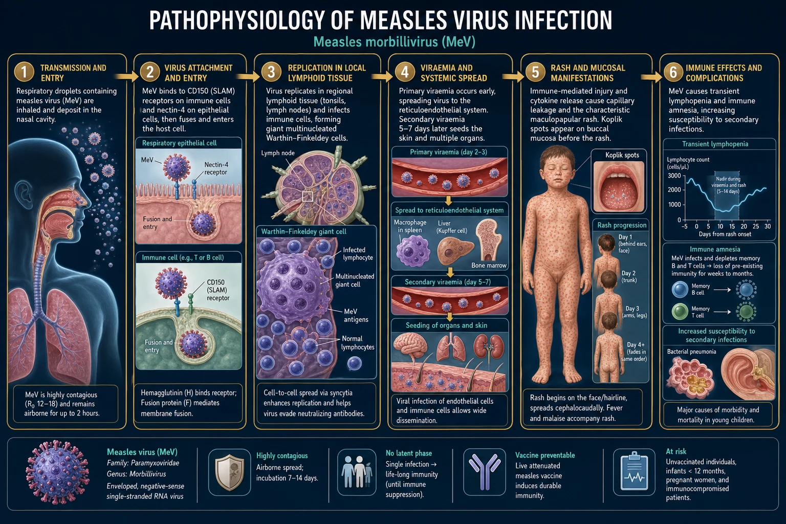
Measles enters the respiratory tract in droplets or aerosols and binds two receptors: CD150 (signalling lymphocyte activation molecule, SLAM) on activated T and B lymphocytes, monocytes, dendritic cells — and nectin-4 (PVRL4) on the basolateral surface of respiratory (and many epithelial) cells. Nectin-4 is also expressed on many cancer cells, which is why attenuated measles virus is being developed as an oncolytic vector.[1]
The H protein attaches to the receptor; the F protein then mediates pH-independent fusion of the viral envelope with the cell membrane. The ribonucleocapsid is released into the cytoplasm, and the viral RNA-dependent RNA polymerase transcribes and replicates the genome in the cytoplasm. Because the virus replicates in immune cells, it can hijack the very cells that should control it: dendritic cells, macrophages, and lymphocytes spread the virus throughout the body during the first and second viraemias. Measles also produces the V and C proteins, which antagonise the host interferon response and allow viral replication to proceed before adaptive immunity is mobilised. This immune evasion explains the high peak viraemia and the dramatic fall in circulating lymphocytes during acute infection. The lymphopenia is so characteristic that a normal or elevated lymphocyte count in a suspected measles case should prompt reconsideration of the diagnosis. [1]
[1]The cascade proceeds in stages:[1]
- Entry and local replication (days 0 to 3) — virus infects respiratory epithelial cells and alveolar macrophages, then drains to regional lymph nodes.
- Primary viraemia (days 2 to 3) — infected lymphocytes and monocytes spread virus to the reticuloendothelial system (spleen, lymph nodes, liver, thymus).
- Secondary viraemia (days 5 to 7) — virus seeds the skin, conjunctivae, respiratory tract, GI tract, and other epithelial surfaces. This is when symptoms escalate.
- Warthin-Finkeldey giant cells — pathognomonic histology: multinucleated giant cells (fused infected epithelial and lymphoid cells) carrying viral inclusion bodies; found in lymphoid tissue, respiratory epithelium, and Koplik-spot lesions.
- Rash (day 14, ~4 days after prodrome onset) — a cell-mediated (type IV) immune attack by cytotoxic T lymphocytes on measles-infected capillary endothelial and epithelial cells. The rash therefore reflects immune clearance, not direct viral damage — which is why an immunocompromised host (no cell-mediated immunity) may have no rash yet severe disease, and why rash appearance coincides with defervescence.
- Koplik spots are the buccal-mucosal analogue: tiny foci of epithelial necrosis with neutrophil infiltrate on an erythematous base, appearing 1 to 2 days before the rash. [1]
The most important recent discovery is measles-induced immune amnesia. Measles infection depletes 11 to 73 percent of the pre-existing antibody repertoire — memory B and T lymphocytes carrying immunity to other pathogens are lost, leaving the host susceptible to otitis, pneumonia, diarrhoea, and other infections for 2 to 3 years after the acute illness. The mechanism appears to be both the depletion of memory B cells and the replacement of the plasma-cell niche after the lymphopenia of acute infection, resulting in a failure to maintain antibody titres to previously encountered pathogens. This explains why measles mortality is increased for years after infection, not just during the acute illness.[4][5]
Why SSPE happens: a defective measles variant (mutations especially in the M matrix protein) persists in neurons, is poorly cytolytic, spreads cell-to-cell, and slowly accumulates over years — eventually triggering a fatal neurodegenerative process. The risk is highest when measles is acquired under 2 years of age, presumably because the immature immune system fails to clear the virus and allows persistence. The M protein is required for viral assembly and budding; when it is mutated, the virus cannot form infectious particles but can still spread directly from cell to cell within the central nervous system, evading antibody-mediated neutralisation.[2]
Clinical Presentation
Measles has a stereotyped four-phase clinical course: incubation, prodrome, exanthem (rash), and recovery.[1][2]
Phase 1 — Incubation (10 to 14 days, range 7 to 21): asymptomatic. The patient becomes infectious from the late incubation period — about 4 days before the rash appears. This asymptomatic infectious period is a major reason why outbreaks are hard to control. [1]
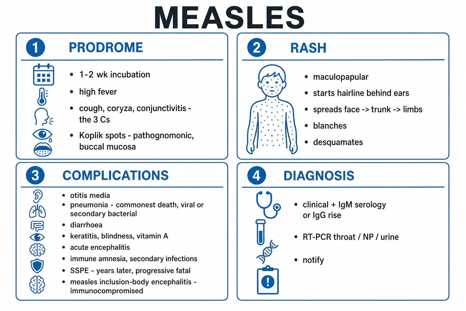
Phase 2 — Prodrome / catarrhal stage (2 to 4 days): the patient looks and feels unwell. The 3 Cs are the high-yield triad: [1]
- Cough — dry, brassy, persistent (viral tracheobronchitis). The cough is often severe and may be the last symptom to resolve.
- Coryza — sneezing, nasal discharge, nasal obstruction; the child has a "runny nose" and sounds congested.
- Conjunctivitis — red, watery, often photophobic; may progress to keratitis, especially with vitamin A deficiency. [1]
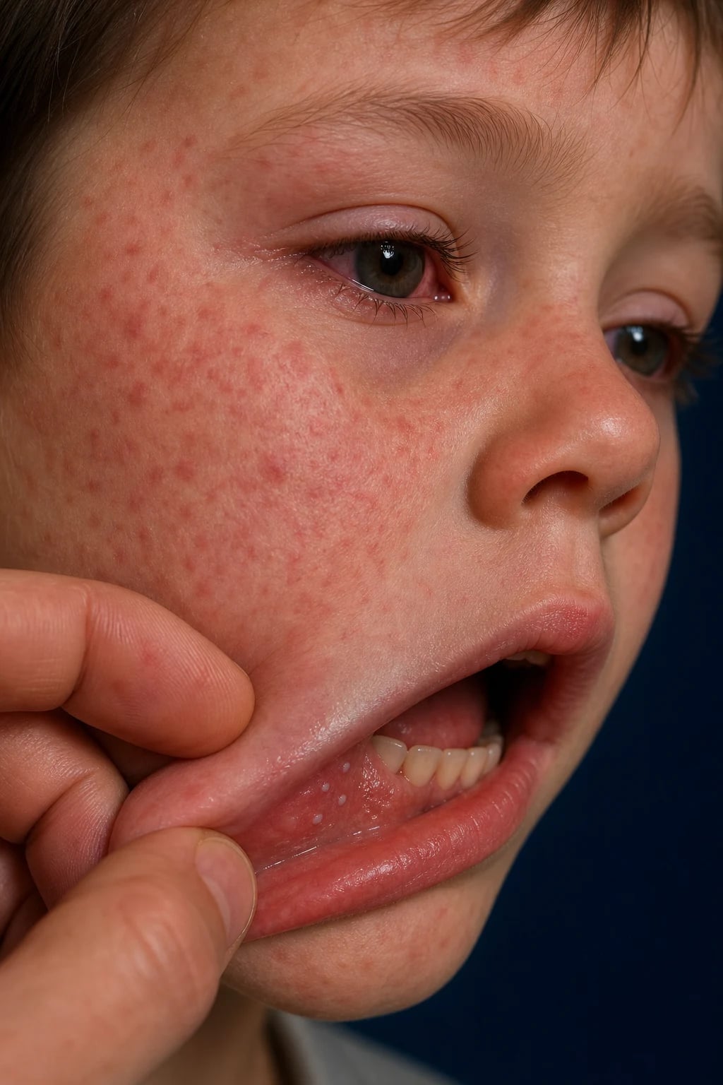
Accompanying features: high fever (often 39 to 40.5 degrees C), malaise, anorexia, myalgia, sometimes mild pharyngitis. The fever often rises progressively to a peak on the day the rash erupts. This "step-ladder" fever pattern is useful but not universal. Koplik spots — pathognomonic — appear on the buccal mucosa opposite the lower second molars during the late prodrome (1 to 2 days before the rash): tiny (1 to 3 mm) bluish-white "grains of salt" papules on a bright-red erythematous base. They fade 1 to 2 days after the rash appears. Koplik spots are sometimes described as resembling "grains of salt on a red carpet". They are diagnostic when present and may appear on any inflamed oral mucosa, but the buccal mucosa opposite the lower molars is the classic site. Some patients have a diffuse erythematous enanthem before the discrete Koplik spots appear. Histologically, Koplik spots represent foci of epithelial necrosis with neutrophilic infiltration and multinucleated giant cells on an erythematous base, mirroring the pathophysiology seen in the skin and lymphoid tissue.[2]
Phase 3 — Exanthem / rash stage: a maculopapular, erythematous, blanching, usually non-pruritic rash that becomes confluent. The distribution is cephalocaudal and is itself diagnostic: [1]
- Day 1 — appears behind the ears and at the hairline on the face, neck, and behind the ears; spreads to the face and upper neck. The rash often first appears where the skin is thin and vascular.
- Day 2 to 3 — spreads down the trunk, becoming confluent on the face. The face may look swollen and intensely erythematous.
- Day 3 to 4 — reaches the limbs (including palms and soles); the face begins to clear. By this stage the rash is often most confluent on the trunk and may have a "stained" appearance. [1]
As the rash appears, the fever begins to defervesce over the next 2 to 3 days (a clue: a still-rising fever with the rash suggests a complication such as secondary bacterial infection). A fine desquamation and brownish staining may follow over the next week. Generalised lymphadenopathy and mild splenomegaly are common. The rash may be more intense in areas of pressure or skin folds. In dark skin, the erythema may be harder to appreciate, but the texture and distribution remain diagnostic. The rash can sometimes be purpuric or haemorrhagic in severe disease or thrombocytopenia.[3]
Phase 4 — Recovery: fatigue and cough may persist for 1 to 2 weeks. The patient is infectious until 4 days after the rash appears (longer in the immunocompromised — the entire illness). [1]
Atypical presentations — what examiners probe deliberately: [1]
- Immunocompromised host (HIV, transplant, chemo, severe primary immunodeficiency): no rash (or atypical sparse rash), because cell-mediated immunity is needed to generate it. Severe giant-cell (Hecht) pneumonia with prominent giant cells on biopsy, measles inclusion-body encephalitis (MIBE) within months. Live MMR is contraindicated. The absence of rash can mislead clinicians, so a high index of suspicion is needed in any immunocompromised patient with fever, cough, and exposure to measles.
- Infants: high fever and toxicity, higher rates of pneumonia and otitis media, secondary bacterial sepsis; maternal antibody has waned by 6 to 9 months leaving a susceptibility gap. Infants under 6 months may have partially protective maternal antibody, producing modified measles, but they can still be severely ill.
- Pregnant woman: higher risk of severe measles, pneumonia, hospitalisation, miscarriage, premature labour, low birthweight — but, unlike rubella, measles is NOT teratogenic and does not cause a congenital malformation syndrome.
- Malnourished / vitamin A deficient child: severe disease, keratitis, corneal ulceration, xerophthalmia and blindness, severe diarrhoea, dehydration, high mortality.
- Adults: more likely to be hospitalised, higher rates of hepatitis, pneumonia, and encephalitis than children.
- Modified measles: as described above, with attenuated symptoms due to partial immunity. It can be mistaken for a non-specific viral illness, yet the patient remains infectious and can seed outbreaks. [1]
Differential Diagnosis
An acute febrile illness with a maculopapular rash is not always measles. The distribution, the prodrome, Koplik spots, and the patient's immune and exposure history are the discriminators. Always consider rubella (especially in pregnancy), parvovirus B19, Kawasaki disease, and drug reactions.[3]
Measles (rubeola)
- 3 Cs prodrome + **Koplik spots** (pathognomonic)
- Rash **head to toe** (cephalocaudal), becomes confluent
- Patient looks unwell, high fever
- Virus: Paramyxoviridae, Morbillivirus
Rubella (German measles)
- **Mild prodrome**, low fever
- **Prominent postauricular and suboccipital lymphadenopathy**
- Rash spreads **rapidly** (face to body in ~1 day), does not coalesce
- **Forscheimer spots** (petechiae on soft palate)
- **Togaviridae; TERATOGENIC (congenital rubella syndrome)**
Roseola infantum (HHV-6)
- **High fever 3 to 5 days, then rash APPEARS AS FEVER FALLS**
- Rash rose-pink macules, starts on **trunk**
- **Infant under 3 years**, otherwise well during fever
- Herpesviridae, HHV-6 (rarely HHV-7)
Erythema infectiosum (Parvovirus B19, 'fifth disease')
- **Slapped-cheek** facial rash, then **lacy reticular** rash on extremities
- Arthritis in adults; **aplastic crisis in haemolytic anaemia**
- **Hydrops fetalis** in pregnancy
- No cough/coryza; low-grade fever
Scarlet fever (group A strep)
- Fine **sandpaper rash**, flexural accentuation, **Pastia lines**
- **Strawberry tongue**, exudative pharyngitis, **no cough/coryza**
- Positive throat swab / rapid strep test
Infectious mononucleosis (EBV)
- **Exudative tonsillitis, posterior cervical lymphadenopathy, splenomegaly**
- Atypical lymphocytes, positive Monospot
- **Ampicillin/amoxicillin rash** is diffuse maculopapular but prodrome differs
Kawasaki disease
- Fever **5 days or more**, strawberry tongue, cracked lips, **oedema/desquamation of hands and feet**
- Bilateral non-purulent conjunctivitis, **polymorphous rash**
- **Coronary artery aneurysm** risk — needs IVIG + aspirin
Drug reaction / DRESS / SJS
- Exposure history (antibiotics, anticonvulsants)
- **Mucosal involvement**, atypical target lesions, epidermolysis in SJS
- Eosinophilia, transaminitis in DRESS
Dengue / enterovirus / meningococcaemia
- Dengue: retro-orbital pain, thrombocytopenia, positive tourniquet test, **mosquito exposure**
- Meningococcaemia: **petechial/purpuric, rapidly progressive**, sepsis
The high-yield differentiating features to commit to memory: Koplik spots = measles; postauricular lymphadenopathy + soft-palate petechiae (Forscheimer) + rapid rash + pregnancy concern = rubella; rash appearing as fever drops = roseola; slapped cheek = parvovirus B19; sandpaper + strawberry tongue = scarlet fever; 5-day fever + cracked lips + coronary risk = Kawasaki. [1]
Examination tips for the bedside: in a child with fever and rash, always inspect the buccal mucosa for Koplik spots before looking at the skin. Koplik spots appear before the rash, so checking the mouth can clinch the diagnosis at the prodromal stage, when isolation and contact tracing are most valuable. In a pregnant woman with a rash, always ask about rubella immunity and parvovirus B19 exposure because both have fetal implications that measles does not. In a child with fever and conjunctivitis, think of Kawasaki disease if the fever has lasted 5 or more days and there are extremity changes or coronary concern. [1]
[3]Other conditions to consider in specific contexts include secondary syphilis (palmar/plantar rash, condylomata lata, positive serology), typhoid fever (rose spots on trunk, relative bradycardia, enteric exposure), leptospirosis (conjunctival suffusion, renal involvement, animal exposure), and primary HIV infection (mononucleosis-like illness, mucocutaneous ulcers, high-risk exposure). These are less common in the classic paediatric measles setting but become important in adults or returning travellers with a rash. The key is always to look for Koplik spots and ask about vaccination and exposure before committing to an alternative diagnosis. If Koplik spots are present, measles is essentially certain; if they are absent but the clinical picture fits, do not rule measles out, especially in the first 1 to 2 days of rash. [1]
Clinical & Bedside Assessment
Vital signs drive severity and complications. Check temperature, respiratory rate, oxygen saturation, hydration, capillary refill, alertness, and look systematically for the complications.[9]
Targeted bedside examination: [1]
- Buccal mucosa — inspect under bright light opposite the lower second/third molars for Koplik spots (pathognomonic if present). Use a tongue depressor and good light; spots may be few and fleeting.
- Conjunctivae and cornea — redness, discharge, photophobia; examine with fluorescein for keratitis/corneal ulcer (vitamin A deficiency link).
- Ears — pneumatic otoscopy for otitis media (bulging, red, immobile tympanic membrane; the commonest bacterial complication).
- Respiratory — signs of pneumonia (tachypnoea, crackles, bronchial breath sounds, effusion), croup (stridor), and hypoxia.
- Abdomen — dehydration from diarrhoea, hepatosplenomegaly.
- Neurology — irritability, meningism, seizures, depressed consciousness (encephalitis), and (in the recovered child seen years later) myoclonus, decline in school performance, motor regression (SSPE).
- Nutrition — weight-for-height, mid-upper-arm circumference, signs of vitamin A deficiency (xerosis, Bitot spots, corneal ulceration).
- Skin — character and distribution of the rash; desquamation; petechiae/purpura (thrombocytopenia, atypical measles, secondary sepsis). [1]
A structured bedside checklist (the "measles ABCDE") helps ensure nothing is missed in a busy clinic or outbreak setting: Airway and croup/stridor; Breathing and oxygenation; Circulation and hydration; Disability and encephalopathy; Exposure and rash distribution plus eye/skin/mouth exam. In addition, always ask about vaccination status, recent exposures, travel, and contact with a pregnant or immunocompromised person. These history points guide isolation duration, contact tracing, and post-exposure prophylaxis. [1]
Assess for the four lethal complications at every contact: pneumonia, dehydration, encephalitis, and keratitis/blindness. [1]
Investigations
Diagnosis is primarily clinical in an outbreak (typical prodrome with Koplik spots plus cephalocaudal rash) but laboratory confirmation is mandatory for sporadic cases, atypical cases, and public-health notification.[1][2]
First-line laboratory confirmation: [1]
- Measles-specific IgM serology — positive from 1 to 2 days after the rash appears, peaks at 1 to 3 weeks, persists for 30 to 60 days. A single positive IgM in someone not recently vaccinated confirms acute measles. (False negatives in the first 1 to 2 days of rash — repeat at 7 to 10 days if suspicion persists; false positives from rheumatoid factor, parvovirus, EBV.) IgM may be delayed or absent in modified measles or after immunoglobulin.
- Measles RNA by RT-PCR — throat swab, nasopharyngeal aspirate, or urine; most sensitive in the first 3 to 4 days of the rash; allows genotyping to identify the outbreak source and distinguish wild-type from vaccine-strain measles. PCR is the preferred test in modified or atypical measles, and in the immunocompromised. In resource-limited settings, measles IgG avidity can help distinguish recent infection (low avidity) from past infection or vaccination (high avidity), but this is not usually available as a rapid test. Genotyping is important for outbreak investigation: it can distinguish wild-type virus from vaccine-strain virus (the latter may be detected in a recently vaccinated person), and it can link cases to a known outbreak strain or importation.
- Salivary measles IgM (where available) — non-invasive, useful in children. [1]
Supporting tests: [1]
- Full blood count — typically leucopenia with lymphopenia (the immune amnesia); sometimes atypical lymphocytes; thrombocytopenia may occur.
- Vitamin A level — in severe or malnourished cases (subclinical deficiency worsens outcomes). However, do not wait for a level to give vitamin A.
- Chest X-ray if pneumonia suspected — interstitial infiltrates (viral giant-cell pneumonia), lobar consolidation (secondary bacterial), hilar adenopathy, sometimes pleural effusion.
- CT brain, EEG, and CSF for suspected encephalitis — CSF shows lymphocytic pleocytosis and may detect measles antibody; EEG in SSPE shows characteristic periodic complexes with burst-suppression; brain MRI shows cortical/subcortical and basal-ganglia changes. [1]
Paired serology — a four-fold rise in measles IgG between acute and convalescent sera (2 to 3 weeks apart) confirms infection when IgM is unobtainable. This is rarely needed now that PCR is widely available but remains useful in retrospective diagnosis or atypical cases.[1]
WHO/CDC case definition (high-yield):[8]
- Suspected case — any person with fever and maculopapular rash AND at least one of cough, coryza, or conjunctivitis.
- Laboratory-confirmed case — suspected case with positive measles IgM (not recently vaccinated), OR measles RNA by RT-PCR, OR a four-fold rise in IgG, OR epidemiologically linked to a laboratory-confirmed case. [1]
Management — Resuscitation
ABCDE first. The two non-negotiable immediate actions on suspecting measles are to ISOLATE and to NOTIFY.[1][7]
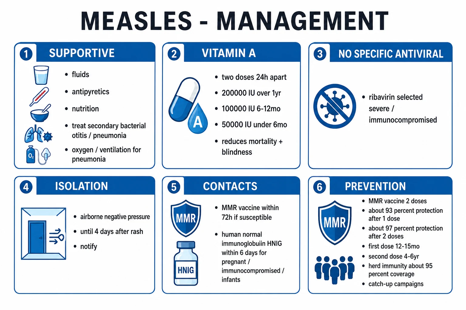
Isolation — airborne precautions: place the patient in a single room, preferably negative-pressure, with FFP3/N95 respiratory protection for staff (measles virus aerosolises and lingers for up to two hours). Isolate for at least 4 days after the rash appears (longer in the immunocompromised — the entire duration of illness). Only staff with documented immunity (two documented MMR doses or positive IgG) should enter. Susceptible staff should not care for the patient; if unavoidable, they should wear full airborne PPE and be excluded from work from day 5 to 21 post-exposure if exposed.[7]
Notify public health immediately (measles is statutorily notifiable worldwide) — it triggers contact tracing and outbreak control. Notification should include the patient's demographics, onset date, rash date, vaccination history, and known contacts. This is not a bureaucratic step; it is a clinical intervention that may prevent further cases.[7]
Resuscitative bundle: [1]
- Oxygen to target SpO2 94 to 98 percent (or 88 to 92 percent in chronic CO2 retainers) for pneumonia or hypoxia.
- IV fluids for dehydration from diarrhoea or inadequate intake — balanced crystalloid, judiciously (beware of fluid overload in pneumonia or myocarditis).
- Treat secondary bacterial infection early — otitis media, pneumonia, sepsis per local guidance.
- Vitamin A on the day of diagnosis to all children (see below).
- Identify and protect contacts — MMR within 72 h of exposure for susceptible contacts; human normal immunoglobulin (HNIG) within 6 days for those in whom live vaccine is contraindicated (pregnancy, immunocompromise, infants under 6 months).[7]
Management — Definitive & Stepwise
There is no specific antiviral of proven benefit in measles. Management is supportive, with vitamin A to every child, treatment of complications, and rigorous infection control and public-health action.[2][6]
Supportive care: [1]
- Antipyretics and analgesia — paracetamol 10 to 15 mg/kg every 4 to 6 h (max 60 mg/kg/day, 4 g/day in adults), or ibuprofen 5 to 10 mg/kg every 6 to 8 h for fever, myalgia, headache. Avoid aspirin in children (Reye syndrome risk).
- Hydration and nutrition — maintain oral intake; IV fluids if dehydrated; nutritional rehabilitation in the malnourished. Continue breastfeeding.
- Eye care — protect the cornea in keratitis/xerophthalmia; antibiotic eye ointment; review by ophthalmology.
- Cough and respiratory support — humidified oxygen, bronchodilators if wheeze; mechanical ventilation for severe pneumonia or ARDS. [1]
Vitamin A — give to ALL children with measles (WHO recommendation; reduces mortality roughly 50 to 60 percent in deficient populations and prevents blindness):[6][7]
- Two doses, 24 h apart:
- 200,000 IU orally if age 12 months or older.
- 100,000 IU orally if age 6 to 11 months.
- 50,000 IU orally if under 6 months.
- A third dose 2 to 4 weeks later in confirmed vitamin A deficiency, xerophthalmia, or persistent malnutrition. [1]
Vitamin A is thought to work by restoring epithelial integrity, supporting immune function, and reducing the severity of keratitis and diarrhoea. The WHO recommendation is to give vitamin A to all children with measles regardless of measured serum levels or national setting, because even subclinical deficiency worsens outcomes and serum levels fall during acute infection. Vitamin A should be given on the day of diagnosis and repeated 24 hours later. If the child has evidence of vitamin A deficiency, xerophthalmia, or persistent malnutrition, a third dose is given 2 to 4 weeks later. High-dose vitamin A can cause transient headache, vomiting, and bulging fontanelle in infants, but these are usually mild and self-limiting; the benefits in measles far outweigh the risks. For children with acute diarrhoea or malabsorption, the oral route is still preferred because the WHO formulation is oil-based and well absorbed even in the presence of gut pathology.
[6]Treatment of complications: [1]
- Otitis media — oral amoxicillin 40 to 90 mg/kg/day in 2 to 3 divided doses for 5 to 10 days (or co-amoxiclav if treatment failure), per local guidance.
- Secondary bacterial pneumonia — empirical CAP therapy (ceftriaxone plus azithromycin for severe; amoxicillin or co-amoxiclav for milder disease); cover pneumococcus, H. influenzae, Staphylococcus aureus.[7]
- Viral (giant-cell / Hecht) pneumonia — supportive; oxygen and ventilation as needed. IV ribavirin is sometimes used in severe life-threatening disease, especially in the immunocompromised (off-label, limited evidence; teratogenic — avoid in pregnancy).
- Acute encephalitis — supportive intensive care; control seizures (IV lorazepam 0.1 mg/kg or rectal diazepam 0.5 mg/kg), treat raised intracranial pressure; IV ribavirin occasionally; the evidence for steroids, IVIG, and plasmapheresis in ADEM/post-measles encephalomyelitis is limited.
- SSPE — no curative treatment; isoprinosine (inosine pranobex) and intraventricular interferon-alpha may slow progression in some; ultimately symptom control and supportive care.
Hospitalisation criteria: any complication (pneumonia, dehydration, encephalitis, severe keratitis, secondary bacterial infection), infants, immunocompromise, malnutrition, pregnancy, or unsafe social situation. In low-resource settings, the threshold for admission should be lower because the risk of complications and mortality is higher, and follow-up may be unreliable. Any child with signs of pneumonia, encephalopathy, dehydration, or severe ocular involvement should be admitted for observation and treatment. [1]
Step-down and discharge once the patient is clinically improving, afebrile, adequately hydrated, tolerating oral intake, and oxygen-independent, with isolation discontinued 4 days after the rash appeared (longer in the immunocompromised). Provide a safety-net to return if breathlessness, altered consciousness, dehydration, or eye pain develop. [1]
Specific Subtypes & Scenarios
- Modified measles — partial immunity gives a mild prodrome and sparse rash; still isolate and confirm (PCR, since IgM may be attenuated). Think of it in recently vaccinated children, infants with maternal antibody, or post-immunoglobulin contacts.[1]
- Atypical measles (historical, in recipients of killed vaccine) — severe, with extremity-first rash, petechiae, severe interstitial pneumonitis; treat supportively; live vaccine replaced killed vaccine.
- Measles in the immunocompromised — no rash, giant-cell pneumonia, MIBE; live MMR contraindicated; HNIG within 6 days of exposure; consider IV ribavirin for severe disease. Prophylaxis with HNIG is also indicated after exposure in some HIV-positive children depending on immune status.
- Measles in pregnancy — severe disease risk; HNIG post-exposure; live MMR contraindicated in pregnancy (give postpartum). Measles is not teratogenic but raises miscarriage and preterm-labour risk. The fetus may be infected, but congenital measles syndrome is not a defined entity.[2]
- Neonatal/infantile measles — HNIG if exposed under 6 months; severe disease possible; vitamin A; supportive care. Breastfeeding is encouraged.
- Measles pneumonia — differentiate viral (giant-cell / Hecht) from secondary bacterial (often pneumococcal, Hib, or staph); cover empirically. Mortality from pneumonia is highest in malnutrition and the immunocompromised.
- Acute and post-measles encephalitis / SSPE / MIBE — see Complications. The key clinical distinction is timing: ADEM within 2 to 14 days of rash; MIBE at 1 to 10 months; SSPE at 5 to 15 years.
- Healthcare-worker exposure — susceptible healthcare workers should be furloughed from day 5 to 21 post-exposure to prevent nosocomial transmission. Those with documented immunity (two MMR doses or positive IgG) can continue to work.
Complications & Pitfalls
Complications are common and severe, especially in the under-5s, adults over 20, the malnourished, the immunocompromised, and pregnant women.[9]
Respiratory
- **Pneumonia** — commonest cause of death (viral giant-cell / Hecht OR secondary bacterial pneumococcal, Hib, staph)
- **Otitis media** — commonest bacterial complication (especially children)
- Laryngotracheobronchitis (**croup**), bronchiolitis
- Sinusitis, activation/reactivation of tuberculosis (TST anergy post-measles)
Gastrointestinal
- **Diarrhoea and dehydration** — worsens malnutrition
- Stomatitis, oral ulcers, anorexia
- Hepatitis (more common in adults)
Eye
- **Keratitis, corneal ulceration, xerophthalmia** (vitamin A deficiency)
- **Blindness** — preventable with vitamin A
Neurological
- **Acute measles encephalitis** (~1 in 1000) — mortality 15 percent, sequelae in 25 to 40 percent
- **Post-infectious encephalomyelitis (ADEM)** — demyelinating, day 2 to 14 after rash
- **MIBE** — immunocompromised, 1 to 10 months later, often fatal
- **SSPE** — years later, fatal in 1 to 3 years
Systemic / other
- **Transient immunosuppression / immune amnesia** — predisposes to other infections for 2 to 3 years
- **Thrombocytopenia** (bleeding, purpura)
- Myocarditis, pericarditis, hepatitis
- **Miscarriage, premature labour, low birthweight** in pregnancy
Pneumonia deserves special emphasis because it is the commonest cause of death. It can be primary viral giant-cell pneumonia (Hecht pneumonia), caused directly by measles virus in alveolar epithelium and macrophages, with interstitial infiltrates and giant cells on biopsy; or secondary bacterial pneumonia, most often pneumococcal, Haemophilus influenzae type b, or Staphylococcus aureus, with lobar consolidation, effusion, or empyema. In practice, the two are often indistinguishable clinically, and empirical antibiotics are indicated for any pneumonia in measles. Hypoxia, respiratory distress, and persistent fever after the rash should be treated as pneumonia until proven otherwise.[9]
Neurological complications are also high-yield. Acute measles encephalitis occurs in about 1 in 1000 cases, with mortality of 15 percent and neurological sequelae in 25 to 40 percent of survivors. ADEM is an immune-mediated demyelination that occurs 2 to 14 days after the rash and is thought to be triggered by molecular mimicry. MIBE is a progressive, usually fatal encephalitis in the immunocompromised, occurring 1 to 10 months after infection, with measles inclusions in neurons and glia. SSPE is the late, fatal complication that every examiner expects: progressive cognitive decline, behavioural change, myoclonus, seizures, EEG periodic complexes with burst-suppression, and raised measles antibody in CSF and serum. It is uniformly fatal in 1 to 3 years. The risk of SSPE is highest when measles is acquired under 2 years of age and may be as high as 1 in 600 to 1 in 1500 in that age group, compared with about 1 in 10,000 overall. This is why protecting infants through maternal vaccination and timely MMR is so important. [1]
Systemic complications include thrombocytopenia (petechiae, purpura, bleeding), myocarditis (arrhythmias, heart failure), hepatitis (transaminitis), and, in pregnancy, miscarriage, premature labour, and low birthweight. The immune amnesia described by Mina means that the increased mortality from measles persists for years after the acute infection, as the immune system relearns protection against previously encountered pathogens.
[2]Classic pitfalls (and how to avoid them):[1][9]
- Misdiagnosing as drug rash or dengue when the prodrome and Koplik spots are missed — examine the buccal mucosa in every febrile child with cough, coryza, and conjunctivitis.
- Using droplet precautions instead of airborne — measles aerosolises; use a negative-pressure room and FFP3/N95.
- Forgetting vitamin A — give to every child on the day of diagnosis.
- Missing secondary bacterial pneumonia — re-evaluate for rising fever, respiratory distress, and new infiltrates.
- Missing SSPE in the child with school decline and myoclonus years after measles — check EEG and CSF/serum measles antibody.
- Giving live MMR in pregnancy or immunocompromise — give HNIG instead for post-exposure prophylaxis.
- Failing to vaccinate contacts within the 72 h (MMR) or 6 day (HNIG) windows.
- Assuming modified measles is not infectious — isolate and confirm with PCR. [1]
Prognosis & Disposition
Most uncomplicated cases recover within 7 to 10 days of the rash. Case fatality in well-nourished, developed settings is 1 to 3 in 1000, but rises to 5 to 10 percent (and higher in refugee/displaced populations) in malnutrition and the immunocompromised. Death is most often from pneumonia (viral or secondary bacterial) or encephalitis.[9]
Poor-prognostic factors: age under 5 or over 20, malnutrition (especially vitamin A deficiency), immunocompromise, pregnancy, no prior vaccination, secondary bacterial infection, delayed vitamin A, and crowded/low-resource settings.[2]
SSPE is uniformly fatal 1 to 3 years from onset.[2]
Lifelong immunity follows natural measles (re-infection is rare); two-dose MMR gives over 97 percent protection.[7]
Disposition is driven by complication severity: home with safety-net for uncomplicated cases; hospital for any complication, infants, immunocompromise, malnutrition, pregnancy, or poor social situation; ICU for severe pneumonia, ARDS, encephalitis, or shock. Discontinue isolation 4 days after the rash appeared (longer if immunocompromised). A clear follow-up plan is essential because complications may develop or worsen after discharge. [1]
Special Populations
- Children (paeds) — weight-based dosing for paracetamol, vitamin A, antibiotics; infants at higher risk; MMR at 9 months in India's Universal Immunisation Programme (6 months in outbreak, then repeated), second dose at 15 to 18 months. Maternal antibody wanes by 6 to 9 months, leaving a susceptibility gap. The first dose at 9 months in India is earlier than in many temperate countries because the risk of exposure is higher and maternal antibody wanes earlier in high-transmission settings. In an outbreak, infants as young as 6 months may receive MMR, but this dose does not count toward routine immunity and must be repeated at 9 months and again at 15 to 18 months.[7]
- Malnutrition / vitamin A deficiency — severe disease, keratitis, blindness, high mortality; vitamin A essential (two doses, third in deficiency); nutritional rehabilitation. Measles and malnutrition form a vicious cycle: measles causes anorexia and diarrhoea, worsening malnutrition; malnutrition impairs immunity, worsening measles.[6]
- Immunocompromised (HIV, transplant, chemo, primary immunodeficiency) — severe/atypical (no rash, giant-cell pneumonia, encephalitis, MIBE); live MMR CONTRAINDICATED; HNIG within 6 days of exposure; consider IV ribavirin for severe disease. Some HIV-positive children with preserved CD4 counts may receive MMR, but this is specialist-directed. In solid-organ and stem-cell transplant recipients, measles can be fulminant, with giant-cell pneumonia and encephalitis; post-exposure prophylaxis with HNIG is standard, and some centres also use ribavirin. Patients on high-dose steroids or rituximab are at particularly high risk because cell-mediated and humoral immunity are both impaired.[2]
- Pregnant women — higher risk of severe measles, miscarriage, preterm labour, low birthweight; NOT teratogenic (unlike rubella); live MMR contraindicated in pregnancy (give postpartum); HNIG within 6 days of exposure. The absence of teratogenicity is a common exam point: rubella causes congenital rubella syndrome, measles does not.[2]
- Newborn of mother with measles — HNIG; supportive care; observe for severe disease; breastfeeding encouraged (maternal antibodies, hydration). Neonatal measles can be severe because the infant's immune system is immature and maternal antibody may be insufficient. Congenital measles is not a defined syndrome (unlike rubella), but intrauterine infection can occur and may result in a neonatal rash or severe disease. Postnatal exposure from the mother or other close contacts is the more common route.
- Healthcare workers — documented two-dose MMR immunity required; susceptible HCW exposed should be excluded from work from day 5 to 21 post-exposure. This is a critical infection-control measure to prevent nosocomial outbreaks.[7]
- Tuberculosis co-infection — measles transiently suppresses cell-mediated immunity, may reactivate TB, and causes tuberculin skin test (TST) anergy for 4 to 6 weeks; live MMR can theoretically interfere with BCG/TST — separate MMR from BCG/TST by about 4 weeks where feasible (give MMR first, delay BCG/TST).[2]
Evidence, Guidelines & Regional Differences
Landmark evidence: [1]
- MMR does NOT cause autism. The 1998 Wakefield Lancet paper was fully retracted in 2010; subsequent large cohort studies, including Hviid et al. 2019 in 657,461 Danish children (Annals of Internal Medicine), found no increased risk of autism with MMR, even in children with a family history of autism or other susceptibility factors. The adjusted hazard ratio was 0.93 (95 percent CI, 0.85 to 1.02).[10]
- Vitamin A reduces measles mortality. Hussey & Klein's 1990 NEJM randomised trial and subsequent Cochrane reviews established two-dose vitamin A as the WHO standard. The trial showed reduced mortality and pneumonia-specific mortality in children with severe measles.[6]
- Immune amnesia. Mina et al. (2015, Science) showed population-level rises in non-measles infectious disease mortality for 2 to 3 years after measles epidemics in England & Wales, the USA, and Denmark. Mina et al. (2019, Science) demonstrated that measles depletes 11 to 73 percent of the pre-existing antibody repertoire, mechanistically explaining immune amnesia.[4][5]
- Vaccine effectiveness — single dose about 93 percent, two doses about 97 percent; herd immunity needs about 92 to 95 percent coverage. The Disneyland 2014-15 outbreak and the UK loss of elimination in 2018 illustrate how small coverage gaps cause large outbreaks. Even a 5 percent drop in two-dose coverage can change a region from elimination to sustained transmission.[7]
Regional guideline differences: [1]
- WHO (2017 position paper) — two doses of measles-containing vaccine; first dose at 9 months (or 6 months in outbreaks, then repeated), second at 15 to 18 months; elimination target. The position paper explicitly recommends vitamin A for all children with measles.
- US (ACIP / CDC) — MMR at 12 to 15 months and 4 to 6 years; two doses. Documented HCW immunity required. Some US states require MMR for school entry, and California's SB277 removed personal-belief exemptions in 2015. Post-exposure prophylaxis with MMR is offered within 72 hours, and HNIG within 6 days, for susceptible contacts in whom live vaccine is contraindicated.
- UK (UKHSA / Green Book) — MMR at 12 to 13 months and 3 years 4 months; aggressive catch-up campaigns after the Wakefield-related coverage collapse (UK lost WHO elimination status in 2018, regained in 2021). Measles is notifiable.
- India (NMC / NTAGI / Universal Immunisation Programme) — MR (measles-rubella) at 9 months and 15 to 18 months; nationwide MR campaign; goal of measles elimination and CRS control. Measles is notifiable under IDSP. [1]
Post-exposure prophylaxis decision algorithm: for a susceptible contact of a measles case, offer MMR vaccine within 72 hours of first exposure if there are no contraindications to live vaccine. For contacts in whom live vaccine is contraindicated — pregnant women, severely immunocompromised patients, and infants under 6 months — give human normal immunoglobulin (HNIG) 0.5 mL/kg IM (maximum 15 mL) within 6 days of first exposure. HNIG may attenuate disease but does not always prevent it; the patient should still be monitored. After delivery or recovery, give MMR to those who are eligible. [1]
Controversies and current issues: vaccine hesitancy (the principal driver of measles resurgence); mandatory vaccination (Italy, France, California SB277, parts of Germany); the route of administration (aerosolised measles vaccine studied, not in routine use); and the global elimination target (verified eliminated in some regions such as the Americas in 2016, then re-established transmission). The COVID-19 pandemic disrupted routine immunisation globally, leading to millions of missed measles doses and a resurgence in outbreaks in low- and middle-income countries. Surveillance quality is also critical: a country cannot claim elimination if it cannot detect cases. The WHO target of measles elimination requires not only high coverage but also sensitive case-based surveillance and rapid outbreak response. [1]
Exam Pearls
The 3 Cs of the measles prodrome
3 Cs
Dry, brassy, persistent (viral tracheobronchitis)
Sneezing, nasal discharge, obstruction
Red, watery, often photophobic
Classic viral exanthems — number and pathogen
1-2-3-4-5-6
Paramyxoviridae, Morbillivirus; Koplik spots; head-to-toe rash
Group A strep; sandpaper rash, strawberry tongue
Togaviridae; postauricular lymphadenopathy; teratogenic
Historical; now considered a staphylococcal toxin
Parvovirus B19; slapped cheek; lacy rash; hydrops fetalis
HHV-6; high fever then rash on defervescence; infant under 3 y
High-yield one-liners examiners reward: [1]
- Measles = Morbillivirus (Paramyxoviridae); enveloped negative-sense single-stranded RNA virus; receptors CD150 (SLAM) on immune cells and nectin-4 on epithelium.
- Prodrome = fever + the 3 Cs (cough, coryza, conjunctivitis) + Koplik spots.
- Koplik spots = pathognomonic; blue-white grains of salt on bright-red buccal mucosa opposite the lower molars; appear BEFORE the rash.
- Rash = maculopapular, cephalocaudal (head to toe), becomes confluent; defervescence with rash.
- Patient MOST infectious in the prodrome — from 4 days before to 4 days after the rash.
- R0 12 to 18 — one of the most contagious human pathogens; herd immunity needs about 95 percent coverage.
- Commonest cause of death = pneumonia (viral giant-cell / Hecht OR secondary bacterial).
- Vitamin A two doses 24 h apart (200,000 IU if over 1 y; 100,000 IU 6 to 12 mo; 50,000 IU under 6 mo) reduces mortality and blindness.[6]
- Warthin-Finkeldey giant cells = pathognomonic histology.
- Acute encephalitis about 1 in 1000; SSPE years later (fatal; persistent defective measles in CNS).
- Immune amnesia (Mina) — measles wipes out memory immunity to other pathogens for 2 to 3 years.[4][5]
- MMR within 72 h for post-exposure prophylaxis; HNIG within 6 days for pregnancy, immunocompromise, infants under 6 months.
- MMR is LIVE — contraindicated in pregnancy and immunocompromise; separate from BCG/TST by ~4 weeks.
- Measles is NOT teratogenic (unlike rubella) but causes miscarriage/preterm.
- MMR does NOT cause autism (Wakefield retracted; Hviid 2019 in 657,461 children).
- Isolate with AIRBORNE precautions (negative pressure, FFP3/N95) — not just droplets.
- Modified measles is still infectious; use PCR because IgM may be delayed.
- Atypical measles (killed vaccine era) = extremity-first rash, severe pneumonitis, rare today.
- Contact tracing is a clinical intervention, not just public-health paperwork.
- In an outbreak, the diagnosis is clinical; in sporadic cases, confirm with IgM or PCR.
- The four lethal complications to reassess at every visit: pneumonia, dehydration, encephalitis, keratitis/blindness.
Self-test: distinguish the maculopapular exanthems
- Head-to-toe rash with 3 Cs and Koplik spots → measles.
- Mild fever + postauricular lymphadenopathy + rapid rash + Forscheimer spots → rubella (teratogenic).
- 3 to 5 days of high fever, then rash AS fever falls → roseola (HHV-6).
- Slapped cheek, then lacy extremity rash; arthritis or aplastic crisis → parvovirus B19 (fifth disease).
- Sandpaper rash, strawberry tongue, no cough → scarlet fever (group A strep).
- 5-day fever, cracked lips, oedema/desquamation of hands/feet, coronary aneurysm risk → Kawasaki disease.[3]
Exam application bank (NEET-PG / INICET)
One-line answer
Measles (rubeola) is a highly contagious, vaccine-preventable acute viral exanthem caused by the measles morbillivirus (Paramyxoviridae), transmitted by respiratory droplets and airborne aerosols. After a 10 to 14 day incubation, a prodrome of high fever with the '3 Cs' (cough, coryza, conjunctivitis) and the pathognomonic Koplik spots (blue-white grains on the buccal mucosa) precedes a maculopapular rash that spreads head to toe. With a basic reproduction number (R0) of 12 to 18 it is one of the most infectious human pathogens; herd immunity needs about 95 percent coverage. Complications are common and severe — otitis media, pneumonia (the commonest cause of death), diarrhoea, keratitis/blindness, acute encephalitis, and the late, fatal subacute sclerosing panencephalitis (SSPE) — and measles induces a transient 'immune amnesia' that erases pre-existing immunity to other pathogens. Diag
Worked stems (answer without another resource)
Stem 1 — Classic presentation. Map symptoms to mechanism; name the first investigation and first treatment step with dose/route if drug therapy is standard. [1]
Stem 2 — Unstable / complicated. List red flags that force immediate resuscitation, theatre, ICU, antidote, or reperfusion — and what you do in the first 15 minutes. [1]
Stem 3 — Atypical group. Elderly, pregnancy, child, or immunocompromised: how presentation and thresholds change. [1]
Stem 4 — Differential trap. Name the three closest mimics and one discriminator for each. [1]
Stem 5 — Disposition. Who goes home with safety-netting, who is admitted, who needs HDU/ICU/theatre, and what follow-up is mandatory. [1]
Rapid viva checklist
- Definition + classification
- Pathophysiology chain
- Bedside signs / criteria
- Score with exact components (if any)
- Emergency bundle
- Definitive therapy with doses
- Complications of disease and of treatment
- Special populations
- Guideline/trial name if classic
- Three exam traps
Coverage self-check
If you cannot answer any stem above from this page alone, re-read the matching section — the page is intended to be self-sufficient for final-prof and NEET-PG/INICET questions on Measles (Rubeola).
Final rapid-fire review: a child with fever, cough, coryza, conjunctivitis, and Koplik spots has measles until proven otherwise. Isolate, notify, test, give vitamin A, and trace contacts. Remember that pneumonia is the killer, SSPE is the late trap, and MMR is the prevention. In pregnancy, use HNIG, not vaccine. In immunocompromise, expect no rash and severe disease. These facts cover the vast majority of measles questions at NEET-PG and INICET. [1]
References
- [1]Rota PA, Moss WJ, Takeda M, de Swart RL, Thompson KM, Goodson JL. Measles Nat Rev Dis Primers, 2016.PMID 27411684
- [2]Moss WJ, Griffin DE. Measles Lancet, 2012.PMID 21855993
- [3]Keighley CL, Saunderson RB, Kok J. Viral exanthems Curr Opin Infect Dis, 2015.PMID 25706914
- [4]Mina MJ, Metcalf CJ, de Swart RL, Osterhaus AD, Grenfell BT. Long-term measles-induced immunomodulation increases overall childhood infectious disease mortality Science, 2015.PMID 25954009
- [5]Mina MJ, Kermack-Phillips A, Adu-Gyamfi CG, et al. Measles virus infection diminishes preexisting antibodies that offer protection from other pathogens Science, 2019.PMID 31672891
- [6]Hussey GD, Klein M. A randomized, controlled trial of vitamin A in children with severe measles N Engl J Med, 1990.PMID 2194128
- [7]World Health Organization. Measles vaccines: WHO position paper, April 2017 - Recommendations Vaccine, 2019.PMID 28760612
- [8]Perry RT, Gacic-Dobo M, Dabbagh A, Mulders MN, Strebel PM, Okwo-Bele JM, Rota PA, Goodson JL. Progress toward regional measles elimination - worldwide, 2000-2014 MMWR Morb Mortal Wkly Rep, 2015.PMID 26562349
- [9]Perry RT, Halsey NA. The clinical significance of measles: a review J Infect Dis, 2004.PMID 15106083
- [10]Hviid A, Hansen JV, Frisch M, Melbye M. Measles, Mumps, Rubella Vaccination and Autism: A Nationwide Cohort Study Ann Intern Med, 2019.PMID 30831578