Infectious Diseases · General Medicine
Osteomyelitis
Also known as Osteomyelitis · Bone infection · Acute haematogenous osteomyelitis · Chronic osteomyelitis · Vertebral osteomyelitis · Diabetic foot osteomyelitis
Osteomyelitis is an infection of bone and bone marrow (myelo = marrow) by microbes (usually bacteria), producing inflammation, bone destruction (osteolysis), necrosis and reactive new bone formation. By pathogenesis (Lew-Waldvogel): haematogenous (bloodstream seeding — children: long-bone metaphysis; adults: vertebrae), contiguous-focus (adjacent wound, ulcer, diabetic foot, surgery) and chronic (sequestrum, involucrum, sinus tract, biofilm). Staphylococcus aureus is the commonest organism across all ages and types. Acute disease presents with localised bone pain, tenderness, swelling and fever; chronic disease with a draining sinus tract and relapsing pain. X-ray may be normal for the first 1 to 2 weeks — MRI is the modality of choice (sensitivity about 90%). The microbiological gold standard is bone biopsy culture (sinus tract culture is contaminated). Management is orthopaedic + infectious diseases co-management: dead bone cannot be cured by antibiotics, so surgical debridement of necrotic/sequestered bone is the cornerstone of chronic disease, combined with prolonged culture-directed antibiotics (4 to 6 weeks acute, 6 weeks plus chronic; add rifampin for staphylococcal biofilm).
On this page & tools
Your progress
Saved locally on this device.
Exam tags
Red flags
Overview & Definition
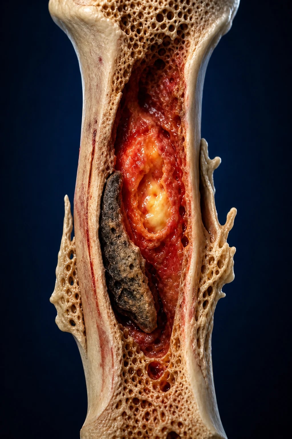
Osteomyelitis is a progressive infection of bone and bone marrow (myelo = marrow) by microbes — usually bacteria, occasionally mycobacteria or fungi — producing inflammation, bone destruction (osteolysis), necrosis, and reactive new bone formation. It is distinguished from septic arthritis (joint infection), cellulitis (superficial soft tissue), and contiguous soft-tissue abscess by involvement of the osseous tissue and medullary cavity.[2]
The clinical skill in osteomyelitis is not recognising the florid case — it is recognising the early case (a normal early X-ray does NOT exclude it), choosing the right imaging (MRI, not a repeat X-ray), securing an accurate microbiological diagnosis (bone biopsy, not sinus tract swab), and understanding that dead, avascular bone cannot be sterilised with antibiotics — surgical source control is the cornerstone of chronic disease. A second recurring theme is the biofilm: sessile bacteria encased in slime on dead bone or implants are 10 to 1000 times more resistant to antibiotics and immune attack, which is why rifampin (which penetrates biofilm) is added for staphylococcal disease, and why infected implants are so hard to cure.[2][7]
Classification
Osteomyelitis is classified along three independent axes — by pathogenesis, by tempo/duration, and by anatomic stage. An examiner will probe all three. [1]
1. By pathogenesis (the Lew-Waldvogel classification)[2] — the classification that decides the empirical antibiotic spectrum and the surgical approach:
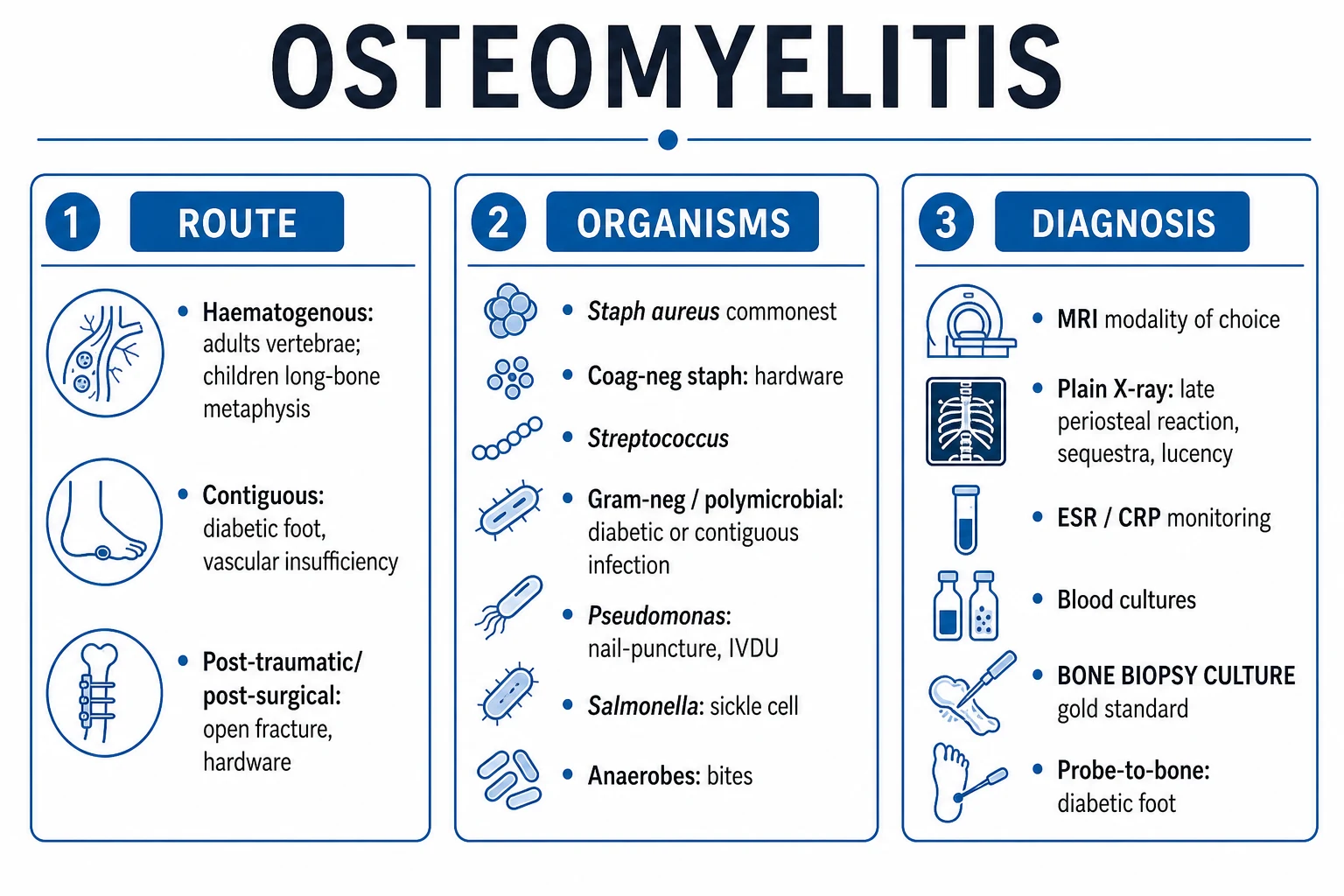
- Haematogenous osteomyelitis — bacteraemic seeding of the richly vascular bone. In children, the long-bone metaphysis (femur, tibia) is the target; in adults, the vertebra (vertebral osteomyelitis / discitis). Single organism, usually Staphylococcus aureus.
- Osteomyelitis from a contiguous focus — spread from adjacent soft tissue (cellulitis, diabetic ulcer, surgical wound, open fracture, joint replacement). Mixed flora including Gram-negatives and anaerobes; needs debridement.
- Chronic osteomyelitis — the disease persists or relapses over months to years; hallmark is dead, separated bone (sequestrum), new periosteal bone (involucrum), cortical cloaca and a draining sinus tract, with biofilm on necrotic tissue or implants. [1]
2. By duration/tempo: [1]
- Acute (within ~2 weeks) — suppurative, neutrophilic, may be cured medically if treated before necrosis.
- Subacute (weeks to months) — less virulent; Brodie abscess (intraosseous abscess with sclerotic rim) is the classic lesion.
- Chronic (months plus) — defined histopathologically by the presence of dead bone and biofilm; relapsing. [1]
3. By anatomic stage (the Cierny-Mader staging system, adult chronic disease)[3] — an anatomic type crossed with a host class:
Anatomic Type I — Medullary
- Infection confined to the intramedullary cavity
- e.g. infected intramedullary nail, haematogenous
- Treatment: intramedullary reaming / exchange
Type II — Superficial
- Surface cortex infected; exposed bone with poor soft-tissue cover
- e.g. anterior tibia after trauma
- Treatment: surface debridement + flap cover
Type III — Localised
- Full-thickness cortical sequestrum, well circumscribed
- Combination of types I and II
- Treatment: en-bloc excision + dead-space management
Type IV — Diffuse
- Circumferential disease, unstable bone
- Periarticular / through-and-through
- Treatment: segmental resection; may need amputation
Host A — Healthy
- No systemic compromise
- Good healing potential
- Standard treatment tolerated
Host B — Compromised
- Local (Bs) or systemic (Ba) compromise
- e.g. diabetes, smoking, vascular disease, malnutrition
- Optimise host before / during surgery
Host C — Treatment worse than disease
- Severe comorbidity; surgery carries unacceptable risk
- Suppressive antibiotics only
- Palliative intent
Epidemiology & Risk Factors
Osteomyelitis is uncommon in the general population but disproportionately affects the very young, the elderly, and the immunocompromised, and its incidence is rising globally with the diabetes pandemic, ageing populations, and increasing orthopaedic implant surgery.[2]
Risk factors and the organisms they favour (high-yield exam pairing):[1][9]
| Risk factor / host | Organism to consider |
|---|---|
| None / otherwise well child or adult | Staphylococcus aureus (always the default) |
| Diabetes, peripheral vascular disease | S. aureus, streptococci, enterobacteriaceae, anaerobes (mixed) |
| Open fracture, post-surgical, implant | S. aureus, coagulase-negative staphylococci, Gram-negatives, anaerobes (mixed) |
| Prosthetic joint / orthopaedic hardware | Coagulase-negative staphylococci, S. aureus (biofilm) |
| IV drug use | Pseudomonas aeruginosa, S. aureus; unusual sites (sternoclavicular, sacroiliac, vertebral) |
| Sickle cell disease | Salmonella (classic exam answer) — but S. aureus still commonest |
| Neonate | Group B streptococcus, E. coli, S. aureus |
| Immunocompromise / neutropenia | Gram-negatives incl. Pseudomonas, fungi (Aspergillus, Candida), atypical mycobacteria |
| Tuberculosis endemic | Mycobacterium tuberculosis (Pott disease of the spine) |
The single most tested pairing is sickle cell disease with Salmonella — but examiners also test whether you know that even in sickle cell disease, S. aureus is still the commonest single organism. The second most tested is IV drug use with Pseudomonas at unusual sites (sternoclavicular, sacroiliac, vertebral).[2]
Pathophysiology
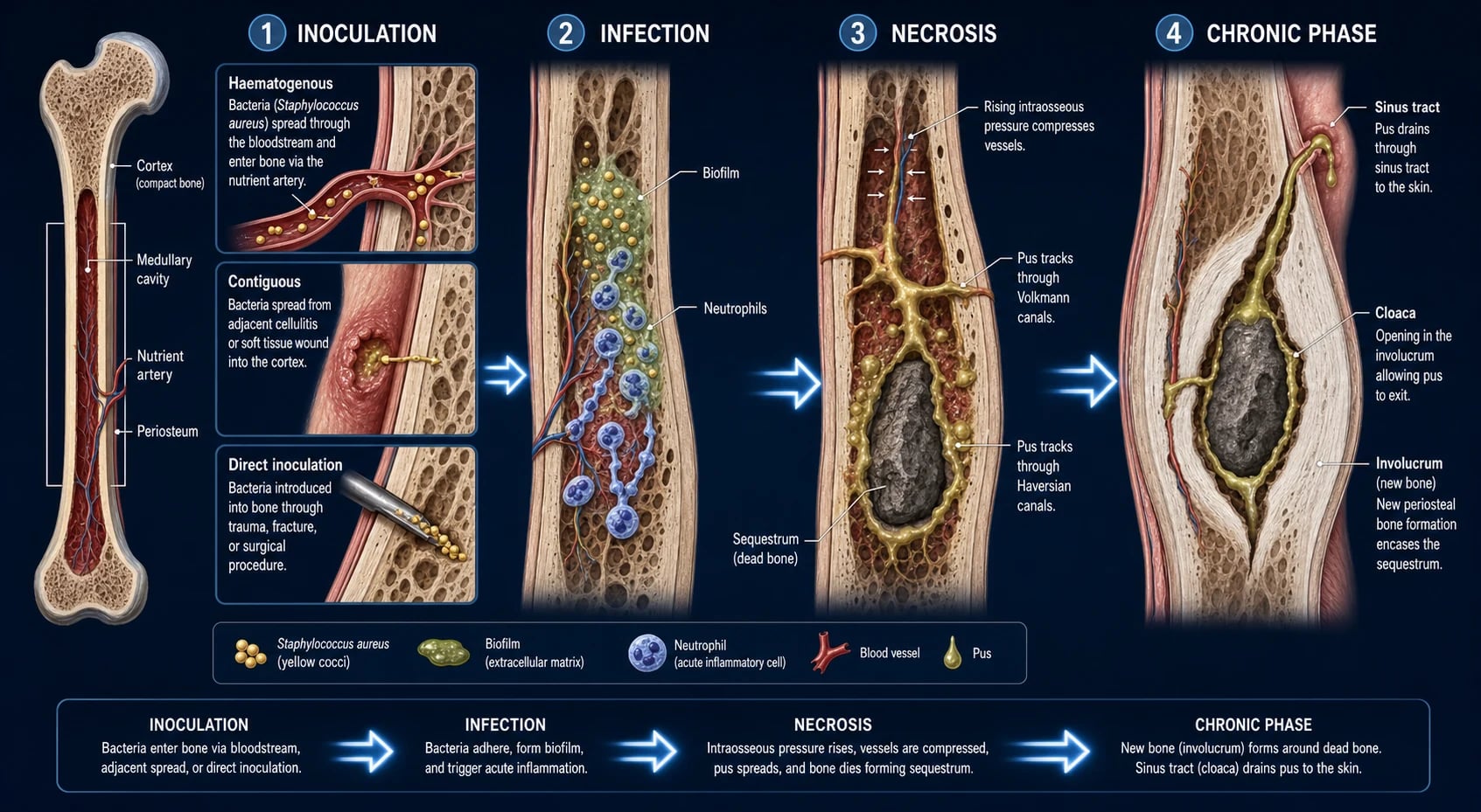
Bone is normally sterile. Infection occurs when microbes reach the medullary cavity or cortex and overwhelm local defences. There are three routes of entry, and the resulting cascade explains every clinical, radiological and therapeutic feature of the disease.[2]
Routes of entry: [1]
- Haematogenous — bacteraemic organisms lodge in the sluggish, fenestrated hair-loop capillaries of the long-bone metaphysis (children) or the vertebral end-plate (adults, via Batson's vertebral venous plexus). Single organism, usually S. aureus.
- Contiguous spread — from an adjacent soft-tissue infection, diabetic ulcer, surgical wound, or open fracture. Mixed flora including Gram-negatives and anaerobes.
- Direct inoculation — penetrating trauma, surgery, or a foreign body (orthopaedic implant) carries organisms directly into the bone. [1]
Why the metaphysis in children? The metaphyseal capillaries make sharp hair-pin loops, have sluggish flow and fenestrated endothelium without a basement membrane — bacteria slow, lodge, and multiply. The physis (growth plate) acts as a barrier: in children older than about 18 months the transphyseal vessels have closed, so infection is confined to the metaphysis and does not reach the joint (except where the metaphysis is intracapsular — hip, shoulder, knee, where septic arthritis can result). In neonates, the transphyseal vessels are still patent, so joint involvement (septic arthritis) is common.[4]
The inflammatory cascade (the mechanism that produces every sign): [1]
- Bacteria adhere to bone matrix via fibronectin and other adhesins, multiply, and form a biofilm (extracellular polymeric slime) on devitalised tissue.
- Macrophages release IL-1, TNF-alpha and IL-6 → fever, malaise, acute-phase response (raised CRP/ESR).
- Neutrophils rush in but cannot clear bacteria protected within necrotic tissue and biofilm; they release proteolytic enzymes that erode bone (osteolysis).
- Rising intraosseous pressure — bone is encased in a rigid cortical envelope that cannot expand — compresses the venous and arterial supply, producing venous stasis, thrombosis and ischaemia.
- Ischaemic cortex dies and detaches as a sequestrum (dead, separated bone) — avascular, so antibiotics and immune cells cannot reach it; it becomes a permanent reservoir of infection.
- The periosteum is elevated by subperiosteal pus and lays down new bone around the sequestrum = the involucrum.
- Pus forces its way out through a hole in the cortex = the cloaca, and tracks to the skin as a sinus tract (the pathognomonic feature of chronic osteomyelitis). [1]
The biofilm is the key to why chronic osteomyelitis and implant infection are so refractory: sessile bacteria within the slime are 10 to 1000 times more resistant to antibiotics and to phagocytosis. This is the rationale for surgical debridement of dead tissue, removal of infected implants, and the addition of rifampin (which uniquely penetrates the biofilm and kills adherent staphylococci) for staphylococcal disease.[7]
Vertebral osteomyelitis follows a distinctive pattern: haematogenous seeding (via Batson's plexus) of the vertebral end-plate, then spread to the adjacent disc and into the opposing vertebral body — hence the classic finding of two adjacent vertebrae and the disc between them. Complications include epidural abscess (a neurological emergency) and paraspinal abscess.[2]
Clinical Presentation
The presentation depends on route, age, host and chronicity, and a high index of suspicion is essential because systemic signs are frequently absent, especially in contiguous-focus, diabetic, elderly and immunocompromised patients.[1][2]
Acute haematogenous osteomyelitis (typically a child): rapid onset localised bone pain over the long-bone metaphysis (distal femur, proximal tibia, distal tibia, proximal humerus), with localised tenderness, swelling, warmth and erythema, fever, malaise, and — in younger children and infants — refusal to use the limb (pseudoparalysis) and limp or refusal to bear weight. The pain is often very precisely localised, unlike the diffuse pain of septic arthritis.[4][8]
Vertebral osteomyelitis (typically an adult): insidious, progressive back pain (often worse at night, unrelated to activity), localised spinal tenderness over the affected vertebra, low-grade or absent fever (up to 50% are afebrile), and a subacute course over weeks. Look for risk factors for bacteraemia (UTI, IV drug use, endocarditis, recent instrumentation, intravascular device). Red flags include radicular pain, limb weakness, sphincter disturbance or saddle anaesthesia — these indicate epidural abscess / cord compression and are a neurological emergency.[2]
Contiguous-focus / diabetic foot osteomyelitis: chronic, localised pain, swelling, erythema and warmth over a non-healing wound or ulcer that lies over a bony prominence (e.g., the metatarsal heads in a neuropathic foot). Probe-to-bone may be positive. Systemic signs are often absent — the neuropathic, ischaemic foot may not mount a fever or leukocytosis, and pain is blunted by neuropathy. Exposed bone in the base of an ulcer is highly suggestive.[5]
Chronic osteomyelitis: relapsing pain over months to years, a draining sinus tract (the pathognomonic feature), low-grade fever, intermittent discharge of pus or bone fragments. The disease may lie dormant for years and flare after minor trauma or intercurrent illness. A change in a chronic sinus (new pain, mass, bleeding) mandates biopsy to exclude Marjolin ulcer (squamous cell carcinoma arising in a chronic sinus tract).[2]
Prosthetic joint / implant-related infection: persistent joint pain, stiffness, wound drainage, sinus tract, or radiographic loosening of the implant. May be early (within 3 months — usually virulent S. aureus or Gram-negative), delayed (3 to 24 months — coagulase-negative staphylococci), or late (over 24 months — usually haematogenous).[7]
Atypical presentations (deliberately tested): [1]
- Elderly / diabetic / immunocompromised: minimal pain, no fever, vague malaise, a non-healing wound, or just unexplained raised inflammatory markers.
- Neonate: may present as sepsis or pseudoparalysis of a limb with no localising signs; multifocal disease is possible.
- IV drug user: back pain (vertebral), sternoclavicular or sacroiliac pain — easy to dismiss as musculoskeletal; screen for endocarditis. [1]
Differential Diagnosis
A painful, swollen limb or an unexplained spinal lesion is not always osteomyelitis. Distinguish the following, each by its discriminating features:[1][9]
Septic arthritis
- Joint, not bone; pain on passive movement
- Synovial fluid: WBC over 50,000, neutrophils, positive culture
- Can COEXIST with osteomyelitis (esp. paediatric hip)
Cellulitis / erysipelas
- Superficial skin infection; well-demarcated (erysipelas)
- No deep bony tenderness; pain on palpation of skin only
- Inflammatory markers only mildly raised relative to signs
Fracture (acute, stress, pathological)
- Trauma history (acute) or overuse/runner (stress)
- Radiographic fracture line; stress fracture MRI oedema limited to one site
- No fever, no raised CRP (unless healing)
Bone tumour — osteosarcoma
- Adolescent/young adult; pain worse, swelling
- Sunburst / Codman triangle / sunray periosteal reaction
- Biopsy essential — radiology can mimic osteomyelitis
Bone tumour — Ewing sarcoma
- Child/young adult; systemic features can mimic infection
- Onion-skin / layered periosteal reaction
- Tissue biopsy for diagnosis
Bone metastasis / myeloma
- Older adult; lytic lesion, often spine or pelvis
- Known primary; raised Ca, anaemia, renal failure (CRAB in myeloma)
- Tissue biopsy; no fever, normal WCC
Charcot neuroarthropathy (diabetes)
- Warm, swollen, erythematous foot; NO ulcer (typically)
- Often bilateral and symmetric; history of neuropathy
- MRI differentiation from osteomyelitis is challenging
Brodie abscess
- Subacute osteomyelitis; chronic local pain
- Intraosseous cavity with sclerotic rim on MRI/X-ray
- Curettage for cure
Vertebral: discitis, metastasis, compression fracture
- Discitis: often sterile/inflammatory; MRI disc signal change
- Metastasis: posterior elements, multiple sites, known primary
- Compression fracture: acute trauma/osteoporosis, no fever
Always consider malignancy (osteosarcoma, Ewing, metastasis) when the radiology is atypical, the age is wrong for haematogenous disease, or there is no response to appropriate therapy — biopsy is mandatory when there is any doubt.[2]
Clinical & Bedside Assessment
Vital signs drive triage. Fever, tachycardia, hypotension, tachypnoea and altered mentation indicate sepsis — apply the Surviving Sepsis hour-1 bundle.[1]
Focused limb examination: [1]
- Localised point tenderness over the affected bone — often very precisely localised in acute haematogenous disease (unlike the diffuse tenderness of septic arthritis or cellulitis).
- Warmth, swelling, erythema over the site; fluctuance suggests subperiosteal pus or abscess.
- Sinus tract with discharge (chronic disease) — note the location and character of the discharge.
- Distal neurovascular status — pulses, capillary refill, sensation, motor power. Neurovascular compromise is a surgical emergency.
- Examine the joint above and below — coexisting septic arthritis is common in children, especially at the hip.
- Refusal to bear weight / pseudoparalysis in infants and non-verbal children — a key sign. [1]
Diabetic foot — the probe-to-bone test: with sterile technique, insert a blunt metal probe through the wound to the base; if hard, gritty bone is felt, the test is positive. In high-prevalence diabetic foot clinics, a positive test has a high positive predictive value (around 90%); sensitivity is highest when bone is visible in the ulcer base. Combine with MRI for confirmation.[5]
Spine examination: point tenderness over the affected spinous processes; a full neurological examination — motor power, sensation (including perineal / saddle area), reflexes, anal tone. Saddle anaesthesia, urinary retention (catheter incontinence), or bilateral leg weakness = cauda equina / epidural abscess — emergency MRI and surgical referral. [1]
Search for the source of bacteraemia: skin and soft-tissue infection, indwelling vascular device, endocarditis (auscultate for murmurs — association with vertebral osteomyelitis is strong), urinary tract infection, dental infection, IV drug use injection sites. [1]
Investigations
Investigations serve three purposes: to confirm infection (bloods, MRI), to identify the organism (cultures, biopsy), and to define the anatomy for surgery (MRI, CT). The plain X-ray may be normal for 1 to 2 weeks — a normal early film never excludes osteomyelitis.[1][2]
Bloods: FBC (leukocytosis), CRP (the best marker for monitoring response — should fall steadily with effective therapy), ESR (slow; may remain elevated for weeks; useful as a baseline), U&E, LFTs, glucose, lactate (sepsis), coagulation. Blood cultures BEFORE antibiotics — positive in about 40% of acute haematogenous disease and 70 to 90% of vertebral osteomyelitis. In suspected endocarditis-associated vertebral disease, add echocardiography.[2]
Imaging — the ladder: [1]
| Modality | Sensitivity | Specificity | Role / timing |
|---|---|---|---|
| Plain X-ray | Low early | Moderate | First-line. Normal for 1 to 2 weeks. Earliest: soft-tissue swelling; then periosteal reaction (7 to 14 days); then lytic lesions (need 30 to 50% bone loss to be visible). Sequestrum appears as a dense sclerotic fragment. |
| MRI | ~90% | ~90% | Modality of choice. Marrow oedema = low T1, high T2/STIR signal. Defines extent, abscess, sequestrum, sinus tract, joint involvement. Gadolinium delineates abscess and necrosis. |
| CT | Moderate | High | Cortical detail — sequestrum identification, surgical planning, guided biopsy. |
| Bone scan (3-phase Tc-99) | High early | Low | Sensitive before X-ray changes; poor specificity (also hot in fracture, tumour, Charcot). White-cell scan better for chronic/implant. |
| PET-CT (18F-FDG) | High | High | Especially useful for prosthetic joint infection and chronic multifocal disease. |
Microbiological diagnosis — the gold standard is deep bone biopsy culture. Bone is obtained at surgical debridement or by percutaneous (often CT-guided) biopsy — not by swabbing the sinus tract. Send for aerobic and anaerobic culture, mycobacteria and fungi if risk factors. Histology confirms the diagnosis (acute: neutrophils in marrow; chronic: plasma cells, lymphocytes, necrotic bone) and excludes malignancy.[2][5]
Probe-to-bone test (diabetic foot) — bedside, see above. The IWGDF/IDSA 2023 guideline recommends combining clinical findings (probe-to-bone, exposed bone) with imaging (MRI) and, where possible, bone culture before committing to prolonged antibiotics.[5]
Investigations in specific scenarios: vertebral disease — MRI + CT-guided biopsy + blood cultures + echocardiography (endocarditis); paediatric — bloods, blood culture, ultrasound of the adjacent joint (effusion), MRI under sedation; prosthetic joint — aspiration of the joint (synovial WCC, culture), blood cultures, PET-CT. [1]
Management — Resuscitation
ABCDE first. If septic or systemically unwell, apply the Surviving Sepsis hour-1 bundle:[1]
- Oxygen to target SpO2 94 to 98% (or 88 to 92% in COPD / CO2 retention risk).
- Blood cultures BEFORE antibiotics (where feasible) — at least two sets, plus local (bone/joint) cultures.
- Empirical IV antibiotics within 1 hour of recognising sepsis.
- Lactate, balanced crystalloid 30 mL/kg if hypotensive or lactate over 2 mmol/L, noradrenaline for fluid-refractory shock; reassess fluid responsiveness before further boluses.
- Analgesia and splint/elevate the affected limb in the position of comfort. [1]
Spinal epidural abscess / cord compression with new neurological deficit is a separate, time-critical emergency: urgent MRI of the whole spine and neurosurgical/orthopaedic referral for decompression within 24 to 48 hours to preserve neurological function. Dexamethasone is sometimes given for cord oedema but is secondary to surgical decompression.[2]
Management — Definitive & Stepwise
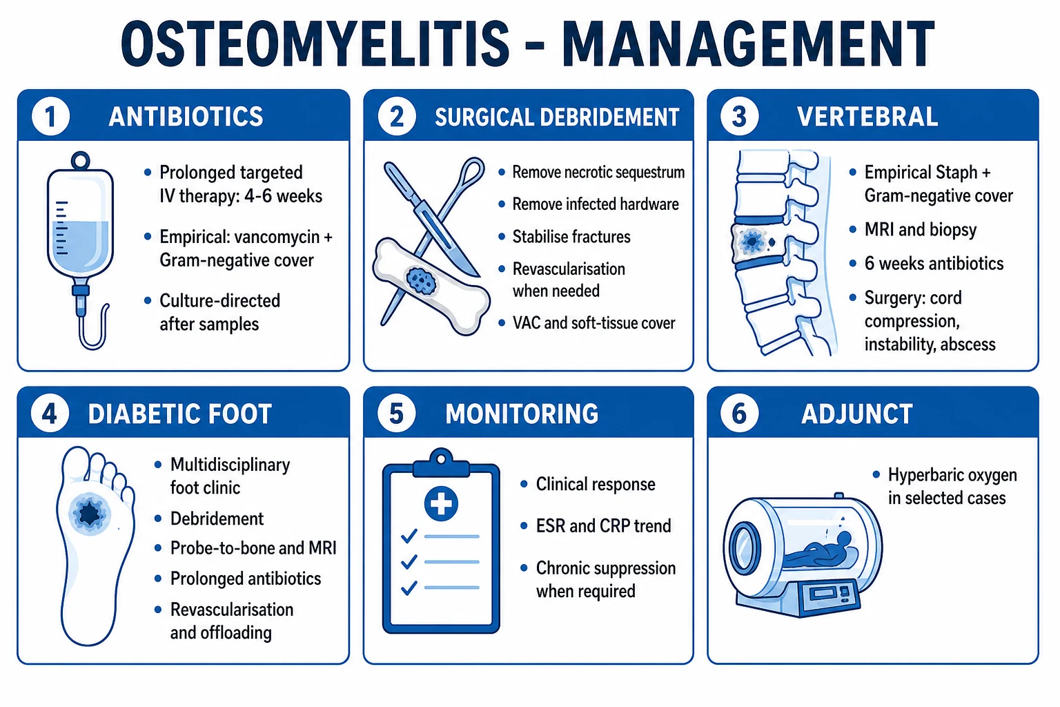
Osteomyelitis is managed by a co-management model: orthopaedic surgery (source control — debridement, stabilisation, soft-tissue cover) plus infectious diseases (culture-directed antibiotics, OPAT, monitoring). The unifying principle: dead bone cannot be cured by antibiotics.[2][3]
Empirical antibiotic choice — must cover Staphylococcus aureus and be tailored to the scenario:[1][9]
| Scenario | Empirical regimen | Rationale |
|---|---|---|
| Acute haematogenous, no risk factors | Flucloxacillin 2 g IV 6-hourly (or cefazolin/cefuroxime) | Cover MSSA |
| MRSA risk, severe beta-lactam allergy, prosthetic | Vancomycin 15 to 20 mg/kg IV 12-hourly (target trough 15 to 20) ± add for MRSA; alternatives teicoplanin, daptomycin | Cover MRSA |
| Contiguous / diabetic foot / contaminated wound | Add Gram-negative cover — ceftriaxone, piperacillin-tazobactam, or ceftazidime; add anaerobic cover (metronidazole) if necrotic | Mixed flora |
| Sickle cell disease | Add Salmonella cover — ceftriaxone or ciprofloxacin | Classic but S. aureus still commonest |
| IV drug use | Cover Pseudomonas — piperacillin-tazobactam or ceftazidime ± aminoglycoside | Pseudomonas common |
| Prosthetic / implant | Vancomycin ± rifampin (after debridement, see below); remove / exchange implant | Biofilm |
Definitive therapy — narrow to culture sensitivities once bone culture returns. For staphylococcal chronic osteomyelitis, add rifampin 600 to 900 mg PO once daily to a beta-lactam or a fluoroquinolone (e.g., ciprofloxacin 750 mg PO 12-hourly + rifampin) for at least 6 weeks — rifampin uniquely penetrates the biofilm and kills adherent staphylococci. The evidence base has been challenged (the Renz 2021 controversy), but the overall evidence and current guidelines still support its use in staphylococcal implant infection.[7]
Duration: [1]
- Acute osteomyelitis: 4 to 6 weeks total (commonly 2 weeks IV, then oral step-down once afebrile, improving, and CRP falling).
- Chronic osteomyelitis: 6 weeks minimum, often longer (up to 6 months in some); guided by clinical response and CRP trend.
- Vertebral osteomyelitis: typically 6 weeks.
- Paediatric acute haematogenous: the Peltola / Pääkkönen data support a shortened IV course (3 to 4 days) and early oral switch — total 3 to 4 weeks is sufficient in uncomplicated cases.[4][8]
IV-to-oral step-down criteria: haemodynamically stable, afebrile for 24 to 48 hours, CRP falling, able to swallow and absorb, and a bioavailable oral agent available (e.g., ciprofloxacin + rifampin, clindamycin, linezolid, doxycycline). OPAT (outpatient parenteral antimicrobial therapy) is a well-validated option for completing the IV course at home.[6]
Surgical management — the principles of source control, applied according to the Cierny-Mader stage:[3]
- Drainage of abscess (subperiosteal, intraosseous, paraspinal).
- Debridement of all necrotic bone to bleeding, healthy bone (the paprika sign — punctate bleeding from viable cortical bone — marks adequate debridement).
- Removal of infected hardware if possible (the biofilm on it is incurable).
- Dead-space management — antibiotic-laden polymethylmethacrylate (PMMA) cement spacer (often loaded with vancomycin + gentamicin), or a biodegradable carrier; later replaced with bone graft and/or a vascularised soft-tissue / muscle flap.
- Stabilisation of any pathological fracture or instability.
- Soft-tissue cover — often by plastic surgery (free or rotational flap), particularly for exposed tibia and diabetic foot.
- Revascularisation if the limb is ischaemic (diabetic / vascular disease) — antibiotics cannot reach an ischaemic limb. [1]
Chronic osteomyelitis refractory to standard therapy: consider hyperbaric oxygen therapy as an adjunct (controversial; not first-line); long-term suppressive antibiotics if the patient is not fit for surgery (Host C).[3]
Prosthetic joint infection pathway (Zimmerli / IDSA model):[7]
- Acute (under 4 weeks, mobile implant): debridement, antibiotics and implant retention (DAIR) + rifampin for staphylococcal disease, 6 weeks; then suppressive.
- Chronic (over 4 weeks, loose implant): two-stage exchange — remove implant, place antibiotic-loaded cement spacer, 4 to 6 weeks of antibiotics, then reimplant. One-stage exchange in selected centres with good soft tissue.
- Not operable: lifelong suppressive antibiotics. [1]
Specific Subtypes & Scenarios
- Acute haematogenous osteomyelitis in children — long-bone metaphysis (distal femur, proximal/distal tibia, proximal humerus); S. aureus; often medical (4 to 6 weeks antibiotics) if no abscess or sequestrum; surgical debridement reserved for subperiosteal pus, sequestrum, or failure to improve in 48 to 72 hours. Peltola / Pääkkönen shortened-IV data underpin the modern approach.[4][8]
- Vertebral osteomyelitis / discitis (adults) — back pain, two adjacent vertebrae and the disc, end-plate oedema; risk of epidural / paraspinal abscess; check for endocarditis; 6 weeks of targeted antibiotics; surgery only for instability, cord compression, or failure of medical therapy.[2]
- Contiguous-focus osteomyelitis (post-trauma, post-surgical) — mixed flora including Gram-negatives and anaerobes; surgical debridement is essential; remove infected hardware if possible.[3]
- Diabetic foot osteomyelitis — multifactorial (neuropathy, vascular disease, pressure ulcer over a bony prominence); mixed flora; probe-to-bone, MRI; combined surgical (resection/amputation) + prolonged antibiotics; revascularisation if ischaemic; multidisciplinary foot team; offloading (total contact cast). The IWGDF/IDSA 2023 guideline provides the framework.[5]
- Chronic osteomyelitis — sequestrum, involucrum, cloaca, sinus tract, biofilm; surgical; risk of Marjolin ulcer.[2][3]
- Brodie abscess (subacute) — intraosseous abscess cavity with sclerotic rim; chronic local pain; surgical curettage.[2]
- Prosthetic joint infection — biofilm; DAIR vs one-/two-stage exchange; rifampin for staphylococci.[7]
- Salmonella osteomyelitis in sickle cell disease — the classic exam answer; but S. aureus still commonest; infarcted bone provides a nidus.[2]
- Neonatal osteomyelitis — Group B streptococcus, E. coli, S. aureus; transphyseal vessels are patent so joint involvement (septic arthritis) is common; often multifocal.[4]
- Tuberculous osteomyelitis (Pott disease of the spine) — chronic, destructive; paraspinal cold abscess; MRI; tissue diagnosis (AFB, culture, PCR); standard anti-tubercular therapy.
Complications & Pitfalls
Disease-related: chronic relapsing infection (inadequate debridement), septic arthritis (extension through physis or cortex), joint destruction and growth arrest / limb-length discrepancy in children (physeal damage), pathological fracture through weakened or lytic bone, epidural abscess / cord compression / cauda equina (vertebral), sepsis, septic shock, metastatic infection (endocarditis, brain abscess), amputation (diabetic foot), death.[2]
Marjolin ulcer — squamous cell carcinoma arising in a chronic draining sinus tract, typically after 10 to 30 years of chronic osteomyelitis. Any change in a chronic sinus (new pain, mass, bleeding, increased discharge) mandates biopsy. The lesion is often aggressive; wide local excision and staging are required.[2]
Secondary amyloidosis (long-standing chronic suppuration) and renal disease from prolonged nephrotoxic antibiotics (vancomycin, aminoglycosides) are long-term complications.[2]
Antibiotic-related complications: vancomycin nephrotoxicity (monitor troughs, renal function), aminoglycoside nephro- and ototoxicity, rifampin hepatotoxicity and potent CYP3A4 induction (reduces efficacy of oral contraceptives, warfarin, DOACs, antiretrovirals — warn and adjust), linezolid myelosuppression, peripheral and optic neuropathy and serotonin syndrome (it is a reversible MAO inhibitor), fluoroquinolone tendinopathy/rupture and QT prolongation.[7]
Classic pitfalls: [1]
- Accepting a normal early X-ray as excluding osteomyelitis — it does not; image with MRI.
- Trusting a sinus tract swab as the organism — it is contaminated; obtain bone biopsy culture.
- Treating chronic osteomyelitis with antibiotics alone — dead bone cannot be sterilised; debride.
- Forgetting to add rifampin for staphylococcal implant / biofilm infection.
- Missing an epidural abscess in a patient with vertebral osteomyelitis and new leg weakness.
- Missing Marjolin ulcer in a chronic sinus that has changed.
- Not screening for endocarditis in vertebral osteomyelitis.
- Not addressing ischaemia before expecting antibiotics to work in the diabetic foot. [1]
Prognosis & Disposition
Outcome depends on organism virulence (S. aureus is most virulent), host (diabetes, immunosuppression, vascular disease all worse), site (vertebral higher mortality), and — above all — time to diagnosis and the adequacy of surgical source control.[1][2]
- Acute haematogenous in children: excellent with prompt therapy — cure rate over 90%; chronicity 5 to 15%.[4]
- Vertebral osteomyelitis: mortality 2 to 12%; neurological recovery depends on speed of decompression for those with epidural extension.[2]
- Diabetic foot osteomyelitis: high recurrence and amputation (10 to 30% if ischaemic or inadequately treated); 5-year mortality approaches 50%, comparable to many cancers.[5]
Prognostic markers of response: falling CRP and clinical improvement (afebrile, less pain, less drainage) indicate response; persistent fever, rising CRP or ongoing drainage suggest inadequate source control and mandate re-debridement.[1]
Disposition: IV antibiotics (often via OPAT) for 2 to 6 weeks, oral step-down to complete a 4 to 6 week (acute) or longer (chronic) course; long-term follow-up — recurrence can occur years later, especially in diabetics and after implant retention. Multidisciplinary follow-up (orthopaedics, infectious diseases, diabetology, podiatry, plastic surgery as indicated). [1]
Special Populations
- Children — weight-based dosing: cefazolin 50 to 100 mg/kg/day IV in 3 divided doses, or flucloxacillin 25 to 50 mg/kg IV 6-hourly; clindamycin or vancomycin if MRSA. Remember the physeal barrier and the risk of coexisting septic arthritis (especially at the paediatric hip, where the metaphysis is intracapsular). The Kocher criteria help distinguish septic arthritis from transient synovitis in the limping child.[4][8]
- Diabetes mellitus — low threshold to image (MRI); probe-to-bone; address ischaemia (revascularisation may be required before antibiotics work); multidisciplinary foot team; offloading (total contact cast); combined surgical + medical therapy.[5]
- Pregnancy — avoid tetracyclines (fetal bone/teeth), fluoroquinolones (relative caution), aminoglycosides (fetal ototoxicity); beta-lactams are safe; weight-based dosing with increased GFR. Surgical decisions balance maternal benefit against fetal risk.
- Elderly — atypical presentation; vertebral disease common; consider malignancy in the differential; lower threshold for imaging; beware drug toxicity with declining renal function (dose-adjust vancomycin, beta-lactams).[1]
- Immunocompromised (neutropenia, transplant, HIV) — broader empirical cover including Pseudomonas, fungi (Aspergillus, Candida), atypical mycobacteria; low threshold for biopsy.
- Sickle cell disease — Salmonella classically; but S. aureus still commonest; infarcted bone is a nidus; cover both.
- Anticoagulated — balance surgical bleeding risk against thromboprophylaxis; bridging may be required; chronic infection itself is prothrombotic.[1]
- IV drug users — Pseudomonas; unusual sites (sternoclavicular, sacroiliac, vertebral); screen for endocarditis and blood-borne viruses; harm reduction.
Evidence, Guidelines & Regional Differences
Landmark evidence and what it changed:[2]
- Lew & Waldvogel, Lancet 2004 — the definitive modern review defining the pathogenic classification (haematogenous vs contiguous vs chronic) that still structures practice.[2]
- Cierny-Mader, Clin Orthop 2003 — the anatomic-host staging system that guides surgical decision-making in adult chronic osteomyelitis.[3]
- Peltola & Pääkkönen, NEJM 2014 — established that short IV courses (3 to 4 days) and early oral switch are safe in uncomplicated paediatric acute osteomyelitis, transforming paediatric practice away from prolonged IV lines.[4]
- IWGDF/IDSA 2023 guideline — the global standard for diabetic foot infection diagnosis and treatment; endorses probe-to-bone, MRI, and a limited duration of antibiotics after surgical resection (no more than 1 week of antibiotics after a clean amputation margin).[5]
- Conterno, Cochrane 2013 — found insufficient high-quality RCT evidence on antibiotic choice and duration for chronic osteomyelitis; practice is therefore largely expert- and guideline-based, not RCT-driven.[6]
- Renz & Zimmerli, 2021 — a critical appraisal of the rifampin in biofilm evidence; concluded that the evidence overall supports its use in staphylococcal implant infections, but flagged methodological concerns and the need for better trials.[7]
Regional guideline differences. India (ICMR/NMC): high community MRSA prevalence — empirical vancomycin + Gram-negative cover (piperacillin-tazobactam or carbapenem) for severe diabetic foot infection; Salmonella emphasis in sickle cell. US (IDSA): empirical vancomycin + cefepime or piperacillin-tazobactam for broad cover; specific IDSA guidance on prosthetic joint, diabetic foot, and vertebral infection. UK (NICE NG19): the diabetic foot pathway with a multidisciplinary foot service; emphasis on probe-to-bone and MRI; rifampin for staphylococcal biofilm. Europe (IWGDF/EWMA): aligned with the global IWGDF framework. Always apply the local antibiogram — empirical choice should reflect local resistance patterns.[5]
Where the evidence is weak: optimal duration of antibiotics for chronic osteomyelitis; rifampin in non-staphylococcal biofilm; hyperbaric oxygen as adjunct; one- vs two-stage exchange for prosthetic joint infection; surgical vs medical management of uncomplicated vertebral osteomyelitis.[6]
Exam Pearls
The chronic osteomyelitis quartet
SICS
dead, separated bone (avascular, dense on X-ray)
new periosteal bone laid down around the sequestrum
hole in the cortex through which pus escapes
the channel draining pus to the skin — pathognomonic
Imaging — when to use what
MIX-B
modality of choice — sensitivity and specificity about 90%
echocardiography for endocarditis in vertebral disease
normal for 1 to 2 weeks; lytic lesions need 30 to 50% bone loss
the microbiological gold standard (NOT sinus tract swab)
Pitfalls that cost marks
DEAD-BONE
it is contaminated; obtain bone biopsy culture
new leg weakness in vertebral disease — emergency MRI + surgery
in chronic disease — debride dead bone
probe-to-bone; revascularise if ischaemic
add rifampin for staphylococcal implant infection
Marjolin ulcer in a chronic sinus — biopsy any change
cover S. aureus first, broaden by scenario
screen for it in vertebral osteomyelitis
Quick self-test — answer in your head, then click to verify
A 14-year-old boy presents with 3 days of fever and progressive, severe pain over the distal thigh, just above the knee, with localised tenderness, warmth and a limp. His X-ray is normal. What is the diagnosis, the next best imaging test, and the empirical antibiotic? (Answer: acute haematogenous osteomyelitis of the distal femoral metaphysis — MRI is the next best test; flucloxacillin or cefazolin IV, adding vancomycin if MRSA risk. Blood cultures and CRP first.)[4][8]
The numbers that examiners ask
One-liners examiners reward:[1][2][7]
- The commonest organism in osteomyelitis at every age and every subtype is Staphylococcus aureus.
- Salmonella is the classic exam answer for sickle cell disease, but S. aureus is still commonest there too.
- IV drug users get Pseudomonas at unusual sites (sternoclavicular, sacroiliac, vertebral).
- Neonates get Group B strep, E. coli, S. aureus and often joint involvement (patent transphyseal vessels).
- Vertebral disease: two adjacent vertebrae and the disc, via Batson's plexus; always screen for endocarditis.
- The microbiological gold standard is bone biopsy culture — sinus tract culture is contaminated.
- Dead bone cannot be cured by antibiotics — surgical debridement is the cornerstone.
- Add rifampin for staphylococcal biofilm / implant infection.
- Marjolin ulcer = SCC in a chronic sinus tract — biopsy any change.
- The paprika sign = punctate bleeding from viable bone = adequate debridement.
- Peltola / Pääkkönen: short IV course and early oral switch in uncomplicated paediatric acute osteomyelitis.
- Probe-to-bone positive diabetic ulcer = osteomyelitis until proven otherwise. [1]
Exam application bank (NEET-PG / INICET)
One-line answer
Osteomyelitis is an infection of bone and bone marrow (myelo = marrow) by microbes (usually bacteria), producing inflammation, bone destruction (osteolysis), necrosis and reactive new bone formation. By pathogenesis (Lew-Waldvogel): haematogenous (bloodstream seeding — children: long-bone metaphysis; adults: vertebrae), contiguous-focus (adjacent wound, ulcer, diabetic foot, surgery) and chronic (sequestrum, involucrum, sinus tract, biofilm). Staphylococcus aureus is the commonest organism across all ages and types. Acute disease presents with localised bone pain, tenderness, swelling and fever; chronic disease with a draining sinus tract and relapsing pain. X-ray may be normal for the first 1 to 2 weeks — MRI is the modality of choice (sensitivity about 90%). The microbiological gold standard is bone biopsy culture (sinus tract culture is contaminated). Management is orthopaedic + infec
Worked stems (answer without another resource)
Stem 1 — Classic presentation. Map symptoms to mechanism; name the first investigation and first treatment step with dose/route if drug therapy is standard. [1]
Stem 2 — Unstable / complicated. List red flags that force immediate resuscitation, theatre, ICU, antidote, or reperfusion — and what you do in the first 15 minutes. [1]
Stem 3 — Atypical group. Elderly, pregnancy, child, or immunocompromised: how presentation and thresholds change. [1]
Stem 4 — Differential trap. Name the three closest mimics and one discriminator for each. [1]
Stem 5 — Disposition. Who goes home with safety-netting, who is admitted, who needs HDU/ICU/theatre, and what follow-up is mandatory. [1]
Rapid viva checklist
- Definition + classification
- Pathophysiology chain
- Bedside signs / criteria
- Score with exact components (if any)
- Emergency bundle
- Definitive therapy with doses
- Complications of disease and of treatment
- Special populations
- Guideline/trial name if classic
- Three exam traps
Coverage self-check
If you cannot answer any stem above from this page alone, re-read the matching section — the page is intended to be self-sufficient for final-prof and NEET-PG/INICET questions on Osteomyelitis.
References
- [1]Hatzenbuehler J, Pulling TJ. Diagnosis and management of osteomyelitis Am Fam Physician, 2011.PMID 22046943
- [2]Lew DP, Waldvogel FA. Osteomyelitis Lancet, 2004.PMID 15276398
- [3]Cierny G 3rd, Mader JT, Penninck JJ. A clinical staging system for adult osteomyelitis Clin Orthop Relat Res, 2003.PMID 12966271
- [4]Paakkonen M. Acute osteomyelitis in children N Engl J Med, 2014.PMID 24693913
- [5]Senneville E, Albalawi Z, van Asten SA, et al. IWGDF/IDSA guidelines on the diagnosis and treatment of diabetes-related foot infections (IWGDF/IDSA 2023) Diabetes Metab Res Rev, 2024.PMID 37779323
- [6]Conterno LO, Turchi MD. Antibiotics for treating chronic osteomyelitis in adults Cochrane Database Syst Rev, 2013.PMID 24014191
- [7]Renz N, Zimmerli W. Controversy about the Role of Rifampin in Biofilm Infections: Is It Justified? Antibiotics (Basel), 2021.PMID 33562821
- [8]Shapiro K, Anand A, Garg I, Agarwal A. Diagnosis and Management of Acute Osteoarticular Infections: Summary of New Guidelines Pediatr Rev, 2025.PMID 40306703
- [9]Carek PJ, Dickerson LM, Sack JL. Diagnosis and management of osteomyelitis Am Fam Physician, 2001.PMID 11430456