Infectious Diseases · General Medicine
Pneumonia (Community-Acquired)
Also known as Pneumonia · Community-acquired pneumonia · CAP · Lobar pneumonia · Atypical pneumonia
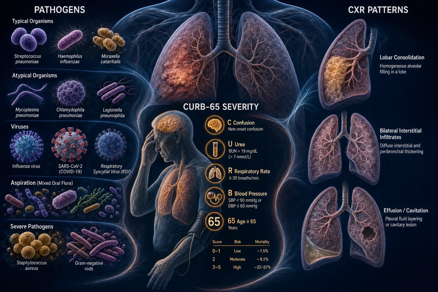
Community-acquired pneumonia (CAP) is an acute infection of the lung parenchyma acquired outside hospital (or within 48 h of admission). Commonest organism Streptococcus pneumoniae; atypicals (Mycoplasma, Chlamydophila, Legionella); viruses (influenza, COVID-19, RSV); Staphylococcus aureus and Gram-negatives in severe/chronic disease; Klebsiella in alcoholics. Presents with productive cough, fever, dyspnoea, pleuritic chest pain and signs of consolidation; atypical pneumonias have a dry cough, prominent systemic features and a normal/mildly abnormal CXR. Diagnosis is clinical plus chest X-ray; severity by CURB-65 (Confusion, Urea over 7, RR over 30, BP under 90/60, age over 65). Treat with antibiotics within 4 hours: low severity amoxicillin/doxycycline; moderate/severe beta-lactam plus macrolide; give oxygen, fluids, cover atypicals. Admit if CURB-65 at least 2; ICU if 3 to 5. Vaccinate (pneumococcal, influenza, COVID-19).
On this page & tools
Your progress
Saved locally on this device.
Exam tags
Red flags
Overview & Definition
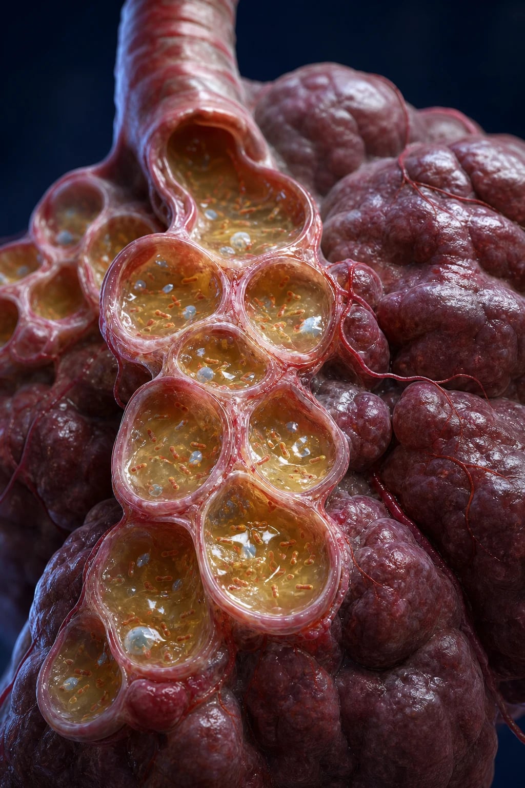
Pneumonia is an acute infection of the lung parenchyma — the alveoli, terminal bronchioles and the surrounding interstitium — that produces inflammatory exudate and consolidation visible (usually) on imaging. It is distinguished from acute bronchitis (infection of the larger airways with no parenchymal involvement, a normal chest X-ray and a predominantly dry or mucoid cough) and from upper respiratory tract infection (coryza, sore throat, no lower-respiratory signs or consolidation).[2]
The clinical skill in pneumonia is not the diagnosis itself — fever, cough and new consolidation on a chest X-ray is straightforward — but recognising severity early, choosing empirical antibiotics that cover typical AND atypical organisms, and remaining alert to viruses (COVID-19, influenza), aspiration, and the complications that turn a curable infection into a lethal one (effusion, empyema, lung abscess, sepsis). The single biggest process lever in hospitalised CAP is early antibiotics — within 4 hours of admission for ward patients and within 1 hour for those with septic shock — because every hour of delay in the septic patient measurably increases mortality.[1][5]
Pneumonia is the leading infectious cause of death worldwide in adults, and in low- and middle-income countries it remains among the top three causes of death at every age. It sits at the intersection of microbiology, respiratory physiology (ventilation–perfusion mismatch and shunt), sepsis physiology, antimicrobial stewardship and preventive medicine (vaccination). A candidate who masters CAP masters a template — recognise, grade, treat early, cover broadly, de-escalate, prevent — that transfers to almost every infectious presentation.[2]
Classification
Pneumonia is classified along four axes — place of acquisition, anatomy/radiology, aetiology, and severity — and each axis changes either the empirical antibiotic choice or the disposition. [1]
By place of acquisition
The acquisition threshold is the classification that matters most for empirical therapy, because it predicts the likely organisms and their resistance patterns:[1]
- Community-acquired pneumonia (CAP) — symptoms and signs plus radiographic consolidation, acquired outside hospital or within the first 48 hours of admission. The pathogens are the "CAP list": S. pneumoniae, H. influenzae, M. pneumoniae, C. pneumoniae, Legionella, and respiratory viruses.
- Hospital-acquired pneumonia (HAP) — pneumonia developing more than 48 hours after admission that was not incubating at the time of admission. Organisms shift toward the enteric Gram-negatives (Klebsiella, E. coli, Enterobacter, Acinetobacter) and S. aureus, including MRSA, reflecting oropharyngeal colonisation by hospital flora.
- Ventilator-associated pneumonia (VAP) — HAP arising more than 48 hours after endotracheal intubation. The microbiology is dominated by multidrug-resistant Gram-negatives (including Pseudomonas aeruginosa) and MRSA; prevention rests on ventilator-care bundles (head-up 30 degrees, daily sedation breaks, oral chlorhexidine, subglottic suctioning, early extubation).
- Aspiration pneumonia — pneumonia following macroaspiration of oropharyngeal or gastric contents (stroke, seizure, reduced GCS, impaired swallow, alcoholism). The dependent segments are involved and the flora is mixed, including anaerobes (Peptostreptococcus, Fusobacterium, Prevotella). The related but distinct aspiration pneumonitis is a chemical injury from sterile gastric acid that may not need antibiotics unless it fails to resolve in 48 hours. [1]
The HCAP (healthcare-associated pneumonia) category — introduced in the 2005 ATS/IDSA guideline for patients with recent hospitalisation, residence in a nursing home, or recent IV antibiotics — was dropped in the 2019 update. Modern empirical therapy is instead guided by local validated risk factors for MRSA and Pseudomonas (prior isolation, recent IV antibiotics, structured predictive scores), not by the old HCAP label, which over-treated many patients.[1][5]
By anatomy and radiology
- Lobar pneumonia — consolidation confined to one lobe or segment with a sharp border; the typical bacterial pattern (S. pneumoniae, K. pneumoniae). The four classical pathological stages — congestion, red hepatization, grey hepatization, resolution — describe the evolution of lobar exudate but are seldom clinically visible.
- Bronchopneumonia — patchy, multifocal, bilateral consolidation distributed around bronchi; seen with S. aureus, H. influenzae, Pseudomonas, and in aspiration.
- Interstitial (atypical) pneumonia — diffuse, often bilateral reticulonodular or ground-glass pattern; the atypicals (Mycoplasma, Chlamydophila, viruses, Pneumocystis). [1]
By aetiology — typical vs atypical
The typical vs atypical distinction is an examiner favourite because it predicts presentation, radiology, and empiric cover: [1]
Typical bacteria
Lobar, abrupt, purulent
- **S. pneumoniae** — commonest CAP organism worldwide
- **H. influenzae** — COPD, structural lung disease
- **Moraxella catarrhalis** — COPD, elderly
- **Klebsiella pneumoniae** — alcoholics, "red-currant-jelly" sputum
- **Staphylococcus aureus** — post-influenza, IV drug use, cavitation
Atypical bacteria
Interstitial, insidious, dry cough
- **Mycoplasma pneumoniae** — young adults, bullous myringitis, cold agglutinins
- **Chlamydophila pneumoniae** — sinusitis, pharyngitis
- **Chlamydophila psittaci** — psittacosis, bird exposure
- **Legionella pneumophila** — water systems, GI symptoms, hyponatraemia
- **Coxiella burnetii** — Q fever, farm animals, parturient sheep
Viruses & opportunists
Bilateral, prominent systemic upset
- **Influenza A/B** — seasonal epidemic, myalgia
- **SARS-CoV-2 (COVID-19)** — bilateral ground-glass, hypoxia
- **RSV, adenovirus, parainfluenza** — children, immunocompromised
- **Pneumocystis jirovecii** — HIV, exertional desaturation
By severity
Severity is graded with named scores reproduced verbatim below: CURB-65 (bedside favourite), CRB-65 (urea-free variant), the Pneumonia Severity Index (PSI/PORT) (more accurate but slower), and the IDSA/ATS severe-CAP criteria (decides ICU). These are not interchangeable — each answers a slightly different question.[3][4]
Epidemiology & Risk Factors
Pneumonia is a leading cause of infectious death worldwide, the most common lethal infection in the developed world, and a particular killer of the very young, the elderly, and the immunocompromised. In adults, Streptococcus pneumoniae is the commonest identifiable pathogen across all severities of CAP, followed by Haemophilus influenzae, the atypicals (Mycoplasma, Chlamydophila), and respiratory viruses; Staphylococcus aureus and Gram-negatives dominate in severe disease, in alcoholics, and post-influenza.[1]
Host and environmental risk factors increase susceptibility by impairing one or more of the normal defences (cough, mucociliary clearance, alveolar macrophages, neutrophil function, or the integrity of the swallow). The high-yield list:[2]
- Age — extremes of life (neonates, the elderly); physiological immune senescence plus declining cough reflex.
- Smoking — the single biggest modifiable risk factor for CAP; impairs mucociliary clearance and alveolar macrophage function and increases pneumococcal and Legionella risk.
- Chronic lung disease (COPD, bronchiectasis) — colonisation with H. influenzae and Moraxella; Pseudomonas in severe bronchiectasis.
- Alcohol misuse — impaired swallow, neutrophil function, and the classic association with Klebsiella.
- Diabetes, CKD, liver disease — impaired neutrophil and humoral immunity.
- Immunocompromise — HIV (risk of Pneumocystis and TB), chemotherapy, transplant, hypogammaglobulinaemia.
- Aspiration risk — stroke, dementia, seizure, reduced GCS, impaired swallow (Parkinson's, motor neuron disease), alcoholism.
- Institutionalisation / recent hospitalisation / recent antibiotics — selects for resistant organisms (MRSA, Pseudomonas, ESBL producers).
- Malnutrition and indoor air pollution (biomass-fuel smoke) — major drivers in low- and middle-income countries. [1]
Risk-factor-to-organism pairings (exam favourite): [1]
| Risk factor / host | Organism to consider |
|---|---|
| Alcohol misuse | Klebsiella pneumoniae, anaerobes, S. pneumoniae |
| Post-influenza | Staphylococcus aureus (often cavitating), H. influenzae |
| COPD / bronchiectasis | H. influenzae, Moraxella catarrhalis, Pseudomonas (severe) |
| Young adult, crowded | Mycoplasma pneumoniae, Chlamydophila |
| Hotel/cooling-tower outbreak | Legionella pneumophila |
| Bird exposure (parrots, poultry) | Chlamydophila psittaci (psittacosis) |
| Farm animals, parturient sheep/cats | Coxiella burnetii (Q fever) |
| HIV / CD4 under 200 | Pneumocystis jirovecii, TB, fungal |
| Neutropenia, haematological malignancy | Gram-negative bacilli, Pseudomonas, fungi |
| Asplenia / sickle-cell | S. pneumoniae, H. influenzae type b (overwhelming post-splenectomy infection) |
Pathophysiology
The lung is normally sterile below the vocal cords, defended by a layered immune system: the cough reflex expels large particles, the mucociliary escalator sweeps debris and microbes cephalad from the small airways, and the alveolar macrophages phagocytose organisms that reach the alveolus, supplemented by neutrophils recruited when the microbial load overwhelms them. Pneumonia occurs when these defences are overwhelmed, bypassed, or defective.[2]
Routes by which pathogens reach the alveoli: [1]
- Microaspiration of oropharyngeal flora — the commonest mechanism in CAP and HAP; small-volume aspiration occurs during sleep even in healthy people but is normally cleared.
- Macroaspiration of gastric or oropharyngeal contents — anaerobes and mixed flora; the basis of aspiration pneumonia.
- Inhalation of aerosolised droplets — the route of Mycoplasma, Mycobacterium tuberculosis, viruses (influenza, SARS-CoV-2), Legionella from contaminated water aerosols, and Coxiella.
- Haematogenous spread — septic emboli from right-sided endocarditis (IV drug use) or distant infection; classically S. aureus.
- Direct extension — from a contiguous infection (subdiaphragmatic abscess, mediastinitis) — rare. [1]
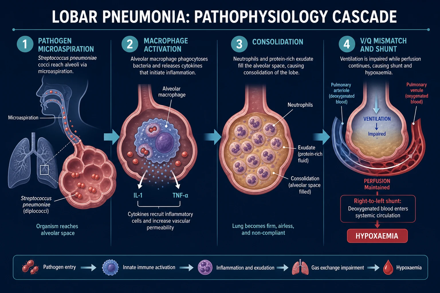
Once in the terminal airway, organisms multiply, triggering alveolar macrophage cytokine release (IL-1, TNF-alpha, IL-6) that recruits neutrophils and a protein-rich exudate into the alveolus. The exudate — fibrin, neutrophils, erythrocytes and organisms — fills the air sacs (consolidation), collapsing alveolar units and flooding gas-exchange surface. This produces ventilation–perfusion mismatch and intrapulmonary shunt — blood flows past non-ventilated, consolidated lung and returns to the left heart deoxygenated, causing the hypoxaemia that is the cardinal physiological lesion of pneumonia. The same cytokines produce the fever, tachycardia, rigors and systemic upset, and — when severe — drive the vasodilation, capillary leak and myocardial depression of septic shock. Resolution follows as macrophages clear the exudate once the organisms are controlled.[2]
Typical (lobar) vs atypical (interstitial): typical organisms multiply in the alveolar space producing a dense lobar exudate; atypicals proliferate in the epithelium and interstitium, producing a less exudative, more interstitial process with disproportionately prominent systemic upset and a paucity of chest signs. Viruses injure the alveolar epithelium and capillary endothelium directly, producing the diffuse alveolar damage of viral pneumonitis and, in severe cases, acute respiratory distress syndrome (ARDS). [1]
The four classical pathological stages of lobar pneumonia — describing the evolution of the lobar exudate over days, and still examiner-favoured despite being seldom clinically visible: [1]
- Congestion (first 24 hours) — vascular engorgement and intra-alveolar fluid with abundant organisms; the lung is heavy, red and boggy.
- Red hepatization (days 2–4) — exudate of fibrin, neutrophils and erythrocytes solidifies the lobe so it resembles liver (hence "hepatization"); air is largely displaced.
- Grey hepatization (days 4–8) — fibrin and a predominantly neutrophil/macrophage exudate give the lung a dry, grey-yellow, friable surface; the organisms are being cleared.
- Resolution (from day 8 onward) — macrophage enzymatic digestion (fibrinolysis) liquefies the exudate, which is coughed up or resorbed; normal architecture is restored without scarring because the alveolar basement membrane stays intact. This scarless resolution is why uncomplicated pneumonia heals without residual damage — a contrast with TB or lung abscess, which leave fibrosis and cavities. [1]
A normal blood pressure does not exclude severe illness or early sepsis. Compensatory vasoconstriction and tachycardia maintain the blood pressure until a large fraction of the intravascular volume has been lost to capillary leak; by the time the pressure falls, the patient is already very sick. Assess perfusion, not pressure — capillary refill, skin temperature, lactate, conscious level and urine output are earlier and more reliable markers of tissue hypoperfusion than the blood pressure alone.[2]
Clinical Presentation
Typical (bacterial) CAP — abrupt onset over hours to a day: fever with rigors, productive purulent or rust-coloured sputum, dyspnoea, and pleuritic chest pain (worse on deep inspiration or coughing) due to visceral and parietal pleural inflammation. Tachypnoea is invariable and is the most sensitive single sign of a lower-respiratory infection. Examination shows the signs of consolidation (below). Herpes labialis reactivation is classically associated with pneumococcal pneumonia.[2]
Atypical CAP — insidious onset over days: dry, non-productive cough, prominent headache, myalgia, fatigue, sore throat and arthralgia, low-grade fever, and few chest signs despite a symptomatic patient. The chest X-ray often looks worse than the patient (or the patient worse than the X-ray) — a high-yield phrase. [1]
Organism-specific pointers (exam pearls): [1]
- Legionella pneumophila — GI symptoms (diarrhoea, nausea, abdominal pain), hyponatraemia, confusion, often severe; source is contaminated water systems, cooling towers, spa pools; diagnose with urinary antigen (serotype 1 only) and a 4-fold rise in paired serology.
- Klebsiella pneumoniae — red-currant-jelly sputum, upper-lobe predilection, cavitation, in alcoholics and diabetics (Friedländer pneumonia).
- Mycoplasma pneumoniae — young adults in epidemics, bullous myringitis, cold agglutinins (IgM anti-I), and extrapulmonary features — erythema multiforme / Stevens-Johnson syndrome, haemolytic anaemia, neurological and cardiac complications.
- Staphylococcus aureus — post-influenza, cavitating lesions, pneumatoceles, IV drug use (haematogenous, septic emboli).
- Chlamydophila psittaci (psittacosis) — bird exposure (parrots, pigeons, poultry), splenomegaly, headache.
- Coxiella burnetii (Q fever) — farm animals, parturient sheep/cats/goats, hepatitis, endocarditis in chronic infection; treat with tetracyclines.
- Pneumocystis jirovecii — insidious dry cough, exertional desaturation, fever in HIV with CD4 under 200; bilateral perihilar interstitial infiltrates; pneumocystis may be the AIDS-defining illness.
- COVID-19 / viral pneumonitis — anosmia/ageusia, silent hypoxia (happy hypoxaemic), bilateral ground-glass opacities, prominent systemic upset and a clotting tendency. [1]
Atypical presentation in the elderly: confusion, falls, functional decline, anorexia, incontinence — fever and cough may be absent, the only sign being a raised respiratory rate or a fall. A lower threshold to admit, investigate and treat is essential; "off legs" or new delirium in an older person mandates a chest X-ray. Pregnancy is another group in which CAP can present atypically and progress rapidly because of physiological immunomodulation and reduced functional residual capacity. [1]
Differential Diagnosis
An acute febrile illness with lung shadowing is not always CAP. The classical mimics, with the features that distinguish each, are:[2]
- Pulmonary embolism (PE) — pleuritic pain and dyspnoea out of proportion to the CXR findings, risk factors (immobility, malignancy, recent surgery, pregnancy, oestrogen), no fever or a low-grade fever only, and a wedge-shaped peripheral infarct on CT. A Consolidation plus pleuritic pain + risk factors = send a D-dimer / CTPA — PE is the most frequently missed mimic.
- Pulmonary oedema — orthopnoea, bilateral crackles, cardiomegaly, upper-lobe blood diversion, Kerley B lines, and a history of cardiac failure; usually afebrile, no purulent sputum, responds to diuresis.
- Tuberculosis — subacute (weeks), weight loss, night sweats, haemoptysis, upper-lobe infiltrates with cavitation, Ghon focus / Ghon complex, risk factors (endemic exposure, HIV, immunosuppression); send sputum for AFB / GeneXpert and isolate.
- Lung abscess — cavitating lesion with an air-fluid level, foul-smelling sputum (anaerobes), often post-aspiration; fever, weight loss, clubbing in chronic cases.
- Lung malignancy (bronchogenic carcinoma, post-obstructive pneumonia) — non-resolving or recurrent pneumonia in the same segment, weight loss, smoking history, hilar mass, haemoptysis; mandates follow-up CXR at 6–8 weeks and CT-bronchoscopy if it doesn't clear.
- Vasculitis with diffuse alveolar haemorrhage (DAH) (ANCA-associated, anti-GBM) — haemoptysis, anaemia, diffuse bilateral alveolar infiltrates, renal failure; a pulmonary-renal syndrome.
- Atelectasis / post-operative collapse — fever is variable, volume loss on CXR, recent surgery, responds to physiotherapy.
- Acute respiratory distress syndrome (ARDS) — refractory hypoxaemia, bilateral opacities not fully explained by effusion/atelectasis, non-cardiogenic origin.
- Drug-induced pneumonitis (amiodarone, methotrexate, nitrofurantoin) — exposure history, eosinophilia, ground-glass pattern. [1]
Always specifically consider TB and PE when the presentation, epidemiology, or radiology is not a straightforward CAP — these are the two most frequently missed diagnoses that change management fundamentally. [1]
"Non-resolving pneumonia" — failure to improve clinically or radiologically within the expected timeframe (typically 4–6 weeks) — has a differential of its own and is a classic long-case stem: wrong antibiotic or resistant organism, endobronchial obstruction (post-obstructive pneumonia — malignancy, foreign body, bronchial carcinoid), TB or atypical mycobacteria, fungal infection (histoplasmosis, aspergillosis, coccidioidomycosis), lung abscess or empyema, immunocompromise (HIV, malignancy), misdiagnosis (PE, vasculitis/DAH, organising pneumonia, eosinophilic pneumonia). The work-up moves to CT chest, bronchoscopy with lavage/biopsy, autoimmune and HIV serology, and interventional sampling. [1]
Clinical & Bedside Assessment
Vital signs drive severity and should be measured and recorded at the first contact: respiratory rate, oxygen saturation, blood pressure, temperature, conscious level (GCS/confusion), and urine output. The most sensitive single marker of a lower-respiratory infection is a raised respiratory rate — a tachypnoeic patient without an obvious explanation has a lower-respiratory problem until proven otherwise.[2]
Signs of consolidation on focused respiratory examination — a high-yield OSCE sequence: [1]
- Reduced chest expansion on the affected side.
- Dullness to percussion over the consolidated area (or over a pleural effusion).
- Bronchial breath sounds (tubular, high-pitched, with a tubular quality) — consolidated lung conducts sound better than air-filled lung.
- Crackles (coarse, localized) from exudate in the small airways.
- Increased vocal resonance / tactile fremitus (the "99" or "one-one-one" transmitted loudly) — consolidated lung transmits voice better; conversely, voice is diminished over an effusion or collapse.
- A pleural rub — a creaking, leather-like sound synchronous with respiration, from inflamed pleural surfaces, often transient and replaced by an effusion. [1]
A silent hemithorax with dullness suggests an effusion or collapse rather than consolidation. Signs of a parapneumonic effusion or empyema — reduced breath sounds, stony dullness, decreased fremitus, and a swinging fever with ongoing inflammatory markers despite antibiotics — should prompt ultrasound and diagnostic pleural aspiration.[2]
Assess for sepsis at the bedside using qSOFA: respiratory rate 22 or more, altered mentation, and systolic blood pressure 100 mmHg or less (2 or more of 3 = high risk of poor outcome) — and look hard for the complications of effusion, empyema, abscess, and shock. [1]
Investigations
First-line investigations in an admitted CAP patient (NICE/ATS-IDSA core set):[1]
- Chest X-ray — mandatory to confirm consolidation and to localise it; assess the pattern (lobar, interstitial, multilobar, cavitation, effusion, pneumatocele). It is also the baseline against which resolution is judged at 6–8 weeks.
- Full blood count — leucocytosis with neutrophilia (typical bacterial); leucopenia or a left shift is a severity marker; eosinophilia suggests drug-induced or eosinophilic pneumonitis.
- C-reactive protein (CRP) — typically markedly elevated in bacterial CAP; serial CRP guides response.
- Urea & electrolytes — urea is a CURB-65 variable; hyponatraemia suggests Legionella or SIADH; AKI marks severity.
- Liver function tests — hepatitis (Legionella, Q fever, sepsis), hypoalbuminaemia (severity).
- Blood cultures — two sets, before antibiotics, in all severe/moderate CAP and whenever bacteraemia is suspected; positive in around 10–20% of hospitalised pneumococcal CAP.
- Sputum Gram stain and culture — in severe disease, ideally before antibiotics; purulent sample preferred; in TB suspects send AFB / GeneXpert regardless.
- Arterial or venous blood gas — in severe disease (hypoxia, confusion, sepsis) for oxygenation, ventilation, and lactate; raised lactate marks tissue hypoperfusion and severity.
- Respiratory viral PCR (influenza A/B, RSV, SARS-CoV-2) — aetiology and isolation decisions.
- Urinary antigen tests — pneumococcal and Legionella serotype 1 — rapid, high specificity, useful in severe CAP and outbreaks.
- Atypical serology — paired (acute and convalescent) Mycoplasma, Chlamydophila, Legionella, Coxiella serology for retrospective diagnosis; a 4-fold rise is diagnostic. [1]
Chest X-ray patterns and what they imply: [1]
| CXR pattern | Implies |
|---|---|
| Lobar consolidation (air bronchograms) | Typical bacteria — S. pneumoniae, Klebsiella |
| Bilateral interstitial | Atypicals (Mycoplasma, Chlamydophila), viruses, Pneumocystis |
| Bilateral ground-glass | COVID-19 / viral pneumonitis, Pneumocystis, DAH |
| Multilobar | Severe CAP; doubles mortality |
| Cavitation | S. aureus, Klebsiella, anaerobes, TB, fungal |
| Pleural effusion | Parapneumonic effusion — assess with ultrasound |
| Pneumatocele | S. aureus (especially children) |
| Round pneumonia | S. pneumoniae in children — may mimic a mass |
Severity scores — reproduced verbatim
CURB-65 — one point each, maximum 5, calculated at the bedside:[3]
- C — Confusion (new disorientation in time, place, or person — abbreviate mental test 8 or below).
- U — Urea over 7 mmol/L.
- R — Respiratory rate 30 breaths/min or more.
- B — Blood pressure: systolic under 90 mmHg OR diastolic 60 mmHg or below.
- (age) 65 — age 65 years or more. [1]
| Score | 30-day mortality (approx) | Disposition |
|---|---|---|
| 0 | 1.5% | Home treatment usually suitable |
| 1 | 2.7% | Home or short-stay, individualise |
| 2 | 9.2% | Hospital admission; consider supervised ward |
| 3 | 14.5% | Hospital; consider ICU |
| 4 | 40% | ICU |
| 5 | 57% | ICU (very high mortality) |
(Where urea is unavailable — primary-care or pre-hospital — the CRB-65 uses the same four clinical variables without urea: a CRB-65 of 0 may be suitable for home treatment, 1 or more warrants hospital assessment, and 2 or more suggests severe disease requiring inpatient care.)[3]
Pneumonia Severity Index (PSI / PORT)[4] — a 20-variable model (demographics, comorbidity, examination, laboratory) that stratifies patients into classes I–V and was designed to identify low-risk patients safe for outpatient care. It is more accurate than CURB-65 at the low-risk end but slower and harder to use at the bedside, and it under-weights young patients who are nonetheless very sick (a young person with hypoxia but a low PSI score).
IDSA/ATS 2019 severe-CAP criteria (for ICU admission):[1]
- At least 1 major criterion → ICU: invasive mechanical ventilation, OR septic shock requiring vasopressors.
- OR at least 3 of 9 minor criteria → ICU: respiratory rate 30 or more, PaO2/FiO2 ratio under 250, multilobar infiltrates, confusion/disorientation, uraemia (BUN 20 mg/dL / 7 mmol/L or more), leucopenia (under 4000), thrombocytopenia (under 100,000), hypothermia (core under 36°C), hypotension requiring aggressive fluid resuscitation. [1]
When to investigate further: bronchoscopy or CT is reserved for non-resolving pneumonia, immunocompromise, suspected malignancy/foreign body, or unusual organisms. Procalcitonin — a calcitonin precursor that rises rapidly and specifically in bacterial infection (and is suppressed by viral infection and corticosteroids) — may help guide antibiotic initiation and duration (stop when it falls by 80% from baseline or below 0.25 ng/mL); it is not required for diagnosis, can be falsely low in early infection and immunosuppressed patients, and should never override clinical judgement in the septic patient. CT pulmonary angiography is indicated when PE is suspected alongside CAP, when the CXR is atypical, or to characterise cavitation/empyema/mass lesions. [1]
Sensitivity, specificity, and the limits of each test — a frequent viva probe. Blood cultures are positive in only 10–20% of pneumococcal CAP and are easily sterilised by a single antibiotic dose, so a negative culture never excludes bacteraemia. Sputum culture is contaminated by upper-airway flora in up to half of samples; a good specimen has fewer than 10 squamous epithelial cells per low-power field and more than 25 neutrophils. The pneumococcal urinary antigen has specificity above 90% but limited sensitivity (~70%); the Legionella urinary antigen detects only serogroup 1 (which causes most disease). Atypical serology requires paired sera 4 weeks apart, so it confirms the diagnosis only retrospectively. [1]
Management — Resuscitation
ABCDE assessment first. Oxygen to target SpO2 94–98% for most adults; use 88–92% if COPD/hypercapnia risk until ABG guides titration.[1]
Sepsis / hour-1 bundle for septic CAP
Apply when hypotension, lactate >2 mmol/L, or organ dysfunction:
- High-flow oxygen to target saturations
- Blood cultures before antibiotics (do not delay abx >1 h in shock for cultures)
- Measure lactate
- IV antibiotics within 1 hour of recognition of sepsis (within 4 h for non-septic admitted CAP)
- Balanced crystalloid 30 mL/kg for hypotension or lactate ≥4, then reassess fluid responsiveness
- Noradrenaline first-line vasopressor for fluid-refractory shock (map ≥65 mmHg) [1]
UK Sepsis Six maps to the same actions (O2, cultures, abx, fluids, lactate, urine output). [1]
Respiratory support ladder
- Nasal cannula / face mask O2
- HFNC or NIV in selected hypoxaemic CAP (caution if imminent intubation need or excess secretions)
- Early ICU for exhaustion, refractory hypoxia, hypercapnic failure, or shock
Immediate investigations alongside resus
CXR (or early CT if unstable/uncertain), VBG/ABG, FBC, U&E, CRP, LFTs, glucose, ECG (MI mimic), viral PCR in season, sputum if productive, urinary antigens when severe/epidemiology suggests Legionella/pneumococcus, HIV test if risk/atypical severe disease.
Worked stem
RR 34, BP 82/50, confused, urea 11, age 70 → CURB-65 high + septic shock physiology → ICU-capable ward, hour-1 antibiotics (dual atypical cover), 30 mL/kg fluid with reassessment, noradrenaline if still shocked, source control if empyema later. [1]
Management — Definitive & Stepwise
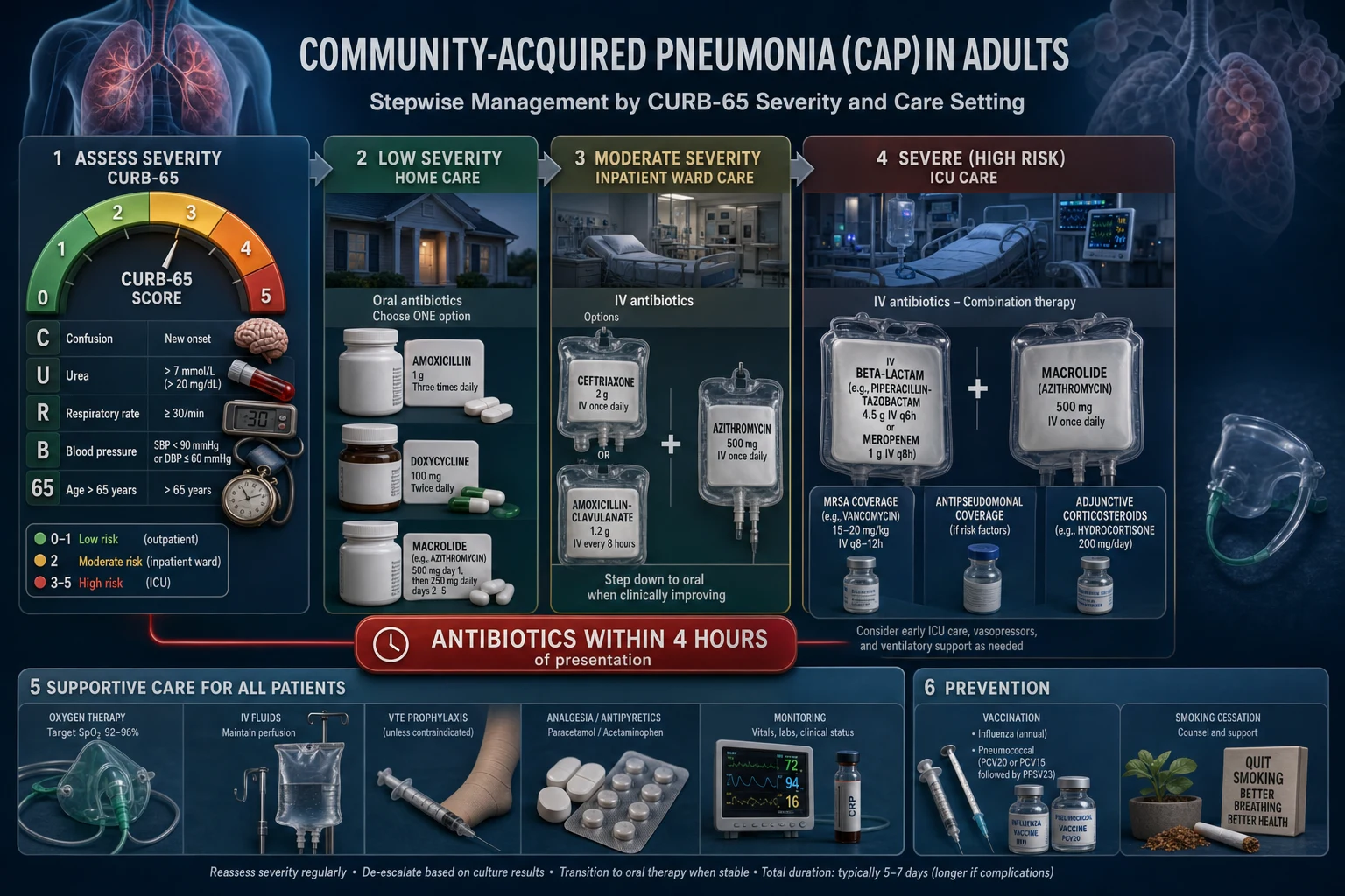
Empirical antibiotics are severity- and setting-driven and must cover both typical AND atypical organisms in all hospitalised patients (the 2019 ATS/IDSA update made this explicit). The drug ladders below follow IDSA/ATS 2019 with NICE/BTS and ICMR deltas.[1][5]
LOW severity — CURB-65 0–1 (home)
- **Oral amoxicillin** 500 mg–1 g TDS (first-line)
- OR **doxycycline** 100 mg BD if penicillin-allergic
- OR **clarithromycin** 500 mg BD / **azithromycin** 500 mg OD
- Duration **5–7 days**
- Macrolide monotherapy only where pneumococcal macrolide resistance is low
MODERATE — CURB-65 2 (ward)
- **IV amoxicillin** 1 g TDS + **oral/IV macrolide** (clarithromycin 500 mg BD)
- OR **IV co-amoxiclav** 1.2 g TDS + macrolide
- OR **IV ceftriaxone** 1–2 g OD + macrolide
- Penicillin-allergic: **IV clarithromycin** ± (ceftriaxone cautiously) or respiratory fluoroquinolone
- Switch to oral when improving; **5–7 days** typical
SEVERE — CURB-65 3–5 / ICU
- **IV co-amoxiclav** 1.2 g TDS OR **IV ceftriaxone** 2 g OD + **IV macrolide** (clarithromycin 500 mg BD)
- Alternative: **respiratory fluoroquinolone** (moxifloxacin/levofloxacin)
- **Add MRSA cover** (vancomycin/linezolid) if risk factors or prior MRSA
- **Add antipseudomonal** cover (piperacillin-tazobactam/meropenem + ciprofloxacin) if bronchiectasis, recent antibiotics, or recent hospitalisation
- Duration **7–14 days**; **14–21 days** for Legionella, MRSA, Pseudomonas
Regional note — India (NMC/ICMR/NCDC): community penicillin resistance in S. pneumoniae is moderate–high and macrolide resistance is rising, so standard empirical therapy for admitted CAP is IV ceftriaxone 2 g OD plus azithromycin 500 mg OD, with co-amoxiclav 1.2 g TDS plus azithromycin as an alternative for low-severity disease; always apply the local antibiogram and de-escalate on culture results.[1]
[1]Duration — 5–7 days in the patient who is clinically improving and afebrile for 48–72 hours. Longer courses are indicated for S. aureus (14 days or more), Pseudomonas, complicated infection, MRSA, and Legionella (14–21 days, or shorter with azithromycin/fluoroquinolone). Procalcitonin-guided shortening is supported but optional.[1]
Corticosteroids — a short course (e.g. hydrocortisone or prednisolone) may reduce mortality and time-to-clinical-stability in severe CAP with sepsis or septic shock, and the effect is largest in the very sick; the benefit is modest and the evidence remains contested, so steroids are not used routinely in mild–moderate CAP but should be considered in ICU-level disease with persistent shock or high inflammatory burden. Avoid in influenza or untreated immunocompromise.[6]
IV-to-oral switch (step-down) — once the patient is haemodynamically stable, clinically improving, afebrile, and able to swallow and absorb oral medication, switch to oral therapy with the same agent class. Discharge is appropriate when the patient is clinically stable, afebrile for 24–48 hours, tolerating oral intake, has a safe social situation, and the inflammatory markers are falling — with a safety-net to review or re-present if not improving within 48 hours.[1]
Supportive care in every hospitalised CAP patient: oxygen to target, adequate hydration (oral or IV), analgesia for pleuritic pain (paracetamol, NSAIDs if not contraindicated — pain splints respiration and worsens atelectasis), venous thromboembolism prophylaxis (LMWH) for immobile patients, physiotherapy only if there is sputum retention, nutritional support, glycaemic control, and monitoring of SpO2, vital signs and urine output. Avoid unnecessary antibiotics in viral pneumonitis where bacterial co-infection is not present. [1]
Specific Subtypes & Scenarios
- Aspiration pneumonia — dependent segments (posterior segments of the upper lobes and apical segments of the lower lobes in a supine patient; basal segments if upright); mixed oral flora including anaerobes (Peptostreptococcus, Fusobacterium, Prevotella); cover with co-amoxiclav 1.2 g TDS IV (or clindamycin 600 mg QDS if penicillin-allergic, plus a Gram-negative agent if severe); address the underlying swallow or reflux (SLT assessment, head-of-bed elevation, PEG consideration). Aspiration pneumonitis (sterile acid injury) may need no antibiotics unless it fails to resolve in 48 hours.[2]
- Post-influenza pneumonia — Staphylococcus aureus (often cavitating, pneumatoceles), H. influenzae, S. pneumoniae; add anti-staphylococcal / anti-MRSA cover (vancomycin or linezolid) plus antiviral oseltamivir if influenza is still active. A rapidly progressive pneumonia with cavitation in the influenza season is post-influenza staphylococcal pneumonia until proven otherwise.
- Atypical pneumonias — Mycoplasma pneumoniae (young adults, cold agglutinins, erythema multiforme/Stevens-Johnson, bullous myringitis); Chlamydophila pneumoniae (sinusitis, pharyngitis); Chlamydophila psittaci (bird exposure, psittacosis); Legionella (water systems, GI symptoms, hyponatraemia, confusion, severe); Coxiella burnetii (Q fever, farm animals, hepatitis). Drug of choice: a macrolide (azithromycin 500 mg OD, clarithromycin 500 mg BD) or doxycycline 100 mg BD; fluoroquinolones are alternatives.
- Pneumocystis jirovecii pneumonia (PCP) — HIV with CD4 under 200, transplant, immunosuppression; insidious dry cough, exertional desaturation, fever, bilateral perihilar interstitial infiltrates (normal CXR early); exertional or exercise SpO2 drop is a clue. Treatment: high-dose co-trimoxazole (trimethoprim 15–20 mg/kg/day + sulfamethoxazole 75–100 mg/kg/day IV in 4 divided doses) for 21 days, plus prednisolone 40 mg BD for 5 days, then taper if hypoxic (PaO2 under 70 mmHg or SpO2 under 92% on room air) to blunt inflammation.
- COVID-19 pneumonia — bilateral ground-glass opacities, silent hypoxia, anosmia; isolation and PCR; treatment bundles include supplemental oxygen (nasal, HFNO, CPAP), dexamethasone 6 mg OD for up to 10 days in hypoxic patients, antivirals (e.g. remdesivir in selected hypoxic patients) and IL-6 receptor antagonists (tocilizab) in rapidly escalating systemic inflammation, plus VTE prophylaxis (marked prothrombotic tendency). Proning is highly effective for refractory hypoxaemia.
- HAP / VAP — broader empiric cover guided by local antibiogram and prior cultures; cover MRSA (vancomycin/linezolid) and multidrug-resistant Gram-negatives (piperacillin-tazobactam, meropenem, or an aminoglycoside ± antipseudomonal beta-lactam); de-escalate on culture results; 7 days is usual unless undrained foci or S. aureus/Gram-negative bacteraemia. Prevention is the ventilator bundle (head of bed 30 degrees, daily sedation breaks and spontaneous breathing trials, oral chlorhexidine, subglottic secretion drainage, DVT and stress-ulcer prophylaxis).
- Lung abscess — cavitation with an air-fluid level, often anaerobic or post-aspiration; prolonged antibiotics (4–6 weeks of a beta-lactam/beta-lactamase inhibitor or clindamycin), postural drainage, and percutaneous or surgical drainage if large, progressive, or unresponsive.
Complications & Pitfalls
Local complications — parapneumonic effusion (the commonest, occurring in up to half of bacterial CAP), empyema (frank pus in the pleural space), lung abscess, necrotising pneumonia (often S. aureus, Klebsiella), pneumatocele (children, S. aureus), bronchopleural fistula, and ARDS in severe bilateral disease.[2]
Systemic complications — sepsis and septic shock (the leading mode of death), acute kidney injury, metastatic infection (endocarditis, meningitis, septic arthritis — especially with bacteraemic pneumococcal disease), new-onset atrial fibrillation in the elderly, myocardial infarction triggered by the septic insult, disseminated intravascular coagulation, hyponatraemia (SIADH or Legionella), and dermal/organ manifestations of atypicals (erythema multiforme, haemolytic anaemia in Mycoplasma). [1]
Parapneumonic effusion — classify the pleural fluid into exudate vs transudate with Light's criteria (exudate if pleural-fluid/serum protein ratio over 0.5, LDH ratio over 0.6, or fluid LDH over two-thirds the upper limit of normal). Categorise as uncomplicated (clear, pH over 7.2, no organisms — antibiotics alone, often resolves), complicated (pH under 7.2, glucose under 40 mg/dL, LDH over 1000, or positive Gram stain/culture — needs chest-tube drainage), or empyema (frank pus — chest tube + surgical drainage). Loculated or large effusions, and any not resolving with tube drainage, warrant surgical drainage (VATS); intrapleural fibrinolytics (tPA/DNase) help multiloculated effusions.[2]
Classic pitfalls: [1]
- Under-treating severe CAP — missing the ICU need because the blood pressure is "OK"; always calculate CURB-65 and apply the ATS/IDSA minor criteria.
- Not covering atypicals — empirical therapy for hospitalised CAP must include a macrolide or a respiratory fluoroquinolone.
- Failing to consider TB or PE in the atypical case — a "non-resolving pneumonia" on standard antibiotics may be TB, a PE infarct, or malignancy.
- Delaying antibiotics for tests — the door-to-needle target is 4 hours; cultures are taken first but never delay the dose.
- Over-investigating mild disease — a CURB-65 0 patient needs little more than a CXR, CRP and safety-net advice.
- Forgetting follow-up CXR at 6–8 weeks in smokers over 50, persistent symptoms, or non-resolving opacity — to exclude underlying malignancy. [1]
Prognosis & Disposition
CURB-65 drives early disposition and correlates with 30-day mortality; PSI/PORT refines low-risk outpatient decisions. Beyond scores, multilobar disease, hypoxia, shock, bacteraemia, comorbidity (COPD, HF, CKD, cancer, diabetes), hyponatraemia, hypoalbuminaemia, and high lactate predict poor outcomes.[3]
CURB-65 (reproduce exactly)
One point each:
- Confusion (new)
- Urea >7 mmol/L
- Respiratory rate ≥30/min
- Blood pressure <90 systolic or ≤60 diastolic
- Age ≥65 [1]
| Score | Risk band | Typical disposition |
|---|---|---|
| 0–1 | Low | Home treatment if social circumstances allow |
| 2 | Moderate | Short-stay / ward admission often |
| 3–5 | High | Admit; 4–5 consider HDU/ICU |
ICU adjuncts (IDSA/ATS severe CAP major/minor criteria concept): septic shock on vasopressors or mechanical ventilation = major → ICU. Multiple minor criteria (RR ≥30, PaO2/FiO2 ≤250, multilobar, confusion, BUN ≥20 mg/dL, leukopenia, thrombocytopenia, hypothermia, hypotension needing fluids) also support higher care. [1]
Disposition detail
- Antibiotics within 4 hours of presentation for admitted CAP; within 1 hour if septic shock
- Switch IV→oral when haemodynamically stable, afebrile-ish, able to absorb oral drugs
- Usual duration 5–7 days if improving (longer for complications, Legionella, Staph, Gram-negatives — pathogen-specific)
- Follow-up CXR at 6–8 weeks if smoker >50, persistent symptoms, or incomplete recovery — exclude malignancy
- Vaccinate before discharge when recovered enough (pneumococcal, influenza, COVID-19)
Prevention
Vaccination is the single most effective population-level intervention against CAP, and examiners reward a candidate who can name the vaccines and their target groups.[1]
- Pneumococcal vaccination — the conjugate vaccines (PCV13, PCV15, PCV20) induce T-cell-dependent immunity and work in infants and immunocompromised hosts; the polysaccharide vaccine (PPSV23) induces T-cell-independent immunity with broader serotype cover but no mucosal memory. Schedules vary by country: PCV in childhood immunisation programmes, and PCV15/20 with or without PPSV23 in adults aged 65 and over, in asplenia, sickle-cell disease, cochlear implants, CSF leaks, chronic heart/lung/liver/renal disease, diabetes, immunocompromise, and smoking adults.
- Influenza vaccination — annual — the most cost-effective individual CAP prevention; reduces influenza, post-influenza bacterial pneumonia, hospitalisation, and death; recommended universally for over-6-month-olds, with priority for the elderly, pregnant women, healthcare workers, and the chronically ill.
- COVID-19 vaccination — reduces severe COVID-19 pneumonia, hospitalisation, and death; current schedules depend on age, risk, and prior infection.
- Haemophilus influenzae type b (Hib) — part of childhood immunisation; important in asplenia and functional hyposplenism.
- Respiratory syncytial virus (RSV) — monoclonal antibody (nirsevimab) for infants and vaccines for older adults and the pregnant.
- Pertussis booster — adults and pregnant women, to prevent whooping-cough-associated pneumonia. [1]
Non-pharmacological prevention: smoking cessation (reverses the mucociliary defect and halves CAP risk within five years), alcohol moderation, good oral hygiene (reduces aspiration-inoculum bacterial load), nutrition and glycaemic control, infection control (hand hygiene, masking in outbreaks, isolation of respiratory cases), fall and swallow assessment in the elderly, head-of-bed elevation 30–45 degrees in intubated patients (VAP bundle), and early mobilisation and pulmonary physiotherapy in the hospitalised. Asplenic patients additionally need lifelong penicillin V prophylaxis (250–500 mg BD) and a standing-alert card/medic-alert bracelet plus urgent empirical self-treatment at first sign of fever. [1]
Special Populations
Elderly
May present with confusion, falls, anorexia, incontinence without fever. Lower threshold to admit. Aspiration risk in stroke/dementia. Check for silent urinary sepsis dual pathology.
Pregnancy
Beta-lactams and macrolides preferred; avoid tetracyclines and usually fluoroquinolones. Treat aggressively — maternal hypoxia harms the fetus. VTE prophylaxis; left lateral tilt when gravid uterus large.
Immunocompromised / HIV
Broaden differential: PJP, CMV, fungi, TB, nocardia, unusual bacteria. For PJP: high-dose trimethoprim-sulfamethoxazole and steroids if hypoxaemic (PaO2 <70 mmHg) per HIV protocols. Early CT and specialist infection input.
Post-influenza
Secondary S. aureus (including MRSA) and pneumococcus — consider anti-MRSA cover if cavitary/necrotising disease or known risk after influenza.
Aspiration risk
Chemical pneumonitis vs bacterial aspiration (oral anaerobes/Gram-negatives). Anaerobic cover is not always routinely required for community aspiration in modern guidance unless empyema/abscess — know local protocol and IDSA nuance.
India / high TB-burden settings (ICMR lens)
Any subacute presentation, upper-lobe disease, haemoptysis, weight loss, or non-resolving pneumonia → evaluate for TB (sputum AFB/NAAT). Do not label everything as CAP and miss smear-positive TB. Stewardship: avoid unnecessary broad antipseudomonal cover without risk factors.
Evidence, Guidelines & Regional Differences
Key changes in the ATS/IDSA 2019 guideline[1][5] — the most widely cited modern CAP guideline and a frequent exam reference:
- Empirical therapy must cover atypical organisms in all hospitalised CAP (beta-lactam + macrolide, OR a respiratory fluoroquinolone).
- The HCAP category was abandoned — empirical MRSA/Pseudomonas cover is now driven by locally validated risk factors (prior isolation, recent IV antibiotics), not nursing-home residence alone.
- Routine blood cultures, sputum culture, urinary antigens, and procalcitonin are not required in all CAP — they are targeted to severe disease, failure of outpatient therapy, and specific pathogens (Legionella, S. aureus, P. aeruginosa).
- Corticosteroids are not routinely recommended for CAP but may be considered in refractory septic shock.
- Follow-up imaging is not routine — reserved for smokers over 50 and non-resolving disease. [1]
Regional empirical differences:[1]
- US (IDSA/ATS 2019) — outpatients: amoxicillin, doxycycline, or a macrolide (in low-resistance areas); inpatients: beta-lactam + macrolide or respiratory fluoroquinolone; ICU: beta-lactam + macrolide (or fluoroquinolone), add MRSA/Pseudomonas cover per risk.
- UK (NICE NG191 / BTS) — community: amoxicillin (doxycycline or clarithromycin if allergic); moderate inpatient: IV amoxicillin + macrolide; severe: co-amoxiclav or cefuroxime + macrolide. Door-to-antibiotic 4-hour target is explicit.
- India (ICMR/NCDC AMR) — high community penicillin and macrolide resistance; admitted CAP: ceftriaxone 2 g OD + azithromycin 500 mg OD; co-amoxiclav + azithromycin for milder disease; always consult the local antibiogram.
- Europe (ERS/ESCMID) — broadly aligned with IDSA/ATS but with regional fluoroquinolone-sparing preference and tiered severity pathways. [1]
Vaccination (the prevention that examiners reward): pneumococcal vaccination — the conjugate vaccines (PCV13, PCV15, PCV20) and the polysaccharide vaccine (PPSV23), given per age/risk schedules (PCV in children and high-risk adults; PPSV23 in adults 65 and over and at-risk groups, with the sequence depending on indication); annual influenza vaccine (the single most effective population-level CAP prevention); COVID-19 vaccination; and, in selected groups, H. influenzae type b, RSV, and pertussis boosters. Smoking cessation reduces CAP risk substantially and reverses the mucociliary defect.[1]
Empirical Antibiotic Exam Table & Worked Stems
Empirical therapy patterns (adult teaching map — always localise to antibiogram)
| Setting | Typical regimen (examples) | Duration idea |
|---|---|---|
| Low-severity outpatient | Amoxicillin 500 mg–1 g TDS OR doxycycline 100 mg BD | 5 days if well |
| Outpatient with comorbidities | Amoxicillin-clavulanate ± macrolide OR respiratory FQ where appropriate | 5 days |
| Non-severe inpatient | Beta-lactam (e.g. amoxicillin or ceftriaxone) + macrolide (azithromycin/clarithromycin) | 5–7 days |
| Severe / ICU | IV beta-lactam + macrolide; add anti-MRSA / antipseudomonal if risk factors | Longer if complicated |
| Aspiration-associated | Often standard CAP cover; add anaerobic cover if abscess/empyema | Individualise |
| Suspected PCP (HIV) | High-dose TMP-SMX; steroids if hypoxic | 21 days typical PCP |
Worked stem — CURB-65 = 3
Confused 70-year-old, urea 9, RR 32, BP 110/70, age 70 → score 3 (C+U+R+age; BP not scoring). Admit, cultures if severe pathway, oxygen, IV dual atypical-covering therapy, reassess for ICU minor criteria (RR, multilobar, PaO2/FiO2).
Worked stem — non-resolving
Treated CAP 3 weeks, still febrile with weight loss and apical cavity in India → TB until proven otherwise, also consider malignancy, abscess, resistant organisms, inadequate dosing, wrong diagnosis (PE, eosinophilic pneumonia).
Worked NEET-PG Stems — Pneumonia
Stem bank (answerable from this topic alone)
- Alcoholic with upper-lobe consolidation and currant-jelly sputum → Klebsiella considered; still cover pneumococcus; watch for abscess/cavitation.
- Hotel air-conditioning outbreak, hyponatraemia, GI symptoms, confusion → Legionella; urinary antigen; macrolide/FQ coverage; notify public health.
- Young adult, bullous myringitis, dry cough, patchy CXR → Mycoplasma; macrolide/doxy.
- Post-influenza necrotising pneumonia → S. aureus/MRSA risk; add anti-MRSA if severe/cavitary.
- HIV with dry cough, hypoxia out of proportion, ground-glass → PJP; TMP-SMX + steroids if PaO2 low.
- CURB-65 = 0, good support → outpatient amoxicillin or doxy; safety-net advice.
- Empyema: pH under 7.2, glucose low → drain; prolonged antibiotics; do not rely on abx alone.
- Non-resolving after 6 weeks in smoker → CT/bronchoscopy for malignancy/TB.
Antibiotic stewardship pearls
- Stop anti-MRSA/antipseudomonal cover at 48 h if cultures negative and no risk factors.
- Switch IV to oral early when stable.
- Duration is usually short if uncomplicated and improving — longer is not better by default.
Exam Pearls
CURB-65 components (verbatim)
CURB-65
New disorientation in time, place, or person
Over 7 mmol/L
30/min or more
Systolic under 90 OR diastolic 60 or below
65 years or older (1 point)
0–1 home, 2 hospital, 3–5 consider ICU
- Red-currant-jelly sputum = Klebsiella; foul-smelling sputum = anaerobes/aspiration.
- Rust-coloured sputum = S. pneumoniae; haemoptysis with weight loss/night sweats = TB or malignancy.
- Legionella triad: GI symptoms (diarrhoea), hyponatraemia, confusion; diagnose with urinary antigen; source is water systems/cooling towers.
- Cold agglutinins + erythema multiforme (Stevens-Johnson) + young adult = Mycoplasma.
- Bullous myringitis = Mycoplasma.
- Bird exposure = Chlamydophila psittaci (psittacosis); farm animals/parturient sheep = Coxiella burnetii (Q fever) — treat with tetracyclines.
- Pneumatoceles in a child = S. aureus; cavitation post-influenza = S. aureus.
- Antibiotics within 4 hours; cover atypicals with a macrolide; duration 5–7 days (longer for Legionella, MRSA, Pseudomonas, S. aureus).
- CURB-65 vs CRB-65 — CRB-65 omits urea; useful in primary care/pre-hospital; CRB-65 of 0 = home care option.
- PSI/PORT is better at identifying low-risk patients but under-weights young hypoxic patients; CURB-65 is the bedside favourite.
- Asplenia + encapsulated organism (pneumococcus, Hib, meningococcus) = overwhelming post-splenectomy infection (OPSI) — vaccinate, prophylactic penicillin, education.
- "Non-resolving pneumonia" — think TB, PE, malignancy, abscess, empyema, immunocompromise, wrong antibiotic.
- Follow-up CXR at 6–8 weeks in smokers over 50, persistent symptoms, or non-resolving opacity — to exclude underlying lung cancer.
- Silent hypoxia (happy hypoxaemic patient) is a hallmark of COVID-19; check SpO2 in every respiratory presentation. [1]
Exam application bank (NEET-PG / INICET)
One-line answer
Community-acquired pneumonia (CAP) is an acute infection of the lung parenchyma acquired outside hospital (or within 48 h of admission). Commonest organism Streptococcus pneumoniae; atypicals (Mycoplasma, Chlamydophila, Legionella); viruses (influenza, COVID-19, RSV); Staphylococcus aureus and Gram-negatives in severe/chronic disease; Klebsiella in alcoholics. Presents with productive cough, fever, dyspnoea, pleuritic chest pain and signs of consolidation; atypical pneumonias have a dry cough, prominent systemic features and a normal/mildly abnormal CXR. Diagnosis is clinical plus chest X-ray; severity by CURB-65 (Confusion, Urea over 7, RR over 30, BP under 90/60, age over 65). Treat with antibiotics within 4 hours: low severity amoxicillin/doxycycline; moderate/severe beta-lactam plus macrolide; give oxygen, fluids, cover atypicals. Admit if CURB-65 at least 2; ICU if 3 to 5. Vaccinate
Worked stems (answer without another resource)
Stem 1 — Classic presentation. Map symptoms to mechanism; name the first investigation and first treatment step with dose/route if drug therapy is standard. [1]
Stem 2 — Unstable / complicated. List red flags that force immediate resuscitation, theatre, ICU, antidote, or reperfusion — and what you do in the first 15 minutes. [1]
Stem 3 — Atypical group. Elderly, pregnancy, child, or immunocompromised: how presentation and thresholds change. [1]
Stem 4 — Differential trap. Name the three closest mimics and one discriminator for each. [1]
Stem 5 — Disposition. Who goes home with safety-netting, who is admitted, who needs HDU/ICU/theatre, and what follow-up is mandatory. [1]
Rapid viva checklist
- Definition + classification
- Pathophysiology chain
- Bedside signs / criteria
- Score with exact components (if any)
- Emergency bundle
- Definitive therapy with doses
- Complications of disease and of treatment
- Special populations
- Guideline/trial name if classic
- Three exam traps
Coverage self-check
If you cannot answer any stem above from this page alone, re-read the matching section — the page is intended to be self-sufficient for final-prof and NEET-PG/INICET questions on Pneumonia (Community-Acquired).
References
- [1]Metlay JP, Waterer GW, Long AC, et al. Diagnosis and Treatment of Adults with Community-acquired Pneumonia. An Official Clinical Practice Guideline of the American Thoracic Society and Infectious Diseases Society of America Am J Respir Crit Care Med, 2019.PMID 31573350
- [2]Prina E, Ranzani OT, Torres A. Community-acquired pneumonia Lancet, 2015.PMID 26277247
- [3]Lim WS, van der Eerden MM, Laing R, et al. Defining community acquired pneumonia severity on presentation to hospital: an international derivation and validation study Thorax, 2003.PMID 12728155
- [4]Fine MJ, Auble TE, Yealy DM, et al. A prediction rule to identify low-risk patients with community-acquired pneumonia N Engl J Med, 1997.PMID 8995086
- [5]Metlay JP, Waterer GW. Update in adult community-acquired pneumonia: key points from the new American Thoracic Society/Infectious Diseases Society of America 2019 guideline Curr Opin Pulm Med, 2020.PMID 32084039
- [6]Eastwood B, et al. Systemic corticosteroids for severe community-acquired pneumonia: a narrative review of mechanisms and critical appraisal of recent trials in ICU Eur Respir Rev, 2026.PMID 42342261