Infectious Diseases · General Medicine
Sepsis & Septic Shock
Also known as Sepsis · Septic shock · Bloodstream infection · Bacteraemia · Systemic inflammatory response syndrome · Severe sepsis
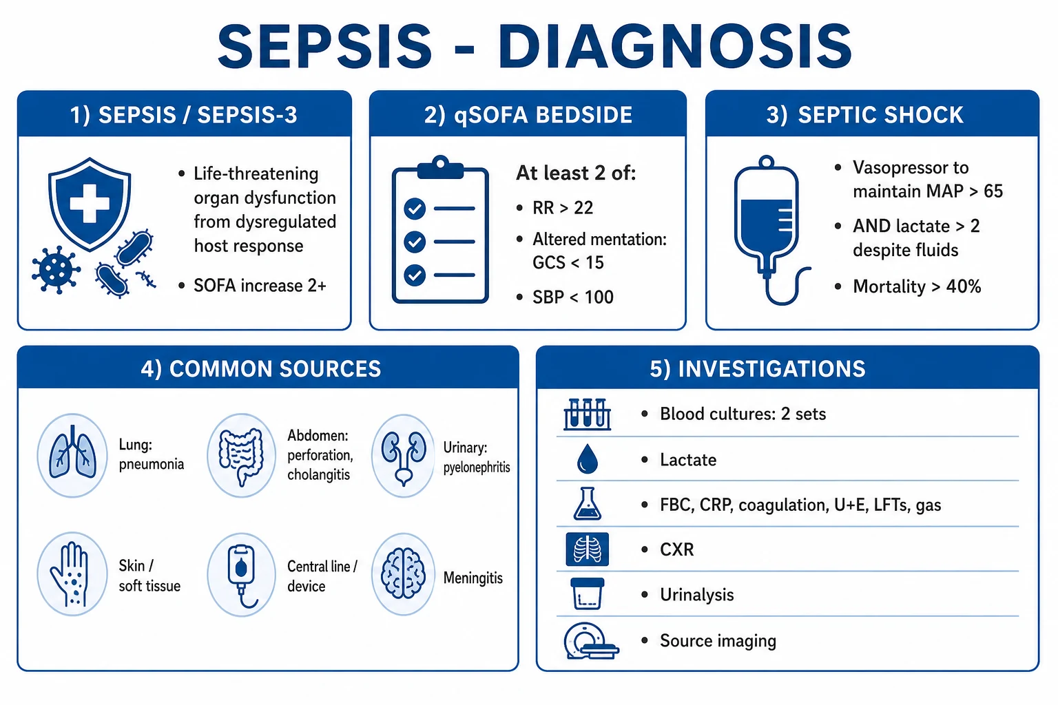
Sepsis is a life-threatening organ dysfunction caused by a dysregulated host response to infection (Sepsis-3 definition, Singer 2016). Operationally it is identified at the bedside as an acute change in total SOFA score of 2 or more points consequent to infection. Septic shock is a subset of sepsis with circulatory and cellular/metabolic abnormalities substantial enough to substantially increase mortality — clinically hypotension requiring vasopressors to maintain a mean arterial pressure of at least 65 mmHg AND a serum lactate over 2 mmol/L despite adequate volume resuscitation, carrying a hospital mortality over 40 percent. Common sources are respiratory, abdominal, urinary, skin/soft tissue and device-related. Recognition is clinical (suspected infection with qSOFA at least 2: RR over 22, altered mentation, SBP under 100), and management is the Surviving Sepsis Campaign Hour-1 bundle — measure lactate, obtain blood cultures, give broad-spectrum antibiotics, give 30 mL/kg crystalloid for hypotension or lactate over 4, and start vasopressors — all within one hour — plus source control and organ support.
On this page & tools
Your progress
Saved locally on this device.
Exam tags
Red flags
Overview & Definition
Sepsis is the commonest preventable cause of in-hospital death worldwide and the archetypal time-critical medical emergency. The clinical skill is early recognition (suspected infection plus a deranged physiological screen — qSOFA at least 2 — plus clinical gestalt) followed by immediate execution of the Hour-1 bundle without waiting for confirmation, because each hour of delay in effective antibiotics and resuscitation measurably increases mortality.[4]
The conceptual shift that defines modern sepsis is the abandonment of SIRS (Systemic Inflammatory Response Syndrome) as a defining criterion and its replacement with organ dysfunction (SOFA). Under the older 1992 (Sepsis-1) and 2001 (Sepsis-2) definitions, sepsis was "infection plus two or more SIRS criteria," severe sepsis was sepsis with organ dysfunction, and septic shock was sepsis with refractory hypotension. SIRS is sensitive but non-specific (present after exercise, pancreatitis, trauma and surgery), it under-detects the genuinely deteriorating patient, and the term "severe sepsis" was redundant (sepsis is organ-dysfunctional by definition). The Sepsis-3 task force (2016) retired SIRS and "severe sepsis," redefined sepsis as life-threatening organ dysfunction caused by a dysregulated host response to infection, and gave an operational bedside handle — an acute change in total SOFA score of 2 or more points consequent to infection.[1]
Septic shock — the most severe phenotype — is defined operationally as sepsis with vasopressor requirement to maintain a mean arterial pressure (MAP) of at least 65 mmHg AND a serum lactate over 2 mmol/L despite adequate volume resuscitation (30 mL/kg of crystalloid). The combination of vasoplegia and a raised lactate identifies a population with a hospital mortality exceeding 40 percent, distinctly worse than sepsis alone.[1]
The three core pathophysiological abnormalities — vasoplegia, capillary leak and cellular hypoxia (mitochondrial dysfunction) — explain every clinical feature and every element of therapy: fluids and vasopressors restore the macrocirculation, source control removes the trigger, and supportive organ support buys time for the host response to reset.[1][2]
Exam application bank (NEET-PG / INICET)
One-line answer
Sepsis is a life-threatening organ dysfunction caused by a dysregulated host response to infection (Sepsis-3 definition, Singer 2016). Operationally it is identified at the bedside as an acute change in total SOFA score of 2 or more points consequent to infection. Septic shock is a subset of sepsis with circulatory and cellular/metabolic abnormalities substantial enough to substantially increase mortality — clinically hypotension requiring vasopressors to maintain a mean arterial pressure of at least 65 mmHg AND a serum lactate over 2 mmol/L despite adequate volume resuscitation, carrying a hospital mortality over 40 percent. Common sources are respiratory, abdominal, urinary, skin/soft tissue and device-related. Recognition is clinical (suspected infection with qSOFA at least 2: RR over 22, altered mentation, SBP under 100), and management is the Surviving Sepsis Campaign Hour-1 bundle [1]
Worked stems (answer without another resource)
Stem 1 — Classic presentation. Map symptoms to mechanism; name the first investigation and first treatment step with dose/route if drug therapy is standard. [1]
Stem 2 — Unstable / complicated. List red flags that force immediate resuscitation, theatre, ICU, antidote, or reperfusion — and what you do in the first 15 minutes. [1]
Stem 3 — Atypical group. Elderly, pregnancy, child, or immunocompromised: how presentation and thresholds change. [1]
Stem 4 — Differential trap. Name the three closest mimics and one discriminator for each. [1]
Stem 5 — Disposition. Who goes home with safety-netting, who is admitted, who needs HDU/ICU/theatre, and what follow-up is mandatory. [1]
Rapid viva checklist
- Definition + classification
- Pathophysiology chain
- Bedside signs / criteria
- Score with exact components (if any)
- Emergency bundle
- Definitive therapy with doses
- Complications of disease and of treatment
- Special populations
- Guideline/trial name if classic
- Three exam traps
Coverage self-check
If you cannot answer any stem above from this page alone, re-read the matching section — the page is intended to be self-sufficient for final-prof and NEET-PG/INICET questions on Sepsis & Septic Shock.
Classification
Sepsis is classified along three axes: the clinical severity ladder (infection -> sepsis -> septic shock), the source (which determines the antibiotic choice and the source-control strategy), and the haemodynamic phenotype (warm vasoplegic vs cold decompensated), which guides resuscitation. [1]
Uncomplicated infection
- Localised infection WITHOUT organ dysfunction
- No SOFA change; qSOFA 0 to 1
- Manage with source-directed therapy; observe for deterioration
Sepsis (Sepsis-3)
- Life-threatening organ dysfunction from a dysregulated host response to infection
- Operational: acute change in total SOFA of 2 or more points due to the infection
- qSOFA at least 2 is the bedside escalation prompt (not the sole screen)
- Hospital mortality approximately 10 percent
Septic shock
- A subset of sepsis with circulatory AND cellular/metabolic abnormalities sufficient to substantially increase mortality
- Operational: vasopressor requirement to maintain MAP at least 65 mmHg AND serum lactate over 2 mmol/L despite adequate fluid resuscitation
- Requires ICU and vasopressor support
- Hospital mortality over 40 percent
The haemodynamic phenotype is the second classification axis and is clinically useful at the bedside: [1]
- Warm shock (early, vasoplegic / distributive) — the patient is warm, flushed, vasodilated with a wide pulse pressure, a bounding pulse and a low diastolic pressure (often under 50 mmHg). Cardiac output is high; systemic vascular resistance is low. This is the dominant picture of early septic shock and is norepinephrine-responsive (restores vascular tone).
- Cold shock (late, decompensated) — the patient is cold, clammy, mottled with prolonged capillary refill (over 3 seconds), a narrow pulse pressure and oliguria. Cardiac output has fallen (myocardial depression, hypovolaemia from capillary leak). This signals decompensation and demands aggressive resuscitation plus an inotrope if cardiac output remains low.[1]
The source classification drives empirical antibiotic selection and the source-control plan and is covered in detail under Management. [1]
Epidemiology & Risk Factors
Sepsis is a leading cause of death worldwide. The Global Burden of Disease analysis (Rudd et al, 2020) estimated 48.9 million incident cases and 11.0 million sepsis-related deaths in 2017, accounting for 19.7 percent of all global deaths. Although age-standardised sepsis mortality fell by about half between 1990 and 2017 (driven by vaccination, clean water and antimicrobials), the absolute burden remains enormous and is disproportionately borne by low- and middle-income countries — 84 percent of sepsis cases occur in these settings, and sub-Saharan Africa, South Asia and East Asia carry the highest age-standardised mortality. Almost half of all sepsis deaths occur in children under 5.[3]
Septic shock carries a hospital mortality of 30 to 40 percent — by the Sepsis-3 operational definition, over 40 percent.[1] The single largest modifiable determinant of that mortality is the time to effective antimicrobial therapy and adequate resuscitation: in the landmark Kumar cohort, each additional hour of hypotension before effective antibiotics was associated with a steady fall in survival, and subsequent data place the marginal mortality cost at roughly 4 percent per hour in septic shock.[4]
In India and much of South Asia, sepsis incidence and mortality are higher than in high-income settings, driven by delays in presentation, limited access to ICU and laboratory support, a high prevalence of malnutrition and comorbidity, and a rapidly rising burden of multidrug-resistant organisms (ESBL-producing Enterobacterales, carbapenem-resistant Acinetobacter and Klebsiella, and MRSA) documented by the ICMR Antimicrobial Resistance Surveillance Network. The National Centre for Disease Control (NCDC) publishes empirical therapy guidance that must be followed locally. Practical adaptations — early oral rehydration, point-of-care lactate, and rational antibiotic stewardship — are essential where infrastructure is limited.
Host risk factors for sepsis: [1]
- Extremes of age — neonates (immature immunity) and the elderly (immunosenescence, comorbidity, frailty). The very young and very old account for a disproportionate share of sepsis deaths.
- Immunocompromise — HIV/AIDS, chemotherapy-induced neutropenia, solid-organ or stem-cell transplant, long-term corticosteroids or other immunosuppressants, biologics (anti-TNF, anti-IL-6, anti-CD20), haematological and solid malignancy, diabetes mellitus, chronic liver disease (cirrhosis), chronic kidney disease, and asplenia / hyposplenism (surgical, sickle-cell, coeliac).
- Chronic organ failure — CKD, COPD, heart failure, cirrhosis.
- Breakdown of barriers and devices — indwelling central venous and arterial lines, urinary catheters, endotracheal tubes, prosthetic material, burns, chronic wounds, pressure ulcers, recent surgery.
- Pregnancy and the puerperium — chorioamnionitis, septic abortion, puerperal sepsis, pyelonephritis of pregnancy.
- Iatrogenic and environmental — broad-spectrum antibiotics (selection pressure for resistant organisms and C. difficile), immunosuppressants, residence in a long-term care facility, and (in resource-limited settings) household air pollution and unsafe water. [1]
Organisms. The microbial spectrum is dominated by Gram-negative bacilli (Escherichia coli, Klebsiella pneumoniae, Pseudomonas aeruginosa, Acinetobacter species) and Gram-positive cocci (Staphylococcus aureus including MRSA, Streptococcus pneumoniae, enterococci, Group A strep). Fungal sepsis (candidaemia) is an important cause in the critically ill, the immunocompromised, and patients with prolonged ICU stay, central lines and total parenteral nutrition. The rising global threat is multidrug resistance — extended-spectrum beta-lactamase (ESBL) producers, carbapenem-resistant Enterobacterales (CRE), Pseudomonas and Acinetobacter resistant to carbapenems, vancomycin-resistant enterococci (VRE) and MRSA — which forces empiric broad-spectrum regimens and worsens outcome.[2]
Pathophysiology
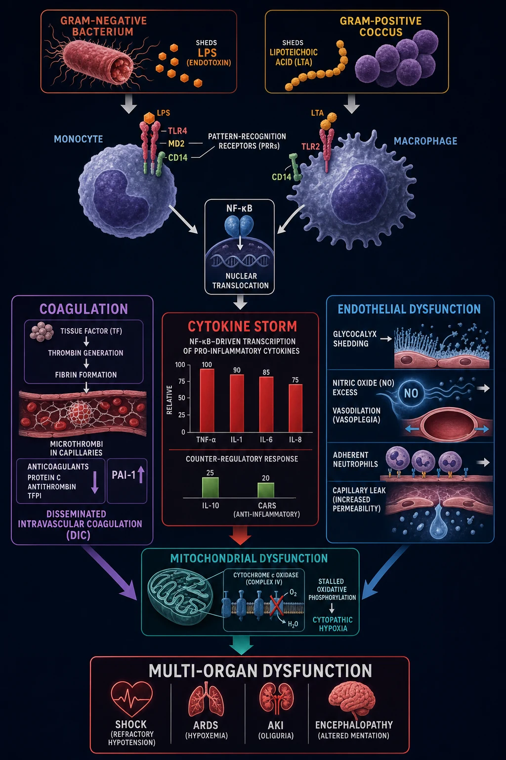
The entire clinical syndrome of sepsis flows from a single organising event — a dysregulated host response in which the normally protective inflammatory, coagulation and endothelial responses to infection escape their regulatory checks, simultaneously over- and under-shooting, and damaging the host. Understanding this cascade explains every clinical feature (vasoplegia, capillary leak, multi-organ failure, lactate rise) and every element of therapy. [1]
1. Initiation: PAMPs and pattern-recognition receptors
The innate immune system does not recognise "sepsis" — it recognises microbial signatures. Pathogens shed conserved pathogen-associated molecular patterns (PAMPs): lipopolysaccharide (LPS/endotoxin) from the outer membrane of Gram-negative bacteria, lipoteichoic acid and peptidoglycan from Gram-positive cell walls, flagellin from motile bacteria, unmethylated CpG DNA from bacteria, beta-glucans from fungi, and viral RNA/DNA. These are detected by pattern-recognition receptors (PRRs) — the Toll-like receptors (TLR4/MD2/CD14 complex for LPS; TLR2 for lipoteichoic acid) and the intracellular NOD-like receptors — on monocytes, macrophages, dendritic cells and endothelial cells. Host tissue damage also releases damage-associated molecular patterns (DAMPs) (HMGB1, histones, mitochondrial DNA, ATP) which activate the same receptors, explaining why non-infectious insults (trauma, pancreatitis, burns) produce a sepsis-like picture.[1]
2. The inflammatory response: SIRS, CARS and their dysregulation
Receptor engagement activates the NF-kB transcription factor pathway, driving synthesis of the pro-inflammatory cytokines — tumour necrosis factor-alpha (TNF-alpha), interleukin-1 beta (IL-1), interleukin-6 (IL-6), interleukin-8 (IL-8) and interferon-gamma (IFN-gamma) — which recruit and activate neutrophils, up-regulate adhesion molecules, generate fever and drive the acute-phase response. Simultaneously, a counter-regulatory anti-inflammatory response (CARS) is mounted, dominated by interleukin-10 (IL-10), the IL-1 receptor antagonist and transforming growth factor-beta (TGF-beta), which dampens immune activation to prevent uncontrolled inflammation. [1]
In health, SIRS (the pro-inflammatory surge) and CARS (the anti-inflammatory brake) are tightly balanced and resolve together as the infection clears. In sepsis, this balance is dysregulated: some patients have uncontrolled hyper-inflammation (early cytokine storm), others develop immunoparalysis (excessive CARS, with reactivation of latent viruses and secondary infections), and most oscillate between the two over the course of illness. This explains why a single anti-cytokine strategy has repeatedly failed in trials — the host response is heterogeneous and time-dependent, not a uniform cytokine excess.[1]
3. Endothelial dysfunction and capillary leak
The cytokine storm directly injures the vascular endothelium and its protective glycocalyx (the carbohydrate-rich lining of the vascular lumen). Glycocalyx degradation, junctional disruption and endothelial apoptosis produce three consequences: [1]
- Nitric oxide (NO) over-production (driven by inducible NO synthase, iNOS) — produces generalised vasodilation (vasoplegia), the haemodynamic hallmark of distributive shock, lowering systemic vascular resistance and the diastolic blood pressure.
- Increased vascular permeability (capillary leak) — albumin and fluid extravasate into the interstitium, causing hypovolaemia, tissue oedema (pulmonary oedema in ARDS, soft-tissue swelling) and haemoconcentration. This is why large fluid volumes are often needed initially, but also why excessive fluid is harmful (positive fluid balance is an independent predictor of mortality).
- Loss of vascular tone regulation and microcirculatory heterogeneity — even when the macrocirculation (MAP) is restored, the microcirculation may remain dysfunctional, with shunted, non-perfused capillaries — the basis for lactate elevation despite an adequate blood pressure.[1]
4. The procoagulant shift and disseminated intravascular coagulation
Sepsis drives a systemic procoagulant, antifibrinolytic state. Cytokines induce tissue factor expression on monocytes and endothelium, activating the extrinsic coagulation cascade and generating thrombin and fibrin, which deposit as microvascular thrombi throughout the capillary bed. Simultaneously, the natural anticoagulant pathways are consumed and down-regulated — activated protein C, antithrombin and tissue-factor-pathway inhibitor (TFPI) fall — and fibrinolysis is suppressed by a marked rise in plasminogen activator inhibitor-1 (PAI-1). The net effect is microvascular thrombosis with consumption of platelets and clotting factors — disseminated intravascular coagulation (DIC) — which causes both ischaemic organ injury (the microthrombi) and bleeding (consumption of platelets and factors). DIC is a strong independent predictor of sepsis mortality.[1]
5. Mitochondrial dysfunction and cytopathic hypoxia
The most counter-intuitive and important pathophysiological insight in modern sepsis is cytopathic hypoxia: in established sepsis, cells become unable to utilise oxygen even when oxygen delivery is adequate. Nitric oxide and reactive oxygen species inhibit cytochrome c oxidase (complex IV) of the electron transport chain, stalling oxidative phosphorylation; cells switch to anaerobic glycolysis and lactate accumulates. This is a dysoxic (type B/C) lactate rise — distinct from the hypoxic (type A) lactate rise of pure hypoperfusion — and it explains why simply maximising oxygen delivery (the rationale of the now-abandoned early goal-directed therapy protocol) does not reliably reverse organ failure. The cell must recover its mitochondrial function for the organ to recover, and this is a matter of time and supportive care, not of more oxygen or more fluid.[1]
Integration: why a normal blood pressure does not exclude sepsis
The three abnormalities — vasoplegia, capillary leak and cellular hypoxia — do not progress in lock-step. A patient can have significant tissue hypoperfusion (rising lactate, falling urine output, confusion) while the systemic blood pressure is still normal, because compensatory vasoconstriction, tachycardia and the stress response maintain MAP until reserve is exhausted. By the time hypotension appears, the patient is already decompensating. This is why sepsis recognition must not wait for hypotension — the qSOFA screen, lactate and clinical gestalt identify the at-risk patient earlier.[1]
[1]Clinical Presentation
Sepsis presents as an acute, evolving, multi-system illness built on a focus of infection. The tempo is hours to days, and the discriminating features are the source, the systemic features, and the atypical presentations in the vulnerable. [1]
Shared systemic features
- Fever or hypothermia — fever (over 38 deg C) is typical, but hypothermia (under 36 deg C) is a grave sign, especially in the elderly and immunocompromised, and predicts higher mortality.
- Tachycardia — over 90 per minute; a heart rate that fails to rise appropriately, or a relative bradycardia in the face of high fever, is concerning.
- Tachypnoea — respiratory rate over 22 is a qSOFA component and often the earliest vital-sign abnormality; it reflects both metabolic acidosis (compensatory Kussmaul breathing) and incipient ARDS.
- Altered mental status — new confusion, agitation, drowsiness or reduced GCS (a qSOFA component) — sepsis-associated encephalopathy (SAE), a reversible metabolic encephalopathy WITHOUT focal neurological signs or CNS infection.
- Reduced urine output — oliguria (under 0.5 mL/kg/h) reflecting renal hypoperfusion and incipient acute kidney injury.
- Skin changes — warm, flushed (early vasoplegia) or cold, clammy, mottled with prolonged capillary refill (over 3 seconds) in decompensated shock. [1]
Source-specific presentations
Respiratory (commonest source)
- Cough, purulent sputum, dyspnoea, pleuritic chest pain
- Tachypnoea, hypoxia, focal crackles/bronchial breathing, dullness
- Organisms: S. pneumoniae, H. influenzae, Legionella, S. aureus (post-viral), Gram-negatives
- CXR: consolidation, multilobar, effusion
Abdominal
- Abdominal pain, localised peritonism, guarding, rigidity; biliary tenderness (Murphy sign); distension
- Sources: perforation (diverticulitis, appendicitis, perforated ulcer), cholangitis, ischaemic bowel, pancreatitis, post-operative
- Organisms: Enterobacterales (E. coli, Klebsiella), anaerobes (Bacteroides), enterococci
- Charcot triad (RUQ pain, fever, jaundice) or Reynolds pentad (+ shock + altered mental state) for cholangitis
Urinary tract
- Dysuria, frequency, urgency, flank pain, suprapubic tenderness; may be silent in the elderly/catheterised
- Sources: pyelonephritis, obstructed infected system (pyonephrosis), catheter-associated UTI
- Organisms: E. coli (commonest), Klebsiella, Proteus, Enterococcus; Pseudomonas if catheter/recurrent
- Urinalysis: nitrites, leucocyte esterase; send culture BEFORE antibiotics
Skin / soft tissue
- Erythema, swelling, warmth, pain; necrotising fasciitis presents with pain OUT OF PROPORTION and rapidly spreading dusky skin, haemorrhagic bullae, cutaneous anaesthesia
- Sources: cellulitis, abscess, necrotising fasciitis, myonecrosis, infected wound/ulcer/burn, diabetic foot
- Organisms: Group A strep, S. aureus (incl MRSA), polymicrobial (type I necrotising fasciitis), Clostridium (gas gangrene)
- LRINEC score over 6 or clinical suspicion -> URGENT surgical exploration
Device / line related
- Fever with no obvious source in a patient with a central line, PICC, cannula, pacemaker, prosthetic joint, dialysis catheter
- Organisms: coagulase-negative staph, S. aureus (incl MRSA), enterococci, Candida
- Action: REMOVE the line (and send the tip for culture), blood cultures from a peripheral site and through the line
CNS / endovascular
- Meningitis: headache, neck stiffness, photophobia, petechial rash (meningococcaemia)
- Endocarditis: new or changing murmur, embolic phenomena (Janeway, Osler nodes, Roth spots, splinter haemorrhages), constitutional features
- Use Duke criteria for endocarditis; ceftriaxone for meningitis dose; add vancomycin if MRSA
Atypical presentation in the elderly and immunocompromised
The classic febrile, tachycardic picture is frequently absent in the most vulnerable patients, and this is the single most common reason sepsis is missed: [1]
- The elderly often present with no fever or with hypothermia, and with new-onset confusion, a fall, incontinence, lethargy, anorexia or simply 'off legs'. A maternal or elderly relative who has 'just gone off' may be in septic shock. The threshold to screen, culture and treat must be very low.
- The immunocompromised (neutropenia, transplant, steroids, HIV, biologics) may have attenuated fever and inflammation, a broader organism differential (opportunists — Pneumocystis, fungi, CMV, mycobacteria), and rapid deterioration. Neutropenic sepsis is a medical emergency — empirical broad-spectrum antibiotics within one hour of presentation, before confirmatory cultures.[2]
- Diabetics, the cirrhotic, and those on beta-blockers or steroids may have blunted tachycardia, masking severity.
Paediatric presentation
Suspect sepsis in any child with fever and altered behaviour, abnormal colour (pale, mottled, ashen), prolonged capillary refill (over 3 seconds), tachypnoea, grunting, increased work of breathing, lethargy or reduced feed intake. Paediatric septic shock is recognised by decompensated (cold) shock — weak peripheral pulses, prolonged capillary refill, mottled or cool skin, hypotension (a late sign in children). The Paediatric Early Warning Score (PEWS) and the NICE 'red flag' criteria for paediatric sepsis support recognition. Fluid boluses are 10 to 20 mL/kg of balanced crystalloid, reassessed after each, and weight-based antibiotic and vasopressor doses apply (see Special Populations).[2]
Maternal and neonatal sepsis
- Maternal / puerperal — chorioamnionitis, puerperal endometritis, septic abortion, pyelonephritis, wound infection, mastitis. The pregnant patient has a physiologically raised baseline RR and HR and a leukocytosis — interpret these cautiously. Group A strep, E. coli, anaerobes and (globally) Gram-negative sepsis dominate. Deliver if the uterus is the source and chorioamnionitis is established. Safe antibiotics in pregnancy: beta-lactams, cephalosporins; avoid aminoglycosides, tetracyclines, chloramphenicol, quinolones where possible.[2]
- Neonatal sepsis — early-onset (under 72 hours) from vertical transmission (Group B strep, E. coli, Listeria monocytogenes), risk factors PROM, maternal GBS colonisation, prematurity, intrapartum fever; versus late-onset (over 72 hours), usually nosocomial (coagulase-negative staph, Klebsiella, S. aureus, Candida). Presents with non-specific features — temperature instability, respiratory distress, poor feeding, lethargy, apnoea, jaundice. Treat empirically within one hour.
Differential Diagnosis
The useful framing is the differential of acute circulatory shock with a systemic inflammatory phenotype — a picture dominated by hypotension, tachycardia, a raised lactate and multi-organ disturbance, where sepsis is the commonest but not the only cause. [1]
SHOCKING
The discriminating features examiners expect, with the bedside tests that separate them: [1]
Septic (distributive) shock
- Early: WARM, flushed, wide pulse pressure, vasodilated; low SVR, high cardiac output
- Identifiable source (lung, abdomen, urine, skin, line); fever or hypothermia
- Leukocytosis or leukopenia; raised lactate; positive cultures (later)
- Responds to fluids, norepinephrine, source control
Cardiogenic shock
- COLD, clammy, narrow pulse pressure; RAISED JVP, pulmonary oedema, gallop, murmur
- ECG: ischaemia/infarct/arrhythmia; echocardiography: poor LV/RV function
- Troponin raised; lung ultrasound: B-lines
- Responds to inotrope (dobutamine), mechanical support, reperfusion
Hypovolaemic / haemorrhagic
- COLD, clammy, narrow pulse pressure; FLAT JVP; history of bleeding, vomiting, diarrhoea, burns
- Low venous lactate (until severe); Hb drop if bleeding; responds to volume and blood
- Bedside ultrasound (FAST, IVC collapse); respond rapidly to fluid/blood
Obstructive shock
- Massive PE: dyspnoea, chest pain, raised JVP, hypoxia, DVT signs; ECG S1Q3T3, right axis; echo dilated RV; CTPA confirms
- Tension pneumothorax: hypoxia, hypotension, tracheal deviation, hyper-resonance, absent breath sounds, raised airway pressure if ventilated — CLINICAL diagnosis, decompress before imaging
- Tamponade: raised JVP, muffled heart sounds, pulsus paradoxus, echo confirms; needle pericardiocentesis
Anaphylactic shock
- Acute onset (minutes) after a trigger (drug, food, sting, contrast); URTICARIA, angioedema, STRIDOR, WHEEZE, hypotension
- Treat IMMEDIATELY: IM adrenaline 0.5 mg (500 micrograms) anterolateral thigh, repeat; oxygen, fluids, chlorphenamine, hydrocortisone
- Raised serum tryptase (within 1 to 2 hours) supports the diagnosis
Adrenal crisis
- Hypotension refractory to fluids and vasopressors; hyponatraemia, hyperkalaemia, hypoglycaemia; pigmentation or steroid history
- Give EMPIRICAL hydrocortisone 200 mg IV stat (do NOT wait for cortisol/Synacthen results in a sick patient)
- Random cortisol under 500 nmol/L or a failed short Synacthen test confirm afterwards
Non-infectious SIRS-mimics
Several non-infectious insults produce a sepsis-like systemic inflammatory picture and must be considered, because the management differs: acute pancreatitis, major trauma, burns, major surgery, transfusion reaction, tumour lysis syndrome, drug reactions (DRESS, neuroleptic malignant syndrome, serotonin syndrome, malignant hyperthermia), heat stroke, thyrotoxic storm, aspiration/chemical pneumonitis, mesenteric ischaemia, acute adrenal crisis, and severe diabetic ketoacidosis. The history, the source and the bedside tests (lipase, drug history, thyroid function, cortisol) usually separate them. Many of these patients still benefit from the sepsis resuscitation principles (fluids, vasopressors, organ support) while the underlying cause is addressed. [1]
Causes of a raised lactate other than sepsis
A raised lactate is not synonymous with sepsis. Distinguish: [1]
- Type A (hypoxic / hypoperfusion) — sepsis, shock (any cause), severe hypoxaemia, mesenteric ischaemia, seizures, severe exercise, shivering.
- Type B (impaired metabolism / clearance) — metformin-associated lactic acidosis, malignancy (Warburg effect), mitochondrial toxicity (antiretrovirals, linezolid, propofol infusion syndrome), severe hepatic failure (impaired lactate clearance), beta-agonists (salbutamol), ethanol, thiamine deficiency, carbon monoxide and cyanide poisoning, inhaled nitric oxide. [1]
A rising or persistently raised lactate in a 'treated' septic patient prompts a search for ongoing hypoperfusion (inadequate resuscitation), an undrained source, bowel ischaemia, a secondary complication (limb ischaemia, mesenteric ischaemia), or hepatic failure.[1]
Clinical & Bedside Assessment
The bedside assessment of suspected sepsis is structured (ABCDE), time-critical, and source-seeking. The goals are to recognise, resuscitate, localise the source, and quantify the severity. [1]
ABCDE priorities
- Airway — patency; protect if reduced GCS (under 8 -> intubation); assess for stridor (anaphylaxis, epiglottitis).
- Breathing — respiratory rate (the earliest and most sensitive vital sign), SpO2, oxygen target 94 to 98 percent (88 to 92 percent in COPD at risk of CO2 retention), auscultation, work of breathing; consider ABG/VBG if hypoxaemic or to measure lactate; mechanical ventilation for respiratory failure, ARDS or GCS under 8.
- Circulation — heart rate, blood pressure and MAP (target at least 65 mmHg), capillary refill time, mottling score, JVP (flat in hypovolaemia, raised in cardiogenic/obstructive), pulses, two large-bore IV cannulae, full monitoring; identify shock phenotype (warm vs cold).
- Disability — GCS / AVPU, pupils, blood glucose (hypoglycaemia is common and dangerous), sepsis-associated encephalopathy.
- Exposure — full examination to find the source (see below); temperature; skin for rashes (meningococcal petechiae, cellulitis, necrotising fasciitis, line sites). [1]
The focused source-seeking examination
This is the highest-yield part of the assessment. Inspect every potential focus: [1]
- Skin — cellulitis, abscess, necrotising fasciitis (pain out of proportion, dusky skin, haemorrhagic bullae, crepitus), infected wounds, ulcers, diabetic foot, burns, surgical sites, pressure ulcers.
- Lines and devices — inspect EVERY central line, PICC, peripheral cannula, dialysis catheter, urinary catheter, drain, pacemaker pocket for erythema, purulence, tenderness; palpate along the track.
- Respiratory — consolidation, effusion, crackles, bronchial breathing.
- Abdomen — peritonism (guarding, rigidity, rebound), biliary tenderness (Murphy sign), distension (obstruction, ileus, ischaemia), bladder (retention), organomegaly, ascites.
- Genitourinary — prostate tenderness, testicular (epididymo-orchitis, torsion), pelvic (PID, septic abortion), retained products.
- CNS — neck stiffness, photophobia, petechiae (meningococcaemia), Kernig/Brudzinski signs; focal signs (abscess).
- Ears, throat, sinuses, teeth — otitis media, quinsy, dental abscess, sinusitis.
- Joints and bones — septic arthritis, osteomyelitis, prosthetic joint infection. [1]
The qSOFA bedside screen (reproduce verbatim)
[1]Bedside observations and severity markers
- Respiratory rate — over 22 is a qSOFA component and the single most sensitive marker of deterioration.
- Blood pressure and MAP — MAP under 65 indicates shock; a falling diastolic and widening pulse pressure signal early vasoplegia even with a preserved systolic.
- Capillary refill time — over 3 seconds (measured on the sternum or finger with the limb at heart level) signals impaired perfusion.
- Mottling score (Ait-Oufella) — grades the extent (0 to 5) of skin mottling around the knee; a higher score and failure to clear predict mortality in septic shock.
- Temperature — fever or hypothermia (the latter graver).
- GCS / confusion — a qSOFA component; new confusion is sepsis until proven otherwise.
- Urine output — catheterise and measure hourly; oliguria (under 0.5 mL/kg/h) signals renal hypoperfusion.
- Lactate — the single best biochemical marker of tissue hypoperfusion; over 2 mmol/L is significant, over 4 mmol/L is severe. [1]
Assessment of fluid responsiveness
Before giving more fluid, assess whether the heart will eject it — excessive fluid is harmful. Bedside methods: passive leg raise (the most reliable; raise the legs to 45 degrees for 60 to 90 seconds and observe the change in stroke volume / pulse pressure / cardiac output), a fluid challenge (250 to 500 mL bolus with reassessment), and (in ventilated patients with an arterial line) pulse pressure variation / stroke volume variation. If the patient is not fluid-responsive, start vasopressors/inotropes rather than fluid-loading.[2]
Investigations
The goals of investigation are to confirm infection, quantify organ dysfunction, localise the source, identify the organism, grade the severity, and exclude mimics. Do NOT delay the Hour-1 bundle while waiting for results — cultures are taken in parallel with bundle delivery, and empirical therapy is started before organism identification. [1]
First-line investigations
- Blood cultures (TWO sets) — before antibiotics if feasible without delaying them (the principle is antibiotics within one hour in septic shock; cultures ideally drawn first but never at the cost of delay). Each set = an aerobic and anaerobic bottle from a separate venepuncture site; minimum 20 mL per set (10 mL per bottle) to maximise yield.
- Full blood count — leukocytosis or leukopenia or a left shift (band forms); thrombocytopenia (a marker of severity and DIC); haemoglobin (acute bleed or anaemia of chronic disease).
- CRP and procalcitonin — CRP is a non-specific acute-phase reactant; procalcitonin is more specific for bacterial infection and can guide antibiotic duration and cessation (a procalcitonin algorithm safely shortens antibiotic courses).
- Urea and electrolytes — creatinine (AKI is a SOFA component); sodium (hyponatraemia in severe sepsis, Legionella), potassium.
- Liver function tests — bilirubin (a SOFA component), AST/ALT, albumin.
- Coagulation — INR, APTT, fibrinogen and D-dimer to screen for DIC (use the ISTH overt-DIC score, below).
- Venous blood gas with lactate — lactate, pH, base excess, glucose; an arterial gas if hypoxaemic or to assess ventilation.
- Glucose — hypoglycaemia is common and dangerous; hyperglycaemia is a stress response.
- Creatinine kinase, troponin, amylase/lipase — as directed by the suspected source.
- Urinalysis and urine culture — nitrites and leucocyte esterase; culture before antibiotics; catheter specimen if necessary.
- Chest X-ray — for a pulmonary source, ARDS, pulmonary oedema, line position.
- Source-directed imaging — CT abdomen/pelvis (peritonism, obstruction, abscess, pancreatic source), CT head then lumbar puncture (meningitis), CT pulmonary angiogram (PE), echocardiography (endocarditis — Duke criteria; cardiac dysfunction), ultrasound (biliary, obstructed renal tract, abdominal collection, line site, lung). [1]
The SOFA score (reproduce verbatim)
The Sequential Organ Failure Assessment (SOFA) score grades six organ systems from 0 (normal) to 4 (severe failure); an acute change in total SOFA of 2 or more points consequent to infection defines sepsis operationally. [1]
| System | 0 | 1 | 2 | 3 | 4 |
|---|---|---|---|---|---|
| Respiration PaO2/FiO2 (mmHg) | over or equal to 400 | under 400 | under 300 | under 200 (with respiratory support) | under 100 (with respiratory support) |
| Coagulation Platelets (x10^9/L) | over 150 | under 150 | under 100 | under 50 | under 20 |
| Liver Bilirubin (umol/L) | under 20 | 20 to 32 | 33 to 101 | 102 to 204 | over 204 |
| CNS GCS | 15 | 13 to 14 | 10 to 12 | 6 to 9 | under 6 |
| Circulation MAP / vasopressors | MAP at least 70 | MAP under 70 | dopamine under or equal to 5 or noradrenaline at most 0.1 | dopamine over 5 or noradrenaline over 0.1 | dopamine over 15 or noradrenaline over 0.5 (all mcg/kg/min) |
| Renal Creatinine (umol/L) or urine output | under 110 | 110 to 170 | 171 to 299 | 300 to 440 OR urine output under 500 mL/day | over 440 OR urine output under 200 mL/day |
(Baseline SOFA is assumed to be 0 in a previously well patient; in a patient with known chronic organ failure, the change from baseline is used.) A SOFA of 2 or more points has an in-hospital mortality over 10 percent.[1]
Lactate — interpretation and re-measurement
A lactate over 2 mmol/L with infection indicates tissue hypoperfusion (the 'severe sepsis' phenotype of the old definitions). A lactate over 4 mmol/L is severe and mandates the 30 mL/kg fluid bolus within the Hour-1 bundle. Re-measure the lactate at 2 to 4 hours; failure to clear by at least 10 percent predicts a higher mortality and prompts reassessment of resuscitation, source control and complications (ongoing ischaemia, bowel infarction). Serial lactate clearance is a practical resuscitation target.[2]
The ISTH overt-DIC score (reproduce verbatim)
[1]Procalcitonin — role and limitations
Procalcitonin rises specifically with bacterial infection (and is suppressed by viral infection via interferon-gamma), making it a useful adjunct to guide the initiation and, more importantly, the duration and cessation of antibiotics — a procalcitonin-based algorithm safely shortens antibiotic exposure without excess adverse outcomes. Limitations: it is also raised after major surgery, trauma, burns, cardiogenic shock and in small-cell lung cancer; it should never override clinical judgement.[2]
Management — Resuscitation
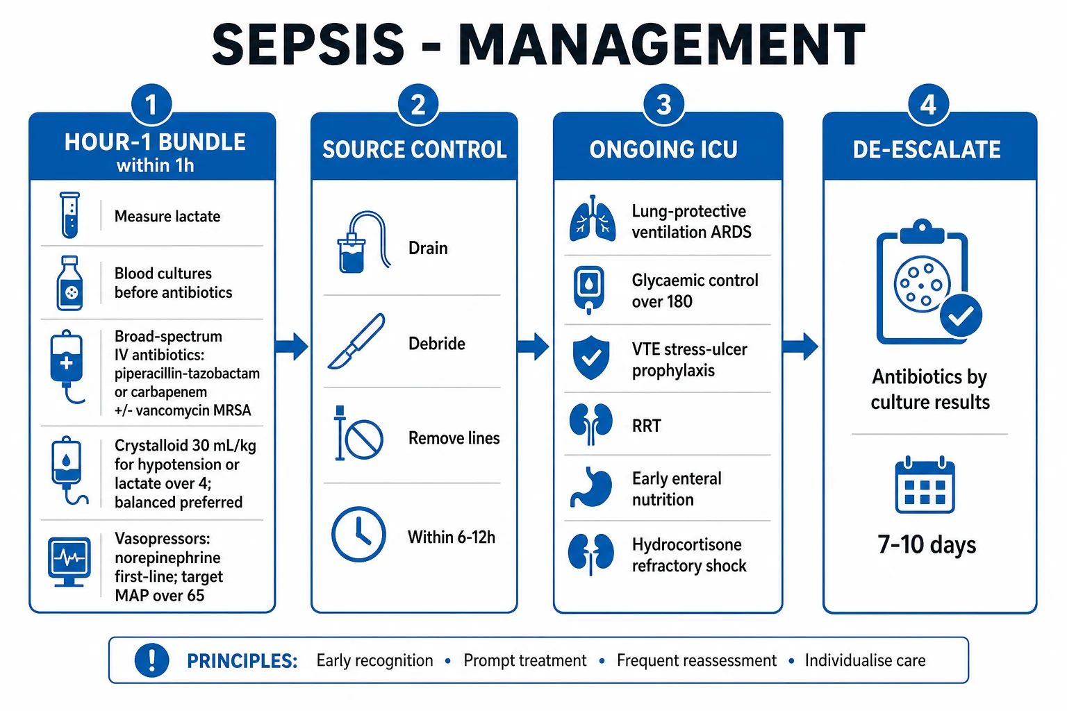
Sepsis is a time-critical emergency. The resuscitation bundle must be delivered concurrently, not sequentially, and within one hour of recognition — the Surviving Sepsis Campaign Hour-1 bundle. The philosophy is: recognise, resuscitate, sample, source-control, support. [1]
The Surviving Sepsis Campaign 2021 Hour-1 bundle (reproduce verbatim)
[1]Oxygen and the airway
Give supplemental oxygen to target SpO2 of 94 to 98 percent (or 88 to 92 percent in COPD at risk of CO2 retention). Escalate to high-flow nasal cannulae or non-invasive ventilation for moderate hypoxaemic respiratory failure; intubate and mechanically ventilate for severe respiratory failure, ARDS, falling GCS (under 8), or inability to protect the airway. In the ventilated patient with sepsis-associated ARDS, use lung-protective ventilation (see Definitive management).[2]
Fluid resuscitation — principles and the evidence
- Fluid of choice: balanced crystalloid (Hartmann's / Ringer's lactate / Plasma-Lyte). The SMART trial (Semler 2018) showed that balanced crystalloids reduce the composite of death, new dialysis or persistent renal dysfunction compared with normal saline in critically ill adults — saline, given in volume, causes hyperchloraemic metabolic acidosis, renal vasoconstriction and acute kidney injury.[8]
- Initial bolus: 30 mL/kg IV (ideal body weight) within the first 3 hours for hypotension or lactate over 4 mmol/L. Reassess after every bolus using the markers of perfusion (MAP, lactate, capillary refill, urine output) and a test of fluid responsiveness (passive leg raise).
- Avoid excessive fluid. A positive fluid balance is an independent predictor of mortality — it worsens pulmonary oedema, ARDS, abdominal compartment syndrome and tissue oedema. The CLOVERS trial (2023) showed that an early restrictive fluid strategy (early vasopressors, less fluid, then fluid only if hypoperfusion persisted) was non-inferior to a liberal fluid strategy in sepsis-induced hypotension.[10]
- Colloids (albumin) may be considered as an adjunct after initial crystalloid resuscitation in patients requiring substantial fluid; starches (HES) are harmful (increased renal failure and mortality) and are contraindicated in sepsis.[2]
Vasopressors
- Norepinephrine (noradrenaline) is first-line — an alpha-agonist (predominantly) that restores vascular tone and MAP; titrate from 0.05 to 1.0 mcg/kg/min to a target MAP at least 65 mmHg.[2]
- Add vasopressin (0.03 units/min fixed dose — a catecholamine-sparing agent) if norepinephrine requirements are rising; it acts via V1 receptors and is relatively catecholamine-independent.
- Adrenaline (epinephrine) as a third-line/additional agent or where norepinephrine is unavailable; it has both alpha and beta activity (greater risk of arrhythmia, lactate rise via beta-2 glycolysis).
- Target MAP: 65 to 70 mmHg. The SEPSISPAM trial (Asfar 2014) found that targeting a higher MAP (80 to 85 mmHg) offered no overall benefit over 65 to 70 mmHg, and may harm patients with chronic hypertension (in whom a higher target was associated with more atrial fibrillation and no mortality benefit).[9]
- Start vasopressors EARLY — if the patient is hypotensive during or after the initial fluid bolus, do NOT wait to finish a full 30 mL/kg before starting norepinephrine; the CLOVERS strategy supports early vasopressor use. A peripheral line is acceptable short-term (forearm vein) while central access is established.[10]
Early source control
Source control — the physical removal of the infective focus — is as important as antibiotics and must be achieved as soon as medically and logistically practical, ideally within 6 to 12 hours of recognition. The principle: drain infected fluid (abscess, empyema, collection), debride infected tissue (necrotising fasciitis, myonecrosis, infarcted bowel), and remove infected devices and lines (central lines, cannulae, urinary catheters, prosthetic material where feasible). An undrained focus is a frequent cause of refractory shock and must be sought actively with imaging. For the surgical abdomen (perforation, ischaemia, cholangitis), surgery or ERCP is definitive and antibiotics are adjunctive.[2]
The legacy of early goal-directed therapy (EGDT)
The protocolised central-venous-oxygen-saturation-targeted (ScvO2 over 70 percent) resuscitation of early goal-directed therapy (EGDT, Rivers 2001) was the dominant paradigm for a decade but was NOT supported by the three large pragmatic trials — ProCESS (2014)[5], ARISE (2014)[6] and ProMISe (2015)[7] — which showed that protocolised EGDT was not superior to usual care and was associated with more fluid, more vasopressors and more central lines. The modern Hour-1 bundle is simpler and centred on lactate, cultures, antibiotics, judicious fluid, and early vasopressors.[2]
Management — Definitive & Stepwise
The definitive management builds on resuscitation with source-directed and resistance-guided antibiotics, source control, organ support, and the prevention of complications. The escalation triggers are explicit. [1]
SEPSIS-Rx
Antibiotic principles
- Broad-spectrum, source-guided, and local-resistance-guided — cover the likely organisms for the source and the patient's risk profile (MRSA, Pseudomonas, ESBL/CRE).
- Within one hour of recognition in septic shock or high-likelihood sepsis — every hour of delay increases mortality.[4]
- IV at maximum dose; reassess daily.
- De-escalate to culture and sensitivity results within 48 to 72 hours — stop redundant agents, narrow to the narrowest effective spectrum.
- Duration: typically 7 to 10 days for most sepsis; procalcitonin-guided shortening is reasonable; longer courses for undrained foci, S. aureus bacteraemia (minimum 14 days, often longer), fungaemia, and the immunocompromised.[2]
Empirical antibiotic regimens (adapt to local resistance)
Undifferentiated community sepsis (adult)
- Piperacillin-tazobactam 4.5 g IV q8h (broad, including pseudomonal and anaerobic cover)
- OR ceftriaxone 2 g IV q24h (if no pseudomonal risk — typical community sepsis)
- ADD vancomycin 15 to 25 mg/kg IV (trough-guided, 15 to 20 mg/L) or linezolid 600 mg IV q12h if MRSA suspected (line infection, severe pneumonia, known colonisation)
- ADD metronidazole 500 mg IV q8h if anaerobic source and a cephalosporin is used (ceftriaxone lacks anaerobic cover)
- ADD antiviral (aciclovir) or antifungal if clinically indicated
Hospital-acquired / ICU / neutropenic sepsis
- Meropenem 1 g IV q8h (broad; covers ESBL, many CRE and Pseudomonas) OR piperacillin-tazobactam 4.5 g IV q8h
- PLUS vancomycin 15 to 25 mg/kg IV (MRSA, line infection)
- Consider empiric antifungal (echinocandin, e.g. caspofungin 70 mg loading then 50 mg IV daily) if candidaemia risk (prolonged ICU, TPN, multiple antibiotics, abdominal source)
- Neutropenic sepsis: anti-pseudomonal beta-lactam WITHIN 1 HOUR; add vancomycin for line infection/haemodynamic instability; G-CSF per protocol; low threshold for imaging and source control
Source-specific cover
- Meningitis / meningococcaemia: ceftriaxone 2 g IV q12h (add vancomycin + ampicillin if listeria/elderly) — meningeal dose
- Necrotising fasciitis: clindamycin 600 to 900 mg IV q8h + penicillin + MRSA cover (vancomycin/linezolid) — see TSS topic
- Endocarditis: benzylpenicillin / vancomycin + gentamicin per Duke organism — prolonged
- Intra-abdominal: piperacillin-tazobactam or ceftriaxone + metronidazole
- Catheter-associated UTI: remove/change catheter; anti-pseudomonal cover
De-escalation
- Review at 48 to 72 hours with cultures
- Stop redundant agents; narrow to the narrowest effective spectrum
- Stop vancomycin if MRSA not isolated; stop antipseudomonal if Pseudomonas not isolated
- Use procalcitonin to guide duration; reassess daily
The vasopressor/inotrope ladder
- Norepinephrine (alpha over beta) — first-line; restores vascular tone and MAP; 0.05 to 1.0 mcg/kg/min, titrate to MAP at least 65.
- Vasopressin 0.03 units/min (fixed dose) — add when norepinephrine requirements are rising; catecholamine-sparing.
- Adrenaline — add as a third-line agent if shock persists; alpha and beta activity; greater risk of arrhythmia and lactate rise.
- Inotrope (dobutamine) 2 to 20 mcg/kg/min — add for persistent low cardiac output / myocardial depression (cold shock, echocardiographic evidence of poor contractility, rising lactate despite an adequate MAP and filling). The septic heart is often depressed (sepsis-induced cardiomyopathy).[2]
Corticosteroids in septic shock
Hydrocortisone is suggested (weak recommendation, low-quality evidence) for adults with septic shock on ongoing vasopressor requirements — not for sepsis without shock. The rationale is that critical-illness-related corticosteroid insufficiency (CIRCI) contributes to vasopressor-refractory shock, and corticosteroids restore vascular responsiveness and reduce the duration of shock. Dose: hydrocortisone 200 mg/day by continuous infusion, OR 50 mg IV every 6 hours; the APROCCHSS trial (Annane 2018) supported hydrocortisone PLUS fludrocortisone 50 mcg/day for 7 days in vasopressor-dependent septic shock with reduced mortality at 90 days. Do not use the short Synacthen test to decide whether to give steroids — give empirical hydrocortisone for refractory shock and taper as the patient weans from vasopressors.[11][12]
Lung-protective ventilation for sepsis-associated ARDS
- Tidal volume 6 mL/kg ideal body weight; plateau pressure under 30 cmH2O.
- Titrate PEEP to optimise oxygenation without compromising venous return.
- Prone positioning for at least 16 hours/day if PaO2/FiO2 under 150 despite optimised ventilation.
- Conservative fluid strategy (aim for even or negative fluid balance once resuscitated).
- Neuromuscular blockade (short course) and ECMO in the most severe, refractory cases.[2]
Supportive measures
- VTE prophylaxis — the critically ill patient is at high VTE risk; give enoxaparin 40 mg SC daily (or unfractionated heparin in renal failure); add mechanical prophylaxis (sequential compression devices).
- Stress-ulcer prophylaxis — a proton-pump inhibitor for patients who are mechanically ventilated for over 48 hours OR have coagulopathy; not routinely for all.
- Glycaemic control — target blood glucose 8 to 10 mmol/L with an insulin infusion if needed; avoid hypoglycaemia and avoid tight glycaemic control (increases hypoglycaemia and mortality — NICE-SUGAR).
- Nutrition — early enteral nutrition (within 24 to 48 hours) trophic, advancing to target; parenteral only if enteral is not feasible.
- Renal replacement therapy — for AKI with refractory hyperkalaemia, acidosis, fluid overload or uraemia; continuous RRT (CRRT) for haemodynamically unstable patients.
- Transfusion — restrictive strategy (transfuse to haemoglobin over 70 g/L; over 80 g/L in myocardial ischaemia); avoid routine erythropoietin and fresh-frozen plasma in the absence of bleeding.[2]
Controversies that are NOT supported
- Vitamin C / thiamine / hydrocortisone ('HAT' or Marik protocol) — the LOVIT trial (Lamontagne 2022) showed that high-dose IV vitamin C increased the risk of death or persistent organ dysfunction compared with placebo in critically ill patients with sepsis; vitamin C is NOT recommended.[13]
- Extracorporeal blood purification (polymyxin B haemoperfusion, CytoSorb) — not routinely supported by evidence.
- Erythropoietin, selenium, IV immunoglobulin (for septic shock without specific indication) — not recommended.
Specific Subtypes & Scenarios
Neonatal sepsis
Early-onset (under 72 hours) — vertical transmission; organisms Group B strep (Streptococcus agalactiae), E. coli, Listeria monocytogenes. Risk factors: PROM over 18 hours, maternal intrapartum fever over 38 deg C, maternal GBS colonisation, prematurity, chorioamnionitis. Presents with non-specific features (temperature instability, respiratory distress, apnoea, poor feeding, lethargy, jaundice, irritability). Empirical therapy: benzylpenicillin + gentamicin (or ampicillin + gentamicin; add cefotaxime if meningitis). Maternal intrapartum GBS prophylaxis (IV benzylpenicillin in labour) reduces early-onset GBS disease. [1]
Late-onset (over 72 hours) — usually nosocomial; organisms coagulase-negative staphylococci (line-related), Klebsiella, S. aureus, Candida. Empirical therapy is guided by the unit's resistance patterns; remove/change central lines.[2]
Paediatric sepsis
Suspect sepsis in any febrile child with altered behaviour, abnormal colour, prolonged capillary refill (over 3 seconds), tachypnoea, grunting or lethargy. Decompensated (cold) shock is recognised by weak pulses, mottled cool skin, prolonged capillary refill and hypotension (a late sign). Resuscitate with 10 to 20 mL/kg boluses of balanced crystalloid, reassessing after each (stop boluses if hepatomegaly, crackles or rising respiratory distress develop), and weight-based antibiotics within one hour: cefotaxime 50 mg/kg IV or ceftriaxone 50 to 75 mg/kg IV (add vancomycin 15 mg/kg if MRSA suspected); add acyclovir if HSV encephalitis is possible. Use weight-based vasopressors (adrenaline for cold shock, noradrenaline for warm shock) and admit to PICU. The FEAST trial (2011, Lancet) showed that, in resource-limited African settings, aggressive fluid boluses increased mortality in febrile children with shock — hence the caution with boluses in settings without intensive-care support, where cautious fluid and early referral are emphasised.[2]
Sepsis in the immunocompromised and neutropenia
Neutropenic sepsis (neutrophil count under 0.5 x10^9/L, or under 1.0 and falling) is a medical emergency — empirical broad-spectrum antibiotics within one hour of presentation, BEFORE confirmatory cultures. Initial regimen: an anti-pseudomonal beta-lactam (piperacillin-tazobactam, ceftazidime or meropenem) as monotherapy; add vancomycin for suspected line infection, haemodynamic instability, severe mucositis, or known MRSA colonisation. Consider empirical antifungal (echinocandin or mould-active azole) in high-risk neutropenia with persistent fever over 4 to 7 days; antiviral (aciclovir) if mucocutaneous HSV. G-CSF per unit protocol; low threshold for CT chest (looking for invasive fungal disease — halo sign, angioinvasion) and bronchoalveolar lavage. Do not delay antibiotics for cultures.[2]
Surgical abdomen as the source
Perforation (diverticular, appendiceal, peptic ulcer), mesenteric ischaemia, cholangitis, post-operative collection, obstructed infected biliary or urinary system — urgent source control is definitive (surgery, ERCP, percutaneous drainage), and antibiotics are adjunctive. Empirical cover must include anaerobes and Enterobacterales (piperacillin-tazobactam or ceftriaxone + metronidazole). Do not delay surgery for 'stabilisation' that cannot be achieved without source control.[2]
Puerperal and obstetric sepsis; septic abortion
Sources: chorioamnionitis, puerperal endometritis, septic abortion, wound infection, mastitis, retained products of conception. Organisms: Group A strep (rapidly progressive, high mortality), E. coli, anaerobes, and (globally) Gram-negative sepsis. Remove retained products (uterine evacuation), explore and debride wounds, broad-spectrum antibiotics including anaerobic and Group A strep cover (piperacillin-tazobactam + clindamycin for toxin suppression if GAS). Deliver the baby if chorioamnionitis is the source and the pregnancy is viable. Safe antibiotics in pregnancy and breastfeeding: beta-lactams, cephalosporins, macrolides; avoid aminoglycosides, tetracyclines, chloramphenicol, and quinolones where possible.[2]
Toxic shock syndrome and meningococcaemia as sepsis phenotypes
- Toxic shock syndrome — a superantigen-mediated cytokine storm; treat with clindamycin (anti-toxin) + beta-lactam + IVIG (1 to 2 g/kg) + urgent source control (remove tampon/foreign body; debride necrotising fasciitis). See the dedicated TSS topic.
- Meningococcaemia — Neisseria meningitidis septicaemia with a petechial/purpuric non-blanching rash progressing to purpura fulminans (DIC) and Waterhouse-Friderichsen syndrome (adrenal haemorrhage); treat with ceftriaxone 2 g IV q12h (meningitis dose) immediately, plus empirical hydrocortisone if adrenal haemorrhage is suspected; give prophylaxis to close contacts (rifampicin, ciprofloxacin or ceftriaxone). [1]
Sepsis in cirrhosis and chronic liver disease
The cirrhotic patient is functionally immunocompromised (reticuloendothelial dysfunction, portosystemic shunting, low complement) and prone to spontaneous bacterial peritonitis (SBP), bacteraemia and infection with Gram-negative organisms. SBP presents with ascites, abdominal pain, fever or encephalopathy; diagnose by an ascitic fluid neutrophil count over 250 cells/mm3; treat with a third-generation cephalosporin (cefotaxime 2 g IV q8h) AND IV albumin (1.5 g/kg day 1, 1 g/kg day 3) to prevent hepatorenal syndrome. Adrenal insufficiency is common in septic shock with cirrhosis — give empirical hydrocortisone for refractory shock. Watch for variceal bleeding risk (coagulopathy, portal hypertension).[2]
Complications & Pitfalls
Early complications
- Refractory vasoplegic shock — mortality from persistent vasodilation, myocardial depression and capillary leak.
- Acute respiratory distress syndrome (ARDS) — bilateral pulmonary infiltrates, refractory hypoxaemia (PaO2/FiO2 under 300); manage with lung-protective ventilation.
- Acute kidney injury — from hypoperfusion, nephrotoxic drugs and inflammation; may require RRT.
- Hepatic dysfunction — cholestatic and hepatocellular; hyperbilirubinaemia.
- Disseminated intravascular coagulation — bleeding and microvascular thrombosis; manage the cause.
- Metabolic — hypoglycaemia or hyperglycaemia; lactic acidosis; electrolyte derangement.
- Ileus, stress ulceration, abdominal compartment syndrome — from fluid overload and splanchnic oedema.
- Sepsis-associated encephalopathy (SAE) — delirium, reduced GCS without CNS infection or focal signs; reversible with resolution of sepsis but associated with longer-term cognitive impairment. [1]
Late complications
- Critical-illness polyneuropathy and myopathy (ICU-acquired weakness) — difficulty weaning from the ventilator, profound limb weakness, slow recovery over months.
- Post-intensive-care syndrome — the triad of physical (weakness, breathlessness), cognitive (impaired memory, executive function) and psychological (PTSD, depression, anxiety) morbidity that persists for months to years.
- Post-sepsis immunoparalysis — increased susceptibility to secondary and nosocomial infections (ventilator-associated pneumonia, C. difficile, candiduria, reactivation of latent viruses — CMV, HSV, EBV).
- Recurrence — survivors are at increased risk of a further sepsis episode in the following year. [1]
Classic errors and pitfalls
- Delay in antibiotics — the single most preventable cause of avoidable death; every hour beyond the first increases mortality.[4]
- Inadequate or delayed source control — an undrained abscess, an infected line left in situ, or infarcted bowel not resected causes refractory shock.
- Excessive fluid — a positive fluid balance worsens pulmonary oedema, ARDS, abdominal compartment syndrome and tissue oxygenation; reassess responsiveness and stop bolusing when the patient is no longer fluid-responsive.
- Delayed vasopressors — waiting to complete the full 30 mL/kg before starting norepinephrine in a profoundly hypotensive patient prolongs hypoperfusion.
- Missing resistant organisms — MRSA, ESBL, CRE, Pseudomonas, Candida — not covered by the empirical regimen in the high-risk patient.
- Forgetting glucose and cortisol — hypoglycaemia is easily missed and dangerous; refractory shock may have an adrenal component.
- Failure to de-escalate antibiotics — prolonged broad-spectrum therapy drives resistance, C. difficile and fungal overgrowth; reassess at 48 to 72 hours.
- Not screening for the source — a 'sepsis of unknown origin' demands active imaging to find and control the focus.
- Over-reliance on a single number — a normal blood pressure, a normal white count, or a normal lactate alone does not exclude sepsis; the trend and the clinical picture decide.
Fluid overload — recognise and avoid
A positive cumulative fluid balance is an independent predictor of mortality in septic shock. Recognise it by rising oxygen requirements, pulmonary crepitations, widespread B-lines on lung ultrasound, abdominal distension and rising ventilator pressures (abdominal compartment syndrome), and peripheral and sacral oedema. Avoid it by reassessing fluid responsiveness before every bolus, starting vasopressors early, and adopting a conservative (de-resuscitative) fluid strategy once the patient is stabilised — guided by perfusion markers and lactate clearance rather than by protocolised volume targets.[10]
Prognosis & Disposition
Case-fatality
- Sepsis: approximately 10 percent in-hospital mortality.
- Septic shock: 30 to 40 percent; by the Sepsis-3 operational definition (vasopressor-dependent hypotension with lactate over 2 after fluids), over 40 percent.[1]
- Predictors of poor outcome: high and non-clearing lactate, high SOFA/APACHE II score, age, immunocompromise, cirrhosis, malignancy, an uncontrollable source, a multidrug-resistant organism, and delayed antibiotics and resuscitation.[3][4]
The effect of time
The single largest modifiable determinant of mortality is the time to effective antimicrobial therapy and adequate resuscitation. In septic shock, each additional hour of hypotension before effective antibiotics is associated with a measurable rise in mortality (of the order of 4 percent per hour in the Kumar cohort).[4] Early recognition and bundle delivery — the entire point of the Hour-1 bundle and the Sepsis Trust pathway — exist to compress this interval.
Disposition
- ICU (level 3): any patient requiring vasopressors, mechanical ventilation, or renal replacement therapy, or with rapidly escalating organ failure.
- HDU (level 2): the patient with single-organ failure or requiring close monitoring but not advanced organ support.
- Ward: the stable septic patient who has been resuscitated, is off vasopressors, and has a controlled source. [1]
Mobilise critical care, the infectious diseases team, microbiology, and (for a surgical source) surgery early. Communicate clearly with the patient and family, including realistic prognostic discussions and escalation plans, and consider goals of care in the setting of advanced comorbidity. [1]
Long-term outcomes — the post-sepsis syndrome
Survivors of sepsis face an increased risk of death and morbidity for months to years afterward — the post-sepsis syndrome. This includes physical impairment (weakness, breathlessness, fatigue — critical-illness polyneuropathy/myopathy), cognitive decline (impaired memory and executive function, especially in the elderly), psychological morbidity (PTSD, depression, anxiety), recurrent infection, and a high rate of rehospitalisation. Survivors have a higher long-term mortality than matched controls. Comprehensive follow-up — rehabilitation, vaccination, functional and cognitive assessment, and management of new or worsened comorbidity — is essential and is increasingly delivered through post-ICU clinics.[3]
Special Populations
Paediatrics
Recognition is harder (non-specific presentation); use the Paediatric Early Warning Score and NICE 'red flag' sepsis criteria. Weight-based fluids (10 to 20 mL/kg boluses, reassess; caution with boluses in resource-limited settings per the FEAST trial) and weight-based drugs: cefotaxime 50 mg/kg IV, ceftriaxone 50 to 75 mg/kg IV, vancomycin 15 mg/kg IV (MRSA), adrenaline 0.1 mcg/kg/min for cold shock, noradrenaline 0.1 mcg/kg/min for warm shock. Admit to PICU. Distinguish from Kawasaki disease in the febrile child with rash.[2]
Pregnancy and the puerperium
The pregnant patient has a physiologically raised baseline respiratory rate and heart rate and a mild leukocytosis — interpret these cautiously, and have a low threshold to investigate and treat. Sources: chorioamnionitis, pyelonephritis, appendicitis (right-lower quadrant pain, often higher in pregnancy), cholecystitis, pneumonia, puerperal sepsis, septic abortion. Safe antibiotics: beta-lactams, cephalosporins, macrolides, nitrofurantoin (avoid near term). Avoid (where possible): aminoglycosides (fetal ototoxicity), tetracyclines (teeth/bone), chloramphenicol (grey baby — historical), quinolones (cartilage — caution), trimethoprim (first trimester — neural tube). Left lateral tilt to relieve aortocaval compression during resuscitation. Deliver if the uterus is the source and chorioamnionitis is established.[2]
The elderly
Blunted fever (may be afebrile or hypothermic); atypical presentation (confusion, falls, incontinence, anorexia, 'off legs'); multiple comorbidities and polypharmacy (beta-blockers blunt the tachycardic response, steroids mask inflammation); a lower threshold to escalate; and a higher mortality. The "new confusion in an elderly patient is sepsis until proven otherwise" rule saves lives. [1]
The immunocompromised
Neutropenia, transplant, HIV with low CD4, steroids, biologics — a broader differential (opportunists — Pneumocystis jirovecii pneumonia, invasive fungal disease, CMV, mycobacteria), empirical broad cover, a low threshold for bronchoalveolar lavage and tissue biopsy, and early antifungal and antiviral coverage. Neutropenic sepsis is treated empirically within one hour before cultures.[2]
Asplenia / hyposplenism
Overwhelming post-splenectomy infection (OPSI) is a medical emergency — encapsulated organisms (Streptococcus pneumoniae, Neisseria meningitidis, Haemophilus influenzae type b, and Capnocytophaga canimorsus after dog bites) cause fulminant, often fatal sepsis with DIC and adrenal haemorrhage. Management: empirical broad-spectrum cover immediately (ceftriaxone; add vancomycin if meningitis), standby emergency antibiotics at home (amoxicillin), and lifelong vaccination (pneumococcal, meningococcal, Haemophilus influenzae type b, influenza annual). See the dedicated asplenia topic. [1]
Chronic liver disease / cirrhosis
Functional immunoparalysis (reticuloendothelial dysfunction, low complement, portosystemic shunting); high risk of spontaneous bacterial peritonitis, bacteraemia, and sepsis with Gram-negatives and enterococci; adrenal insufficiency is common in septic shock with cirrhosis (give empirical hydrocortisone); coagulopathy and variceal bleeding risk complicate management; albumin for SBP prevents hepatorenal syndrome; prognosis is worse than in the non-cirrhotic.[2]
Anticoagulated and dialysis-dependent
Manage DIC (the ISTH overt-DIC score) — transfuse platelets and plasma for bleeding or before invasive procedures; reversal of therapeutic anticoagulation if life-threatening bleeding (warfarin: vitamin K + prothrombin complex concentrate; DOAC: andexanet alfa or PCC; heparin: protamine). Adjust antibiotic doses for renal function (meropenem, piperacillin-tazobactam, vancomycin (level/trough-guided), ceftriaxone needs no adjustment; linezolid needs no adjustment). Vasopressors are safe in dialysis patients — do not withhold them. Ensure VTE prophylaxis despite bleeding risk (use mechanical prophylaxis if pharmacological is contraindicated). [1]
Evidence, Guidelines & Regional Differences
Landmark trials and guidelines
The Surviving Sepsis Campaign 2021 guidelines (Evans, PMID 34599691)
The SSC 2021 guidelines are the global standard. Key recommendations:[2]
- Hour-1 bundle — lactate, cultures, broad-spectrum antibiotics, 30 mL/kg crystalloid for hypotension or lactate over 4, vasopressors — within one hour.
- Balanced crystalloids preferred over normal saline for resuscitation (strong).
- Norepinephrine first-line vasopressor; add vasopressin; target MAP at least 65 mmHg (strong).
- Restrictive transfusion (haemoglobin over 70 g/L); avoid fresh-frozen plasma in the absence of bleeding.
- Hydrocortisone 200 mg/day suggested for adults with septic shock on ongoing vasopressor requirements.
- Lung-protective ventilation for ARDS; conservative fluid strategy.
- Glycaemic target 8 to 10 mmol/L; early enteral nutrition; VTE and stress-ulcer prophylaxis.
- Do NOT use vitamin C (LOVIT, 2022). [1]
Regional deltas
[1] [1] [1]Current controversies
- Vitamin C / thiamine / hydrocortisone (HAT) — the LOVIT trial (2022) showed harm from high-dose vitamin C; the HAT protocol is not recommended.[13]
- Extracorporeal blood purification (polymyxin B haemoperfusion, CytoSorb) — not routinely supported.
- The optimal MAP target — 65 to 70 mmHg is standard; a higher target may help selected patients with chronic hypertension but is not routine.[9]
- Point-of-care ultrasound (POCUS)-guided resuscitation — increasingly used to assess cardiac function, volume status and the source.
- Restrictive vs liberal fluids — the modern trend is restrictive (early vasopressors), supported by CLOVERS.[10]
Exam Pearls
- Sepsis = life-threatening organ dysfunction from a dysregulated host response to infection (Sepsis-3); operationally an acute SOFA increase of 2 or more points. SIRS is retired as a defining criterion.[1]
- Septic shock = vasopressor-dependent hypotension (MAP at least 65) AND lactate over 2 despite adequate fluids; mortality over 40 percent.[1]
- qSOFA = RR over 22, GCS under 15, SBP under 100 (2 of 3); bedside escalation tool, not sole screen.[1]
- Hour-1 bundle: lactate, blood cultures, broad-spectrum antibiotics, 30 mL/kg crystalloid for hypotension/lactate over 4, vasopressors — all within one hour.[2]
- Norepinephrine is first-line vasopressor; target MAP at least 65; add vasopressin; hydrocortisone 200 mg/day for refractory shock.[2]
- Balanced crystalloids preferred (SMART); avoid excessive fluid (CLOVERS — restrictive non-inferior); starches (HES) contraindicated.[8][10]
- Lactate over 4 or failure to clear by 10 percent at 2 to 4 hours predicts mortality; re-measure.[2]
- Antibiotics within one hour in septic shock; every hour delay increases mortality (~4 percent per hour, Kumar 2006).[4]
- Source control (drain, debride, remove lines) is as important as antibiotics; aim for within 6 to 12 hours.[2]
- Common sources in order: lung, abdomen, urinary tract, skin/soft tissue, line/device, CNS, endovascular.[3]
- Suspect adrenal crisis in refractory vasopressor-dependent shock; give empirical hydrocortisone.[11]
- DIC in sepsis: low platelets, low fibrinogen, raised PT/APTT, raised D-dimer; treat the cause (sepsis), support with blood products only if bleeding/procedure.
- Sepsis-associated encephalopathy = delirium/reduced GCS without CNS infection or focal signs.
- Warm shock (early vasoplegic) vs cold shock (late decompensated) — guides resuscitation.
- Procalcitonin guides antibiotic duration, not the diagnosis of sepsis.
References
- [1]Singer M, Deutschman CS, Seymour CW, et al. The Third International Consensus Definitions for Sepsis and Septic Shock (Sepsis-3) JAMA, 2016.PMID 26903338
- [2]Evans L, Rhodes A, Alhazzani W, et al. Surviving sepsis campaign: international guidelines for management of sepsis and septic shock 2021 Intensive Care Med, 2021.PMID 34599691
- [3]Rudd KE, Johnson SC, Agesa KM, et al. Global, regional, and national sepsis incidence and mortality, 1990-2017: analysis for the Global Burden of Disease Study Lancet, 2020.PMID 31954465
- [4]Kumar A, Roberts D, Wood KE, et al. Duration of hypotension before initiation of effective antimicrobial therapy is the critical determinant of survival in human septic shock Crit Care Med, 2006.PMID 16625125
- [5]The ProCESS Investigators. A randomized trial of protocol-based care for early septic shock N Engl J Med, 2014.PMID 24635773
- [6]The ARISE Investigators and the ANZICS Clinical Trials Group. Goal-directed resuscitation for patients with early septic shock N Engl J Med, 2014.PMID 25272316
- [7]Mouncey PR, Osborn TM, Power GS, et al. (ProMISe Trial Investigators). Trial of early, goal-directed resuscitation for septic shock N Engl J Med, 2015.PMID 25776532
- [8]Semler MW, Self WH, Wanderer JP, et al. (SMART Investigators and the Pragmatic Critical Care Research Group). Balanced Crystalloids versus Saline in Critically Ill Adults N Engl J Med, 2018.PMID 29485925
- [9]Asfar P, Meziani F, Hamel JF, et al. (SEPSISPAM Investigators). High versus low blood-pressure target in patients with septic shock N Engl J Med, 2014.PMID 24635770
- [10]The National Heart, Lung, and Blood Institute Prevention and Early Treatment of Acute Lung Injury Clinical Trials Network (Self WH, et al.; CLOVERS). Early Restrictive or Liberal Fluid Management for Sepsis-Induced Hypotension N Engl J Med, 2023.PMID 36688507
- [11]Annane D, Sebille V, Charpentier C, et al. Effect of treatment with low doses of hydrocortisone and fludrocortisone on mortality in patients with septic shock JAMA, 2002.PMID 12186604
- [12]Annane D, Renault A, Brun-Buisson C, et al. (APROCCHSS Trial). Glucocorticoids with or without Fludrocortisone in Septic Shock N Engl J Med, 2018.PMID 30179381
- [13]Lamontagne F, Maslove DM, Boyd JC, et al. (LOVIT Investigators). Intravenous Vitamin C in Adults with Sepsis in the Intensive Care Unit N Engl J Med, 2022.PMID 35704292
- [14]Rhodes A, Evans LE, Alhazzani W, et al. Surviving Sepsis Campaign: International Guidelines for Management of Sepsis and Septic Shock: 2016 Intensive Care Med, 2017.PMID 28101605