Infectious Diseases · Infectious Diseases
Tuberculosis
Also known as TB · Mycobacterium tuberculosis infection · Koch's disease · Consumption (historical) · Phthisis (historical)
Tuberculosis (TB) is a chronic granulomatous infection caused by Mycobacterium tuberculosis (Mtb), an acid-fast aerobic bacillus that classically attacks the lungs (pulmonary TB, 70-80 percent) but can affect any organ (extrapulmonary TB). It exists in two states: latent TB infection (LTBI) — the bacilli are contained by host immunity in a granuloma, the patient is asymptomatic and non-infectious; and active TB disease — replication resumes, symptoms develop, and the patient becomes infectious. Globally TB causes about 10 million new cases and 1.4 million deaths per year and is the leading cause of death from a single infectious agent. Diagnosis rests on microbiology — sputum AFB smear (Ziehl-Neelsen or auramine), mycobacterial culture (the gold standard), and rapid molecular NAAT (Xpert MTB/RIF or Xpert Ultra, which simultaneously detects rifampicin resistance); the interferon-gamma release assay (IGRA) or tuberculin skin test (TST/PPD) detects latent infection. Treatment of drug-susceptible active pulmonary TB is the six-month RIPE regimen — rifampicin (RIF) 10 mg/kg daily max 600 mg, isoniazid (INH) 5 mg/kg daily max 300 mg, pyrazinamide (PZA) 25 mg/kg daily max 2 g, and ethambutol (EMB) 15-20 mg/kg daily for 2 months (intensive), then rifampicin + isoniazid for 4 months (continuation), with pyridoxine (vitamin B6) 25 mg daily to prevent INH-induced peripheral neuropathy. Drug-resistant TB — MDR (resistant to at least INH + RIF), XDR (MDR plus resistance to any fluoroquinolone and at least one of bedaquiline/linezolid), and pre-XDR (MDR plus fluoroquinolone resistance) — requires all-oral longer regimens (18-20 months) built around bedaquiline 400 mg daily for 2 weeks then 200 mg three-times-weekly, linezolid 600 mg daily, levofloxacin 750 mg daily, and/or delamanid 100 mg twice daily. LTBI is treated with shorter rifapentine-based regimens — 3HP (INH + rifapentine weekly for 12 weeks) or 1HP (INH + rifapentine daily for 4 weeks) — to prevent progression to active disease. BCG vaccination at birth protects children against severe forms (miliary TB, TB meningitis) but has variable efficacy against adult pulmonary TB.
On this page & tools
Your progress
Saved locally on this device.
Practise this topic
Exam tags
Red flags
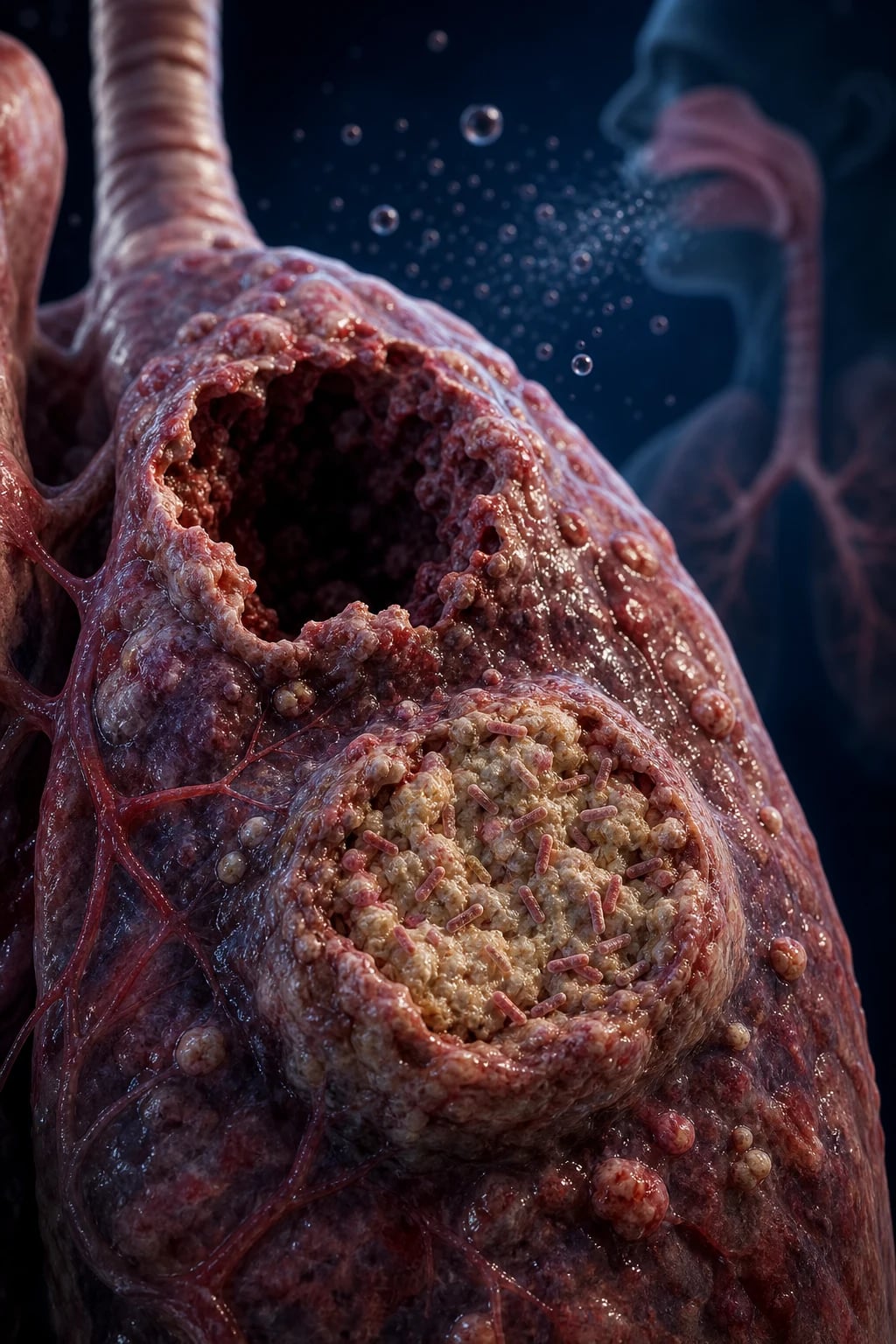
Overview and Definition
Tuberculosis (TB) is a chronic, granulomatous infectious disease caused by members of the Mycobacterium tuberculosis complex — predominantly Mycobacterium tuberculosis itself (Koch's bacillus, discovered 1882), with M. bovis (cattle, unpasteurised milk — now rare in countries with pasteurisation), M. africanum (West Africa) and the live-attenuated vaccine strain M. bovis BCG as related members.[2][3]
The organism is a slender, slightly curved, aerobic, non-motile, non-spore-forming, acid-fast bacillus (AFB). The waxy, lipid-rich, mycolic-acid-containing cell wall is responsible for the key microbiological properties: it resists Gram staining (so Ziehl-Neelsen or auramine-rhodamine fluorescence are used), is resistant to desiccation, and survives for weeks on surfaces and in droplet nuclei. M. tuberculosis is an obligate aerobe — it grows best in well-oxygenated tissue, which is why the apical and posterior segments of the upper lobes, the renal cortex, and the growing ends of long bones are the classic sites of reactivation disease.[2]
Two clinical states are fundamental to the disease and must be distinguished:[2][3]
- Latent TB infection (LTBI) — the bacilli are contained in a granuloma by host cell-mediated immunity, the patient is asymptomatic, non-infectious, and has a normal chest X-ray, but the tuberculin skin test (TST/PPD) or interferon-gamma release assay (IGRA) is positive. About one quarter of the world population is estimated to have LTBI (WHO), and 5-10 percent of immunocompetent LTBI-positive individuals will progress to active disease over their lifetime (much higher in HIV and other immunosuppression).
- Active TB disease — replication breaks out of granuloma control, symptoms develop, the patient becomes infectious, and there is radiological and/or microbiological evidence of disease. Without treatment, case-fatality is about 50 percent over 5-10 years; smear-positive pulmonary TB in HIV co-infection is fatal in over 80 percent without treatment.[2]
By site of disease: pulmonary TB (about 70-80 percent of cases) involves the lung parenchyma and is the infectious form (transmitted by airborne droplet nuclei, 1-5 micrometres, that remain suspended in air for hours); extrapulmonary TB (EPTB, 20-30 percent) involves any other organ — lymph nodes (scrofula, commonest EPTB site), pleura, meninges (TBM), bones and joints (Pott's spine), genitourinary tract, abdomen, pericardium, and miliary (disseminated). Pulmonary and extrapulmonary disease may co-exist, particularly in HIV co-infection and in children.[8]
Why TB matters globally: TB is a top-10 cause of death worldwide and the leading cause of death from a single infectious agent (overtaking COVID-19 in 2023). WHO estimates about 10 million new (incident) cases per year, about 1.4 million deaths per year (including 160 000 HIV-positive), and about 0.5 million incident rifampicin-resistant (RR) cases per year (of which about 80 percent are MDR). The End TB Strategy targets a 90 percent reduction in TB incidence and 95 percent reduction in TB mortality by 2035 (versus 2015 baseline).[2][3]
Classification
TB is classified by clinical state (LTBI vs active), by anatomical site (pulmonary vs extrapulmonary), by radiographic pattern (primary vs post-primary, miliary), and by drug-resistance pattern (each driving a different management pathway).[2][3][4]
Primary TB
- First infection in a non-immune host (typically children in TB-endemic countries)
- Ghon focus — peripheral subpleural mid/lower-zone parenchymal granuloma (often the right middle or lower lobe)
- Ghon complex = Ghon focus + ipsilateral hilar/mediastinal lymphadenopathy
- Usually asymptomatic, heals with calcification (Ranke complex)
- Progressive primary TB in young children and HIV — cavitation, lymphadenopathy, complications (atelectasis, effusion, miliary)
Post-primary (reactivation) TB
- Reactivation of latent infection, often years after primary
- Apical and posterior segments of the upper lobes (high oxygen tension)
- Cavitation, fibrocaseous disease, healed with fibrosis and volume loss
- Strongly symptomatic — cough, fever, weight loss, haemoptysis
- Adult-type TB, infectious, the form that drives transmission
Miliary (disseminated) TB
- Lymphohaematogenous dissemination — 'millet-seed' nodules (1-5 mm) throughout lungs and often other organs
- Risk groups — young children, HIV, anti-TNF, transplant, malnutrition
- Fever, weight loss, hepatosplenomegaly, pancytopenia, choroidal tubercles
- CXR — diffuse 1-5 mm nodules; CT better; may have normal CXR early
- Mortality 20-30 percent even with treatment
Lymph node TB (scrofula)
- Commonest extrapulmonary site in children and in HIV
- Painless, matted cervical lymphadenopathy (most often posterior triangle)
- Caseating granulomas on FNA; AFB smear often negative; GeneXpert on FNA useful
- Surgical excision for cold abscess, chemotherapy for 6 months (RIPE 2 + RI 4)
- Differential — lymphoma, sarcoid, NTM, secondary malignancy
MDR / XDR / pre-XDR / TDR
- MDR — resistant to at least INH and rifampicin
- pre-XDR — MDR plus resistance to any fluoroquinolone
- XDR — MDR plus resistance to any fluoroquinolone AND at least one of bedaquiline/linezolid (WHO 2021 definition)
- TDR — totally drug resistant (no effective standardised regimen, contested term)
- RR-TB — rifampicin-resistant (often a proxy for MDR pending INH testing)
Drug-resistance classification (WHO 2021):[4]
- Mono-resistant — resistant to one first-line drug only (usually INH).
- Poly-resistant — resistant to two or more first-line drugs other than the INH + RIF combination.
- Rifampicin-resistant (RR-TB) — resistant to rifampicin, regardless of other resistances.
- Multidrug-resistant (MDR-TB) — resistant to at least INH and RIF.
- Pre-extensively drug-resistant (pre-XDR-TB) — MDR/RR-TB plus resistance to any fluoroquinolone.
- Extensively drug-resistant (XDR-TB) — MDR/RR-TB plus resistance to any fluoroquinolone AND resistance to at least one of bedaquiline or linezolid (the WHO 2021 revised definition).
- Totally drug-resistant (TDR) — reported sporadically from India and elsewhere; no standardised effective regimen; the term is not formally endorsed by WHO. [1]
By site (the extrapulmonary taxonomy, Sandgren et al. 2013):[8]
- Pleural TB — second most common extrapulmonary site, often young adults, lymphocytic exudative effusion, pleural fluid ADA over 40 U/L supportive; biopsy shows caseating granuloma.
- TB meningitis (TBM) — most severe form; basal meningitis with cranial nerve palsies, hydrocephalus, vasculitis, stroke; highest mortality of all forms.
- Osteoarticular TB — Pott's spine (thoracolumbar, vertebral body, gibbus deformity), hip, knee; cold abscess (paravertebral/psoas).
- Genitourinary TB — sterile pyuria, scrotal swelling in males, infertility, dysmenorrhoea and tubo-ovarian mass in females, renal involvement with calyceal blunting.
- Abdominal TB — peritoneal (ascites with high ADA, matted mesenteric lymphadenopathy), ileocaecal (ulcero-constrictive disease — the commonest site of abdominal TB, mimics Crohn's and malignancy), nodal, visceral.
- TB pericarditis — subacute pericardial effusion with lymphocytic exudate, often progressing to constrictive pericarditis.
- Cutaneous TB — scrofuloderma, lupus vulgaris, tuberculid, warty TB. [1]
Epidemiology and Risk Factors
TB is a global pandemic of low- and middle-income countries, with South-East Asia (44 percent), Africa (24 percent), and the Western Pacific (17 percent) accounting for about 85 percent of cases (WHO 2023). Eight countries — India (27 percent), Indonesia (10), China (7), the Philippines (7), Pakistan (6), Nigeria (6), Bangladesh (3.5), and DRC (3.1) — account for two-thirds of all cases.[2][3]
Global burden numbers (WHO 2023 estimates, illustrative): [1]
- Incident cases: about 10 million per year (range 9-11 million); incident rate: about 134 per 100 000 population.
- Deaths: about 1.4 million per year (including 160 000 HIV-positive); TB is the leading cause of death from a single infectious agent globally.
- Latent TB: estimated one-quarter of the world population (~2 billion).
- MDR/RR-TB: about 0.5 million new cases per year; about 3-4 percent of new and 18-20 percent of previously treated cases have MDR-TB.
- HIV-associated TB: about 700 000 incident cases per year (7 percent of total); TB is the leading cause of death in people with HIV. [1]
Host risk factors for progression from LTBI to active disease (relative risk vs general population):[2][3]
- HIV infection — the single most powerful risk factor: 5-10 percent annual risk of progression (vs 5-10 percent lifetime in immunocompetent); CD4 count under 200 cells/microL carries the highest risk; also atypical presentations (negative TST/IGRA, disseminated/miliary disease, normal CXR with positive sputum).
- Malnutrition and underweight — globally the most important modifiable risk factor (about 2.2 million TB cases attributable); BMI under 18.5 is a strong predictor; diabetes mellitus triples TB risk and is the leading metabolic risk factor.
- Anti-TNF therapy (infliximab, adalimumab, etanercept) — reactivation rate up to 4-12 times higher; screen with IGRA/PPD before starting; consider LTBI therapy in the latent-positive.
- Other immunosuppression — glucocorticoids (over 15 mg/day prednisolone for over 4 weeks), chemotherapy, solid-organ and stem-cell transplant, haematological malignancy, chronic kidney disease and haemodialysis, silicosis.
- Silicosis — increases TB risk about 3-fold; remains a major occupational risk in mining and quarrying.
- Intravenous drug use (IVDU) — HIV, hepatitis C, and direct immunosuppression compound risk; outbreaks documented.
- Prisons and detention — high transmission, late diagnosis, MDR outbreaks; active case-finding in prisons is WHO policy.
- Healthcare workers in TB-endemic settings — annual IGRA conversion rates 2-10 percent; occupational screening recommended.
- Age — young children under 5 (especially under 2) have the highest risk of progression from primary infection to active disease (and of severe forms — miliary, TBM); adolescents and young adults in high-burden settings.
- Recent infection — household and close contacts of a smear-positive pulmonary case have about 2 percent annual risk of developing active disease; window-period TB (developing disease during the first 1-2 years of infection) accounts for the bulk of secondary cases.
- Alcohol misuse, smoking, indoor air pollution, HIV (in pregnancy), vitamin D deficiency — modest independent risk factors. [1]
Social determinants — poverty, overcrowding, undernutrition, indoor air pollution, and limited access to diagnosis and treatment are the upstream drivers of the TB epidemic; the WHO End TB Strategy pillars address these alongside biomedical tools.[3]
Pathophysiology
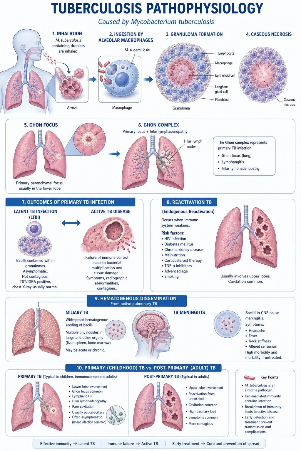
The pathophysiology of TB is a timeline of host-pathogen interaction that explains both primary disease, latent infection, and reactivation.[2][3]
1. Transmission and primary infection. An infectious patient with smear-positive pulmonary or laryngeal TB expels droplet nuclei (1-5 micrometres) by coughing, sneezing, speaking, or singing; these remain suspended in air for hours, and a susceptible contact inhales them to reach the terminal alveoli. The infectiousness of a source is proportional to the smear grade and the duration of contact; a single cough generates about 3000 droplet nuclei, and a sneeze about 1 million. BCG-vaccinated and previously-infected contacts may have partial immunity, but in non-immune hosts the bacilli are phagocytosed by alveolar macrophages. Inside the macrophage the bacilli resist intracellular killing by several mechanisms — cord factor (trehalose-6,6'-dimycolate) inhibits macrophage activation, sulfatides (sulfolipids) prevent phagosome-lysosome fusion, the ESX-1 secretion system releases ESAT-6 and CFP-10 antigens, and LAM scavenges oxygen radicals — and multiply logarithmically over 2-8 weeks before cell-mediated immunity is established.[2]
2. The cell-mediated immune response — Th1, IFN-gamma, TNF-alpha, granuloma. Infected macrophages present Mtb antigens (ESAT-6, CFP-10, Ag85) on MHC class II to CD4+ T-helper-1 (Th1) cells, which release interferon-gamma (IFN-gamma). IFN-gamma activates macrophages, enhancing phagolysosome fusion, nitric oxide production, and intracellular killing. TNF-alpha (from macrophages and T cells) is essential for granuloma maintenance. Together, these cytokines orchestrate the formation of a caseating granuloma (tubercle) — a structured aggregate of epithelioid macrophages, multinucleated Langhans giant cells, surrounding CD4+ and CD8+ T cells, and a central area of caseous (cheese-like) necrosis. The caseum has a low oxygen tension, low pH, and abundant lipid debris — a hostile environment for the bacilli that dormantly survive within the granuloma for decades. This is the immunopathological basis of latent TB infection.[2]
3. Latent TB — containment and the risk of reactivation. A balanced Th1 response contains the bacilli indefinitely in the granuloma; the patient is asymptomatic, non-infectious, and has normal radiology. The lifetime risk of reactivation is 5-10 percent in immunocompetent individuals (greatest in the first 2 years, then a long tail), but rises dramatically with HIV, anti-TNF, malnutrition, and other immunosuppression. Cortisol and TNF-alpha blockade can both destabilise the granuloma.[2]
4. Reactivation (post-primary) TB. When the cell-mediated response wanes (HIV, anti-TNF, malnutrition, age, diabetes, alcohol, smoking), the granuloma caseates and liquefies, releasing viable bacilli into the surrounding tissue, where the higher oxygen tension and better nutrients allow rapid extracellular replication. The apical and posterior segments of the upper lobes (and the superior segment of the lower lobe) are the preferred sites of reactivation because of the higher V/Q ratio (higher PaO2) in these regions. The caseum liquefies, drains into a bronchus, and produces a cavity — the radiological hallmark of post-primary TB and the source of airborne infectiousness.[2]
5. Haematogenous and lymphogenous spread — miliary and extrapulmonary disease. In early infection, bacilli reach the hilar lymph nodes (lymphogenous spread) and the systemic circulation (haematogenous spread), seeding the apices, kidneys, bone epiphyses, meninges, and other sites with microscopic foci. In the immunocompetent these foci are contained by granulomas; in the immunocompromised (especially HIV, anti-TNF, malnourished children), these foci may reactivate simultaneously, producing miliary (disseminated) TB — a radiograph studded with 1-5 mm nodules of caseating granulomas throughout both lungs and often the liver, spleen, bone marrow, choroid, and meninges. The presence of choroidal tubercles on fundoscopy is pathognomonic.[2][8]
6. Why HIV-TB is dangerous. HIV directly depletes CD4+ T cells, the central orchestrators of granuloma integrity. Patients with CD4 under 200 have atypical, disseminated, and severe TB, negative TST/IGRA in up to 50 percent (anergy), and a much higher risk of IRIS after ART initiation. The same high-burden CD4 effect explains the diagnostic difficulty and the fulminant course.[2][3]
7. Drug resistance — the molecular basis. Rifampicin resistance is conferred primarily by mutations in the rpoB gene (RNA polymerase beta subunit), most often at codons 507-533. Isoniazid resistance is conferred by mutations in katG (most common, S315T, which abolishes INH activation by catalase-peroxidase), inhA (promoter, confers low-level resistance), and ahpC. Fluoroquinolone resistance arises in gyrA (DNA gyrase). Xpert MTB/RIF Ultra detects rpoB mutations within 2 hours and is the frontline rapid molecular test in most high-burden settings.[4]
Clinical Presentation
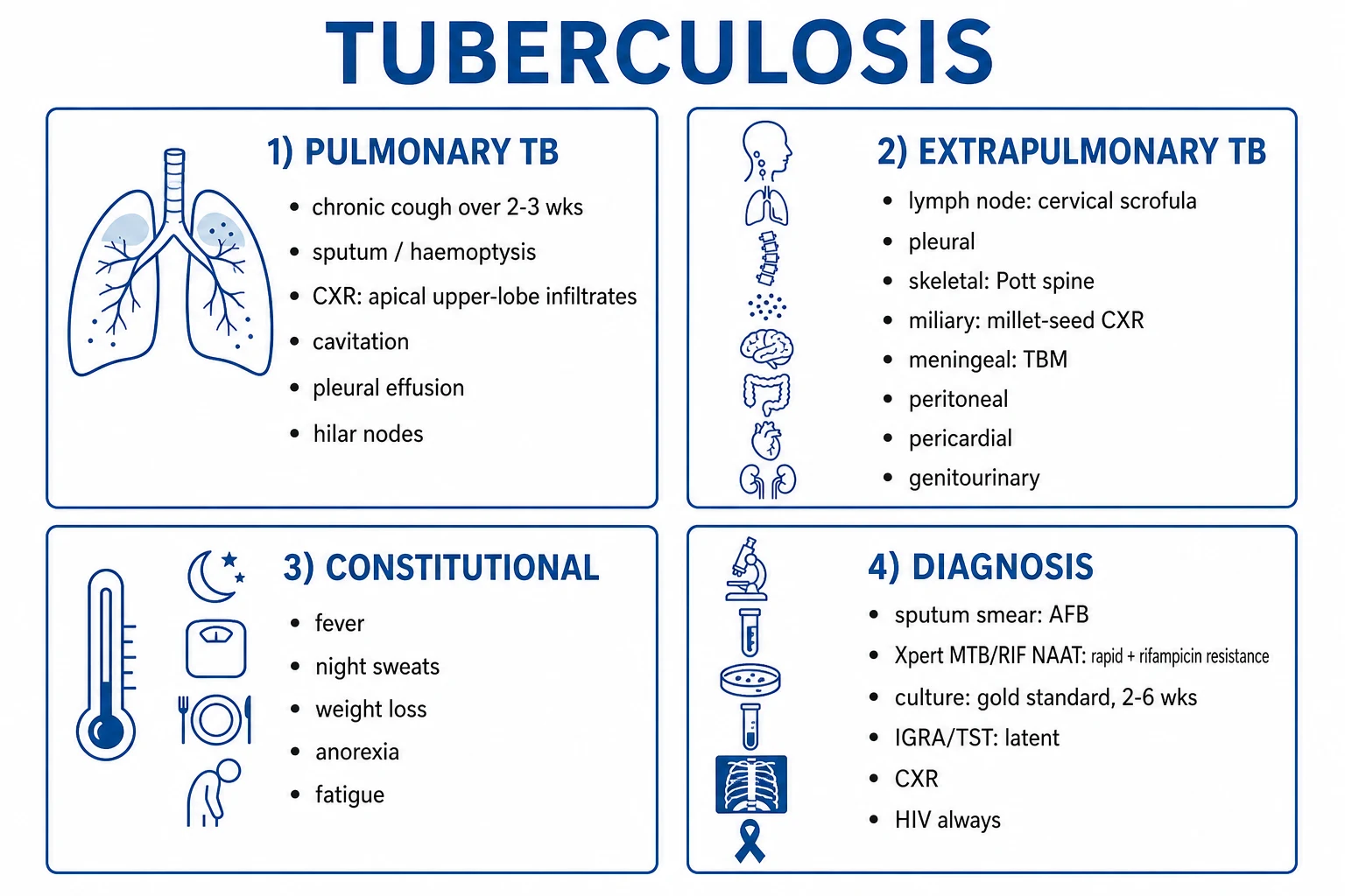
The classic triad of pulmonary TB is cough, fever, and weight loss, but presentation is highly variable and atypical in children, the elderly, the immunocompromised, and in extrapulmonary disease.[2][3]
Constitutional / systemic — low-grade fever (often evening, with night sweats), weight loss (often profound, "consumption"), fatigue, anorexia, and in women, amenorrhoea. The duration is chronic, often weeks to months, distinguishing TB from acute bacterial pneumonia.[2]
Pulmonary / respiratory: [1]
- Cough (the cardinal symptom) — initially dry, then productive of mucoid or purulent sputum; cough more than 2 weeks is the WHO symptom screen. The cough may become blood-tinged, streaky, or frankly haemoptytic (rupture of a vessel in a cavity wall, or Rasmussen aneurysm).
- Haemoptysis — massive (more than 200 mL in one expectoration or more than 600 mL/24 h) is life-threatening and is caused by erosion of a bronchial artery or a Rasmussen aneurysm (pulmonary artery aneurysm in the wall of a chronic cavity).
- Chest pain — pleuritic, from pleural involvement or subpleural inflammation.
- Dyspnoea — in extensive disease, miliary TB, pleural effusion, or TB-associated ARDS.
- Lymphadenopathy — cervical, supraclavicular, axillary, or inguinal; classically painless, matted, with or without caseation and sinus formation (scrofula).[8]
Extrapulmonary presentations (one quarter to one half of cases, more in HIV):[8]
- TB meningitis (TBM) — subacute (1-2 weeks) prodrome of headache, low-grade fever, malaise, progressing to meningism, photophobia, vomiting, altered mental status, cranial nerve palsies (especially VI, but also III, IV, VII from basal exudate), seizures, stroke (from obliterative endarteritis of perforators at the base of the brain), and hydrocephalus. The British Medical Research Council (BMRC) TBM staging is the prognostic standard: Stage I (prodromal, GCS 15, no focal signs), Stage II (meningism + altered mental status or focal signs, GCS 11-14), Stage III (deep coma, GCS under 11, dense neurological deficits). Mortality is about 20 percent in Stage I, 30-40 percent in Stage II, and over 60 percent in Stage III.[5]
- Pleural TB — pleuritic chest pain, dyspnoea, low-grade fever; lymphocytic exudative effusion; pleural fluid ADA over 40 U/L, interferon-gamma over 140 pg/mL, lymphocyte/neutrophil ratio over 0.75; pleural biopsy (image-guided) showing caseating granuloma is diagnostic.
- TB pericarditis — subacute pericardial effusion, fever, chest pain, pericardial rub; lymphocytic pericardial exudate; progresses to constrictive pericarditis in 20-40 percent despite treatment; pericardiectomy may be needed.
- Abdominal TB — ileocaecal (most common; right iliac fossa pain, mass, subacute obstruction, mimics Crohn's and right-sided colonic cancer); peritoneal (abdominal distension, ascites with high ADA, matted mesentery); nodal (mesenteric/retroperitoneal adenopathy, may calcify); visceral (hepatosplenomegaly, splenic abscess, pancreatic TB).
- Bone and joint TB (Pott's spine) — insidious back pain, gibbus deformity, paraspinal or psoas cold abscess, neurological deficit from spinal cord compression; lower thoracic and upper lumbar are the commonest levels; MRI is the imaging gold standard; the triad of Gibbus + paravertebral shadow on CXR + disc-space narrowing is classical.
- Genitourinary TB — sterile pyuria (urine with leucocytes but negative standard culture; send for mycobacterial culture), haematuria, flank pain, renal calcification, ureteric stricture, contracted bladder; in males, epididymo-orchitis, scrotal sinus, infertility; in females, tubo-ovarian mass, infertility, menstrual disturbance.
- Miliary TB — fever, weight loss, hepatosplenomegaly, pancytopenia, choroidal tubercles on fundoscopy; CXR with diffuse 1-5 mm nodules ("millet-seed"); may initially have a normal CXR (CT more sensitive); may progress to TB-associated ARDS, DIC, and multi-organ failure.
- Cutaneous TB — scrofuloderma (overlying lymph node), lupus vulgaris (face, apple-jelly nodules on diascopy), orificial TB (mucocutaneous around orifices in advanced disease), warty TB (tuberculous verrucosa cutis), tuberculid (hypersensitivity eruptions).
Differential Diagnosis
Active pulmonary TB shares features with many conditions; the diagnosis is microbiological, not clinical. The exam skill is to think of TB and order sputum AFB smear, NAAT (Xpert), and culture in the right patients.[2][3]
Lung cancer
- Risk factors: smoking, older age, weight loss, haemoptysis
- Imaging: spiculated mass, may cavitate (especially squamous), hilar/mediastinal nodes
- Diagnosis: contrast CT, bronchoscopy + biopsy; PET-CT for staging
- TB can mimic cancer and vice versa — biopsy and AFB essential
- Bacteriological confirmation required before chemotherapy/radiotherapy
Sarcoidosis
- Bilateral hilar lymphadenopathy, non-caseating granulomas, multisystem
- Negative sputum AFB and NAAT; raised ACE; biopsy shows non-caseating granuloma
- Distinguishing from TB critical — steroids worsen TB
- Pulmonary function: restrictive, reduced DLCO
- Could mask TB and reactivate under immunosuppression
Fungal infections
- Histoplasmosis, coccidioidomycosis, blastomycosis, aspergillosis, mucormycosis
- Geography: Ohio/Mississippi valleys (histoplasmosis); southwest US (coccidioidomycosis)
- Aspergilloma: fungus ball in pre-existing cavity (TB cavity, sarcoid cavity)
- Diagnose: serum/urine antigen, serology, biopsy with special stains
- Endemic mycoses mimic TB granulomas; NTM also relevant
Nocardia / NTM
- Nocardia: Gram-positive branching, weakly acid-fast — cavitating pneumonia in cell-mediated immunodeficiency
- NTM (M. avium complex, M. kansasii, M. abscessus) — chronic cavitary disease in COPD/older smokers or disseminated in HIV
- Distinguishing requires culture and molecular speciation (HPLC, MALDI-TOF, 16S rRNA)
- Treatment differs significantly from TB
- Treat as TB only if M. tuberculosis confirmed
Community-acquired pneumonia
- Acute (days), high fever, productive purulent sputum, lobar consolidation on CXR
- Responds to standard antibiotics (amoxicillin, beta-lactam + macrolide)
- Persistent consolidation despite antibiotics — consider TB, lung cancer, abscess
- Atypical pneumonia (Mycoplasma, Legionella) may be chronic
- Send sputum for AFB if any doubt
Lymphoma / other malignancy
- Lymphadenopathy, weight loss, B symptoms, mediastinal mass
- Diagnose with biopsy (core or excisional) — NEVER start RIPE on suspicion alone
- Steroids without TB therapy cause catastrophic progression
- Miliary TB can mimic lymphoma on CXR
- Tissue for AFB, culture, and histology together
Other important differentials to consider: lung abscess (cavitation with air-fluid level, foul sputum, post-aspiration), silicosis and coal worker's pneumoconiosis (occupational history, progressive massive fibrosis), bronchiectasis (chronic productive cough, CT diagnosis), atypical pneumonia (Legionella, Mycoplasma, Chlamydophila) that does not respond to standard antibiotics, HIV-associated opportunistic infections (Pneumocystis, cryptococcosis, histoplasmosis, Kaposi sarcoma), and constrictive pericarditis (other causes — post-cardiac surgery, radiation, malignancy). [1]
Clinical and Bedside Assessment
The clinical examination of suspected TB is a systematic search for pulmonary findings, lymph nodes, signs of extrapulmonary disease, and complications.[2]
General inspection — chronic illness, wasting (BMI often under 18.5 in advanced disease), pallor of anaemia of chronic disease, low-grade fever (palpable warmth), scars of past surgery (thoracoplasty, lobectomy for TB in the pre-antibiotic era), and — crucial for vaccination history — the BCG scar on the left deltoid (or right deltoid, depending on country).[2]
Hands, arms, neck — clubbing (chronic pulmonary TB, bronchiectasis from post-TB fibrosis), anaemia, erythema nodosum (TB-associated, sarcoid-associated), scrofuloderma (overlying a cold abscess). [1]
Lymph nodes — palpate cervical (anterior and posterior triangles, supraclavicular), supraclavicular, axillary, epitrochlear, and inguinal groups. TB lymphadenopathy is painless, rubbery, and matted (groups of nodes stuck together); caseation with cold abscess and sinus formation is late; isolated matted supraclavicular node (Virchow's node), although classically associated with gastric cancer, is described in TB. The "cold abscess" of TB fluctuates but is not warm or red (vs pyogenic abscess). [1]
Chest — [1]
- Inspection — asymmetry, supraclavicular/infra-clavicular hollowing (chronic upper-lobe fibrosis), scars of past surgery, tachypnoea.
- Palpation — reduced expansion on the affected side, tactile fremitus increased over consolidation, reduced over effusion.
- Percussion — dull over consolidation, effusion, fibrosis; hyper-resonant over a large cavity or compensatory hyperinflation.
- Auscultation — crackles (post-tussive) over active disease, bronchial breathing over consolidation, amphoric breathing over a large cavity, reduced breath sounds over effusion, pleural rub in dry pleurisy. [1]
Cardiovascular — pericardial rub, distant heart sounds, raised JVP, pulsus paradoxus (pericardial effusion/constriction), tachycardia of anaemia or fever. [1]
Abdomen — hepatosplenomegaly (miliary TB, HIV, lymphoma), ascites (peritoneal TB — shifting dullness, fluid thrill, "doughy" abdomen from matted mesentery), right iliac fossa mass (ileocaecal TB), psoas abscess (bulge in the groin, positive psoas sign). [1]
Spine — kyphosis (gibbus deformity of Pott's disease), paraspinal swelling (cold abscess), tenderness on percussion, limited spinal flexion; perform a neurological examination to detect cord compression (motor, sensory, sphincter).[8]
Joints — hip, knee, ankle — swelling, sinus formation, restricted range. [1]
Neurological — neck stiffness, Kernig's, Brudzinski's, cranial nerves (especially VI, III, IV, VII), fundoscopy (papilloedema in TBM, choroidal tubercles in miliary TB), tone, power, sensation, reflexes, plantar response, cerebellar signs.[5]
Fundoscopy — choroidal tubercles (Bouchut's tubercles) are yellowish-white, 0.5-3 mm lesions at the posterior pole — pathognomonic of miliary TB; optic neuritis is a side effect of ethambutol. [1]
Investigations
Investigations are organised by clinical question (does the patient have active TB? What is the resistance profile? Is there latent TB? Is there extrapulmonary disease?).[2][3][4]
1. Imaging. [1]
- Chest X-ray (CXR) — the first imaging test in suspected pulmonary TB. Primary TB: small peripheral mid/lower-zone opacity, hilar/paratracheal lymphadenopathy (the Ghon complex). Post-primary TB: apical and posterior upper-lobe, or superior lower-lobe, infiltrates, cavitation, fibrosis, volume loss, mediastinal shift towards the fibrosis. Miliary: diffuse 1-5 mm nodules throughout both lungs; may be normal early (CT more sensitive). Pleural effusion, empyema, pericardial effusion may be present. Caution: a normal CXR does not exclude active pulmonary TB — up to 10-15 percent of culture-positive pulmonary TB have a normal CXR, especially in HIV and in early disease. Healed TB — fibronodular opacities, calcified granulomas (Ghon focus), calcified hilar nodes (the Ranke complex), calcified pleural plaques, apical caps.
- Contrast-enhanced CT chest — more sensitive than CXR; tree-in-bud opacities, centrilobular nodules, cavitation, mediastinal/hilar nodes (with central necrosis), miliary nodules (when CXR is normal), tree-in-bud endobronchial spread.
- MRI spine — for suspected Pott's disease; vertebral body collapse, paravertebral abscess, epidural extension with cord compression.
- CT/MRI brain — for suspected TBM; basal meningeal enhancement, hydrocephalus, tuberculomas, infarcts from vasculitis.
- PET-CT — not routine; useful for occult disseminated disease and paradoxical response assessment. [1]
2. Microbiology — the diagnostic gold standard. [1]
- Sputum — three sputum samples, ideally early-morning, on consecutive days (WHO standard), at least 8 hours apart including one early-morning sample. Induced sputum (hypertonic saline nebulisation) for those who cannot expectorate. Gastric aspirate in children.
- Ziehl-Neelsen (ZN) or auramine-rhodamine (fluorescent) smear — rapid (within hours); smear-positive if more than 10 000 bacilli per mL; does not distinguish M. tuberculosis from NTM; does not detect drug resistance; sensitivity is 50-80 percent in culture-positive pulmonary TB, lower in HIV and extrapulmonary disease.
- Xpert MTB/RIF or Xpert MTB/RIF Ultra (Cepheid GeneXpert) — automated, cartridge-based, real-time PCR NAAT; detects MTB complex in 2 hours and rifampicin resistance (rpoB mutation) with sensitivity about 90 percent for smear-positive and 70-80 percent for smear-negative TB; the WHO-recommended first-line rapid test; the Ultra is more sensitive and uses a larger sample volume (a 50-microL reaction chamber); the trace call (very low MTB detection) needs clinical correlation.
- Mycobacterial culture (the gold standard) — Löwenstein-Jensen solid medium (4-8 weeks), MGIT (mycobacterial growth indicator tube) liquid medium (1-3 weeks, automated BACTEC system); confirms viability, identifies species, and allows drug susceptibility testing (DST); the only test that confirms live bacilli and the basis for phenotypic DST.
- Phenotypic DST — culture-based, agar proportion or MGIT, against isoniazid, rifampicin, ethambutol, pyrazinamide, fluoroquinolones, injectables (amikacin, kanamycin, capreomycin), bedaquiline, linezolid, clofazimine, delamanid.
- Molecular line-probe assays (LPA) — GenoType MTBDRplus (Hain Lifescience) and similar — detect mutations in rpoB, katG, inhA, gyrA, rrs within 24-48 hours, on positive cultures or directly on smear-positive sputum; the WHO-recommended second-line DST method in high-burden settings.
- Whole-genome sequencing (WGS) — the most comprehensive method for species identification, resistance prediction, and transmission clustering; increasingly used in high-income settings and outbreak investigation. [1]
3. Latent TB diagnosis (LTBI). [1]
- Tuberculin skin test (TST / Mantoux / PPD) — intradermal injection of 0.1 mL of 5 tuberculin units (TU) of PPD on the volar forearm; read at 48-72 hours; induration diameter measured (erythema alone does not count); positive cut-offs: over 5 mm (HIV, recent contact, immunosuppressed, fibrotic CXR changes), over 10 mm (recent immigrants from high-burden countries, IVDU, residents/employees of high-risk settings, mycobacteriology lab personnel, children under 4, those with comorbidities), over 15 mm (no risk factors). The TST is sensitive (75-90 percent) but not specific in BCG-vaccinated populations (BCG causes false-positive in the first 10-15 years).
- Interferon-gamma release assay (IGRA) — QuantiFERON-TB Gold Plus (QFT-Plus) and T-SPOT.TB — measures IFN-gamma release from T cells exposed to M. tuberculosis-specific antigens (ESAT-6, CFP-10); single visit, internal positive and negative controls, not affected by BCG; preferred in BCG-vaccinated populations; false-negative in HIV, immunosuppression, very young children. [1]
4. Histopathology and ancillary tests. [1]
- Biopsy (lymph node, pleural, lung, bone, liver, bone marrow) — caseating granuloma is highly suggestive but not pathognomonic (also seen in NTM, some fungi, sarcoidosis in the non-caseating form); send tissue for AFB, mycobacterial culture, NAAT, and histology simultaneously.
- Pleural fluid analysis — lymphocytic exudate (Light's criteria), protein over 30 g/L, glucose often low, ADA over 40 U/L (sensitivity about 90 percent, specificity 85-90 percent in high-burden settings), IFN-gamma over 140 pg/mL; pleural fluid AFB is rarely positive (under 10 percent); pleural biopsy is the gold standard.
- CSF analysis in suspected TBM — lymphocytic pleocytosis (100-500 cells, but can be polymorphonuclear in early TBM), protein 1-5 g/L (very high), glucose under 2.5 mmol/L (or CSF:serum glucose under 0.5); CSF AFB smear sensitivity under 20 percent; culture 40-80 percent; Xpert MTB/RIF Ultra 50-90 percent (the highest yield of any test in TBM); the CMRI (Thwaites) diagnostic score integrates clinical, CSF, and imaging features.
- Ascitic fluid (peritoneal TB) — exudate, lymphocyte-predominant, high protein, high ADA (over 36 U/L), low SAAG.
- Pericardial fluid (TB pericarditis) — lymphocytic exudate, high protein, low glucose, high ADA and IFN-gamma; pericardial biopsy is the gold standard.
- Urine for TB (genitourinary TB) — three early-morning urine samples for mycobacterial culture; yield is low (sensitivity 30-50 percent) but specific.
- HIV testing — mandatory in every newly diagnosed TB patient (WHO policy).
- Baseline bloods — CBC, U&E, LFT, HIV, hepatitis B and C, fasting glucose, vitamin D, pregnancy test (women of childbearing age); TST/IGRA and CXR are baseline for LTBI screening. [1]
5. Specialised imaging and procedures — bronchoscopy with bronchoalveolar lavage (BAL) and transbronchial biopsy for smear-negative pulmonary TB; image-guided biopsy (CT/US-guided) for nodes, pleura, mass lesions, bone lesions; laparoscopy and peritoneal biopsy for ascites; liver and bone-marrow biopsy for disseminated TB. [1]
Management — Resuscitation
Active TB is rarely a resuscitation emergency, but several scenarios require immediate action.[3]
Massive haemoptysis (more than 200 mL in one expectoration or more than 600 mL/24 h) — this is the most life-threatening acute TB emergency: [1]
- Airway and breathing first — position the patient lying on the side of the bleeding lung (if known), head-down, with high-flow oxygen and suction ready; intubate with a large-bore (over 8 mm) endotracheal tube if airway threatened; consider selective intubation of the non-bleeding lung with a double-lumen tube (anaesthesia) and single-lung ventilation.
- Circulation — two large-bore IV cannulae, cross-match and group-and-save, transfuse as needed (target Hb 8-10 g/dL; coagulopathy correction with FFP, platelets, vitamin K).
- Stop the bleeding — bronchial artery embolisation (BAE) is the first-line interventional procedure; surgical resection (lobectomy or pneumonectomy) for refractory or recurrent bleeding; tranexamic acid nebulised and IV is an adjunct.
- Antitubercular therapy — initiate or continue RIPE (the cavernostomy that allowed the bleeding does not reverse in hours, but the infective component is addressed).
- Rescue — if BAE not available, rigid bronchoscopy with iced saline, adrenaline, or laser/cautery; intubation with balloon tamponade; last-resort emergency surgical resection. [1]
TB-associated ARDS and severe hypoxaemia (miliary, miliary-TBM combined) — ICU admission, lung-protective ventilation (tidal volume 6 mL/kg ideal body weight, plateau pressure under 30 cmH2O, PEEP), prone ventilation, ECMO as a bridge; start/continue RIPE (the ARDS improves with anti-TB therapy over weeks, not hours); adjunctive corticosteroids (prednisolone 1-2 mg/kg/day or methylprednisolone 2 mg/kg/day) in TBM (with dexamethasone taper) and possibly miliary TB with severe hypoxaemia.[5]
Airway — TB epiglottitis and laryngeal TB — rare but life-threatening; ENT/airway emergency, heliox, nebulised adrenaline, IV steroids, early intubation or tracheostomy under controlled conditions; start RIPE; avoid aerosolising procedures. [1]
TB-IRIS (immune reconstitution inflammatory syndrome) in HIV-co-infected patients starting ART — unmasking IRIS (subclinical TB becomes clinical after ART) or paradoxical IRIS (known TB worsens after ART); fever, lymphadenopathy, effusions, cerebral or abdominal mass lesions; do not stop ART or RIPE; add corticosteroids (prednisolone 1-5 mg/kg/day for 2-4 weeks then taper) for severe IRIS; NSAIDs for mild IRIS; delay ART for 2-8 weeks after starting RIPE in patients with CD4 under 50 (the SATURN and REALITY trials inform timing). [1]
Severe drug-induced hepatitis on RIPE — stop INH, RIF, PZA immediately (keep EMB and streptomycin as a 3-drug regimen if necessary), monitor LFTs, re-introduce one drug at a time as LFTs fall (RIF first, then INH, then PZA); in severe hepatitis with synthetic dysfunction (INR over 1.5, encephalopathy) — manage as acute liver failure (N-acetylcysteine, transplant centre referral). [1]
Isolation — place the patient in an airborne infection isolation (AII) room (negative pressure, 6-12 air changes per hour, exhaust to outside or HEPA-filtered), wear N95/FFP2 respirators (fit-tested), limit transport, continue until the patient is on effective therapy, is clinically improving, and has three consecutive negative sputum smears (or per local policy; WHO allows discharge on effective therapy even with positive smear if community exposure is controlled).[3]
Management — Definitive and Stepwise
The drug regimens are organised by indication: drug-susceptible active pulmonary/extrapulmonary TB, drug-resistant TB, and LTBI.[1][4]
1. Drug-susceptible active pulmonary TB — the six-month RIPE regimen (standard 2024 WHO/ATS/CDC/IDSA).[1]
- Intensive phase (2 months) — four drugs daily:
- Rifampicin (RIF) 10 mg/kg orally daily, maximum 600 mg per dose
- Isoniazid (INH) 5 mg/kg orally daily, maximum 300 mg per dose
- Pyrazinamide (PZA) 25 mg/kg orally daily, maximum 2 g per dose (or 1.5 g if under 50 kg)
- Ethambutol (EMB) 15-20 mg/kg orally daily (15 mg/kg if 3-times-weekly, 18 mg/kg if 5-day-a-week, 20 mg/kg if 7-day-a-week)
- Pyridoxine (vitamin B6) 25 mg daily is given with INH to prevent peripheral neuropathy (in those at risk — diabetes, HIV, alcohol, malnutrition, pregnancy, breastfeeding).
- Continuation phase (4 months) — two drugs daily:
- Rifampicin 10 mg/kg daily, maximum 600 mg
- Isoniazid 5 mg/kg daily, maximum 300 mg
- Total duration 6 months for pulmonary TB; 7-10 months for TB meningitis (some guidelines extend to 12); 9-12 months for bone and joint TB.
- Alternative shorter regimen (4 months, 2024 ATS/CDC/IDSA): rifapentine + moxifloxacin + INH + PZA daily for 2 months then rifapentine + moxifloxacin for 2 months — non-inferior to 6-month RIPE in non-cavitary smear-negative disease.
- All-oral daily DOT (directly observed therapy) is the WHO standard of care; 3-times-weekly DOT is an alternative once adherent, under direct supervision; self-administered therapy (SAT) is acceptable in stable, adherent patients in low-burden settings.
- Sputum smear and culture follow-up at 2 months (end of intensive), 5 months (end of continuation), and 6 months (end of therapy); if the 2-month smear is still positive, extend the intensive phase by 1-2 months; if sputum is still positive at 5 months or culture positive at 6 months, treat as treatment failure — send for DST, re-evaluate for resistance, and consider MDR-TB.
- Adjunctive corticosteroids — TB meningitis (dexamethasone 0.15 mg/kg/day IV or orally, taper over 6-8 weeks), TB pericarditis (prednisolone 1-2 mg/kg/day, taper over 6-12 weeks), and possibly severe miliary TB with ARDS. The Thwaites trial established the mortality benefit in TBM.[5]
2. Latent TB infection (LTBI) — treat to prevent progression to active disease. [1]
- High-risk groups to screen and treat — household and close contacts of active pulmonary TB, HIV-positive, anti-TNF therapy start, transplant, dialysis, silicosis, immunosuppression, recent converters (TST/IGRA within 2 years).
- 3HP (12-dose weekly) — isoniazid 15 mg/kg weekly (maximum 900 mg) + rifapentine 600-900 mg weekly (over 50 kg = 900 mg) for 12 weeks (CDC, WHO, PREVENT TB trial); non-inferior to 9 months of INH, with higher completion; requires DOT or careful self-administered supervision; rifapentine interacts with many drugs (oral contraceptives, warfarin, ART).
- 1HP (4-week daily) — isoniazid 300 mg daily + rifapentine 600 mg daily for 4 weeks (BRIEF-TB / ACTG 5279); equivalent efficacy to 9 months INH, safe in HIV on efavirenz.
- 4R (4 months rifampicin daily) — alternative when INH-resistant LTBI suspected.
- 6H or 9H (6 or 9 months INH monotherapy) — isoniazid 5 mg/kg daily, maximum 300 mg, plus pyridoxine 25 mg daily; 9 months is most effective, 6 months is acceptable; monitor for hepatotoxicity (especially over age 35, alcohol, viral hepatitis).
- 3HR (3 months INH + RIF daily) — WHO-endorsed alternative to 6-9H. [1]
3. Drug-resistant TB — bedaquiline-based, all-oral, longer or shorter regimens.[4]
- Shorter all-oral regimen (6-9 months, BPaLM/BPaL) — the Nix-TB and ZeNix trials established the 6-month BPaL regimen (bedaquiline + pretomanid + linezolid) for XDR-TB and MDR-TB that is fluoroquinolone-resistant; the endTB and TB-PRACTECAL trials added moxifloxacin (BPaLM) to shorten further; WHO 2023 BPaLM/BPaL guidance — 6-9 months, bedaquiline 400 mg daily for 2 weeks then 200 mg three-times-weekly (TIW), linezolid 600 mg daily (lower doses 300-600 mg to manage toxicity), pretomanid 200 mg daily, ± moxifloxacin 400 mg daily or levofloxacin 750 mg daily.
- Longer all-oral regimen (18-20 months) — for MDR-TB without fluoroquinolone resistance, the WHO 2019 longer regimen is built on:
- Bedaquiline 400 mg daily for 2 weeks then 200 mg TIW (or 200 mg daily, max 6 months then TIW) for 6-12 months (replace the injectable).
- Levofloxacin 750 mg daily (or moxifloxacin 400 mg daily) for the entire regimen.
- Linezolid 600 mg daily (or 300 mg if toxicity).
- Clofazimine 100 mg daily (or 50 mg daily).
- Plus 2-3 additional drugs — ethambutol, delamanid 100 mg BD, cycloserine/terizidone, PAS, carbapenems with clavulanic acid, pyrazinamide — to make 4-5 effective drugs for the intensive phase.
- Delamanid — 100 mg twice daily (BD) for 6 months; the nitroimidazooxazole class like pretomanid; used in MDR-TB regimens when other options limited; binds QT-prolonging drugs (bedaquiline, moxifloxacin); monitor ECG.
- MDR-TB monitoring — monthly sputum culture and DST, monthly LFT, ECG at baseline and at 2, 4, 8, 12, 16, 20 weeks (QTcF), visual acuity and colour vision at baseline and monthly for EMB and linezolid, neuropathy screen for INH and linezolid, lactate for linezolid, thyroid function for PAS and ethionamide, audiology for injectables (rarely used now). [1]
4. Adjunctive measures and monitoring. [1]
- Pyridoxine (vitamin B6) 25 mg daily with INH in those at risk (diabetes, HIV, alcohol, malnutrition, pregnancy, breastfeeding, CKD).
- DOT (directly observed therapy) — the WHO standard of care to support adherence; videosupervision is an emerging alternative.
- Adherence support — patient education, pill counts, urine INH testing, peer support, enablers (transport, food).
- Side-effect monitoring — LFTs at baseline and at 2 weeks, then monthly (or symptom-driven); visual acuity and colour vision at baseline and monthly for EMB; uric acid for PZA; CBC for linezolid; ECG for bedaquiline, delamanid, moxifloxacin; audiology for injectables.
- Treatment interruption and re-start — short interruption (under 2 weeks) — continue; long interruption — restart full regimen, re-evaluate DST.
- BCG vaccination — live-attenuated M. bovis, given at birth (or to older children if not at birth) in most TB-endemic countries; prevents severe forms (miliary, TBM) in children (70-80 percent efficacy); variable efficacy against adult pulmonary TB (0-80 percent in trials); new vaccines in development — the M72/AS01E vaccine showed 49.7 percent efficacy against active pulmonary TB in M. tuberculosis-infected adults in the final analysis of the phase 2b trial, and WHO has recommended its use in adults in high-burden settings pending phase 3 results.[6][7]
Stepwise Protocol
A time-structured, bedside-anchored protocol for the first 2 weeks, intensive phase (months 1-2), continuation phase (months 3-6), and follow-up (months 7-12 and beyond). [1]
Week 0 — diagnosis, isolation, and initiation: [1]
- Suspect TB from the symptom screen (cough over 2 weeks, fever, night sweats, weight loss) and epidemiology.
- Order — CXR, three sputum samples for AFB smear, Xpert MTB/RIF Ultra NAAT, and mycobacterial culture; HIV test; bloods (CBC, U&E, LFT, vitamin D, fasting glucose, hepatitis B/C); pregnancy test in women of childbearing age; baseline ECG if MDR-TB regimen planned.
- Isolate in a negative-pressure AII room; staff wear fit-tested N95/FFP2 respirators; patient wears a surgical mask if leaving the room; visitors restricted.
- Start empirical RIPE if AFB smear-positive (with high clinical suspicion); await NAAT and DST for the resistant-TB pathway.
- Contact-trace — list household and close contacts from the 2 months before diagnosis; screen each contact with symptom screen + CXR + TST/IGRA; treat LTBI in those positive; test each for HIV. [1]
Weeks 1-2 — stabilisation, side-effect review, HIV/ART coordination: [1]
- Daily RIPE under DOT or with adherence support.
- Daily review for drug toxicity — fever/rash (drug hypersensitivity), jaundice/nausea (hepatitis), visual change (ethambutol), peripheral neuropathy (isoniazid — add or increase pyridoxine), arthralgia/gout (pyrazinamide — uric acid), tinnitus/hearing loss (injectables — rarely used now).
- LFT at 2 weeks — if transaminases raised, manage as drug-induced hepatitis.
- If HIV-positive — start RIPE; plan ART start at 2-8 weeks (CD4-guided); check for drug-drug interactions (rifampicin with protease inhibitors, efavirenz; rifapentine with efavirenz and others).
- If MDR-TB confirmed or suspected on Xpert — switch to MDR-TB regimen; send to a specialist centre; ECG monitoring for QTcF. [1]
Months 1-2 — intensive phase: [1]
- Daily RIPE (or MDR-TB regimen) for 2 months.
- Sputum at 2 months — AFB smear and culture; if smear still positive, extend intensive phase by 1-2 months and re-evaluate.
- Symptom review and weight at every visit (or DOT session).
- LFT monthly; ECG for MDR-TB; visual acuity monthly for EMB. [1]
Months 3-6 — continuation phase (drug-susceptible): [1]
- Rifampicin + isoniazid daily for 4 months (total 6 months).
- Monthly clinical review — symptoms, weight, adherence, side effects.
- Sputum at 5 months and at 6 months (end of therapy) — confirm smear and culture conversion.
- Final review at end of therapy — symptom resolution, weight gain, CXR improvement, sputum conversion.
- CXR at end of therapy — for documentation of residual disease (fibrosis, scarring). [1]
Months 7-12+ — post-treatment follow-up and relapse surveillance: [1]
- Review at 3, 6, and 12 months after end of therapy — symptoms, weight, CXR if symptoms recur; relapse is most common in the first 2 years.
- If symptoms recur — re-evaluate for relapse, drug resistance, or alternative diagnosis; send sputum for AFB and DST.
- Long-term follow-up — lung function tests (restrictive pattern from fibrosis), post-TB bronchiectasis, post-TB chronic respiratory disease (an under-recognised entity); HIV-positive patients — lifelong follow-up. [1]
LTBI protocol — 3HP weekly for 12 weeks (CDC/WHO): [1]
- Week 0 — baseline LFT, hepatitis screen, drug interaction screen, pregnancy test; start INH 15 mg/kg (max 900 mg) weekly + rifapentine 600-900 mg weekly.
- Weeks 1-11 — weekly DOT or video-DOT; symptom review for hepatotoxicity, flu-like syndrome, drug interactions.
- Week 12 — completion review; LFT only if symptomatic. [1]
MDR-TB protocol — longer all-oral regimen (18-20 months): [1]
- Month 0-2 — start the 4-5 drug regimen; ECG weekly for 2 weeks then monthly; LFT monthly; visual acuity for linezolid and EMB monthly; sputum culture monthly.
- Months 3-6 — culture conversion expected by 6 months; continue intensive drugs until culture negative for 6 months (or per protocol).
- Months 7-18/20 — continuation phase with 2-3 drugs (often levofloxacin + clofazimine + EMB or PZA).
- End of therapy — monthly sputum for 2 years (relapse risk highest in first year). [1]
Contact tracing (the public health pillar): [1]
- Household and close contacts — symptom screen + CXR + TST/IGRA; LTBI treatment in latent-positive; active TB treatment in those symptomatic or with abnormal CXR.
- High-burden settings — window-period prophylaxis (isoniazid or rifapentine-based LTBI treatment for new converters); mass screening in prisons, mines, healthcare facilities. [1]
Subtypes and Scenarios
Latent TB infection (LTBI) — positive TST or IGRA, asymptomatic, normal CXR, no microbiological evidence; treat with 3HP, 1HP, 4R, or 6/9H to prevent progression. [1]
Active pulmonary TB — symptomatic, microbiologically confirmed (smear, NAAT, or culture); treat with RIPE 2 + RI 4 (6 months total); RIPE 2 + RI 7-10 for TBM; RIPE 2 + RI 9-12 for bone/joint. [1]
Miliary (disseminated) TB — fever, weight loss, hepatosplenomegaly, pancytopenia, choroidal tubercles, CXR miliary pattern; RIPE 2 + RI 4 (sometimes 7-10); adjunctive corticosteroids in severe ARDS; HIV test; LP for meningeal involvement. [1]
TB meningitis (TBM) — RIPE 2 + RI 7-10 (some guidelines 9-12 months); adjunctive dexamethasone 0.15 mg/kg/day IV or orally, taper over 6-8 weeks (the Thwaites trial — significant mortality benefit in all stages, including HIV).[5] Manage hydrocephalus (medical with acetazolamide/frusemide, or surgical with VP shunt), seizures (anti-epileptics), stroke (aspirin), cranial nerve palsies (rehabilitation).
Pott's spine (spinal TB) — MRI diagnosis (vertebral body collapse, paravertebral abscess, epidural extension); RIPE 2 + RI 9-12; surgical decompression for neurological deficit, instability, or large cold abscess (Hong Kong operation — anterior decompression and fusion); bracing for spinal stability. [1]
Abdominal TB — RIPE 2 + RI 4-7; surgery for obstruction, perforation, or diagnostic uncertainty; ileocaecal resection may be needed for fibro-stenotic disease. [1]
Genitourinary TB — RIPE 2 + RI 4 (or longer for severe); nephrectomy for non-functioning kidney with persistent infection; ureteric stenting for stricture; anticipate bladder contraction (small-capacity bladder). [1]
TB pericarditis — RIPE 2 + RI 4; adjunctive prednisolone 1-2 mg/kg/day for 6-12 weeks reduces progression to constriction (the IMPI trial showed no mortality benefit but reduced constriction); pericardiocentesis for tamponade; pericardiectomy for chronic constriction. [1]
Scrofula (cervical lymph node TB) — RIPE 2 + RI 4; FNA for diagnosis (GeneXpert on FNA useful); surgical excision for cold abscess; avoid early surgery for active disease (increases sinus formation). [1]
MDR-TB — bedaquiline-based 18-20-month all-oral regimen (or 6-9-month BPaL/BPaLM for fluoroquinolone-resistant).[4]
XDR-TB — BPaL (bedaquiline + pretomanid + linezolid) for 6-9 months (Nix-TB, ZeNix); delamanid-based regimen as alternative. [1]
HIV-associated TB — RIPE 2 + RI 4 for drug-susceptible; ART started at 2-8 weeks (CD4-guided); cotrimoxazole prophylaxis for CD4 under 200; drug interaction management (rifampicin with efavirenz, dolutegravir with rifapentine); TB-IRIS managed with corticosteroids (do not stop ART or RIPE). [1]
Paediatric TB — RIPE 2 + RI 4 (doses by weight — INH 10 mg/kg, RIF 15 mg/kg, PZA 35 mg/kg, EMB 20 mg/kg); disseminated and TBM require longer therapy; BCG at birth prevents severe forms; paucibacillary disease — culture is often negative, diagnosis is clinical + TST/IGRA + CXR + contact history; childhood TB scoring system used in high-burden settings. [1]
TB in pregnancy — RIPE is safe in pregnancy; streptomycin is contraindicated (ototoxicity to the fetus); pyrazinamide is considered safe by WHO but avoided in some US guidelines (use INH + RIF + EMB 2 months then INH + RIF + EMB 7 months, 9 months total); breastfeeding — safe on RIPE + pyridoxine; the infant should receive BCG at birth if not previously vaccinated; active TB in the mother is not a contraindication to breastfeeding if on effective therapy. [1]
IRIS (immune reconstitution inflammatory syndrome) — occurs in HIV-positive patients starting ART (especially when CD4 under 50 or when ART is started within 2-4 weeks of RIPE); unmasking IRIS (new clinical TB) or paradoxical IRIS (worsening known TB); prednisolone 1-5 mg/kg/day for 2-4 weeks then taper for severe IRIS; NSAIDs for mild; continue both ART and RIPE; delay ART for 2-8 weeks in patients with CD4 under 50 if tolerated. [1]
Complications
Massive haemoptysis (more than 200 mL in one expectoration or more than 600 mL/24 h) — see Resuscitation; bronchial artery embolisation; surgical resection as rescue. [1]
Miliary TB and ARDS — see Resuscitation; lung-protective ventilation; corticosteroids; ECMO. [1]
Hydrocephalus (TBM) — non-communicating (obstructive) or communicating (CSF absorption failure), develops in up to 80 percent of TBM; VP shunt or EVD for non-communicating; medical management (acetazolamide + frusemide) trial first in communicating. [1]
Drug-induced hepatitis (the most common serious RIPE side effect) — transaminases over 3 times ULN with symptoms (nausea, vomiting, jaundice, abdominal pain), or over 5 times ULN without symptoms — stop RIPE, monitor LFTs, re-introduce one drug at a time as LFTs fall (RIF first, then INH, then PZA), keep EMB as a base; in severe hepatitis with synthetic dysfunction (INR over 1.5, encephalopathy) — manage as acute liver failure. [1]
Optic neuritis / retrobulbar neuritis (ethambutol) — red-green colour blindness first, then reduced visual acuity, central scotoma; dose-related, usually at over 15 mg/kg for over 2 months; stop EMB immediately, refer ophthalmology; usually reversible. [1]
Gout and hyperuricaemia (pyrazinamide) — inhibits renal urate excretion; asymptomatic hyperuricaemia is common; acute gouty arthritis in susceptible patients — NSAIDs, colchicine, switch PZA if severe. [1]
Peripheral neuropathy (isoniazid) — INH depletes pyridoxine; symmetrical distal "stocking-glove" paraesthesia and weakness; prevent with pyridoxine 25 mg daily; treat with pyridoxine 50-100 mg daily. [1]
Hepatotoxicity (multiple drugs) — INH, RIF, PZA, EMB, ethionamide, PAS, fluoroquinolones, linezolid; monitor LFTs at baseline and at 2 weeks, then monthly in at-risk patients; stop hepatotoxic drugs and re-introduce. [1]
Hearing loss and vestibular toxicity (aminoglycosides — amikacin, kanamycin, capreomycin) — ototoxicity is irreversible; audiology at baseline and monthly; rarely used now in the all-oral era; streptomycin is contraindicated in pregnancy. [1]
QT prolongation (bedaquiline, delamanid, moxifloxacin, clofazimine) — additive risk; ECG at baseline and at 2, 4, 8, 12, 16, 20 weeks; hold drugs if QTcF over 500 ms; correct hypokalaemia and hypomagnesaemia; avoid other QT-prolonging drugs (ondansetron, macrolides, methadone, fluoxetine). [1]
Lactic acidosis and peripheral neuropathy (linezolid) — mitochondrial toxicity; monitor lactate, peripheral nerve examination, visual acuity; dose-reduce or stop if severe. [1]
Rifampicin drug interactions — potent inducer of CYP3A4, P-gp, and UGT1A1 — reduces levels of warfarin, oral contraceptives, protease inhibitors, efavirenz (mild), dolutegravir (needs doubling), methadone, corticosteroids, tacrolimus, cyclosporine, oral hypoglycaemics, statins, theophylline, azole antifungals; always check interactions before prescribing. [1]
IRIS — see Subtypes. [1]
Prognosis and Disposition
Prognosis in drug-susceptible active pulmonary TB is excellent with RIPE adherence: sputum culture conversion in 80-90 percent by 2 months; relapse rate under 5 percent; treatment success (cure or completion) about 85-90 percent globally (WHO 2023).[3]
Prognosis is worse with: [1]
- HIV co-infection (mortality 20-30 percent during therapy, higher with advanced immunosuppression and severe IRIS).
- MDR/XDR-TB — treatment success 60-70 percent for MDR (bedaquiline-based), 40-50 percent for XDR; older regimens with injectables had 30-50 percent.
- TB meningitis — mortality 20-30 percent in Stage I, 30-50 percent in Stage II, over 60 percent in Stage III; neurological sequelae in 30-50 percent of survivors.[5]
- Miliary TB with ARDS — mortality 20-30 percent; with multi-organ failure, much higher.
- Pott's spine with neurological deficit — permanent paralysis in 10-30 percent without timely surgery.
- Malnutrition, advanced age, comorbidities, alcoholism, and drug resistance all worsen outcome.
Post-TB lung disease (PTLD) is increasingly recognised: bronchiectasis, fibrosis, restrictive pattern, pulmonary hypertension, cor pulmonale, recurrent infections, haemoptysis, and reduced quality of life; pulmonary rehabilitation, bronchodilators, and pulmonary-hypertension-targeted therapy are emerging. [1]
Disposition: [1]
- Inpatient — for severe disease, complications, miliary/TBM, MDR-TB initiation, HIV co-infection with severe immunosuppression, social barriers, drug toxicity requiring monitoring.
- Outpatient DOT — once stable, sputum conversion expected, and adherence support in place; most patients complete therapy as outpatients.
- Public health follow-up — notifiable disease in most countries; contact tracing initiated; DST reviewed at diagnosis and at 2-5 months (if smear still positive); treatment outcome reported to the national TB programme (cure, completion, failure, loss to follow-up, death). [1]
Special Populations
Children — paucibacillary, often culture-negative; diagnosis is clinical + TST/IGRA + CXR + contact history; childhood TB scoring system in high-burden settings; RIPE 2 + RI 4 with weight-based dosing (INH 10 mg/kg, RIF 15 mg/kg, PZA 35 mg/kg, EMB 20 mg/kg); disseminated and TBM require longer therapy; BCG at birth prevents severe forms; TST/IGRA sensitivity lower under 5 years. [1]
Pregnancy — RIPE is safe in pregnancy; streptomycin is contraindicated (ototoxicity); pyrazinamide is considered safe by WHO (preferred) but avoided in some US guidelines; breastfeeding is safe with RIPE + pyridoxine; infant should receive BCG at birth; active TB in pregnancy increases maternal and fetal risk; treat promptly. [1]
HIV — RIPE 2 + RI 4 for drug-susceptible; ART at 2-8 weeks (CD4-guided); drug interactions (rifampicin with protease inhibitors, efavirenz; dolutegravir needs dose adjustment with rifapentine); cotrimoxazole prophylaxis for CD4 under 200; TB-IRIS is a major challenge; HIV-TB integration is WHO policy. [1]
MDR-TB — bedaquiline-based 18-20-month all-oral regimen (or 6-9-month BPaL/BPaLM for fluoroquinolone-resistant); specialist centre; drug interactions with ART (bedaquiline with efavirenz, lopinavir; linezolid with serotonergic drugs); ECG monitoring; pregnancy — bedaquiline and delamanid use is off-label and individualised. [1]
Refugees, migrants, and the homeless — screen with symptom + CXR + TST/IGRA on entry to low-burden countries; treat LTBI in positive; active TB treated with DOT and social support; language and cultural barriers addressed. [1]
Immunocompromised — anti-TNF start: TST/IGRA + CXR + LTBI treatment if latent-positive; transplant: pre-transplant screening and post-transplant surveillance; haematological malignancy, chemotherapy: clinical vigilance, low threshold for CXR/AFB/IGRA. [1]
Healthcare workers — occupational screening at hire and annually in high-burden settings; post-exposure screening after unprotected exposure; N95/FFP2 respirator fit-testing; BCG for those persistently TST/IGRA-negative in high-burden settings (controversial). [1]
Prisoners — active case-finding on entry and at intervals; DOT and isolation for infectious cases; LTBI treatment in positives; outbreaks are a sentinel for MDR-TB. [1]
Evidence and Guidelines
Key guidelines and statements (every exam candidate should know):[1][2][3][4]
- WHO Global TB Report 2023 — global epidemiology (10 million incident, 1.4 million deaths), End TB Strategy milestones, MDR-TB burden.
- ATS/CDC/IDSA/ERS 2016 Treatment of Drug-Susceptible TB (Nahid, Dorman, Alipanah, et al.) — the RIPE 2 + RI 4 standard; the 2019 drug-resistant update (Nahid, Mase, Battista, et al.); the 2023 update on LTBI diagnosis and treatment.[1]
- WHO 2020/2022/2023 Drug-Resistant TB Guidelines — bedaquiline-based 6-9-month BPaL/BPaLM for fluoroquinolone-resistant MDR/XDR; 18-20-month all-oral longer regimen for other MDR-TB; BPaL is the new standard for XDR-TB.[4]
- Thwaites trial 2004 (NEJM) — adjunctive dexamethasone reduces mortality in TBM (all stages; HIV-positive and -negative).[5]
- PREVENT TB trial 2011 (Sterling et al., NEJM) — 3HP weekly for 12 weeks non-inferior to 9 months INH; the basis for current LTBI standard.
- BRIEF-TB / ACTG 5279 (Swindells et al., NEJM 2019) — 1HP daily for 4 weeks equivalent to 9 months INH; safe in HIV on efavirenz.
- Nix-TB (Conradie et al., NEJM 2020) — BPaL (bedaquiline + pretomanid + linezolid) for 6-9 months in XDR-TB; ZeNix refined linezolid dosing.
- TB-PRACTECAL (Nyang'wa et al., Lancet 2022) — BPaLM (BPaL + moxifloxacin) for 6 months non-inferior to 9-12-month longer regimen for fluoroquinolone-resistant MDR-TB.
- endTB trial (Guglielmetti et al., NEJM 2023) — bedaquiline + delamanid + linezolid + moxifloxacin (BDLM) regimens for 9 months non-inferior to 9-12-month longer regimen for MDR-TB; the endTB-Q and endTB-BDQ-shorten trials inform duration.
- STREAM Stage 1 and 2 (Nunn et al., NEJM 2019; Goodall et al., Lancet Resp Med 2024) — 9-11 month short MDR-TB regimen (bedaquiline-based) non-inferior to longer regimen.
- M72/AS01E vaccine trials (Van Der Meeren 2018, Tait 2019) — 49.7 percent efficacy in M. tuberculosis-infected adults; WHO has recommended it for adults in high-burden settings pending phase 3 results.[6][7]
- WHO Global TB Report 2023 — Sandgren et al. 2013 — extrapulmonary TB epidemiology in Europe (lymphatic 25 percent, pleural 22 percent, bone/joint 12 percent, genitourinary 10 percent, meningeal 6 percent, abdominal 11 percent, miliary 2 percent, other 12 percent).[8]
Regional deltas (the high-yield exam-level differences): [1]
- WHO/End TB Strategy — pillar 1 (integrated, patient-centred care), pillar 2 (bold policies and supportive systems), pillar 3 (intensified research and innovation); 90 percent incidence reduction, 95 percent mortality reduction, zero TB-affected families facing catastrophic costs by 2035.
- US (CDC/ATS/IDSA) — RIPE 2 + RI 4 standard; shorter 4-month rifapentine-moxifloxacin regimen (2023) for eligible adults; TST or IGRA for LTBI diagnosis; 3HP or 1HP for LTBI treatment; bedaquiline-based regimens for MDR-TB.
- Europe (ECDC/ERS) — RIPE 2 + RI 4 standard; LTBI screening of migrants from high-burden countries; T-SPOT or QFT-Plus for IGRA; bedaquiline-based for MDR-TB.
- India (NTEP/RNTCP, ICMR) — RIPE 2 + RI 4 standard; PCMC (programmatic management of drug-resistant TB) with bedaquiline and delamanid; Nikshay (electronic TB notification); mass screening with mobile X-ray units (active case-finding); BPaL/BPaLM implemented in select centres (2023-24); TB vaccine trial networks (M72) in India.
- UK (NICE) — RIPE 2 + RI 4 standard; 3HP or 6/9H for LTBI; screening of new entrants from high-burden countries; MDR-TB treatment in specialised centres. [1]
Current controversies — M72/AS01E phase 3 rollout; shorter MDR-TB regimens (4-6 months); universal drug-susceptibility testing at diagnosis; bedaquiline use in pregnancy; BPaL in children; bedaquiline universal use in all MDR-TB; tuberculosis preventive therapy for all HIV-positive regardless of TST/IGRA; vaccine pipeline (M72, BCG revaccination, MTBVAC). [1]
Exam Pearls
- RIPE for 2 months (RIF 10 mg/kg, INH 5 mg/kg, PZA 25 mg/kg, EMB 15-20 mg/kg daily) + RI for 4 months — total 6 months for pulmonary TB; 9-12 months for TBM and bone TB; give pyridoxine (vit B6) 25 mg daily with INH to prevent neuropathy.[1]
- Xpert MTB/RIF Ultra — first-line rapid NAAT; detects MTB and rifampicin resistance in 2 hours; sensitivity 90 percent smear-positive, 70-80 percent smear-negative; rifampicin resistance → MDR-TB until proven otherwise.[4]
- MDR-TB = resistant to at least INH and RIF; XDR-TB = MDR plus resistance to any fluoroquinolone AND at least one of bedaquiline/linezolid; pre-XDR-TB = MDR plus fluoroquinolone resistance (WHO 2021).[4]
- 3HP for LTBI — INH 15 mg/kg (max 900 mg) + rifapentine 600-900 mg weekly for 12 weeks; non-inferior to 9 months INH; safe in HIV on efavirenz; drug interactions limit use in some ART.[2]
- Ghon focus + hilar lymphadenopathy = Ghon complex (primary TB); Ghon focus + Ranke complex (calcified focus + calcified node) = healed primary TB; apical/posterior upper-lobe cavitation = post-primary (reactivation) TB.
- Miliary TB — millet-seed 1-5 mm nodules on CXR; choroidal tubercles on fundoscopy; hepatosplenomegaly, pancytopenia, fever, weight loss; think of HIV, anti-TNF, malnutrition, children.
- TB meningitis — basal meningitis with cranial nerve palsies (especially VI), hydrocephalus, stroke, vasculitis; CSF: lymphocytic pleocytosis, very high protein, very low glucose; start RIPE + adjunctive dexamethasone (Thwaites regimen) 0.15 mg/kg/day, taper over 6-8 weeks.[5]
- Pott's spine — lower thoracic/upper lumbar, vertebral body collapse, gibbus deformity, paravertebral/psoas abscess; MRI is the imaging gold standard; surgical decompression if neurological deficit or instability.
- Side effects (the exam favourites) — INH → hepatitis, peripheral neuropathy (give B6); RIF → orange body fluids, hepatitis, drug interactions (CYP3A4 inducer); PZA → hyperuricaemia/gout, hepatitis; EMB → optic neuritis (red-green colour blindness first), reduced visual acuity; streptomycin → ototoxicity (avoid in pregnancy); bedaquiline/delamanid/moxifloxacin → QT prolongation; linezolid → lactic acidosis, peripheral/optic neuropathy, cytopenias.
- IGRA (QFT-Plus, T-SPOT) preferred in BCG-vaccinated; TST still used in children under 5 and where IGRA unavailable; induration cut-offs: over 5 mm (high-risk), over 10 mm (moderate-risk), over 15 mm (low-risk).
- BCG at birth — protects against miliary TB and TBM in children (70-80 percent efficacy); variable efficacy against adult pulmonary TB; new M72/AS01E vaccine shows 49.7 percent efficacy against adult pulmonary TB in phase 2b trials.[6][7]
- Extrapulmonary TB — lymph node (scrofula) commonest, then pleural, bone/joint (Pott's), genitourinary, meningeal, abdominal, pericardial, miliary.[8]
- HIV-TB — RIPE 2 + RI 4; ART 2-8 weeks after RIPE start; cotrimoxazole prophylaxis for CD4 under 200; IRIS — corticosteroids, do not stop ART/RIPE; drug interactions (rifampicin, rifapentine with ART) need attention.
- Drug interactions — rifampicin induces CYP3A4 — reduces warfarin, OCP, protease inhibitors, methadone, tacrolimus, steroids, azoles; switch to rifabutin in HIV if available; avoid OCP and use barrier methods.
RIPE — first-line TB drugs and their key side effects
RIPE
10 mg/kg daily max 600 mg; orange body fluids; CYP3A4 inducer (warfarin, OCP, protease inhibitors); hepatotoxic
5 mg/kg daily max 300 mg; hepatotoxic; peripheral neuropathy (give pyridoxine 25 mg daily); drug-induced lupus; CYP2C19 inhibitor
25 mg/kg daily max 2 g; hepatotoxic; hyperuricaemia/gout; arthralgia; photosensitivity
15-20 mg/kg daily; optic neuritis (red-green colour blindness, central scotoma); check visual acuity monthly
Exam application bank (NEET-PG / INICET)
One-line answer
Tuberculosis (TB) is a chronic granulomatous infection caused by Mycobacterium tuberculosis (Mtb), an acid-fast aerobic bacillus that classically attacks the lungs (pulmonary TB, 70-80 percent) but can affect any organ (extrapulmonary TB). It exists in two states: latent TB infection (LTBI) — the bacilli are contained by host immunity in a granuloma, the patient is asymptomatic and non-infectious; and active TB disease — replication resumes, symptoms develop, and the patient becomes infectious. Globally TB causes about 10 million new cases and 1.4 million deaths per year and is the leading cause of death from a single infectious agent. Diagnosis rests on microbiology — sputum AFB smear (Ziehl-Neelsen or auramine), mycobacterial culture (the gold standard), and rapid molecular NAAT (Xpert MTB/RIF or Xpert Ultra, which simultaneously detects rifampicin resistance); the interferon-gamma r
Worked stems (answer without another resource)
Stem 1 — Classic presentation. Map symptoms to mechanism; name the first investigation and first treatment step with dose/route if drug therapy is standard. [1]
Stem 2 — Unstable / complicated. List red flags that force immediate resuscitation, theatre, ICU, antidote, or reperfusion — and what you do in the first 15 minutes. [1]
Stem 3 — Atypical group. Elderly, pregnancy, child, or immunocompromised: how presentation and thresholds change. [1]
Stem 4 — Differential trap. Name the three closest mimics and one discriminator for each. [1]
Stem 5 — Disposition. Who goes home with safety-netting, who is admitted, who needs HDU/ICU/theatre, and what follow-up is mandatory. [1]
Rapid viva checklist
- Definition + classification
- Pathophysiology chain
- Bedside signs / criteria
- Score with exact components (if any)
- Emergency bundle
- Definitive therapy with doses
- Complications of disease and of treatment
- Special populations
- Guideline/trial name if classic
- Three exam traps
Coverage self-check
If you cannot answer any stem above from this page alone, re-read the matching section — the page is intended to be self-sufficient for final-prof and NEET-PG/INICET questions on Tuberculosis.
[1]References
- [1]Sotgiu G, Nahid P, Loddenkemper R, et al. The ERS-endorsed official ATS/CDC/IDSA clinical practice guidelines on treatment of drug-susceptible tuberculosis Eur Respir J, 2016.PMID 27587550
- [2]Pai M, Behr MA, Dowdy D, et al. Tuberculosis Nat Rev Dis Primers, 2016.PMID 27784885
- [3]Furin J, Cox H, Pai M. Tuberculosis Lancet, 2019.PMID 30904262
- [4]Lange C, Dheda K, Chesov D, et al. Management of drug-resistant tuberculosis Lancet, 2019.PMID 31526739
- [5]Thwaites GE, Nguyen DB, Nguyen HD, et al. Dexamethasone for the treatment of tuberculous meningitis in adolescents and adults N Engl J Med, 2004.PMID 15496623
- [6]Tait DR, Hatherill M, Van Der Meeren O, et al. Final Analysis of a Trial of M72/AS01(E) Vaccine to Prevent Tuberculosis N Engl J Med, 2019.PMID 31661198
- [7]Van Der Meeren O, Hatherill M, Nduba V, et al. Phase 2b Controlled Trial of M72/AS01(E) Vaccine to Prevent Tuberculosis N Engl J Med, 2018.PMID 30280651
- [8]Sandgren A, Hollo V, van der Werf MJ. Extrapulmonary tuberculosis in the European Union and European Economic Area, 2002 to 2011 Euro Surveill, 2013.PMID 23557943