Infectious Diseases · General Medicine
Typhoid & Enteric Fever (Salmonella Typhi)
Also known as Typhoid fever · Enteric fever · Paratyphoid fever · Salmonella Typhi
Enteric (typhoid) fever is a prolonged systemic bacteraemic illness caused by Salmonella enterica serovars Typhi and Paratyphi A, B, C, transmitted faecal-orally through contaminated food and water. It is endemic in South Asia (India, Pakistan, Bangladesh), sub-Saharan Africa and parts of SE Asia, with an incubation of 7 to 14 days. The clinical signature is a step-ladder (stepwise-rising) fever with relative bradycardia (Faget sign), dull headache, constipation then 'pea-soup' diarrhoea, rose spots, splenomegaly and a coated tongue. Untreated it progresses over weeks to intestinal perforation and haemorrhage (Peyer's patch necrosis, week 3 to 4), severe typhoid with encephalopathy, myocarditis and hepatitis. Blood culture is the gold standard (first week); bone marrow culture is the most sensitive, remaining positive even after antibiotics; the Widal test is unreliable alone. Treatment is culture-guided: ciprofloxacin (where susceptible), ceftriaxone/cefixime for quinolone-resistant strains, azithromycin for XDR, and dexamethasone (Hoffman regimen) for severe typhoid. Prevention: Vi conjugate (Typbar-TCV), Ty21a and Vi polysaccharide vaccines, safe water and food hygiene.
On this page & tools
Your progress
Saved locally on this device.
Exam tags
Red flags
Overview & Definition
Enteric fever is a prolonged systemic, bacteraemic illness caused by Salmonella enterica serovars Typhi (typhoid) and Paratyphi A, B and C (paratyphoid). The two are clinically indistinguishable — paratyphoid tends to run a slightly milder course — and are managed identically. Typhoid is a strictly human pathogen with no animal reservoir, transmitted faecal-orally through food or water contaminated by the urine or faeces of an acutely ill patient or a chronic carrier.[1]
The incubation period is 7 to 14 days (range 3 to 60 days depending on inoculum). The infective dose is approximately 10⁵ to 10⁹ organisms — much lower when gastric acidity is reduced (antacids, proton-pump inhibitors, achlorhydria, post-gastrectomy, or in infants). The illness unfolds in characteristic weekly phases, and the clinical skill is not missing the indolent, systemic, stepwise febrile illness with relative bradycardia and abdominal features that distinguishes it from the abrupt fever of most other infections — then confirming with blood culture and treating with culture-guided antibiotics in an era of rapidly spreading drug resistance.[2]
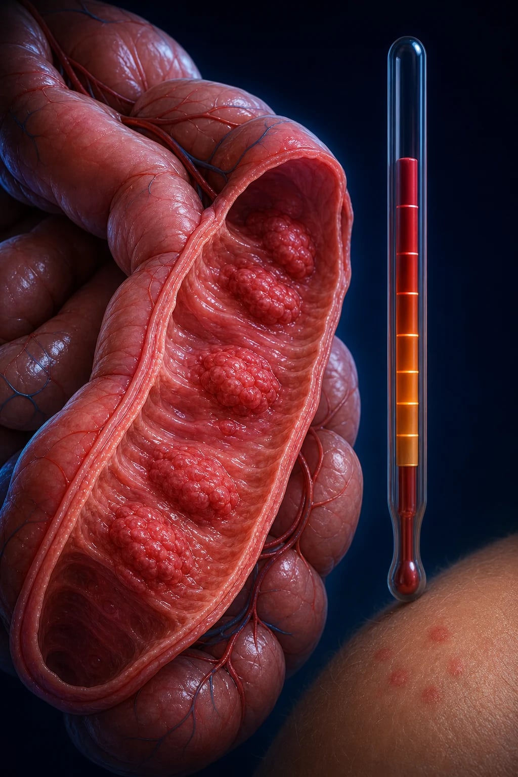
A note on naming: 'typhoid' derives from Greek typhos (smoke, stupor), reflecting the clouded mental state of untreated disease. It is unrelated to typhus (a rickettsial, vector-borne illness) — a classic exam confusion. The umbrella term enteric fever covers both typhoid and paratyphoid. [1]
Classification
Enteric fever is classified along three axes — by serovar (aetiology), by severity and clinical syndrome, and by antimicrobial susceptibility profile, which is now the single most important determinant of therapy. [1]
Uncomplicated typhoid
Illness without organ failure or septic complications
- Fever, headache, abdominal symptoms; no shock, no encephalopathy
- Tolerates oral intake; managed as outpatient or ward
- Responds to oral antibiotics (7 to 14 days)
- Mortality under 1% with appropriate therapy
Severe typhoid
Toxic illness with altered mentation or organ dysfunction
- Delirium, obtundation, coma ('typhoid state') — toxic encephalopathy
- Shock, myocarditis (tachycardia, hypotension, gallop)
- Severe hepatitis with jaundice; DIC
- **Add dexamethasone (Hoffman regimen)** — cuts mortality from ~40% to ~10%
- Mortality 10-30% even with treatment
Typhoid with perforation
Week 3-4 surgical emergency
- Sudden severe abdominal pain, rigid (board-like) abdomen
- Free gas under diaphragm on erect CXR
- Resuscitate + broad-spectrum antibiotics + urgent laparotomy
- Mortality 10-30% despite surgery
Typhoid with haemorrhage
Week 3, from sloughed Peyer's patch ulcers
- Melaena, haematemesis or fresh blood per rectum
- Drop in haemoglobin; may be massive and life-threatening
- Most settle with transfusion, fluids, NBM, PPI
- Surgery reserved for uncontrolled bleeding
Chronic carrier
Persistent gallbladder carriage over 12 months
- Stool/urine culture positive beyond 1 year
- Classically women over 40 with gallstones
- Gallbladder biofilm is the reservoir
- Treat: prolonged ciprofloxacin +/- cholecystectomy; exclude from food-handling
By antimicrobial susceptibility (the axis that drives empirical choice):[6]
- Fully susceptible — sensitive to first-line agents (ampicillin, chloramphenicol, co-trimoxazole) and fluoroquinolones. Increasingly rare.
- Multidrug-resistant (MDR) — resistant to ampicillin, chloramphenicol AND co-trimoxazole (classical plasmid-mediated resistance, common in South Asia since the 1990s).
- Fluoroquinolone-resistant (Nalidixic-acid resistant, NAR) — the dominant phenotype in South Asia today; quinolones fail clinically. Use a third-generation cephalosporin.
- Extensively drug-resistant (XDR) — resistant to first-line drugs, fluoroquinolones AND third-generation cephalosporins, susceptible essentially only to azithromycin and carbapenems. First described in Hyderabad, Pakistan, 2016, and now exported globally via returning travellers.[6]
Epidemiology & Risk Factors
Typhoid is a disease of poverty — it thrives where clean water, sanitation and food hygiene are inadequate. The Global Burden of Disease 2017 study estimated approximately 11 to 21 million cases and 110,000 to 160,000 typhoid deaths worldwide each year, with the heaviest burden in South Asia (India, Pakistan, Bangladesh), sub-Saharan Africa and parts of Southeast Asia.[3]
Host and environmental risk factors:[1]
- Travel to / residence in an endemic region — the single biggest risk; essential history in any returned traveller with fever. Pakistan-origin XDR is a specific travel-related concern.
- Contaminated food and water — shellfish from sewage-contaminated water, raw produce washed in contaminated water, street food, ice made from unsafe water.
- Reduced gastric acidity — antacids, proton-pump inhibitors (PPIs), histamine-2 blockers, prior gastric surgery, achlorhydria (pernicious anaemia), extremes of age — lower the infective dose.
- Household or close contact with a case or a chronic carrier.
- Immunocompromise — HIV/AIDS (especially with low CD4), immunosuppressive therapy.
- Functional asplenia or haemoglobinopathy — sickle-cell disease (which also predisposes to Salmonella osteomyelitis), congenital asplenia, postsplenectomy.
- Gallstones — the key risk factor for the chronic carrier state (the gallbladder biofilm on stones provides a protected niche).
- Age — in endemic areas the burden is highest in school-age children (5 to 15 years); in younger children the disease is often atypical and bacteraemic.
- Endemic institutional settings — refugee camps, overcrowded housing, schools and dormitories with shared sanitation. [1]
The chronic carrier and 'Typhoid Mary': approximately 2 to 5% of survivors develop persistent gallbladder colonisation, shedding S. Typhi in stool for years. The historical archetype is Mary Mallon ('Typhoid Mary'), an asymptomatic Irish-American cook who, in the early 1900s, transmitted typhoid to over 50 people across several New York households and was eventually forcibly quarantined for life on North Brother Island. The carrier state remains a public-health and food-handler problem today.[1]
Pathophysiology
Typhoid is fundamentally a disease of intracellular survival — S. Typhi lives, replicates and disseminates inside macrophages, evading the humoral immune response that produces the antibodies the Widal test measures. Understanding this cascade explains every clinical and laboratory feature. [1]
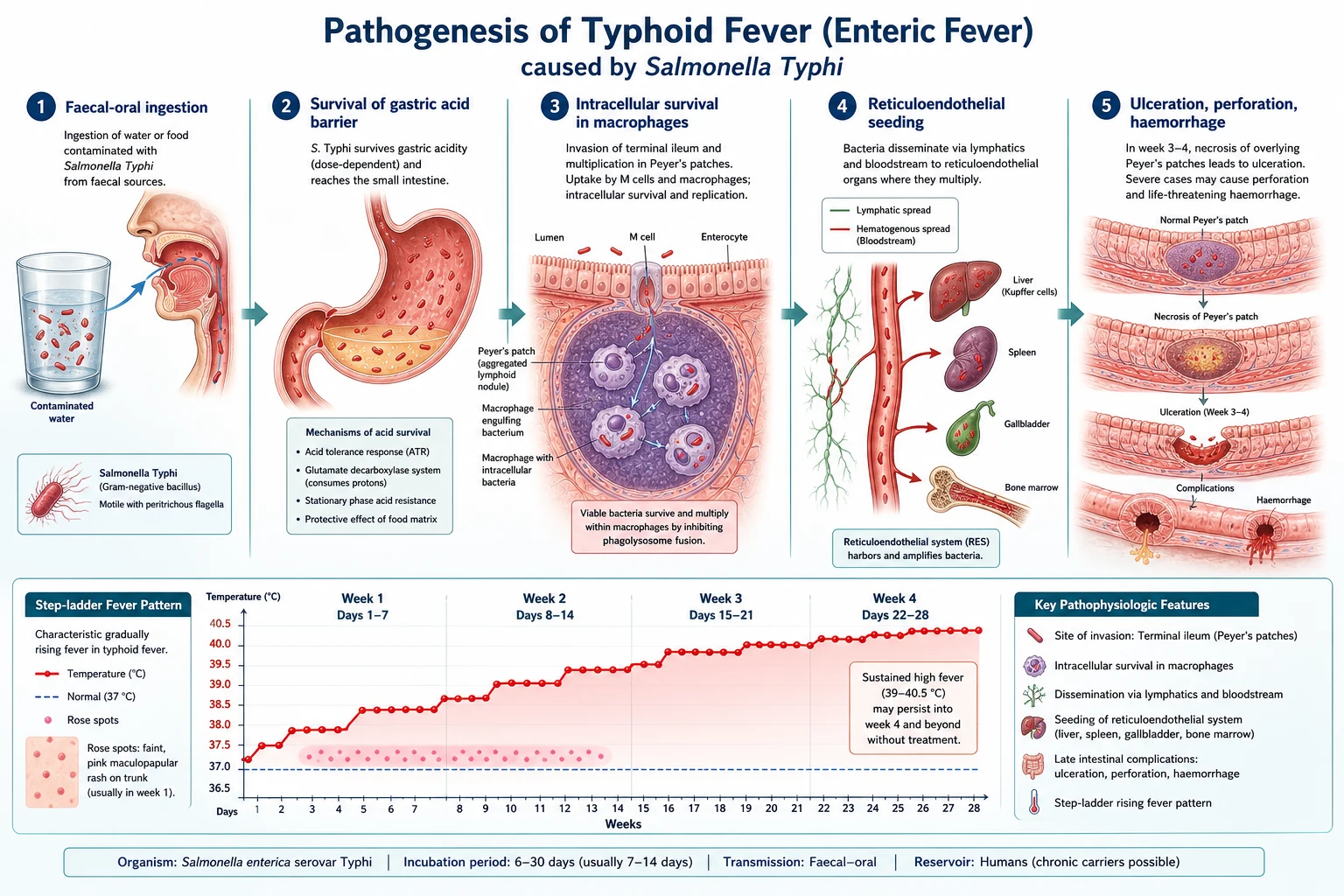
The cascade in molecular detail:[8]
-
Ingestion and gastric survival. Organisms are swallowed in contaminated food or water. The gastric acid barrier is the first line of defence, but S. Typhi survives via an acid-tolerance response (ATR) that induces acid-shock proteins, allowing passage into the small intestine. Reduced gastric acidity (PPIs, antacids, achlorhydria, post-vagotomy/post-gastrectomy, infancy) dramatically lowers the infective dose. [1]
-
Invasion of Peyer's patches. In the terminal ileum, bacteria attach to and are taken across the epithelium by specialised microfold (M) cells overlying the Peyer's patches (aggregated lymphoid follicles). They are then engulfed by macrophages and dendritic cells in the lamina propria. The SPI-1 (Salmonella pathogenicity island 1) type III secretion system injects effector proteins (SipA, SipC, SopE) that induce macropinocytosis and bacterial uptake. [1]
-
Intracellular survival and replication. Inside macrophages, S. Typhi resides in a modified phagosome — the Salmonella-containing vacuole (SCV) — that resists fusion with lysosomes and avoids killing. The SPI-2 type III secretion system injects effectors (SpiC, SifA) that divert normal endosomal trafficking. The Vi capsular polysaccharide masks the somatic O antigen from complement and antibody, and interferes with oxidative killing and autophagy. The bacteria replicate within this protected niche — this is why the disease is prolonged, why humoral antibodies are diagnostic but not protective, and why bone marrow culture remains positive even after antibiotics (organisms are sequestered intracellularly).[8]
-
First (silent) bacteraemia and reticuloendothelial seeding. Bacteria drain via mesenteric lymph nodes into the thoracic duct and then the portal and systemic circulation (first bacteraemia). They are cleared by the reticuloendothelial system (RES) — liver (Kupffer cells), spleen, bone marrow, and the gallbladder — where they continue to multiply intracellularly. The patient is asymptomatic during this incubation phase. [1]
-
Second (symptomatic) bacteraemia and cytokine release. After sufficient intracellular multiplication (typically around day 7 to 14), organisms are released back into the bloodstream in large numbers (second bacteraemia), coinciding with the onset of symptoms. Infected macrophages release pro-inflammatory cytokines — IL-1, IL-6, TNF-α and IFN-γ — which produce the stepwise-rising fever, headache, myalgia and systemic upset. The hallmark relative bradycardia is thought to reflect direct effects of circulating endotoxin and cytokines on the cardiac conduction system (a vagally-mediated response). The same cytokine cascade, when overwhelming, drives severe typhoid with toxic encephalopathy, myocarditis and shock — the target of dexamethasone therapy.[4]
-
Gallbladder re-infection and Peyer's patch necrosis. During the second bacteraemia the gallbladder is re-infected (either haematogenously or via bile). S. Typhi multiplies in the gallbladder and is re-excreted into the bile, re-invading the Peyer's patches — now hyperplastic then necrotic. In weeks 3 to 4 these necrotic patches slough and ulcerate, producing the two surgical emergencies of typhoid: intestinal perforation (through the ulcerated, necrotic lymphoid tissue of the terminal ileum) and intestinal haemorrhage (from erosion of a vessel in an ulcer base). The ulceration is characteristically longitudinal (parallel to the long axis of the bowel) — distinguishing typhoid ulcers from tuberculous (transverse) ulcers.[1]
-
Immune response and recovery. Recovery is governed principally by cell-mediated immunity — a Th1 response with macrophage activation and interferon-gamma — that eventually clears intracellular organisms. The humoral response (anti-O, anti-H and anti-Vi antibodies) is the basis of the Widal test but is not protective on its own, which is why antibody titres do not correlate well with protection and why the Vi polysaccharide vaccine is only modestly efficacious compared with the conjugate.[5]
Clinical Presentation
Typhoid unfolds in characteristic weekly phases. The tempo is indolent — a slow climb over the first week, in marked contrast to the abrupt rigors of malaria, bacteraemic pneumonia or meningococcaemia. [1]
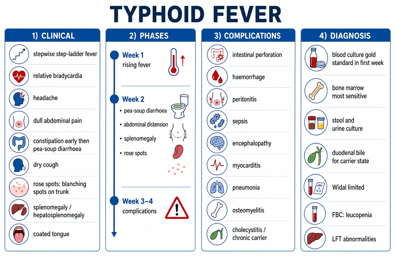
Week 1 — the step-ladder fever. The illness begins insidiously with malaise, headache (often frontal and severe), anorexia, and a fever that rises in a stepwise pattern — climbing each afternoon/evening, falling partially by morning, then climbing higher the next day, until it plateaus at around 39 to 40°C by the end of week 1. Constipation (not diarrhoea) is typical early; a dry cough is common and may mislead toward a respiratory diagnosis; epistaxis occurs in some. The relative bradycardia is now evident: the pulse is slower than the fever would predict (pulse–temperature dissociation, the Faget sign).[1]
Week 2 — the classical picture. The fever is now sustained and high. The abdomen becomes distended and tender, and the constipation gives way to 'pea-soup' diarrhoea — loose, greenish, foul-smelling stools. Rose spots appear: 2 to 4 mm blanching salmon-pink macules, found in crops of fewer than 20 on the upper abdomen, lower chest and back. They are bacterial (septic) emboli to dermal capillaries, not petechiae — they blanch with pressure and fade in 3 to 4 days as new crops appear. The tongue is coated white with red edges and prominent papillae ('typhoid tongue'). Splenomegaly (and often hepatosplenomegaly) is now palpable. A faint maculopapular rash may be seen on the trunk. [1]
Weeks 3 to 4 — toxicity and complications. If untreated, the patient becomes increasingly toxic: confused, delirious, picking at the bedclothes ('carphology' or 'flos salvationum'), and may lapse into the muttering, stuporose 'typhoid state'. The abdomen is more distended (ileus). This is the danger window for intestinal perforation (sudden severe abdominal pain, rigid abdomen, absent bowel sounds, free gas) and intestinal haemorrhage (melaena, haematemesis, drop in haemoglobin). Myocarditis, hepatitis with jaundice, acute cholecystitis, pneumonia, meningitis (rare), and osteomyelitis (especially in sickle-cell disease) may appear.[1][2]
Step-ladder fever
The single most characteristic sign
- Fever rises daily in steps, with morning remissions
- Plateaus at 39-40°C by end of week 1
- Persists for 2-3 weeks if untreated
- Contrast: malaria (abrupt, periodic); dengue (saddle-back)
Relative bradycardia (Faget sign)
Pulse-temperature dissociation
- Pulse slower than the fever would predict
- e.g. pulse 70-80 at 39.5°C
- Also seen in leptospirosis, brucellosis, Q fever, legionella, drug fever
- Typhoid is the classic exam answer
Rose spots
Bacterial emboli to skin
- 2-4 mm blanching salmon-pink macules
- Trunk (upper abdomen, lower chest, back)
- Crops of fewer than 20; fade in 3-4 days
- Biopsy yields S. Typhi — septic emboli, not petechiae
Coated 'typhoid tongue'
White coating, red edges
- Thick white-grey coating with red margins
- Prominent papillae
- Halitosis
- Resolves with defervescence
Atypical presentations (deliberately tested)
- Elderly — fever may be low-grade or absent; confusion predominates and may be mistaken for dementia or delirium; multiple organ involvement and higher mortality; a lower threshold to culture, admit and treat.
- Children — higher fever, more diarrhoea and vomiting, febrile seizures, faster progression to perforation; jaundice and hepatitis are more prominent; the presentation may mimic acute gastroenteritis or septicaemia.
- Immunocompromised (HIV, transplant) — severe, prolonged or relapsing disease, higher bacteraemia, atypical organ involvement, and a higher index of suspicion for concomitant opportunistic infection.
- Pregnancy — higher maternal and fetal risk (miscarriage, preterm labour); treat aggressively with pregnancy-safe antibiotics.
- Returned traveller — typhoid must be in the differential of any fever in a traveller returning from South Asia, Africa or SE Asia, even months after return (the incubation can be long). Always exclude malaria first (thick/thin films, antigen test) — the two are co-endemic and may coexist. [1]
Differential Diagnosis
Typhoid is a febrile systemic illness in a returned traveller or endemic resident, and it sits at the centre of the "fever in a traveller" differential. The can't-miss mimics are malaria, tuberculosis, brucellosis, leptospirosis, dengue and other enteric and rickettsial fevers. [1]
Falciparum malaria
- Abrupt high fever with rigors, periodicity
- Splenomegaly, anaemia, thrombocytopenia, jaundice
- Travel to malaria zone; thick/thin film or malaria antigen
- Co-endemic with typhoid — ALWAYS exclude malaria first; the two may coexist
Tuberculosis (miliary / abdominal)
- Chronic fever, weight loss, night sweats, cough
- Abdominal mass, ascites (peritoneal TB); hepatosplenomegaly (miliary)
- CXR (miliary 'snowstorm'), Mantoux/IGRA, AFB / GeneXpert
- Slower tempo than typhoid; no rose spots
Brucellosis
- Undulant fever, arthralgia, hepatosplenomegaly, sweats
- Occupational (abattoir workers, vets) or unpasteurised dairy
- Blood culture (prolonged incubation), Brucella serology (SAT)
- Causes sacroiliitis, spondylitis, endocarditis
Leptospirosis
- Febrile with conjunctival suffusion, severe myalgia (calves)
- Jaundice, renal failure, thrombocytopenia (Weil disease)
- Exposure: contaminated water, farm animals, rodents
- IgM ELISA; doxycycline; penicillin in severe disease
Dengue
- High fever, retro-orbital headache, bone pain, rash
- Thrombocytopenia, leucopenia, positive tourniquet test
- Saddle-back fever pattern; NS1 antigen (early), IgM (late)
- Bleeding risk — AVOID NSAIDs/aspirin
Scrub typhus / rickettsial fever
- Eschar at inoculation site, regional lymphadenopathy, rash
- Rural/agricultural exposure (mite bite)
- Doxycycline response (often dramatic)
- Weil-Felix or IgM IFA
Infectious mononucleosis
- Fever, exudative pharyngitis, posterior cervical lymphadenopathy
- Splenomegaly, atypical lymphocytes on film
- Monospot positive; EBV IgM
- Caution: ampicillin causes a maculopapular rash
Melioidosis
- Burkholderia pseudomallei; SE Asia / northern Australia
- Pneumonia with septicaemia, abscesses (multiple organs)
- Soil/water exposure (esp. after rains); diabetes risk
- Ceftriaxone/meropenem then eradication phase
The exam trap: typhus ≠ typhoid. Typhus is rickettsial (louse/flea/mite-borne), has an eschar and rash, and responds to doxycycline. Typhoid is Salmonella Typhi, faecal-oral, rose spots, treated with cephalosporins/azithromycin. The names are similar; the diseases are entirely different. [1]
Clinical & Bedside Assessment
The focused assessment rests on three pillars: the fever pattern, the pulse–temperature relationship, and a careful abdominal and skin examination. None of these requires a laboratory, and the classical picture can be recognised at the bedside. [1]
History. Establish the tempo (indolent stepwise rise over days), the travel/residence history (South Asia, Pakistan, Africa, SE Asia), food and water exposure (street food, untreated water, shellfish, ice), sick contacts or a known carrier in the household, vaccination status, and any antibiotic pre-treatment (which blunts cultures). Note the symptom evolution — constipation early, 'pea-soup' diarrhoea later, dry cough, frontal headache. [1]
Fever pattern. Plot the temperature over several days. The step-ladder (stepwise-rising) pattern — daily climbs with morning remissions, plateauing around day 7 — is the classical signature. It is distinct from the abrupt fever of malaria or the saddle-back of dengue. [1]
Relative bradycardia (Faget sign). At the bedside, the pulse is slower than the fever would predict. As a rule of thumb, temperature in °C above 37°C should raise the pulse by roughly 8 to 10 beats/min per degree; a pulse of 75 at 39.5°C is relative bradycardia. It is also seen in leptospirosis, brucellosis, Q fever, legionellosis and drug fever, but typhoid is the classic exam answer. [1]
Abdomen. Look for soft distension, diffuse tenderness (especially in the right iliac fossa over the terminal ileum and caecum where the Peyer's patches are), a palpable spleen (and sometimes liver), and the coated 'typhoid tongue'. In week 3 to 4, examine specifically for peritoneal signs — rebound tenderness, guarding, rigidity (board-like abdomen), and absent bowel sounds — which signal intestinal perforation. [1]
Skin. Examine the upper abdomen, lower chest and back for rose spots: faint 2 to 4 mm blanching salmon-pink macules in crops of fewer than 20. They blanch with a glass slide (diascopy). Do not mistake them for the petechiae of meningococcaemia or the rash of dengue. [1]
Other systems. Listen to the chest for the dry cough / bronchitis. Examine for jaundice (hepatitis), neck stiffness (meningism, rare), signs of shock (cool peripheries, prolonged capillary refill, hypotension — severe typhoid), and arrhythmias or a gallop rhythm (myocarditis). Perform a rectal examination for melaena (intestinal haemorrhage). [1]
Monitoring. In the admitted patient, monitor hourly urine output, pulse, BP, temperature, respiratory rate and conscious level (GCS), and serial abdominal examinations — the deterioration into perforation or haemorrhage can be rapid. [1]
Investigations
The microbiological diagnosis is culture-based; serology (Widal) and rapid tests are adjuncts. The choice of sample depends on the week of illness and whether the patient has been pre-treated. [1]
Cultures — the diagnostic mainstay
Blood culture
Gold standard (first week)
- Sensitivity 60-90% in first week; falls thereafter
- Take ~10 mL (adults) in aerobic AND anaerobic bottles BEFORE antibiotics
- Confirm diagnosis AND allows susceptibility testing (critical for XDR)
- Repeat if initial negative and suspicion high
Bone marrow culture
Most sensitive (~90%)
- Highest yield of any sample, including after antibiotics started
- Organisms sequestered intracellularly in RES macrophages
- Preferred when patient is pre-treated or blood cultures negative
- Pair with bone marrow for histology if TB/dengue also in differential
Stool culture
Week 2-3 and carrier detection
- Positive in second to third week and in chronic carriers
- Use selenite-F enrichment broth then MacConkey / DCA (non-lactose fermenter)
- Negative stool does not exclude acute typhoid
- Three consecutive negative stools needed to clear a food-handler
Urine culture
Week 2-3 (less often)
- Positive in second to third week, less sensitive than stool
- Useful in chronic carrier screening
- Pyuria may occur (organism in urinary tract)
Duodenal bile / string test
Chronic carrier work-up
- Bile cultured via duodenal capsule ('string test') or at ERCP
- Highest yield for gallbladder carriage
- Used when stool cultures are negative but carrier suspected
Serology and rapid tests
Widal test. A tube agglutination test detecting anti-O (somatic) and anti-H (flagellar) antibodies. Anti-O (IgM) appears in the acute illness; anti-H rises later and persists. A single titre is unreliable in endemic regions, where background antibodies (from past infection, vaccination, and cross-reacting enterobacteria such as Citrobacter which shares the O-12 antigen) are common. Cut-offs are population-specific — commonly anti-O 1:80 and anti-H 1:160 in India, but higher in some regions. The only supportive pattern is a four-fold rise in titre between acute and convalescent sera taken 10 to 14 days apart. Over-reliance on a single Widal is a classic pitfall and drives inappropriate antibiotic use.[1][2]
Rapid diagnostic tests (Typhidot, Tubex). Detect IgM (Typhidot) or anti-O IgM via inhibition (Tubex). Their sensitivity and specificity are variable across populations; they should never be used as the sole basis for diagnosis or treatment. Newer real-time PCR assays targeting S. Typhi DNA (e.g. tviB, staG) offer high specificity but variable sensitivity, and are not yet routine in most settings. [1]
Supportive bloods and imaging
- Full blood count — leucopenia (total WCC under 4 ×10⁹/L) with a relative lymphocytosis and eosinopenia (often zero eosinophils) is classical but not invariable; a normal or low-normal WCC is common. A rising leucocytosis with neutrophilia suggests a complication (perforation, secondary infection, intestinal necrosis). Mild thrombocytopenia and anaemia of chronic disease are common.
- Liver function tests — mild transaminitis (AST often greater than ALT) and a raised alkaline phosphatase; overt jaundice and cholestasis indicate typhoid hepatitis (a poor prognostic marker).
- Inflammatory markers — CRP and ESR are raised.
- Electrolytes and renal function — hyponatraemia (from SIADH or gut losses), hypokalaemia (from diarrhoea), and acute kidney injury in severe disease or shock.
- Coagulation — mild prolongation of PT/INR; overt DIC in severe disease.
- Chest X-ray — may show bronchitis, patchy pneumonitis ('typhoid pneumonia'), or, critically, free gas under the right hemidiaphragm on an erect film if perforation has occurred.
- Ultrasound / CT abdomen — hepatosplenomegaly, thickened ileal wall, mesenteric lymphadenopathy, and (in perforation) free intraperitoneal gas and fluid. CT is more sensitive than plain films for subtle perforation and excludes surgical mimics. [1]
Management — Resuscitation
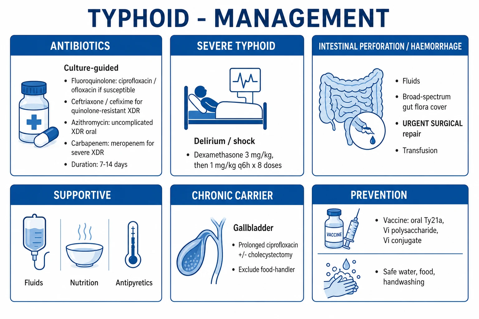
Most patients with typhoid are haemodynamically stable and can be managed with oral antibiotics as outpatients. Resuscitation is reserved for the severe, dehydrated, septic or complicated patient. [1]
ABCDE. Secure the airway, give oxygen to maintain SpO₂ at 94 to 98%, establish IV access, and treat dehydration or shock with balanced crystalloid (Hartmann's or Ringer's lactate). Caution in suspected myocarditis — avoid fluid overload; reassess responsiveness (passive leg raise, IVC, pulse-pressure variation) before further boluses. [1]
Antipyretics and comfort. Paracetamol (1 g PO/PR every 6 hours in adults; 10 to 15 mg/kg per dose in children) is the antipyretic of choice. Avoid NSAIDs if thrombocytopenia or bleeding risk is present. Tepid sponging helps the fever. [1]
Cultures before antibiotics. Take blood cultures (and bone marrow culture if available, especially if pre-treated) before the first antibiotic dose — but never delay antibiotics beyond one hour in septic or severe disease. [1]
Empirical IV antibiotics within one hour in severe/septic typhoid. Do not wait for susceptibilities.[1]
Correct electrolytes (hyponatraemia, hypokalaemia) and treat any coexisting malaria (always re-examine films if the patient deteriorates). [1]
Dexamethasone for severe typhoid. If the patient has severe typhoid — defined as delirium, obtundation, coma, shock, or other features of toxic encephalopathy — give the Hoffman regimen: dexamethasone 3 mg/kg IV over 30 minutes loading, then 1 mg/kg IV every 6 hours for 8 doses (24 hours total). This reduces mortality from approximately 40 to 50% down to around 10% by damping the macrophage cytokine cascade.[4]
Surgical referral. Any patient with peritoneal signs, a rigid abdomen, free gas, or uncontrolled bleeding needs immediate surgical assessment — see Complications. [1]
Management — Definitive & Stepwise
Antibiotic choice is culture- and susceptibility-guided, and empirical therapy must account for the local resistance pattern and the patient's travel history (especially Pakistan/XDR). Resistance has progressively eroded the value of chloramphenicol, ampicillin, co-trimoxazole and fluoroquinolones, so cephalosporins and azithromycin now dominate empirical therapy in endemic regions. [1]
Antibiotic ladder by susceptibility
Fully susceptible (rare)
- Oral ciprofloxacin 500-750 mg BD for 7-10 days
- OR ofloxacin 400 mg BD
- Historical: chloramphenicol 500 mg QID, amoxicillin, co-trimoxazole (now rarely appropriate empirically)
- Defervescence in 3-5 days
Quinolone-resistant (common in South Asia)
- IV ceftriaxone 2-3 g OD for 7-14 days
- OR oral cefixime 200 mg BD for 7-14 days
- OR azithromycin 500-1000 mg OD
- Review susceptibilities at 48-72 hours
XDR (Pakistan-origin clone)
Resistant to first-line + quinolones + 3rd-gen cephalosporins
- Uncomplicated: oral azithromycin 500-1000 mg OD for 7 days
- Severe: IV meropenem 1 g every 8 hours
- Step down to azithromycin once improving
- Always confirm with culture and susceptibilities
Severe typhoid (any susceptibility)
- IV ceftriaxone 2-3 g OD (or meropenem if XDR)
- PLUS dexamethasone Hoffman regimen
- Duration 14 days; longer for metastatic infection
- ICU monitoring for organ support
In South Asia, fluoroquinolone resistance is now so prevalent that empirical ciprofloxacin is inappropriate; ICMR/NCDC India recommends empirical IV ceftriaxone or oral cefixime, with azithromycin for XDR. Pakistan has deployed mass vaccination campaigns with Typbar-TCV to control the XDR outbreak.[1]
Duration, step-down and discharge
Duration: 7 to 14 days for uncomplicated disease; 14 days or longer for severe typhoid, metastatic infection (osteomyelitis, endocarditis), or immunocompromised hosts. Shorter courses (5 to 7 days of azithromycin or fluoroquinolone) are acceptable in fully susceptible, uncomplicated disease where follow-up is assured.[1]
IV-to-oral switch (step-down): once the patient is haemodynamically stable, afebrile for 24 to 48 hours, improving clinically, and able to swallow and absorb oral medication, switch to an oral agent of the same class. Discharge when clinically stable, afebrile, tolerating oral intake and antibiotics, and with a safe social situation — with a safety-net to re-present if fever, abdominal pain or bleeding recur within 48 hours.[1]
Specific complications and their management
Intestinal perforation (week 3 to 4, approximately 1 to 3% of cases) is the most feared surgical complication. Management: aggressive IV fluid resuscitation, nasogastric decompression, broad-spectrum antibiotics covering gut flora including anaerobes and Gram-negatives (e.g. ceftriaxone + metronidazole, or piperacillin-tazobactam, or a carbapenem), and urgent laparotomy with primary closure of the perforation(s) (usually in the terminal ileum) or resection with anastomosis (or stoma if the patient is grossly contaminated or unstable), plus thorough peritoneal lavage. Mortality is 10 to 30% despite surgery, and higher with delayed presentation.[1][2]
Intestinal haemorrhage (approximately 2 to 10%, week 3) from an eroded vessel in a sloughed Peyer's patch ulcer. Most episodes are self-limited. Management: IV fluids, blood transfusion for significant loss, correction of coagulopathy, nil by mouth, and a proton-pump inhibitor. Surgery is reserved for massive or uncontrolled bleeding. Re-bleeding may occur.[1]
Chronic carrier eradication. A chronic carrier (stool/urine culture positive beyond 12 months) is treated with prolonged ciprofloxacin 750 mg BD for 4 to 6 weeks (where susceptible) or high-dose amoxicillin for 6 weeks, combined with cholecystectomy if gallstones are present (the gallbladder biofilm on stones is the protected reservoir). Eradication must be confirmed by three consecutive negative stool cultures taken at least one month apart after antibiotics are completed. Food-handlers and healthcare workers are excluded from work until clearance.[1]
Relapse (approximately 5 to 10%) — recurrence of typical symptoms 1 to 3 weeks after apparent recovery, usually milder than the initial illness. Treat with the same antibiotic to which the original isolate was susceptible.[1]
Public health. Typhoid is notifiable in most jurisdictions. Trace household and close contacts, offer vaccination to contacts travelling to endemic areas, and exclude food-handlers, healthcare workers and handlers of children/unwell adults until three consecutive negative stool cultures (taken at least 48 hours apart, after any antibiotics).[5]
Specific Subtypes & Scenarios
-
Paratyphoid fever (S. Paratyphi A) — clinically indistinguishable from typhoid but often milder and shorter, with fewer complications (perforation is less common). Diagnosed only by culture. Managed identically. Increasingly recognised as a cause of travel-associated enteric fever, partly because the Vi vaccines protect against Typhi but not Paratyphi A.[1]
-
Severe typhoid — the syndrome of toxic encephalopathy (delirium, obtundation, coma), shock, myocarditis, severe hepatitis, or DIC. Untreated mortality is 30 to 50%; dexamethasone (Hoffman regimen) is the single intervention that changes this.[4]
-
XDR typhoid — the Pakistan-origin clone (first described Hyderabad, 2016), now spread across South Asia and exported globally via travellers. Resistant to first-line agents, fluoroquinolones and third-generation cephalosporins; treat uncomplicated XDR with oral azithromycin and severe XDR with IV meropenem. Always culture and confirm susceptibilities.[6]
-
Typhoid in pregnancy — higher maternal and fetal morbidity (miscarriage, preterm labour). Treat aggressively with pregnancy-safe antibiotics (ceftriaxone, azithromycin); avoid fluoroquinolones (theoretical cartilage risk) and co-trimoxazole in the first trimester (folate antagonist, teratogenic risk) and near term (kernicterus risk).[1]
-
Paediatric typhoid — shorter incubation, more GI symptoms, higher fever, febrile seizures, faster progression to perforation, and more prominent hepatitis/jaundice. Cefixime (10 to 15 mg/kg/day in two divided doses) or azithromycin (10 to 20 mg/kg/day) are preferred; ceftriaxone (75 to 100 mg/kg/day) for severe disease. Typhoid Conjugate Vaccine (TCV) is now WHO-recommended from 6 months of age in endemic countries.[5]
-
Chronic carrier — gallbladder colonisation, typically on gallstones, in approximately 2 to 5% of survivors (classically women over 40). The reservoir for ongoing transmission. Treat with prolonged antibiotics and cholecystectomy.[1]
Complications & Pitfalls
Intestinal: perforation (week 3 to 4, surgical emergency, mortality 10 to 30%); haemorrhage (week 3, usually self-limited but may be massive); paralytic ileus; acute cholecystitis (and chronic cholecystitis driving the carrier state). [1]
Systemic: severe typhoid with toxic encephalopathy (delirium, coma); myocarditis (arrhythmias, heart failure, cardiogenic shock — beware fluid overload); hepatitis and jaundice (typhoid hepatitis); pneumonia and secondary bacterial infection; meningitis (rare, more in children); cerebellar ataxia, Guillain-Barré syndrome, encephalomyelitis (neurological, post-infectious); osteomyelitis and septic arthritis (especially in sickle-cell disease); acute kidney injury, glomerulonephritis, haemolytic-uraemic syndrome; DIC and thrombocytopenia; aplastic anaemia (rare); relapse (5 to 10%); and the chronic carrier state.[1][2]
Classic pitfalls: [1]
- Over-reliance on a single Widal titre — it neither confirms nor excludes typhoid in an endemic region, and drives inappropriate antibiotic use. Always culture.
- Empirical fluoroquinolones in South Asia — resistance is so high that ciprofloxacin will fail; use ceftriaxone or cefixime empirically.
- Missing intestinal perforation in a patient whose fever is 'settling' — sudden abdominal pain, rigidity or free gas in week 3 to 4 is perforation until proven otherwise.
- Under-dosing or short-coursing antibiotics — drives relapse and resistance; complete the full course.
- Failing to treat XDR appropriately — a returning traveller with Pakistan-acquired typhoid needs azithromycin or meropenem, not ciprofloxacin or ceftriaxone.
- Not adding dexamethasone in severe typhoid with encephalopathy — a preventable mortality.
- Forgetting the chronic carrier — a food-handler with persistent stool positivity needs eradication, not reassurance.
- Confusing typhoid with typhus — the names are similar; the diseases (and their treatments) are different. [1]
Prognosis & Disposition
Treated uncomplicated typhoid: case fatality under 1%; defervescence in 3 to 5 days with an appropriate antibiotic. Full recovery is usual.[1]
Untreated typhoid: illness lasts 3 to 4 weeks; case fatality 10 to 20%, rising to 30% or more in severe disease with encephalopathy or perforation. The untreated natural history is the basis for the classical weekly phases. [1]
Severe typhoid with encephalopathy: mortality 30 to 50% without steroids, reduced to approximately 10% with the Hoffman dexamethasone regimen.[4]
Intestinal perforation: mortality 10 to 30% despite surgery; higher with delayed presentation, multiple perforations, or gross peritoneal contamination.[1]
Relapse: approximately 5 to 10%; the second episode is usually milder and responds to the original antibiotic. [1]
Chronic carriage: in approximately 2 to 5% of survivors, especially women over 40 with gallstones; lasts years without eradication and is the reservoir of ongoing transmission. [1]
Disposition: [1]
- Outpatient management is appropriate for uncomplicated typhoid in a patient who tolerates oral intake and antibiotics, is haemodynamically stable, not immunocompromised, pregnant or a young child, has no peritoneal or bleeding signs, and has reliable follow-up and a safe social situation.
- Admit (ward) if any of: inability to tolerate oral therapy; persistent vomiting or dehydration; high fever with toxicity; pregnancy; immunocompromise; age under 2; suspected complication; or inadequate social support.
- ICU for severe typhoid with encephalopathy, shock, myocarditis, DIC, or intestinal perforation requiring surgery. [1]
Special Populations
-
Children — shorter course, more GI symptoms, higher fever, febrile seizures, faster progression to perforation. Cefixime (10 to 15 mg/kg/day PO in two divided doses) or azithromycin (10 to 20 mg/kg/day PO) for uncomplicated disease; ceftriaxone (75 to 100 mg/kg/day IV) for severe disease. Typhoid Conjugate Vaccine (TCV) from 6 months of age is now the WHO-recommended prevention.[5]
-
Pregnancy — higher maternal and fetal morbidity (miscarriage, preterm labour). Treat aggressively with ceftriaxone or azithromycin. Avoid fluoroquinolones and co-trimoxazole (first trimester and near term).[1]
-
Elderly — atypical presentation (confusion predominates, fever may be low-grade or absent), comorbidity, higher mortality, and a lower threshold to admit and treat with IV therapy. Look for complications (perforation, myocarditis, hepatitis) at presentation. [1]
-
Immunocompromised (HIV, transplant, chemotherapy) — severe, prolonged or relapsing disease, higher bacteraemia, and a higher index of suspicion for concomitant opportunistic infection. Longer therapy (14 to 21 days); consider secondary prophylaxis in advanced HIV with relapse. [1]
-
Sickle-cell disease / functional asplenia / haemoglobinopathy — high risk of invasive Salmonella including osteomyelitis (the classic 'salmonella osteomyelitis' of sickle-cell disease, though S. Typhi is a less common cause than non-typhoidal Salmonella). Aggressive IV therapy; seek metastatic foci.[1]
-
Gallstones — the key risk factor for the chronic carrier state. Consider cholecystectomy for eradication. Screen food-handlers. [1]
-
Food-handlers, healthcare workers, handlers of infants or immunocompromised persons — exclude from work until three consecutive negative stool cultures (taken at least 48 hours apart, after antibiotics). The 'Typhoid Mary' principle.[1]
Evidence, Guidelines & Regional Differences
Landmark evidence
-
GBD 2017 Typhoid and Paratyphoid Collaborators (Lancet Infect Dis 2019) quantified the global burden at approximately 11 to 21 million cases and 110,000 to 160,000 deaths annually, with the heaviest burden in South Asia and sub-Saharan Africa, and highlighted the school-age child as the peak incidence and transmission group.[3]
-
Hoffman et al. (NEJM 1984) — the definitive trial of high-dose dexamethasone (3 mg/kg loading, then 1 mg/kg every 6 hours for 8 doses) in severe typhoid, reducing mortality from 43% to 10% in chloramphenicol-treated patients with delirium, obtundation or coma. This regimen remains the global standard for severe typhoid.[4]
-
Carey et al. (eLife 2023) — genomic analysis of over 13,000 S. Typhi isolates documenting the global spread of the H58 (haplotype 58) clone and the Pakistan-origin XDR strain carrying an IncY plasmid conferring resistance to first-line agents, fluoroquinolones and third-generation cephalosporins — susceptible only to azithromycin and carbapenems.[6]
-
Bentsi-Enchill et al. (Clin Infect Dis 2019) and WHO 2018 Position Paper — revised the global typhoid vaccination policy, recommending the Vi conjugate vaccine (Typbar-TCV) for routine immunisation from 6 months of age in endemic countries, with Gavi-supported introduction campaigns.[5]
-
Kumar Rai et al. (Lancet Infect Dis 2022) — phase 3 trial confirming the safety and immunogenicity of the Vi-DT (PedaTyph) conjugate vaccine, expanding the conjugate vaccine options for endemic regions.[7]
WHO 2018 vaccination policy recommends the Vi–tetanus toxoid conjugate vaccine (Typbar-TCV) for routine childhood immunisation (single dose from 6 months) in endemic countries, with a catch-up campaign up to 15 years; Gavi supports introduction. The older Ty21a (live oral, 4 capsules, from 6 years) and Vi capsular polysaccharide (single IM dose, from 2 years, booster every 3 years) remain alternatives for travellers, but TCV is preferred for infants and endemic-region programmes because of its immunogenicity in young infants and longer protection.[5]
Regional deltas in empirical therapy:[1][6]
- India (ICMR/NCDC) — fluoroquinolone resistance is so high that empirical ciprofloxacin is inappropriate; empirical IV ceftriaxone (or oral cefixime) is standard, with azithromycin for XDR.
- Pakistan — the XDR epicentre; empirical ceftriaxone with early switch to azithromycin if XDR suspected or confirmed; mass TCV vaccination campaigns.
- UK / Europe / US (returning travellers) — empirical IV ceftriaxone until susceptibilities return; azithromycin for XDR step-down; isolate and notify public health for contact tracing.
- Africa — variable resistance; empirical ceftriaxone or cefixime, with culture guidance.[2]
Controversies
- Widal test use — its poor accuracy in endemic regions means it should be discarded as a sole diagnostic tool, yet it remains widely used; rapid IgM tests and PCR are emerging but not yet universally available or validated.[1]
- Azithromycin vs cefixime for XDR — both are effective in uncomplicated XDR; azithromycin achieves higher intracellular concentrations and is increasingly preferred.[6]
- Steroid use outside severe typhoid — the Hoffman regimen is established for severe typhoid (encephalopathy), but its role in moderate disease without encephalopathy is unproven and not recommended.[4]
- Antibiotic-first vs early surgery for perforation — perforation is a surgical emergency; there is no role for antibiotic-only management of an established perforation.
Exam Pearls
STEP-FEVER
BONE-MARROW
- Step-ladder fever + relative bradycardia + rose spots + splenomegaly = typhoid (the clinical tetrad examiners reward).
- Blood culture = gold standard (first week, before antibiotics); bone marrow = most sensitive (even after antibiotics). The Widal is at best supportive (four-fold rise) — never diagnostic alone.
- Typhoid ulcers are longitudinal (parallel to the long axis of the bowel); tuberculous ulcers are transverse. Peyer's patches are in the terminal ileum — hence the right iliac fossa tenderness.
- Rose spots are bacterial (septic) emboli to dermal capillaries — they blanch, they are on the trunk, and there are fewer than 20.
- The 'pea-soup' diarrhoea of week 2 follows the constipation of week 1.
- XDR S. Typhi = Pakistan-origin clone, resistant to first-line + fluoroquinolones + third-gen cephalosporins — treat with azithromycin (oral) or meropenem (IV).
- Severe typhoid = dexamethasone (Hoffman: 3 mg/kg load + 1 mg/kg q6h × 8 doses) — mortality drops from ~40% to ~10%.
- Chronic carrier = gallbladder (gallstones); treat with prolonged ciprofloxacin ± cholecystectomy; exclude food-handlers until 3 negative stool cultures. The archetype is 'Typhoid Mary' Mallon.
- Vaccines: Ty21a (live oral, from 6 years), Vi polysaccharide (IM, from 2 years), Vi conjugate / Typbar-TCV (from 6 months — WHO-preferred). None protects well against Paratyphi A.
- Typhoid ≠ typhus — typhus is rickettsial, vector-borne (louse/flea/mite), has an eschar, and is treated with doxycycline. The names are similar; the diseases are entirely different.
- Relative bradycardia is also seen in leptospirosis, brucellosis, Q fever, legionellosis and drug fever — but typhoid is the classic exam answer.
- Ciprofloxacin resistance in South Asia is so high it should not be used empirically there — use ceftriaxone or cefixime.
- Paratyphoid A is clinically indistinguishable from typhoid but milder; treat the same way. Note the Vi vaccines do not protect against Paratyphi A.
- Infective dose is lowered by achlorhydria (PPIs, antacids, post-gastrectomy, pernicious anaemia) — a classic risk-factor question. [1]
Exam application bank (NEET-PG / INICET)
One-line answer
Enteric (typhoid) fever is a prolonged systemic bacteraemic illness caused by Salmonella enterica serovars Typhi and Paratyphi A, B, C, transmitted faecal-orally through contaminated food and water. It is endemic in South Asia (India, Pakistan, Bangladesh), sub-Saharan Africa and parts of SE Asia, with an incubation of 7 to 14 days. The clinical signature is a step-ladder (stepwise-rising) fever with relative bradycardia (Faget sign), dull headache, constipation then 'pea-soup' diarrhoea, rose spots, splenomegaly and a coated tongue. Untreated it progresses over weeks to intestinal perforation and haemorrhage (Peyer's patch necrosis, week 3 to 4), severe typhoid with encephalopathy, myocarditis and hepatitis. Blood culture is the gold standard (first week); bone marrow culture is the most sensitive, remaining positive even after antibiotics; the Widal test is unreliable alone. Treatmen
Worked stems (answer without another resource)
Stem 1 — Classic presentation. Map symptoms to mechanism; name the first investigation and first treatment step with dose/route if drug therapy is standard. [1]
Stem 2 — Unstable / complicated. List red flags that force immediate resuscitation, theatre, ICU, antidote, or reperfusion — and what you do in the first 15 minutes. [1]
Stem 3 — Atypical group. Elderly, pregnancy, child, or immunocompromised: how presentation and thresholds change. [1]
Stem 4 — Differential trap. Name the three closest mimics and one discriminator for each. [1]
Stem 5 — Disposition. Who goes home with safety-netting, who is admitted, who needs HDU/ICU/theatre, and what follow-up is mandatory. [1]
Rapid viva checklist
- Definition + classification
- Pathophysiology chain
- Bedside signs / criteria
- Score with exact components (if any)
- Emergency bundle
- Definitive therapy with doses
- Complications of disease and of treatment
- Special populations
- Guideline/trial name if classic
- Three exam traps
Coverage self-check
If you cannot answer any stem above from this page alone, re-read the matching section — the page is intended to be self-sufficient for final-prof and NEET-PG/INICET questions on Typhoid & Enteric Fever (Salmonella Typhi).
References
- [1]Qamar FN, Hussain W, Qureshi S, et al. Salmonellosis Including Enteric Fever Pediatr Clin North Am, 2022.PMID 34794677
- [2]Mahmoud A, Oluyemisi A, Uwishema O, et al. Recent advances in the diagnosis and management of typhoid fever in Africa: A review Int J Health Plann Manage, 2023.PMID 36457176
- [3]GBD 2017 Typhoid and Paratyphoid Collaborators. The global burden of typhoid and paratyphoid fevers: a systematic analysis for the Global Burden of Disease Study 2017 Lancet Infect Dis, 2019.PMID 30792131
- [4]Hoffman SL, Punjabi NH, Kumala S, et al. Reduction of mortality in chloramphenicol-treated severe typhoid fever by high-dose dexamethasone N Engl J Med, 1984.PMID 6361558
- [5]Bentsi-Enchill AD, Marks F, Zulu I, et al. Revised Global Typhoid Vaccination Policy Clin Infect Dis, 2019.PMID 30767001
- [6]Carey ME, McCann N, Dyson ZA, et al. Global diversity and antimicrobial resistance of typhoid fever pathogens: Insights from a meta-analysis of 13,000 Salmonella Typhi genomes Elife, 2023.PMID 37697804
- [7]Kumar Rai G, Banjara MR, Tandukar S, et al. Safety and immunogenicity of the Vi-DT typhoid conjugate vaccine in healthy volunteers in Nepal: an observer-blind, active-controlled, randomised, non-inferiority, phase 3 trial Lancet Infect Dis, 2022.PMID 34942090
- [8]Guo J, Wang Y, Li Y, et al. Virulence factors of Salmonella Typhi: interplay between the bacteria and host macrophages Arch Microbiol, 2025.PMID 40095029