Nephrology · General Medicine
Acid-Base Disorders
Also known as Acid-base disorders · Acid-base disturbances · Metabolic acidosis · Metabolic alkalosis · Respiratory acidosis · Respiratory alkalosis
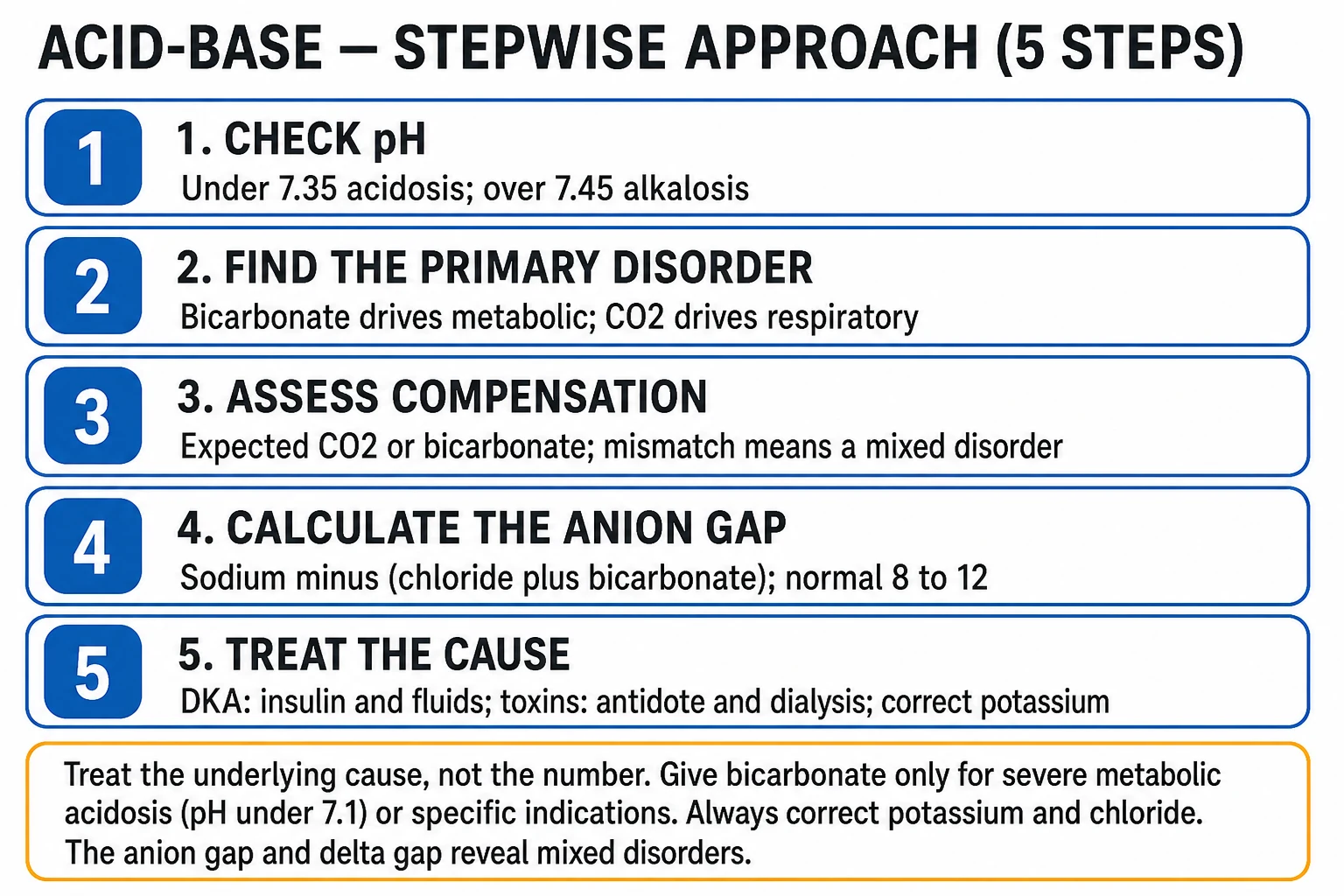
Acid-base disorders arise from disturbance of the bicarbonate-carbon-dioxide buffer system. There are four primary disorders: metabolic acidosis (low pH, low bicarbonate) — raised anion gap from ketoacidosis, lactic acidosis, renal failure or toxins (MUDPILES) or normal gap from diarrhoea and renal tubular acidosis (HARDUP); metabolic alkalosis (high pH, high bicarbonate — vomiting, diuretics); respiratory acidosis (low pH, high CO2 — COPD, opiates); respiratory alkalosis (high pH, low CO2 — anxiety, pain, sepsis, altitude). The stepwise approach is check the pH, identify the primary disorder, assess compensation (Winter's and the respiratory rules), calculate the anion gap, then treat the cause. Bicarbonate is reserved for severe acidosis (pH under 7.1 to 7.15 with instability), hyperkalaemia with ECG change, and tricyclic overdose. Always correct potassium and chloride.
On this page & tools
Your progress
Saved locally on this device.
Exam tags
Red flags

Overview & Definition
Acid-base homeostasis holds the arterial pH between 7.35 and 7.45 — equivalent to a free hydrogen-ion concentration of only 35 to 45 nmol/L. The body defends this narrow window because enzyme function, ion-channel gating, electrolyte distribution and drug-protein binding are all exquisitely pH-sensitive.[1]
Three layers of defence cooperate. Chemical buffers (bicarbonate, phosphate, haemoglobin, proteins) act within seconds. The lungs adjust CO2 within minutes through alveolar ventilation. The kidneys regenerate bicarbonate and excrete acid as ammonium and titratable acid over hours to days — they are slow but powerful, and can normalise the pH of a chronic disturbance almost completely.[1][2]
A primary disorder is the process that first moves the pH; compensation is the secondary response that pulls the pH back toward (but never beyond) normal. The recurring clinical errors are treating the number rather than the cause, and missing a mixed disorder that is hidden by a deceptively near-normal pH — salicylate toxicity is the classic trap. A disciplined, stepwise approach converts any blood gas into a diagnosis.[3]
Classification
Every acid-base disturbance is one — or a mixture — of four primary disorders, defined by which variable changed and which way the pH moved:[1]
Metabolic acidosis
Low pH, low HCO3
- **Primary fall in bicarbonate** from gain of acid (lactate, ketones, toxins) or loss of bicarbonate (diarrhoea, RTA)
- Respiratory compensation: hyperventilation to **lower PaCO2** (Kussmaul breathing)
- **Divide by anion gap** — HIGH gap (MUDPILES) vs NORMAL gap (HARDUP)
- **Treat the cause**; bicarbonate only for pH under 7.1 to 7.15 with instability
Metabolic alkalosis
High pH, high HCO3
- **Primary rise in bicarbonate** from loss of H+ (vomiting, diuretics) or gain of alkali
- Respiratory compensation: hypoventilation to **raise PaCO2** (limited by hypoxia)
- **Divide by urine chloride** — responsive (under 10 to 20: vomiting) vs resistant (over 20: aldosterone)
- **Correct chloride and potassium**; treat the cause
Respiratory acidosis
Low pH, high CO2
- **Primary rise in PaCO2** from alveolar hypoventilation
- Causes: COPD, opiates, neuromuscular disease, chest-wall disease, airway obstruction
- Renal compensation raises HCO3 — **1 per 10 mmHg in acute, 4 per 10 in chronic**
- **Improve ventilation** (reverse opiates, NIV, intubation); oxygen target 88 to 92% in COPD
Respiratory alkalosis
High pH, low CO2
- **Primary fall in PaCO2** from alveolar hyperventilation
- Causes: anxiety, pain, hypoxia, sepsis, pregnancy, salicylates, altitude, pulmonary embolism
- Renal compensation lowers HCO3 — **2 per 10 mmHg in acute, 5 per 10 in chronic**
- **Treat the cause**; rebreathing only in benign acute hyperventilation

Metabolic acidosis is further divided by the anion gap, because the mechanism of the gap localises the differential diagnosis instantly:[5]
- High anion-gap acidosis — an unmeasured acid (lactate, ketones, toxins, sulphates/phosphates of renal failure, oxalate, formate) has accumulated. The chloride stays normal, so the gap widens. Mnemonic MUDPILES.
- Normal (hyperchloraemic) anion-gap acidosis — bicarbonate has been lost (diarrhoea) or a tubule fails to reabsorb/regenerate it (RTA, acetazolamide); the kidney holds electroneutrality by retaining chloride, so the gap stays normal but chloride rises. Mnemonic HARDUP. [1]
Metabolic alkalosis is divided by the urine chloride, which tells you whether the kidney is chloride-hungry (responsive to saline) or autonomously wasting chloride (resistant):[6]
- Chloride-responsive (urine Cl under 10 to 20 mmol/L) — volume- and chloride-depleted states: vomiting, remote diuretic use, villous adenoma, cystic fibrosis, post-hypercapnia. Corrects with normal saline and KCl.
- Chloride-resistant (urine Cl over 20 mmol/L) — mineralocorticoid excess: primary hyperaldosteronism, Cushing, exogenous mineralocorticoid, Bartter and Gitelman syndromes, severe hypokalaemia. Saline does not correct it; treat the underlying cause. [1]
Respiratory disorders are divided by duration into acute (minutes to hours, chemical buffering only) and chronic (days, renal compensation has developed) — the compensation rules differ, and this is how you tell a COPD patient with long-standing hypercapnia from one who has acutely decompensated. [1]
Epidemiology & Risk Factors
Acid-base disorders are ubiquitous in hospital — virtually every critically ill patient has one. The commonest single disorder on the wards is metabolic alkalosis (vomiting, diuretics), but in ICU the picture shifts toward lactic acidosis and respiratory failure.[1]
Headline numbers
Risk factors and the disorders they favour (a high-yield table): [1]
| Risk factor / host | Disorder to consider |
|---|---|
| Sepsis, shock, hypoperfusion, burns, mesenteric ischaemia | Lactic acidosis (type A — tissue hypoxia) |
| Type 1 diabetes, infection, missed insulin, new-onset diabetes | Diabetic ketoacidosis (high gap) |
| Chronic kidney disease, acute kidney injury | Uraemic acidosis (high gap when advanced) |
| Windscreen washer, antifreeze, illicit alcohol; fingernail polish remover | Methanol (visual disturbance) or ethylene glycol (oxalate crystals, AKI) |
| Overdose of aspirin, salicylate-containing products | Salicylate toxicity (mixed resp alkalosis + high-gap acidosis) |
| Metformin in renal failure, HIV drugs (stavudine), cancers | Lactic acidosis (type B — no tissue hypoxia) |
| Iron overdose, isoniazid, toluene (glue sniffing) | High-gap acidosis |
| Profuse diarrhoea, high-output stoma, ureteroenteric fistula | Normal-gap acidosis (HARDUP) |
| Persistent vomiting, nasogastric suction, bulimia, diuretics | Metabolic alkalosis (chloride-responsive) |
| COPD, opiates, neuromuscular disease, obesity-hypoventilation | Respiratory acidosis |
| Anxiety, panic, pain, pregnancy, high altitude, pulmonary embolism | Respiratory alkalosis |
Pathophysiology
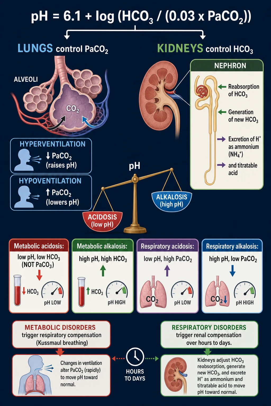
The bicarbonate buffer dominates the extracellular space because it is open at both ends — CO2 is exhaled by the lungs and bicarbonate is regenerated by the kidneys — so it can buffer enormous acid loads. The relationship is fixed by the Henderson-Hasselbalch equation:[1]
[1]Mechanism of metabolic acidosis — there are only two ways bicarbonate falls: a gain of acid that consumes it (every H+ buffered removes one HCO3, generating CO2 that the lungs excrete), or a direct loss of bicarbonate. When the offending acid is hydrochloric (or bicarbonate is lost to diarrhoea), the kidney retains chloride to preserve electroneutrality and the anion gap stays normal. When the offending acid is an organic anion that the kidney cannot rapidly excrete (lactate, beta-hydroxybutyrate, sulphate, formate, oxalate), chloride does not rise and the anion gap widens — the basis of the gap-based classification.[3][5]
Mechanism of metabolic alkalosis — generation needs a source of alkali or a route for H+ loss: vomiting (loss of gastric HCl), diuretics (electrogenic sodium reabsorption drives H+ secretion in the collecting duct), milk-alkali syndrome (exogenous bicarbonate), or contraction alkalosis (diuretic-driven water loss leaving bicarbonate behind). Maintenance requires two perpetuating factors that the bedside clinician must correct: chloride depletion (the collecting duct cannot reclaim bicarbonate without chloride) and hypokalaemia (which stimulates ammoniagenesis, H+ secretion, and renal acid excretion). This is why metabolic alkalosis is chloride-responsive in the volume-depleted patient and why it persists until both K+ and Cl are corrected.[6]
Mechanism of respiratory disorders — PaCO2 is set by the balance between CO2 production (metabolism) and alveolar ventilation. PaCO2 = (VCO2 / VA) x k. Anything that lowers ventilation (COPD, opiates, neuromuscular weakness, airway obstruction, CNS depression) raises PaCO2 → respiratory acidosis. Anything that raises ventilation (pain, anxiety, hypoxia via peripheral chemoreceptors, sepsis, progesterone, salicylates, pulmonary embolism, altitude) lowers PaCO2 → respiratory alkalosis.[2]
The four layers of compensation, with their speed:[1]
- Chemical buffers (seconds) — bicarbonate, haemoglobin, phosphate, intracellular proteins; the first responder.
- Respiratory adjustment (minutes) — the lungs compensate for a metabolic disorder by changing PaCO2 (hyperventilate for acidosis, hypoventilate for alkalosis). Limited by hypoxia and ventilatory capacity.
- Renal HCO3 reabsorption and acid excretion (hours to days) — the kidneys compensate for a respiratory disorder and for chronic metabolic disturbances; they reclaim filtered bicarbonate, generate new bicarbonate (by excreting H+ as ammonium and titratable acid — phosphate), and can eventually near-normalise the pH.
- Bone buffering (months to years) — chronic metabolic acidosis dissolves hydroxyapatite, releasing calcium and phosphate that buffer H+; this is why chronic acidosis (CKD, RTA) causes osteoporosis and growth failure. [1]
Why bicarbonate can paradoxically worsen acidosis — the buffering reaction is HCO3 + H+ to H2CO3 to H2O + CO2. The CO2 diffuses across cell membranes and the blood-brain barrier faster than bicarbonate, so if alveolar ventilation cannot clear the CO2, intracellular and CNS acidosis worsens even as the arterial pH rises. This is the principal reason bicarbonate is restricted, not routine.[1]
Clinical Presentation
Acid-base disturbances are usually asymptomatic in themselves — the symptoms are those of the cause (DKA, sepsis, vomiting) and of the pH shift. The cardinal signs are: [1]
Metabolic acidosis — the body tries to blow off CO2, producing the deep, rapid, sighing Kussmaul respiration (a clinical sign worth knowing on sight). Patients feel fatigued, dyspnoeic and, as the pH falls, hypotensive and confused. Severe acidosis (pH under 7.1) depresses myocardial contractility, blunts catecholamine responsiveness, and causes vasodilation — a vicious spiral in shock. Hyperkalaemia may produce ECG changes.[3]
Metabolic alkalosis — weakness and fatigue from hypokalaemia, paraesthesia and tetany from ionised hypocalcaemia (alkalaemia increases calcium binding to albumin), and arrhythmias. Severe alkalosis shifts the oxyhaemoglobin dissociation curve left (reduced tissue O2 delivery) and causes coronary and cerebral vasoconstriction. [1]
Respiratory acidosis — headache (cerebral vasodilation from CO2), drowsiness, confusion, coarse asterixis (the flap), and at extreme levels CO2 narcosis with papilloedema and coma. Cyanosis appears late. The patient looks hypopnoeic rather than hyperpnoeic. [1]
Respiratory alkalosis — light-headedness, perioral and acral paraesthesia (from alkalaemia-induced hypocalcaemia), carpopedal spasm in acute cases; chronic cases are often asymptomatic (the kidney has compensated). [1]
Cause-specific clues at the bedside: [1]
- DKA — Kussmaul breathing, fruity (ketotic) breath, dehydration, abdominal pain (may mimic an acute abdomen), vomiting.
- Toxic alcohol — altered mental state, visual disturbance and optic disc hyperaemia (methanol), flank pain and calcium oxalate crystalluria (ethylene glycol), a history of alcohol misuse or access to solvents.
- Salicylate — tinnitus, hyperpnoea, sweating, vomiting, agitation progressing to seizures and coma; a mixed respiratory alkalosis + metabolic acidosis pattern.
- Uraemia — fetor, pallor, pruritus, pericardial rub, asterixis, oliguria.
- Opiate — pinpoint pupils, hypoventilation, coma.
- COPD — barrel chest, pursed-lip breathing, prolonged expiration, cachexia, cyanosis. [1]
Atypical presentations — the elderly may present with confusion rather than dyspnoea, and have a blunted ventilatory response (so the Kussmaul sign is muted and the CO2 climbs dangerously). The pregnant patient runs a physiological chronic respiratory alkalosis (progesterone-driven hyperventilation) with a compensatory low bicarbonate (18 to 21) and a mildly alkalaemic pH (~7.44) — do not misread this as metabolic acidosis. The chronic CO2 retainer (COPD) sits with a near-normal pH and a very high bicarbonate — a "normal" PaCO2 in such a patient may represent acute decompensation. Diabetic patients may have euglycaemic DKA (especially with SGLT2 inhibitors), where glucose is modestly raised but ketones and the gap are high. [1]
Differential Diagnosis
The differential is the differential of the cause, localised by the anion gap (for metabolic acidosis) and the urine chloride (for metabolic alkalosis). [1]
High anion-gap metabolic acidosis — MUDPILES, with the distinguishing feature of each:[3][5]
| Cause | Distinguishing feature |
|---|---|
| Methanol | Visual disturbance, optic disc hyperaemia/blindness; high osmolar gap; metabolised to formic acid |
| Uraemia (renal failure) | Raised creatinine, low eGFR, history of CKD; sulphates and phosphates accumulate |
| Diabetic ketoacidosis | Hyperglycaemia, ketones (beta-hydroxybutyrate), dehydration, Kussmaul |
| Propylene glycol / Paraldehyde / Phenformin | Medication/toluene history; propylene glycol is a drug vehicle (lorazepam, IV benzos) |
| Iron / INH (isoniazid) | Overdose history; iron — GI bleeding and hepatotoxicity; INH — seizures |
| Lactic acidosis | Raised lactate (>2 high-gap, >4 severe; type A hypoxia/shock vs type B no hypoxia) |
| Ethylene glycol | Calcium oxalate crystalluria, AKI, flank pain; high osmolar gap; antifreeze |
| Salicylates | Tinnitus, mixed respiratory alkalosis + high-gap acidosis; tachypnoea |
Normal anion-gap (hyperchloraemic) metabolic acidosis — HARDUP:[4]
| Cause | Distinguishing feature |
|---|---|
| Hyperalimentation (TPN) / acid load | History of parenteral nutrition |
| Acetazolamide (and acid load) | Carbonic anhydrase inhibitor; bicarbonaturia, hypokalaemia |
| Renal tubular acidosis | Persistent nongap acidosis with normal anion gap; type 1 distal (hypokalaemic, urine pH over 5.5), type 2 proximal (hypokalaemic, Fanconi), type 4 (hyperkalaemic, hypoaldosteronism) |
| Diarrhoea | History; negative urine anion gap (kidney excreting ammonium appropriately) |
| Ureteroenteric fistula | Post-urological surgery; bowel absorbs and excretes chloride in exchange for bicarbonate |
| Pancreatic fistula / Saline | Pancreatic secretions rich in bicarbonate lost; large-volume normal saline (high chloride drives bicarbonate consumption) |
How to tell GI (diarrhoea) from renal (RTA) loss in nongap acidosis — measure the urine anion gap (Na + K minus Cl). In diarrhoea the kidney appropriately increases ammonium (an unmeasured cation) excretion, so the urine chloride rises and the urine anion gap is negative. In renal tubular acidosis the kidney cannot excrete ammonium, so the urine anion gap is positive.[4]
Metabolic alkalosis — chloride-responsive vs resistant:[6]
- Chloride-responsive (urine Cl under 10 to 20): vomiting, nasogastric suction, remote diuretic use, villous adenoma, cystic fibrosis, post-hypercapnia, exogenous alkali with volume depletion. Corrects with normal saline + KCl.
- Chloride-resistant (urine Cl over 20): hypertensive — primary hyperaldosteronism, Cushing, renovascular disease, exogenous mineralocorticoid (licorice); normotensive — Bartter syndrome, Gitelman syndrome, severe hypokalaemia. Does not correct with saline — treat the cause. [1]
Mixed disorders to recognise (the exam favourites):[1]
- Salicylate toxicity — respiratory alkalosis + high-gap metabolic acidosis (often with a near-normal pH).
- CPR / cardiac arrest — combined respiratory acidosis (apnoea) + metabolic (lactic) acidosis.
- Vomiting + uraemia — metabolic alkalosis + metabolic acidosis.
- Sepsis in COPD — metabolic (lactic) acidosis + respiratory acidosis.
- DKA with Kussmaul respiration that exceeds the requirement — metabolic acidosis + respiratory alkalosis.
- Pregnancy with sepsis — chronic respiratory alkalosis + acute metabolic acidosis. [1]
Clinical & Bedside Assessment
Begin with the ABC, vital signs and a focused volume-status examination. The single most useful observation is the respiratory pattern: deep, rapid, sighing breathing is Kussmaul (compensating for metabolic acidosis); shallow, slow breathing suggests respiratory acidosis (primary hypoventilation); rapid breathing with a normal or high pH suggests respiratory alkalosis.[3]
Volume status decides the cause of metabolic alkalosis (depleted = responsive) and whether the patient with metabolic acidosis is in shock (lactic). Look for tachycardia, hypotension, dry mucosae, reduced skin turgor, slow capillary refill (hypovolaemia) versus raised JVP, oedema, basal crackles (hypervolaemia). Assess perfusion, not pressure alone — lactate and capillary refill. [1]
Named signs and manoeuvres: [1]
- Kussmaul respiration — deep, rapid, sighing; metabolic acidosis.
- Asterixis (the flap) — arms extended, wrists dorsiflexed; flapping tremor at 3 to 5 Hz; severe CO2 retention or uraemia.
- Trousseau sign — inflate sphygmomanometer above systolic for 3 min; carpopedal spasm — latent tetany from hypocalcaemia (alkalaemia).
- Chvostek sign — tap the facial nerve anterior to the ear; twitching of facial muscles — latent tetany.
- Pinpoint pupils — opiate toxicity (respiratory acidosis). [1]
Toxidrome screen — a deliberate, structured search for the cause: methanol (visual disturbance), ethylene glycol (oxalate crystals in urine, AKI, flank pain), salicylate (tinnitus, mixed gas), opiate (pinpoint pupils, hypoventilation), iron (GI bleed, hepatotoxicity), INH (seizures). Read the drug chart — diuretics, metformin, ACE inhibitors, ARBs, SGLT2 inhibitors, acetazolamide, salicylates. [1]
Recognise the patient who needs escalation now: pH under 7.1, potassium over 6.5 with ECG change, PaCO2 rising with falling GCS, a high anion-gap with high osmolar gap, or a mixed disorder — these go to a high-dependency or ICU bed. [1]
Investigations
The arterial (or venous) blood gas is the investigation. Modern analysers give pH, PaCO2, PaO2, HCO3, base excess, and often lactate in under a minute. Venous gases are acceptable for most metabolic questions (the venous pH runs ~0.03 lower and PaCO2 ~4 to 6 mmHg higher than arterial) — use arterial when you need oxygenation or a precise PaCO2.[1]
Normal values to memorise: pH 7.35 to 7.45; PaCO2 35 to 45 mmHg (4.7 to 6.0 kPa); PaO2 80 to 100 mmHg (on room air); HCO3 22 to 26 mmol/L; base excess minus 2 to plus 2 mmol/L; anion gap 8 to 12 mmol/L. [1]
The systematic six-step approach
This is the framework every examiner wants reproduced verbatim — practising it on every gas is how you stop missing mixed disorders:[1][5]
- pH — under 7.35 acidaemia; over 7.45 alkalaemia.
- Primary disorder — if HCO3 and PaCO2 moved in the same direction (both low = acidosis, both high = alkalosis), the bicarbonate drives the primary disorder → metabolic. If they moved in opposite directions, the PaCO2 drives it → respiratory. (If PaCO2 is high and pH is low = respiratory acidosis; PaCO2 low and pH high = respiratory alkalosis.)
- Compensation — apply the expected-formula rules below. If compensation is incomplete the disorder is still evolving; if it exceeds expectation, or the pH crosses normal, a mixed disorder is present.
- Anion gap — Na minus (Cl + HCO3); normal 8 to 12 (correct for albumin).
- Delta gap / delta ratio — unmask a second disorder in the high-gap acidosis.
- Treat the cause — the gas only localises; the diagnosis comes from glucose, ketones, lactate, renal function, salicylate, osmolar gap, drug history. [1]
Compensation formulas — reproduced verbatim
[1]The anion gap and the delta ratio
Anion gap = Na minus (Cl + HCO3). Unmeasured anions (proteins, phosphate, sulphate, organic anions) normally exceed unmeasured cations (K, Ca, Mg) by 8 to 12 mmol/L.[5]
- Albumin correction — albumin is the main unmeasured anion; the normal AG falls by 2.5 mmol/L for every 10 g/L below 40. In hypoalbuminaemia (cirrhosis, nephrotic, critically ill), a "normal" AG of 8 may hide a high-gap acidosis — always correct.
- Delta ratio = (AG minus 12) / (24 minus HCO3). In a pure high-gap acidosis each mmol of HCO3 consumed generates one mmol of unmeasured anion, so the rise in AG equals the fall in HCO3 and the ratio is ~1.
- Under 0.4 — an additional normal-gap acidosis (e.g. a high-gap process plus diarrhoea).
- 0.4 to 0.8 — a combined high-gap + nongap process.
- Around 1 — pure high-gap metabolic acidosis.
- Over 2 — an additional metabolic alkalosis (the HCO3 did not fall as expected because a second process is raising it; classic in vomiting + DKA). [1]
Other investigations
- Osmolar gap — measured minus calculated osmolality (2 x Na + glucose + urea, or 2 x Na + glucose + 1.2 x ethanol); normal under 10 mOsm/kg. High gap + high osmolar gap = toxic alcohol (the parent alcohol is osmotically active).[3]
- Urine anion gap — (Na + K minus Cl); negative in diarrhoea (appropriate ammonium excretion), positive in RTA (impaired ammonium excretion).[4]
- Urine chloride — divides metabolic alkalosis (responsive under 10 to 20 vs resistant over 20).[6]
- Beta-hydroxybutyrate — the dominant ketone in DKA (more sensitive than the nitroprusside test, which detects acetoacetate but not beta-hydroxybutyrate).
- Lactate — defines lactic acidosis (over 2 high-gap, over 4 severe); rising lactate despite resuscitation is an ominous sign.
- Salicylate level — for any unexplained high-gap acidosis with tinnitus or a mixed gas.
- Renal function, glucose, albumin, calcium — define uraemic, hyperglycaemic, and hypoalbuminaemic contributions.
- ECG — for hyperkalaemia (peaked T, flattened P, prolonged PR, wide QRS, sine wave) and to monitor alkalaemia-induced arrhythmia.
Management — Resuscitation
ABCDE first. Airway and breathing: give oxygen to target SpO2 94 to 98% for most adults, but 88 to 92% in the chronic CO2 retainer (COPD) — over-oxygenation worsens hypercapnia via V/Q mismatch and the Haldane effect (oxygen displaces CO2 from haemoglobin). Consider NIV (bilevel) for decompensated respiratory acidosis; intubate if the CO2 is rising with a falling GCS or failing respiratory effort.[2]
Circulation: IV access, treat shock. For septic or cardiogenic lactic acidosis apply the Surviving Sepsis hour-1 bundle — oxygen, blood cultures, broad-spectrum antibiotics within 1 hour, lactate, balanced crystalloid 30 mL/kg, noradrenaline for fluid-refractory shock. (The UK Sepsis Six adds hourly urine output.) Use balanced crystalloids (Hartmann, Plasma-Lyte) in preference to large-volume normal saline — the SMART trial showed balanced solutions reduce AKI and Major Adverse Kidney Events, because saline's high chloride load itself causes a hyperchloraemic acidosis.[3]
The two cardinal rules that govern every acid-base resuscitation:[1]
- Treat the underlying cause, not the pH. Insulin and fluids fix DKA; fomepizole and dialysis fix toxic alcohol; resuscitation fixes lactic acidosis; reversal of opiates fixes narcotic hypoventilation. The pH follows the cause.
- Correct potassium and chloride throughout. Insulin shifts potassium into cells; fixing alkalosis shifts it back. Hypokalaemia kills the patient with DKA or alkalosis. Hypochloraemia perpetuates alkalosis. [1]
The restricted indications for sodium bicarbonate — only in these settings:[1]
- pH under 7.1 to 7.15 WITH haemodynamic instability (catecholamines are less effective at extreme acidosis; a small upward pH shift can restore pressor responsiveness).
- Hyperkalaemia with ECG change (shifts K+ back into cells).
- Tricyclic antidepressant overdose with QRS widening — the sodium load, more than the alkalaemia, stabilises the myocardial membrane.
- Severe renal acidosis pending dialysis. [1]
In all other acidosis — including the vast majority of lactic acidosis — bicarbonate does not improve outcomes and may harm (paradoxical intracellular acidosis from CO2 generation, hypernatraemia, volume overload, hypokalaemia, overshoot alkalosis).[3]
Management — Definitive & Stepwise
Definitive management is cause-specific. The following are the protocols the examiner expects you to be able to reproduce with drug, dose, route, timing and rationale. [1]
High-anion-gap metabolic acidosis — by cause
Diabetic ketoacidosis — the prototype. The three-pillar protocol:[7]
- Fluids — 0.9% saline 10 to 15 mL/kg in the first hour, then reassess; switch to balanced crystalloid with the addition of dextrose (5 to 10%) once glucose falls under 14 mmol/L (250 mg/dL). Avoid overly rapid fluid — cerebral oedema (especially in children) is the feared complication.
- Insulin — continuous IV infusion 0.1 unit/kg/h (or 0.14 unit/kg/h after a 0.1 unit/kg bolus, if no bolus given). Aim for a fall in glucose of 3 to 4 mmol/L/h and a rise in HCO3 and pH.
- Potassium — insulin drives K+ into cells, and the acidosis correction releases it back — net effect a fall. If K+ under 3.3, hold insulin and give 20 to 40 mmol/h IV K+. If 3.3 to 5.2, add 20 to 30 mmol/L to the fluids. If over 5.2, hold K+ and recheck in 2 h.
- Bicarbonate is NOT indicated unless pH under 6.9 with haemodynamic instability — and even then it is debated.
- Identify and treat the trigger (infection, infarct, non-compliance, new-onset diabetes). Monitor glucose hourly, electrolytes and gas 2-hourly, and conscious state (cerebral oedema). [1]
Lactic acidosis — resuscitate, restore perfusion, treat sepsis with source control and antibiotics, and reverse any causative drug (metformin in AKI — stop and consider dialysis). Bicarbonate does not improve outcomes (BICAR-ICU, JAMA 2018). If the cause is shock, mortality tracks the shock.[3]
Toxic alcohol ingestion (methanol, ethylene glycol) — time-critical. The toxic metabolites (formate from methanol; glycolate and oxalate from ethylene glycol) cause the injury; the parent alcohol is comparatively harmless, so blocking its metabolism is the core strategy:[3]
- Fomepizole 15 mg/kg IV loading then 10 mg/kg every 12 h for 4 doses, then 15 mg/kg every 12 h until the level is under 20 mg/dL and the acidosis has resolved. (If fomepizole is unavailable, an ethanol infusion to maintain blood ethanol 100 to 150 mg/dL.)
- Cofactors — thiamine 100 mg IV and pyridoxine 50 mg IV daily (ethylene glycol), folinic acid (leucovorin) 1 mg/kg (methanol, to accelerate formate metabolism).
- Haemodialysis — for severe acidosis (pH under 7.25 to 7.30), renal failure, level over 50 mg/dL, or visual/cerebral involvement. Removes the parent alcohol and metabolites and corrects the acidosis.
- Supportive: treat seizures, correct hypocalcaemia only if symptomatic (avoid over-replacement, which drives oxalate precipitation), fomepizole continues during dialysis (it is dialysable — increase frequency). [1]
Salicylate toxicity — alkalinise the urine and dialyse:[3]
- Activated charcoal 50 g if ingestion within 1 to 2 h.
- Urinary alkalinisation — sodium bicarbonate 1 to 2 mmol/kg IV to keep urine pH 7.5 to 8.0; this traps salicylate as the ionised salt, preventing tubular reabsorption and enhancing excretion. Keep potassium corrected (hypokalaemia prevents alkalinisation of the urine — the kidney excretes H+ to conserve K+).
- Haemodialysis — for level over 100 mg/dL (acute), over 60 to 70 (chronic), severe acidosis, encephalopathy, pulmonary oedema, or renal failure. If intubating, alkalinise aggressively first — intubation removes the compensatory hyperventilation and the acidosis can worsen rapidly. [1]
Uraemic acidosis — treat the renal failure; haemodialysis for severe acidosis (AEIOU). In chronic CKD, oral sodium bicarbonate to keep HCO3 over 22 (BICRA trial — slows eGFR decline). [1]
Normal-anion-gap metabolic acidosis — by cause
Diarrhoea — treat the GI cause; replace bicarbonate and potassium. Renal tubular acidosis — bicarbonate/citrate replacement and potassium:[4][8]
- Type 1 (distal) — oral bicarbonate 1 to 2 mmol/kg/day (or potassium citrate), plus potassium; corrects acidosis, reduces nephrocalcinosis and stones.
- Type 2 (proximal) — high-dose bicarbonate 10 to 15 mmol/kg/day (because the proximal tubule wastes it) plus potassium; treat the underlying cause (myeloma, carbonic anhydrase inhibitors, Fanconi).
- Type 4 (hypoaldosteronism) — hyperkalaemic; treat hyperkalaemia and give fludrocortisone 0.05 to 0.2 mg/day if aldosterone-deficient; withdraw offending drugs (ACE inhibitors, ARBs, NSAIDs, spironolactone, calcineurin inhibitors). [1]
Metabolic alkalosis
Chloride-responsive — 0.9% normal saline (corrects volume and chloride) plus potassium chloride (corrects the perpetuating hypokalaemia). Stop the cause (antiemetics, stop diuretic). The alkalosis usually corrects over 24 to 48 h. Acetazolamide 250 to 500 mg can be used if volume-overloaded (it induces bicarbonaturia) — but expect a kaluresis and hypokalaemia.[6]
Chloride-resistant — saline does not work. Treat the underlying mineralocorticoid excess: spironolactone/eplerenone for hyperaldosteronism; resect an adenoma; address Cushing. Bartter and Gitelman — potassium-sparing diuretics (amiloride) and magnesium replacement. [1]
Respiratory acidosis
Treat the hypoventilation. Reverse the reversible (naloxone 0.4 to 2 mg IV for opiates, repeated as needed; flumazenil for benzodiazepines with caution). Treat COPD exacerbation with bronchodilators, steroids, antibiotics, controlled oxygen (target SpO2 88 to 92%). If PaCO2 is rising with a falling pH and GCS, NIV (bilevel) — the standard for decompensated COPD. Intubate if NIV fails, the airway is unprotected, or the patient is comatose. Do not correct chronic hypercapnia rapidly — the compensated high bicarbonate will overshoot into a post-hypercapnic metabolic alkalosis (arrhythmia, seizures).[2]
Respiratory alkalosis
Treat the cause. Reassure the anxious patient; manage pain; correct hypoxia (give oxygen for the PE or pneumonia that is driving hyperventilation); treat sepsis, fever, salicylate toxicity. Rebreathing (paper bag) is reserved for benign acute hyperventilation syndrome — never use when hypoxia is the driver, as it worsens hypoxaemia. [1]
Specific Subtypes & Scenarios
The high-anion-gap acidoses (MUDPILES) in detail
- Lactic acidosis — divided into type A (tissue hypoxia: shock, sepsis, mesenteric ischaemia, burns, severe anaemia, CO poisoning, exercise) and type B (no clinical hypoxia: metformin in renal failure, malignancy, HIV/NRTIs, thiamine deficiency, toxins, idiopathic). Lactate over 2 defines it; over 4 is severe. The prognosis tracks the cause — type A from septic shock carries mortality over 50%.[3]
- Diabetic ketoacidosis — ketogenesis from insulin deficiency and glucagon excess. Beta-hydroxybutyrate dominates (the nitroprusside test misses it). Beware euglycaemic DKA (SGLT2 inhibitors, pregnancy). See the protocol above.[7]
- Uraemic acidosis — sulphates and phosphates accumulate; rarely a high-gap acidosis until eGFR under 20 to 25. Oral bicarbonate and dialysis.
- Toxic alcohols — methanol (formate, blindness), ethylene glycol (glycolate, oxalate crystals, AKI), diethylene glycol, propylene glycol. The osmolar gap is the key early clue; fomepizole + dialysis is the rescue.[3]
- Salicylates — direct respiratory centre stimulation (respiratory alkalosis) plus uncoupling and Krebs-cycle inhibition (high-gap acidosis). Mixed gas, tinnitus, alkalinise and dialyse.[3]
- Other — iron (hepatotoxicity, GI bleed), isoniazid (seizures, pyridoxine-dependent), toluene (hippuric acid, glue sniffing).
The normal-gap acidoses (HARDUP) and renal tubular acidosis
Type 1 RTA (distal)
Alpha-intercalated cell failure
- Failure to secrete H+ via H+-ATPase — urine pH stays OVER 5.5 in acidosis
- **Hypokalaemic** (K+ wasting in the collecting duct)
- Nephrocalcinosis, kidney stones (alkaline urine, hypercalciuria, low citrate)
- Treatment: bicarbonate 1 to 2 mmol/kg/day + potassium
Type 2 RTA (proximal)
Proximal bicarbonate wastage
- Lowered bicarbonate reabsorption threshold; Fanconi syndrome (glycosuria, aminoaciduria, phosphaturia)
- **Hypokalaemic**; urine pH variable — can acidify once HCO3 falls below threshold
- Causes: myeloma, ifosfamide, carbonic anhydrase inhibitors, inherited
- Treatment: high-dose bicarbonate 10 to 15 mmol/kg/day + potassium
Type 4 RTA
Hypoaldosteronism
- Deficient aldosterone (or resistance) — impaired distal Na+ reabsorption and H+/K+ secretion
- **Hyperkalaemic** (the distinguishing feature); urine pH UNDER 5.5
- Causes: diabetic nephropathy, ACE inhibitors/ARBs, NSAIDs, spironolactone, adrenal insufficiency
- Treatment: treat hyperkalaemia; fludrocortisone 0.05 to 0.2 mg/day if aldosterone-deficient
Metabolic alkalosis in detail
- Chloride-responsive (urine Cl under 10 to 20): vomiting, nasogastric suction, remote diuretic use, post-hypercapnia, villous adenoma, cystic fibrosis, exogenous alkali with volume depletion. Correct with saline + KCl.
- Chloride-resistant (urine Cl over 20): hypertensive — primary hyperaldosteronism (Conn), secondary hyperaldosteronism (renovascular), Cushing, exogenous mineralocorticoid (licorice, carbenoxolone), Liddle syndrome (note: Liddle has LOW aldosterone); normotensive — Bartter (loop of Henle, mimics loop diuretic), Gitelman (distal tubule, mimics thiazide, with hypomagnesaemia and hypocalciuria). Treat the cause; potassium-sparing diuretics, magnesium replacement.[6]
Mixed disorders — recognise the patterns
- Salicylate — respiratory alkalosis + high-gap metabolic acidosis.
- CPR / arrest — respiratory acidosis + metabolic acidosis.
- Vomiting + uraemia — metabolic alkalosis + metabolic acidosis.
- Sepsis + COPD — metabolic acidosis + respiratory acidosis.
- Pregnancy + sepsis — chronic respiratory alkalosis + acute metabolic acidosis.
- DKA with over-compensation — metabolic acidosis + respiratory alkalosis. [1]
Complications & Pitfalls
Complications of severe acidosis (pH under 7.1 to 7.2):[1]
- Cardiovascular — reduced myocardial contractility, arterial vasodilation, attenuation of catecholamine responsiveness (pressors fail), venoconstriction pooling blood centrally, arrhythmia, hypotension.
- Metabolic — hyperkalaemia (H+ shifts into cells, K+ out), insulin resistance, impaired glycolysis.
- Neurological — altered mental state, coma.
- Respiratory — respiratory muscle fatigue (the patient cannot maintain Kussmaul forever), then CO2 retention and arrest.
- Chronic acidosis — bone dissolution (osteoporosis, fractures, growth failure in children), muscle catabolism, hypoalbuminaemia. [1]
Complications of severe alkalosis (pH over 7.6): [1]
- Hypokalaemia (H+/K+ exchange drives K+ into cells) — arrhythmia, weakness, potentiation of digoxin.
- Ionised hypocalcaemia (alkalaemia increases calcium binding to albumin) — tetany, seizures, prolonged QT.
- Left-shift of the oxyhaemoglobin curve — reduced tissue oxygen delivery.
- Cerebral and coronary vasoconstriction — reduced perfusion.
- Hypoventilation (respiratory compensation) — atelectasis, hypoxia.
- Hepatic encephalopathy in cirrhosis (alkalaemia increases ammonium crossing into the brain). [1]
Complications of over-rapid correction of chronic respiratory acidosis — the kidney has spent days generating bicarbonate; if PaCO2 is suddenly normalised (intubation), the high bicarbonate persists and the patient swings into a post-hypercapnic metabolic alkalosis with seizures, arrhythmia, and hypokalaemia. Correct chronic hypercapnia slowly. [1]
The classic pitfalls (do not ship these):[1][5]
- Treating the number, not the cause — giving bicarbonate for a pH of 7.20 in lactic acidosis without resuscitating the shock.
- Missing a mixed disorder — a near-normal pH in salicylate toxicity read as "no acid-base problem."
- Using bicarbonate for lactic acidosis — no outcome benefit; may worsen intracellular acidosis.
- Failing to correct potassium in DKA before/with insulin — precipitating dangerous hypokalaemia.
- Not correcting the anion gap for albumin — missing a high-gap acidosis in the hypoalbuminaemic ICU patient.
- Misclassifying a delta-gap disorder — over-reading the anion gap without checking the delta ratio, missing an additional alkalosis or nongap acidosis.
- Over-oxygenating the chronic CO2 retainer — driving hypercapnia and CO2 narcosis.
- Interpreting a pregnant woman's gas without accounting for the physiological respiratory alkalosis — mislabelling her low bicarbonate as metabolic acidosis. [1]
Prognosis & Disposition
Prognosis tracks the cause, not the pH number. Lactic acidosis from septic or cardiogenic shock carries a mortality of 50 to 70%; mortality rises steeply once pH is under 7.1. DKA, by contrast, now has a mortality under 1% with protocolised care (higher in the elderly and in cerebral oedema). Toxic alcohol mortality depends on the time to fomepizole and dialysis — early treatment is essentially curative, late treatment leaves blindness (methanol) or renal failure (ethylene glycol). Salicylate mortality is low with alkalinisation and dialysis but rises sharply with delayed intubation without alkalinisation. Respiratory disorders track the underlying ventilatory condition.[3][7]
Disposition: [1]
- ICU / HDU — pH under 7.1, failing compensation (rising CO2 with falling GCS), shock requiring vasopressors, mixed disorders, severe toxin ingestion.
- Nephrology / toxicology referral — for dialysis decisions (the AEIOU indications: Acidosis refractory, Electrolytes refractory — hyperkalaemia, Ingestion dialysable — lithium, salicylate, methanol, ethylene glycol, metformin, Overload — pulmonary oedema, Uraemia — pericarditis, encephalopathy).
- Ward — corrected and stable disorders (DKA resolving, alkalosis corrected).
- Safety-net — recheck gas and electrolytes at defined intervals; mixed disorders and toxins can evolve fast. [1]
Chronic metabolic acidosis (CKD, distal RTA) is usually stable but, untreated, erodes bone and muscle and stunts growth in children — give oral bicarbonate to keep HCO3 over 22 (BICRA trial showed slowing of CKD progression). [1]
Special Populations
Pregnancy — the mother runs a physiological chronic respiratory alkalosis: progesterone-driven hyperventilation lowers PaCO2 to 28 to 32 mmHg, with renal compensation dropping HCO3 to 18 to 21 mmol/L and a mildly alkalaemic pH (7.44). A "normal" PaCO2 of 40 in late pregnancy may indicate respiratory compromise, and a normal bicarbonate of 24 may mask a metabolic alkalosis. The fetus suffers acidosis at higher maternal pH than the mother tolerates — treat maternal acidosis aggressively.[2]
Elderly — blunted ventilatory and renal responses, atypical presentation (confusion rather than dyspnoea), polypharmacy (diuretics, ACE inhibitors, salicylates, metformin), and higher mortality at any given pH. Salicylate toxicity is particularly lethal in the elderly (chronic intoxication). Lower threshold to admit, monitor and treat. [1]
Chronic kidney disease — chronic metabolic acidosis from reduced ammoniagenesis. Oral sodium bicarbonate to keep HCO3 over 22 (BICRA trial, JASN 2020 — slowed eGFR decline modestly in stage 3 to 4 CKD). Watch sodium load and volume. [1]
Chronic CO2 retainers (COPD) — oxygen target 88 to 92%; use controlled oxygen (Venturi 24 to 28%) and consider NIV early for decompensated respiratory acidosis (pH under 7.35, PaCO2 over 6.5 kPa). Do not over-oxygenate (V/Q mismatch + Haldane effect worsens hypercapnia). [1]
Children — DKA is the leading cause of severe metabolic acidosis in the young; weight-based fluids (10 to 20 mL/kg aliquots) and insulin (0.05 to 0.1 unit/kg/h), with a particular fear of cerebral oedema (avoid rapid fluid or sodium). Salicylate toxicity (with viral illness, Reye-like), inborn errors of metabolism (organic acidaemias, lactic acidosis), and severe diarrhoea are the other paediatric causes. [1]
Diabetic on SGLT2 inhibitor — risk of euglycaemic DKA (near-normal glucose with high ketones/gap), especially with illness, surgery, or low-carbohydrate intake. Check ketones in any unwell diabetic patient regardless of glucose. [1]
Evidence, Guidelines & Regional Differences
Landmark trials and what they changed: [1]
- BICAR-ICU (JAMA 2018) — sodium bicarbonate in critically ill patients with severe metabolic acidosis did not reduce the primary composite outcome of death at 28 days or the presence of at least one organ failure at 7 days, though a subgroup with acute kidney injury showed a mortality benefit. Reinforces that bicarbonate is not routine.
- BICRA (de Brito-Ashurst, JASN 2020) — oral sodium bicarbonate in stage 3 to 4 CKD slowed eGFR decline modestly; supports bicarbonate supplementation in CKD acidosis.
- SMART (Semler, NEJM 2018) — balanced crystalloids versus saline in critically ill adults; balanced solutions reduced AKI and Major Adverse Kidney Events, including saline-induced hyperchloraemic acidosis.
- Kitabchi / ADA consensus (Diabetes Care 2009) — the modern DKA protocol: fluids first, then insulin infusion, potassium replacement, and bicarbonate only for pH under 6.9.[7]
Guidelines — the Adrogue & Madias NEJM 1998 two-part review remains the international reference framework for management.[1][2] Resuscitation bundles follow Surviving Sepsis 2021 (the hour-1 bundle). NICE NG51 (UK sepsis) and the UK Sepsis Six apply in the UK.
Regional deltas — US/UK practice emphasises balanced crystalloids and protocolised DKA; Surviving Sepsis drives resuscitation. The diagnostic framework (six-step approach, Winter's formula, delta ratio) is universal. In India and resource-limited settings, the principles are identical but peritoneal dialysis is a viable RRT modality for toxic alcohol and severe metabolic acidosis where haemodialysis access is limited, and empirical therapy must respect local antibiograms (ICMR/NCDC). The NMC/Indian MBBS emphasis is on the systematic approach and the high-yield differentials (DKA, sepsis, diarrhoea, toxic alcohol from illicit liquor) — the same framework reproduced here.
Controversies — the Stewart (strong ion difference) approach offers a mechanistically deeper account (the independent variables are the strong ion difference, total weak acid, and PaCO2) but yields the same clinical answers in most cases and is rarely needed at MBBS level. The bicarbonate threshold in DKA (pH under 6.9, or under 7.0?) and the value of alkalinisation in salicylate poisoning remain debated, but the conservative consensus — treat the cause, reserve bicarbonate for instability and specific indications — is stable.[1]
Exam Pearls
Two mnemonics that decide the metabolic acidosis answer
MUDPILES / HARDUP
visual disturbance, optic disc hyperaemia; metabolised to formic acid
renal failure; sulphates and phosphates accumulate
hyperglycaemia, ketones, dehydration, Kussmaul
drug vehicle, solvents, biguanide
overdose; iron — hepatotoxicity; INH — seizures
type A hypoxia/shock vs type B no hypoxia
calcium oxalate crystalluria, AKI; antifreeze
tinnitus; mixed resp alkalosis + high-gap acidosis
parenteral nutrition / acid load
carbonic anhydrase inhibitor; bicarbonaturia
type 1 distal, type 2 proximal, type 4 hyperkalaemic
negative urine anion gap (GI loss)
post-urological surgery; bowel reabsorbs Cl, loses HCO3
bicarbonate-rich secretions lost; high-volume saline
- Anion gap = Na minus (Cl + HCO3); normal 8 to 12; subtract 2.5 for every 10 g/L albumin below 40.
- Winter's formula: expected PaCO2 = 1.5 x HCO3 + 8 (plus or minus 2), for metabolic acidosis.
- Delta ratio = (AG minus 12) / (24 minus HCO3): under 0.4 nongap component; around 1 pure high-gap; over 2 metabolic alkalosis.
- Respiratory compensation for metabolic alkalosis: PaCO2 = 0.7 x HCO3 + 20; or rises 0.6 per 1 mmol/L rise in HCO3.
- Respiratory acidosis: HCO3 rises 1 per 10 (acute), 4 per 10 (chronic).
- Respiratory alkalosis: HCO3 falls 2 per 10 (acute), 5 per 10 (chronic).
- Urine chloride divides metabolic alkalosis: under 10 to 20 responsive (vomiting), over 20 resistant (aldosterone).
- Salicylate = mixed respiratory alkalosis + high-gap metabolic acidosis; alkalinise urine and dialyse if severe.
- Toxic alcohol = high anion gap + high osmolar gap; fomepizole + dialysis; calcium oxalate crystals (ethylene glycol), visual disturbance (methanol).
- RTA type 1 distal (hypokalaemic, urine pH over 5.5, nephrocalcinosis); type 2 proximal (hypokalaemic, Fanconi); type 4 (hyperkalaemic, hypoaldosteronism).
- Bicarbonate reserved for: pH under 7.1 to 7.15 with instability, hyperkalaemia with ECG change, tricyclic overdose with QRS widening — NOT routine for lactic acidosis or DKA.
- DKA protocol: fluids, insulin 0.1 unit/kg/h, potassium; hold insulin if K+ under 3.3; bicarbonate only if pH under 6.9.
- Pregnancy = physiological chronic respiratory alkalosis; HCO3 18 to 21, pH around 7.44.
- COPD oxygen target 88 to 92%; chronic respiratory acidosis compensation — HCO3 rises 4 per 10 mmHg PaCO2.
- Treat the cause, not the number; always correct potassium and chloride. [1]
Exam application bank (NEET-PG / INICET)
One-line answer
Acid-base disorders arise from disturbance of the bicarbonate-carbon-dioxide buffer system. There are four primary disorders: metabolic acidosis (low pH, low bicarbonate) — raised anion gap from ketoacidosis, lactic acidosis, renal failure or toxins (MUDPILES) or normal gap from diarrhoea and renal tubular acidosis (HARDUP); metabolic alkalosis (high pH, high bicarbonate — vomiting, diuretics); respiratory acidosis (low pH, high CO2 — COPD, opiates); respiratory alkalosis (high pH, low CO2 — anxiety, pain, sepsis, altitude). The stepwise approach is check the pH, identify the primary disorder, assess compensation (Winter's and the respiratory rules), calculate the anion gap, then treat the cause. Bicarbonate is reserved for severe acidosis (pH under 7.1 to 7.15 with instability), hyperkalaemia with ECG change, and tricyclic overdose. Always correct potassium and chloride.
Worked stems (answer without another resource)
Stem 1 — Classic presentation. Map symptoms to mechanism; name the first investigation and first treatment step with dose/route if drug therapy is standard. [1]
Stem 2 — Unstable / complicated. List red flags that force immediate resuscitation, theatre, ICU, antidote, or reperfusion — and what you do in the first 15 minutes. [1]
Stem 3 — Atypical group. Elderly, pregnancy, child, or immunocompromised: how presentation and thresholds change. [1]
Stem 4 — Differential trap. Name the three closest mimics and one discriminator for each. [1]
Stem 5 — Disposition. Who goes home with safety-netting, who is admitted, who needs HDU/ICU/theatre, and what follow-up is mandatory. [1]
Rapid viva checklist
- Definition + classification
- Pathophysiology chain
- Bedside signs / criteria
- Score with exact components (if any)
- Emergency bundle
- Definitive therapy with doses
- Complications of disease and of treatment
- Special populations
- Guideline/trial name if classic
- Three exam traps
Coverage self-check
If you cannot answer any stem above from this page alone, re-read the matching section — the page is intended to be self-sufficient for final-prof and NEET-PG/INICET questions on Acid-Base Disorders.
References
- [1]Adrogue HJ, Madias NE. Management of life-threatening acid-base disorders. First of two parts N Engl J Med, 1998.PMID 9414329
- [2]Adrogue HJ, Madias NE. Management of life-threatening acid-base disorders. Second of two parts N Engl J Med, 1998.PMID 9420343
- [3]Kraut JA, Madias NE. Lactic acidosis N Engl J Med, 2015.PMID 25760366
- [4]Kraut JA, Madias NE. Differential diagnosis of nongap metabolic acidosis: value of a systematic approach Clin J Am Soc Nephrol, 2012.PMID 22403272
- [5]Kraut JA, Madias NE. Serum anion gap: its uses and limitations in clinical medicine Clin J Am Soc Nephrol, 2007.PMID 17699401
- [6]Gennari FJ. Pathophysiology of metabolic alkalosis: a new classification based on the centrality of stimulated collecting duct ion transport Am J Kidney Dis, 2011.PMID 21849227
- [7]Kitabchi AE, Umpierrez GE, Miles JM, Fisher JN. Hyperglycemic crises in adult patients with diabetes Diabetes Care, 2009.PMID 19564476
- [8]Kashoor I, Batlle D. Proximal renal tubular acidosis with and without Fanconi syndrome Kidney Res Clin Pract, 2019.PMID 31474092