Nephrology · General Medicine
Acute Kidney Injury
Also known as AKI · Acute renal failure · ARF · Acute tubular necrosis · ATN · Pre-renal uraemia
Acute kidney injury (AKI) is a rapid (hours to days) decline in kidney function defined by KDIGO as a serum creatinine rise of 0.3 mg/dL (26.5 micromol/L) or more within 48 hours, a creatinine 1.5 times baseline or more within 7 days, or urine output under 0.5 mL/kg/h for 6 hours or more. It is classified into pre-renal (hypoperfusion, around 60 per cent), intrinsic/renal (acute tubular necrosis, glomerular, interstitial, vascular) and post-renal (obstruction, around 5 per cent). Commonest intrinsic cause is acute tubular necrosis (ATN) from ischaemia or nephrotoxins. Presents with oliguria, fluid overload, uraemia, hyperkalaemia; diagnose with creatining trend, FENa, urine sediment, renal ultrasound. Management: treat the cause, restore perfusion with balanced crystalloid, stop nephrotoxins (NSAIDs, ACE inhibitors, aminoglycosides, contrast), manage hyperkalaemia and acidosis, and dialyse for AEIOU (Acidosis, Electrolytes, Ingestion, Overload, Uraemia). Mortality 10 per cent uncomplicated to over 50 per cent in ICU; survivors carry increased risk of future CKD.
On this page & tools
Your progress
Saved locally on this device.
Practise this topic
Exam tags
Red flags

Overview & Definition
Acute kidney injury (AKI) is a syndrome of rapid (hours to days) deterioration in kidney function resulting in failure to maintain fluid, electrolyte and acid-base homeostasis and to excrete nitrogenous waste. The internationally agreed KDIGO 2012 definition (which superseded the older RIFLE and AKIN schemes) is any one of the following:[1]
- Serum creatinine rise of 0.3 mg/dL (26.5 micromol/L) or more within 48 hours; OR
- Serum creatinine 1.5 times the known or presumed baseline, occurring within the prior 7 days; OR
- Urine volume under 0.5 mL/kg/h for 6 hours or more. [1]
The older name acute renal failure (ARF) is now avoided — "injury" recognises that even small, reversible creatinine rises carry significant prognostic weight (mortality and future CKD risk), and "failure" implied an all-or-nothing state that understated the spectrum.[5]
The clinical skill in AKI is not the diagnosis (a creatinine trend makes it) but finding and reversing the cause, recognising the life-threatening complications (hyperkalaemia, acidosis, fluid overload, uraemia), and knowing when to dialyse. A normal or only slightly raised creatinine early does not exclude significant AKI — creatinine is a late, lagging marker because it depends on muscle mass; a patient may have lost 50 per cent of GFR before creatinine clearly rises. [1]
Classification
AKI is classified in two complementary ways — by anatomical/physiological cause (the scheme that drives the bedside work-up) and by severity using KDIGO staging (which drives monitoring and disposition). [1]
By cause (the pre-/intra-/post-renal framework):[1]
- Pre-renal (about 60 per cent) — reduced renal perfusion with an intrinsically intact kidney that recovers rapidly when perfusion is restored. Causes: hypovolaemia (haemorrhage, vomiting, diarrhoea, burns, diuretics), decreased effective circulating volume (heart failure, cirrhosis, sepsis, nephrotic syndrome), and renal hypoperfusion from drugs (NSAIDs constrict the afferent arteriole; ACE inhibitors/ARBs dilate the efferent arteriole).
- Intrinsic / renal (about 35 per cent) — disease within the kidney. Sub-divided by the compartment affected: vascular (renal artery thrombosis/embolism, atheroembolism, renal vein thrombosis, malignant hypertension, HUS/TTP, vasculitis), glomerular (acute glomerulonephritis, rapidly progressive GN), tubular (acute tubular necrosis, ATN — the single commonest intrinsic cause, from ischaemia or nephrotoxins), and interstitial (acute interstitial nephritis, AIN — drug-induced or infection).
- Post-renal (about 5 per cent) — urinary obstruction below the renal pelvis that is correctable if relieved early. Causes: BPH, prostate/cervical/bladder cancer, retroperitoneal fibrosis, bilateral stones, neurogenic bladder, blocked catheter. Always exclude this first because it is the most rapidly reversible and the easiest to miss. [1]
Pre-renal (prerenal azotaemia)
- Reduced perfusion, intact tubules; rapidly reversible with fluids
- FENa under 1 per cent, BUN/Cr over 20:1, urine osmolality over 500
- Urine sodium under 20, concentrated urine; bland sediment
- Causes: hypovolaemia, heart failure, sepsis, NSAIDs, ACE inhibitors
Intrinsic — ATN (commonest)
- Tubular cell injury from ischaemia or nephrotoxin; takes days–weeks to recover
- FENa over 2 per cent, BUN/Cr 10–20:1, urine osmolality under 350
- Muddy brown granular casts, renal tubular epithelial cells
- Causes: prolonged hypotension, aminoglycosides, contrast, rhabdomyolysis
Intrinsic — glomerular / vascular
- Inflammation within glomeruli or vessels; urgently biopsy and immunosuppress
- Dysmorphic RBCs, RBC casts, proteinuria; FENa variable
- RPGN: creatinine rises over days–weeks; systemic features (rash, lung haemorrhage, sinusitis)
- Causes: ANCA vasculitis, anti-GBM, lupus nephritis, post-infectious GN
Intrinsic — AIN
- Drug-induced or infective interstitial inflammation; stop offending drug
- WBC casts, eosinophiluria, sterile pyuria; haematuria and mild proteinuria
- Triad of fever, rash, eosinophilia (often incomplete)
- Causes: penicillins, PPIs, NSAIDs, sulphonamides, rifampicin, 5-flucytosine
Post-renal (obstruction)
- Urinary outflow obstruction; exclude first with bladder scan and ultrasound
- Can be anuric; palpable bladder, BPH, pelvic malignancy, single kidney
- Hydronephrosis on ultrasound (may be absent in early or retroperitoneal fibrosis)
- Causes: BPH, prostate/cervical cancer, stones, retroperitoneal fibrosis, neurogenic bladder
By severity (KDIGO 2012 staging — reproduce verbatim):[1]
| Stage | Serum creatinine | Urine output |
|---|---|---|
| 1 | 1.5 to 1.9 times baseline, OR 0.3 mg/dL (26.5 micromol/L) or more increase | Under 0.5 mL/kg/h for 6 to 12 hours |
| 2 | 2.0 to 2.9 times baseline | Under 0.5 mL/kg/h for 12 hours or more |
| 3 | 3.0 times baseline, OR increase to 4.0 mg/dL (353.6 micromol/L) or more, OR initiation of renal replacement therapy, OR in patients under 18 years a fall in eGFR to under 35 mL/min/1.73 m² | Under 0.3 mL/kg/h for 24 hours or more, OR anuria for 12 hours or more |
(Use the worse of the creatinine and urine-output criteria. The baseline is the lowest known creatinine in the prior 7 days; if unknown, estimate from a stable prior value.) [1]

AKI vs CKD — distinguishing acute from chronic. AKI and CKD can coexist (acute-on-chronic) and the distinction matters for prognosis and reversibility. Points towards chronic kidney disease: small echogenic kidneys on ultrasound, normocytic anaemia, hyperphosphataemia with hypocalcaemia, raised parathyroid hormone, established hypertension, and a long-standing high creatinine in prior records. An AKI shows a rising creatinine over days, normal or large kidneys (unless CKD coexists), and often a clear precipitant. When in doubt, treat as AKI (potentially reversible) while confirming. [1]
Rapidly progressive glomerulonephritis (RPGN) sits within the intrinsic group and denotes a glomerular cause of AKI in which creatinine rises over weeks to a few months with active urinary sediment (dysmorphic RBCs, RBC casts). It is a nephrological emergency requiring urgent biopsy and immunosuppression (cyclophosphamide and glucocorticoids, ± plasma exchange for anti-GBM) because delayed treatment causes irreversible scarring. [1]
Epidemiology & Risk Factors
AKI is common. A meta-analysis of 318 cohort studies (Susantitaphong et al, 2013) reported a pooled adult hospital incidence of 21.6 per cent (95 per cent CI 19.3 to 24.1) using KDIGO-equivalent definitions, with AKI-associated mortality of 23.9 per cent in adults; incidence was higher in ICU (over 50 per cent in septic shock) and lower in the community.[1] AKI is therefore not a niche nephrology problem — every medical admission must be screened for it.
Patient risk factors (a vulnerable kidney fails first):[1][1]
- Age over 65 (reduced renal reserve, comorbidity, polypharmacy)
- Pre-existing CKD (the single strongest risk factor — a small insult on a damaged kidney precipitates AKI)
- Diabetes mellitus (diabetic nephropathy, autonomic neuropathy, volume depletion)
- Heart failure (chronic low perfusion, RAAS activation)
- Cirrhosis (hepatorenal physiology)
- Sepsis (the commonest single cause of AKI in hospital)
- Hypovolaemia of any cause
- Myeloma / paraproteinaemia (cast nephropathy, light-chain toxicity)
- Cancer (chemotherapy nephrotoxicity, tumour lysis, obstruction) [1]
Drug and exposure precipitants (memorise these):[1]
- NSAIDs — block prostaglandin-mediated afferent arteriolar dilation in volume depletion
- ACE inhibitors and ARBs — dilate the efferent arteriole, dropping glomerular pressure
- Aminoglycosides (gentamicin, amikacin, tobramycin) — accumulate in proximal tubule cells; non-oliguric AKI after 5 to 7 days; monitor trough levels
- Vancomycin — dose-related AKI, worse with piperacillin-tazobactam
- Amphotericin B — direct tubular toxicity; deoxycholate form worst
- Radiocontrast — iodinated contrast, risk within 24 to 72 hours
- Calcineurin inhibitors (ciclosporin, tacrolimus) — afferent vasoconstriction
- Chemotherapy (cisplatin, ifosfamide, methotrexate) and targeted agents
- Statins (rhabdomyolysis) and antivirals (aciclovir, tenofovir, foscarnet) [1]
Setting determines the commonest cause: in the community, pre-renal volume depletion (gastroenteritis, diuretics, heart failure) and obstruction predominate; on the ward, sepsis, perioperative hypotension, heart failure and nephrotoxic drugs; in ICU, septic shock, multi-organ failure and major surgery dominate. [1]
AKI — key numbers
Pathophysiology
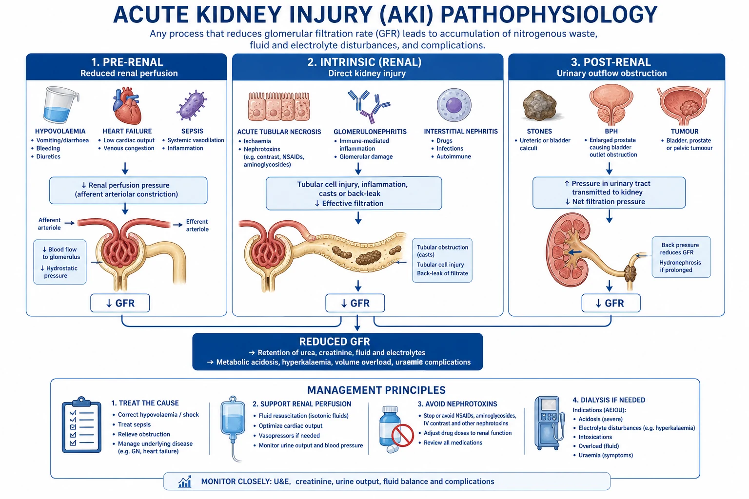
The common final pathway of all AKI is a fall in glomerular filtration rate (GFR), but the mechanism differs by cause — and understanding the mechanism explains why pre-renal AKI recovers in hours while ATN takes days to weeks. [1]
Pre-renal azotaemia — intact tubules, falling perfusion. GFR depends on glomerular capillary hydrostatic pressure, which is maintained by autoregulation: prostaglandins dilate the afferent arteriole and angiotensin II constricts the efferent arteriole. When perfusion drops (hypovolaemia, low cardiac output, sepsis), the tubules are intact and avidly reabsorb sodium and water to preserve circulating volume — hence a low urine sodium (under 20), concentrated urine (osmolality over 500), low FENa (under 1 per cent) and high BUN/Cr (over 20:1). Recovery is rapid once perfusion is restored, because no structural damage has occurred — but untreated pre-renal AKI progresses to ischaemic ATN within hours to days.[1]
Why NSAIDs and ACE inhibitors/ARBs precipitate AKI — the autoregulation trap. In a well-perfused kidney these drugs are harmless; in a volume-depleted patient whose GFR depends entirely on prostaglandins (afferent dilation) and angiotensin II (efferent constriction), blocking either one collapses glomerular pressure: NSAIDs constrict the afferent arteriole, ACE inhibitors/ARBs dilate the efferent arteriole. Both convert a compensated pre-renal state into AKI. This is the "triple whammy": NSAID plus ACE inhibitor plus diuretic in a volume-depleted patient is a classic AKI precipitant.[1]
Acute tubular necrosis (ATN) — structural tubular injury. ATN follows ischaemia (prolonged pre-renal insult — haemorrhage, sepsis, hypotension, cardiac arrest) or a direct nephrotoxin (aminoglycosides, contrast, heavy metals, myoglobin, haemoglobin, tumour lysis products). Four interlocking mechanisms drive the loss of GFR:[1]
- Tubular cell injury and apoptosis — ischaemia depletes ATP; the proximal tubule (S3 segment in the outer medulla, the most metabolically active and least oxygenated zone) is most vulnerable. Cells lose their brush border, detach from the basement membrane and shed into the lumen.
- Cast obstruction — detached cells and Tamm-Horsfall protein form muddy brown granular casts that block the tubular lumen, raising intratubular pressure and opposing filtration.
- Back-leak of filtrate — loss of tubular integrity allows glomerular filtrate to leak back across the damaged epithelium into the interstitium, so "filtered" solute never reaches the urine.
- Intrarenal vasoconstriction — injured tubules release cytokines and activate tubuloglomerular feedback (adenosine, via the macula densa), constricting the afferent arteriole and further dropping GFR. Endothelial injury, microvascular congestion and the inflammatory response perpetuate the cycle. [1]
Because tubular cells must regenerate (a process taking days to weeks), ATN recovers slowly and predictably passes through an oliguric phase followed by a polyuric/diuretic phase (as recovering tubules cannot concentrate urine) before GFR normalises. [1]
Pigment nephropathy (rhabdomyolysis and haemolysis). Myoglobin (from crushed or injured muscle) and free haemoglobin (from haemolysis) are freely filtered, then endocytosed by proximal tubule cells where they release free iron, generating reactive oxygen species, depleting ATP and triggering cast formation with Tamm-Horsfall protein (especially when urine is acidic and volume-depleted). Dipstick is positive for "blood" but microscopy shows no red cells — the tell-tale dissociation. CK over 5000 IU/L defines clinically significant rhabdomyolysis with high AKI risk.[5]
Contrast-induced AKI. Iodinated contrast causes renal medullary hypoxia (contrast increases blood viscosity and medullary oxygen demand) plus direct tubular toxicity and free-radical injury, producing ATN within 24 to 72 hours, typically non-oliguric. [1]
Tumour lysis syndrome. Rapid tumour cell kill (haematological malignancy after chemo, or a bulky treatment-sensitive tumour) releases uric acid, potassium and phosphate. Urate and calcium-phosphate crystals obstruct and injure tubules; hyperkalaemia and hyperphosphataemia are the immediate threats. [1]
Why a "normal" creatinine does not exclude AKI. Creatinine is a lagging, muscle-mass-dependent marker: a small elderly patient may have lost 50 per cent of GFR and still show a creatinine within the "normal" laboratory range. Urine output and trend are more sensitive than a single creatinine value. [1]
Clinical Presentation
AKI is frequently asymptomatic and detected on routine biochemistry. When symptomatic, the symptoms are those of uraemia, fluid overload and electrolyte disturbance, superimposed on the features of the precipitating cause. [1]
General features of AKI itself: oliguria (under 0.5 mL/kg/h) or anuria, fluid overload (dyspnoea, oedema, hypertension), and the uraemic syndrome — anorexia, nausea, vomiting, confusion, asterixis (flap), pericarditic chest pain, friction rub, pruritus, bruising, and in severe cases seizures and coma. [1]
Cause-specific clues (the history and examination that crack the diagnosis):[1][1]
- Hypovolaemia / pre-renal: thirst, dry mucosae, reduced skin turgor, tachycardia, hypotension or postural drop, oliguria, cold peripheries, slow capillary refill. History of vomiting, diarrhoea, haemorrhage, burns, diuretic overuse, or diabetic ketoacidosis.
- Sepsis: fever or hypothermia, rigors, hypotension, warm or mottled peripheries, source signs (pneumonia, pyelonephritis, peritonitis, line infection). Sepsis is the commonest single cause of hospital-acquired AKI.
- Obstructive (post-renal): anuria or fluctuating output, palpable bladder, enlarged prostate on PR examination, pelvic mass, single kidney, prior pelvic/retroperitoneal surgery or radiotherapy, neurogenic bladder (spinal cord disease, diabetes). Straining, hesitancy, weak stream and nocturia suggest BPH.
- Nephrotoxin exposure: detailed drug chart review — recent contrast imaging, NSAIDs, ACE inhibitors/ARBs, aminoglycosides, amphotericin, chemotherapy, tenofovir, aciclovir; recreational or occupational toxins.
- Acute interstitial nephritis: the classic triad of fever, rash and eosinophilia (often incomplete — only a minority have all three); onset days to weeks after a new drug (penicillin, PPI, NSAID, sulphonamide, rifampicin); may have arthralgia and eosinophiluria.
- Glomerulonephritis / vasculitis: haematuria (smoky or cola-coloured urine), oedema and hypertension (nephritic syndrome), proteinuria; systemic features — rash, palpable purpura (vasculitis), arthralgia, mouth ulcers, sinusitis, haemoptysis/lung cavities (granulomatosis with polyangiitis), haemoptysis (anti-GBM / Goodpasture), abdominal pain and diarrhoea (HUS).
- Rhabdomyolysis: muscle pain, weakness, swelling, dark (tea- or cola-coloured) urine; history of trauma, prolonged immobility, statin, cocaine, seizure, exertion, burns or electrical injury.
- Tumour lysis syndrome: high tumour burden (leukaemia, lymphoma, bulky solid tumour) within 48 to 72 hours of starting chemotherapy or steroids; hyperkalaemia, hyperphosphataemia, hypocalcaemia, hyperuricaemia.
- Atheroembolism: after vascular intervention (catheter, angiography, vascular surgery) or spontaneously; livedo reticularis, blue toes, eosinophilia, hypocomplementaemia; AKI may evolve over weeks.
- Hepatorenal syndrome: cirrhosis with ascites, jaundice, hypoalbuminaemia; AKI that fails to improve after albumin challenge. [1]
Atypical presentation in the elderly: AKI often presents as confusion, falls, functional decline, incontinence or drug toxicity rather than oliguria — older patients have reduced renal reserve, polypharmacy (the vulnerable kidney) and blunted thirst, so a lower threshold to check creatinine is essential. [1]
AKI in CKD (acute-on-chronic): a patient with known CKD develops AKI from a small insult (an NSAID, an episode of dehydration, a contrast study) that would not damage a normal kidney. Differentiate acute-on-chronic (potentially recoverable) from end-stage disease (small kidneys, anaemia, mineral bone disease). [1]
Differential Diagnosis
The immediate task is to place the patient in pre-renal, intrinsic or post-renal, then refine within intrinsic. Use the history, urine biochemistry (FENa, urine sodium, urine osmolality, BUN/Cr) and urine sediment together — no single test is definitive. [1]
Pre-renal vs intrinsic (ATN) — the classic distinction:[1]
| Index | Pre-renal | Intrinsic (ATN) |
|---|---|---|
| FENa (fractional excretion of sodium) | Under 1 per cent | Over 2 per cent |
| Urine sodium | Under 20 mmol/L | Over 40 mmol/L |
| Urine osmolality | Over 500 mOsm/kg | Under 350 (isosthenuric, near 300) |
| BUN/creatinine ratio | Over 20:1 | 10 to 20:1 |
| Urine sediment | Bland, hyaline casts | Muddy brown granular casts, renal tubular cells |
| Response to fluid challenge | Recovers (urine output rises) | No improvement |
Calculating FENa: FENa (per cent) = (urine sodium × serum creatinine) / (serum sodium × urine creatinine) × 100. A spot simultaneous urine and serum sample is enough. Caveat: FENa is invalid after recent diuretics (which force sodium excretion regardless of cause) — in that setting use fractional excretion of urea (FEurea), where under 35 per cent suggests pre-renal. FENa is also unreliable in CKD, glycosuria (osmotic diuresis), and salt-wasting states. [1]
Distinguishing the intrinsic subtypes by sediment (the sediment is the "poor man's biopsy"):[1]
- Muddy brown granular casts and renal tubular epithelial cells — acute tubular necrosis.
- Dysmorphic RBCs and RBC casts — glomerulonephritis (a glomerular bleed; RBC casts are pathognomonic of glomerular disease).
- WBC casts, eosinophiluria, sterile pyuria — acute interstitial nephritis (eosinophiluria is Hansel-stain positive; also consider papillary necrosis, tuberculosis, urinary tract infection).
- Pigmented granular casts, dipstick-positive blood but no RBCs — rhabdomyolysis (myoglobin) or haemolysis (haemoglobin).
- Bland sediment with leucocytes — consider obstruction, atypical infection, or pre-renal disease.
- Crystals — urate (tumour lysis), calcium oxalate (ethylene glycol), envelope-shaped calcium phosphate (hyperparathyroidism). [1]
When obstruction MUST be actively excluded: anuria, single kidney or renal transplant, recent pelvic or retroperitoneal surgery, prostate or pelvic malignancy, neurogenic bladder, or bilateral flank pain with stones. A bladder scan and renal ultrasound within 24 hours (immediately if obstructed and infected — a urological emergency). Hydronephrosis on ultrasound confirms obstruction, but its absence does not exclude early obstruction, retroperitoneal fibrosis, or encasement of ureters by tumour. [1]
AKI vs CKD — re-visited: small echogenic kidneys, normocytic anaemia, hyperphosphataemia/hypocalcaemia, raised PTH, and long-standing stable creatinine favour chronic. A clear precipitant with a rising creatinine, normal-sized kidneys and recent decline favours acute (or acute-on-chronic). Always look for a previous creatinine in the records. [1]
Clinical & Bedside Assessment
The bedside assessment answers three questions in order: is the patient volume-depleted or overloaded, what is the cause, and is there a life-threatening complication? [1]
Volume-status examination (decides the fluid challenge):[1]
- Hypovolaemia (give fluid): tachycardia, hypotension or postural drop (systolic fall over 20 mmHg or diastolic over 10 on standing), cool peripheries, slow capillary refill (over 2 seconds), reduced skin turgor, dry axillae and mucosae, low JVP, sunken eyes, oliguria.
- Hypervolaemia (avoid fluid, consider diuretic or dialysis): raised JVP, bilateral basal crackles, peripheral and sacral oedema, ascites, gallop rhythm, hypertension, S3. [1]
Haemodynamic target: restore a mean arterial pressure of 65 mmHg or more to maintain renal perfusion; in hypertensive or chronically vasoconstricted patients a higher target may be needed. Assess perfusion, not just pressure: lactate, capillary refill, mottling and urine output are the markers that matter — a normal blood pressure does not exclude shock. [1]
Targeted search for the cause: [1]
- Septic screen — temperature, source examination (chest, urine, abdomen, lines, wounds, meninges), blood cultures, lactate.
- Abdominal examination — palpable bladder (obstruction), renal masses (tumour, polycystic), ascites (cirrhosis/HRS), abdominal aortic aneurysm.
- Rectal/vaginal examination — enlarged prostate, pelvic mass, cervical or rectal malignancy.
- Skin — rash, purpura (vasculitis), livedo and blue toes (atheroembolism), petechiae, cellulitis (source).
- Drug chart review — go through every drug: stop NSAIDs, hold ACE inhibitors/ARBs, review aminoglycoside levels, antifungals, chemotherapy, antivirals; check for recent contrast and over-the-counter NSAID use (patients often forget ibuprofen/naproxen).
- Cardiac examination — signs of heart failure (low-output pre-renal AKI), pericardial rub (uraemic pericarditis), new murmur (endocarditis).
- Neurology — confusion, asterixis (uraemic flap), seizures (uraemia or hypertensive encephalopathy). [1]
Always check potassium with a venous gas at presentation and review the ECG — hyperkalaemia kills AKI patients before any other complication. [1]
Investigations
First-line investigations in every AKI:[1][1]
- Urea, electrolytes, creatinine, eGFR — establish the baseline and trend (compare with any prior result; this is the single most useful piece of data).
- Venous bicarbonate (or venous gas) — metabolic acidosis; a pH under 7.1 to 7.15 or bicarbonate under 12 is severe.
- Sodium, potassium, chloride — hyperkalaemia, hyponatraemia, anion-gap acidosis.
- Calcium, phosphate, magnesium — hypocalcaemia, hyperphosphataemia.
- Full blood count — anaemia (haemolysis, CKD), eosinophilia (AIN), leucocytosis (sepsis), thrombocytopenia (HUS/TTP, sepsis, DIC), fragmented red cells and schistocytes (HUS/TTP).
- CRP, blood cultures, lactate — sepsis work-up.
- Creatine kinase (CK) — rhabdomyolysis (CK over 5000 indicates high risk).
- Urinalysis and microscopy — protein, blood, leucocytes, nitrites, glucose; sediment is the key discriminator (see Differential Diagnosis).
- Urine electrolytes (sodium and creatinine) with a simultaneous serum sample — to calculate FENa (or FEurea if on diuretics).
- ECG — hyperkalaemia (peaked T waves, flattened/absent P waves, prolonged PR, widened QRS, sine wave), pericarditis (diffuse ST elevation, PR depression).
- Renal ultrasound — kidney size (small suggests CKD; large suggests obstruction, polycystic, infiltrative), hydronephrosis (obstruction), and to confirm two kidneys before biopsy.
- Bladder scan — post-void residual, retention.
- Drug levels — gentamicin/vancomycin troughs.
- Hepatic screen and clotting — coagulopathy, cirrhosis/HRS; group and save. [1]
Second-line (cause-directed) investigations — requested when intrinsic disease is suspected: serum immunoglobulins, serum free light chains and electrophoresis (myeloma); complement C3/C4 (low in lupus, post-infectious GN, cryoglobulinaemia); autoantibodies — ANA, ANCA (MPO/PR3), anti-GBM — for glomerulonephritis/vasculitis; hepatitis B/C and HIV serology (related GN); antistreptolysin O and anti-DNase B (post-streptococcal GN); myoglobin, lactate dehydrogenase, haptoglobin, blood film (rhabdomyolysis/haemolysis); urine for eosinophils (Hansel stain, AIN); renal biopsy (the definitive test for unexplained intrinsic AKI, especially suspected RPGN). [1]
The fractional excretion of sodium (FENa) — formula and interpretation:[1]
FENa (per cent) = (Urine Na x Serum Cr) / (Serum Na x Urine Cr) x 100 [1]
- Under 1 per cent — pre-renal azotaemia (intact tubular sodium retention)
- Over 2 per cent — intrinsic (ATN; damaged tubules cannot retain sodium)
- Misleading after diuretics — use FEurea = (Urine Urea x Serum Cr) / (Serum Urea x Urine Cr) x 100, where under 35 per cent supports pre-renal. [1]
Urine sediment — reproduce the high-yield associations:[1]
- Muddy brown granular casts + renal tubular epithelial cells → ATN
- Dysmorphic RBCs and RBC casts → glomerulonephritis
- WBC casts and eosinophiluria → acute interstitial nephritis
- Sterile pyuria → obstruction, tuberculosis, interstitial nephritis
- Pigmented casts, dipstick blood but no RBCs → rhabdomyolysis/haemolysis [1]
BUN/creatinine ratio thresholds: over 20:1 supports pre-renal (the kidney avidly reabsorbs urea with sodium and water); 10 to 20:1 favours intrinsic. (In SI units, multiply BUN by 2.14 to convert mg/dL of urea nitrogen to urea in mmol/L; the ratio itself is dimensionless and threshold is the same.) [1]
Hyperkalaemia ECG changes — reproduce in order of severity:[1]
- Peaked (tented) T waves — earliest, "pinched" base, narrow
- Flattening or loss of P waves
- Prolonged PR interval
- Widening of the QRS complex
- Sine wave — pre-arrest; the ECG "melts" into a sinusoidal pattern
- Ventricular fibrillation / asystole [1]
The urgent biochemistry threshold is potassium over 6.0 mmol/L (or over 5.5 with ECG changes) — treat immediately; do not wait for a confirmatory repeat. [1]
KDIGO staging — reproduced verbatim
(Creatinine criterion uses the ratio to baseline; urine output is mL/kg/h. Stage by the worst of the two.) [1]
Management — Resuscitation

The first priority in every AKI is the ABCDE primary survey, then a cause-directed resuscitation bundle that applies to all patients, with hyperkalaemia treated as a separate emergency if present. [1]
Five things you do for EVERY AKI (the universal bundle):[1][1]
- Stop nephrotoxic drugs — NSAIDs, ACE inhibitors, ARBs, aminoglycosides (unless essential, then therapeutic drug monitoring), iodinated contrast, metformin (lactic acidosis risk), and review herbal/over-the-counter agents.
- Review and adjust all drug doses for renal function — vancomycin, digoxin, low-molecular-weight heparins, morphine/metabolites, beta-blockers, sulphonylureas, gabapentin/pregabalin; consult a renal drug-dosing chart or pharmacist.
- Exclude and relieve obstruction — bladder scan; if in retention, catheterise; arrange renal ultrasound within 24 hours (immediately if septic obstruction — a urological emergency).
- Optimise haemodynamics — target mean arterial pressure 65 mmHg or more; treat hypotension with fluid if volume-depleted, or vasopressors if vasodilated (sepsis).
- Monitor — strict fluid balance, daily weights, urine output hourly (catheter if oliguric or unstable), and daily U&E and creatinine (more often if unstable or hyperkalaemic). [1]
Resuscitation of pre-renal / hypovolaemic AKI: a fluid challenge with balanced crystalloid (Hartmann's or Plasma-Lyte) 250 to 500 mL over 15 to 30 minutes (10 to 15 mL/kg), then reassess fluid responsiveness (passive leg raise, pulse-pressure variation, IVC, JVP) and urine output before giving more. Avoid hydroxyethyl starch (CHESS-AKI/6S trials showed increased AKI and mortality) and avoid albumin outside cirrhosis; 0.9 per cent saline in large volumes causes hyperchloraemic metabolic acidosis and may worsen AKI, hence balanced crystalloid preferred.[1]
Resuscitation of septic AKI — the Surviving Sepsis hour-1 bundle:[1]
- Oxygen to target saturation 94 to 98 per cent (or 88 to 92 per cent in COPD)
- Blood cultures before antibiotics (do not delay antibiotics for cultures)
- Broad-spectrum IV antibiotics within 1 hour
- Lactate; remeasure if raised
- Balanced crystalloid 30 mL/kg bolus for septic shock or lactate over 4
- Noradrenaline (norepinephrine) as first-line vasopressor for fluid-refractory shock, titrated to MAP 65 mmHg or more; vasopressin as second line [1]
Hyperkalaemia — a medical emergency. If potassium is over 6.0 mmol/L or any ECG change is present, treat immediately and simultaneously:[1]
- Calcium gluconate 10 per cent, 10 mL IV over 5 to 10 minutes — to stabilise the myocardial membrane (no effect on potassium level; repeat if ECG changes persist). Calcium chloride is an alternative via a central line.
- Insulin-dextrose — 10 units of soluble (regular) insulin IV with 25 g (50 mL of 50 per cent) dextrose IV over 15 to 30 minutes; lowers potassium by driving it into cells. Check blood glucose at 30 minutes, 1, 2, 3 and 6 hours (risk of hypoglycaemia for up to 6 hours).
- Salbutamol 10 to 20 mg nebulised (or 0.5 mg IV) — adjunctive; shifts potassium into cells via beta-2 stimulation; useful if insulin/dextrose contraindicated.
- Sodium bicarbonate — only if a significant metabolic acidosis coexists (pH under 7.1 to 7.15); slow to act and limited effect on potassium in isolation.
- Potassium removal — sodium polystyrene sulfonate 15 to 30 g orally or rectally (slow; hours to act); newer binders (patiromer, sodium zirconium cyclosilicate) have a faster onset. Dialysis if refractory or ongoing potassium release. [1]
Why fluid overload is harmful and colloids are avoided. A positive fluid balance worsens outcomes in AKI: it causes pulmonary oedema, raises intra-abdominal and renal venous pressure, and may delay recognition of ongoing injury. Colloids (albumin, hydroxyethyl starch) offer no benefit over crystalloid in most AKI, and starch increases AKI and mortality (CHESS-AKI, 6S trials). Treat volume, not a number. [1]
Management — Definitive & Stepwise
The definitive management of AKI follows a four-step ladder; only stage 3 / dialysis-requiring AKI needs critical care.[1]
The four-step AKI ladder: [1]
- Treat the cause — fluids for pre-renal, antibiotics for sepsis, relieve obstruction, stop nephrotoxins, immunosuppression for RPGN, steroids for severe AIN, rasburicase for tumour lysis.
- Optimise haemodynamics — fluid if depleted, vasopressors if vasodilated, target MAP over 65; inotropes if cardiogenic.
- Manage complications — hyperkalaemia (ladder above), metabolic acidosis, fluid overload, and nutrition.
- Renal replacement therapy when indicated (AEIOU, below). [1]
Hyperkalaemia treatment ladder (drug, dose, route, rationale) — reproduced:[1]
- Calcium gluconate 10 per cent — 10 mL IV over 5 to 10 minutes — membrane stabilisation (no effect on K level); repeat if ECG changes persist
- Insulin 10 units + 50 per cent dextrose 50 mL IV — shifts K into cells; onset 15 to 30 minutes, duration 4 to 6 hours; monitor glucose
- Salbutamol 10 to 20 mg nebulised — shifts K into cells (beta-2); adjunctive
- Sodium bicarbonate (1.26 per cent or 8.4 per cent) — for acidosis; slow, modest K-lowering
- Potassium binders — sodium polystyrene sulfonate 15 to 30 g orally/rectally; patiromer or sodium zirconium cyclosilicate (faster); slow onset
- Dialysis — definitive removal; for refractory hyperkalaemia [1]
Metabolic acidosis: sodium bicarbonate is given if pH is under 7.1 to 7.15 (or bicarbonate under 12) — target a pH over 7.15 to 7.20; avoid in volume overload (dialyse instead). Severe acidosis impairs cardiac contractility and vasopressor responsiveness. [1]
Fluid overload and pulmonary oedema: sit the patient up, give oxygen, loop diuretic (furosemide 40 to 80 mg IV) if still responsive (diuretics do not treat AKI but can convert oliguric to non-oliguric AKI and ease fluid management), and dialyse if refractory. [1]
Nutrition: enteral nutrition early; adequate calories (25 to 30 kcal/kg/day) and protein 0.8 to 1.0 g/kg/day in non-dialysed AKI (higher, 1.0 to 1.5 g/kg/day, on RRT); restrict sodium, potassium and phosphate. Avoid protein restriction (malnutrition increases mortality). [1]
Relieving obstruction: a urethral catheter for bladder-outlet obstruction; a percutaneous nephrostomy or retrograde ureteric stent for ureteric obstruction. Watch for post-obstructive diuresis (polyuria after relief from obstruction, due to tubular dysfunction) — replace fluid and electrolytes to avoid hypovolaemia and a secondary AKI. [1]
Specific disease-directed therapies (the right drug for the right cause): [1]
- Acute interstitial nephritis — stop the offending drug; give a short course of prednisolone 1 mg/kg/day (max 60 mg) tapering over 2 to 4 weeks if severe or biopsy-confirmed; biopsy if not recovering.
- Rapidly progressive glomerulonephritis — urgent renal biopsy; methylprednisolone pulses (500 mg to 1 g IV daily for 3 days) followed by oral prednisolone plus cyclophosphamide (or rituximab for ANCA vasculitis); plasma exchange for anti-GBM disease and severe pulmonary haemorrhage. Do not delay immunosuppression for biopsy in the sick patient.
- Rhabdomyolysis — aggressive isotonic saline to target urine output 200 to 300 mL/h until CK falls; consider sodium bicarbonate to alkalinise urine (evidence mixed); treat compartment syndrome, hyperkalaemia, hypocalcaemia (only if symptomatic).
- Tumour lysis syndrome — aggressive hydration before and during chemotherapy; rasburicase (0.15 to 0.2 mg/kg) for established or high-risk hyperuricaemia (breaks down uric acid to soluble allantoin; contraindicated in G6PD deficiency); allopurinol 300 mg daily for prophylaxis; manage hyperkalaemia and hyperphosphataemia.
- Hepatorenal syndrome — albumin challenge (1 g/kg/day for 2 days) to confirm diagnosis; treat with terlipressin (1 to 2 mg IV every 4 to 6 hours) plus albumin, or noradrenaline in ICU; definitive treatment is liver transplant.
- Contrast-induced AKI — isotonic saline hydration (1 mL/kg/h for 6 to 12 hours pre- and post-procedure); hold metformin and nephrotoxins; use lowest possible contrast volume, iso-osmolar or low-osmolar agents. N-acetylcysteine and bicarbonate are NOT effective (PRESERVE trial).[4]
Indications for renal replacement therapy — AEIOU (reproduce verbatim):[2][3]
- A — Acidosis — severe, refractory metabolic acidosis (pH under 7.1 to 7.15)
- E — Electrolytes — refractory hyperkalaemia (K over 6.5 or persistently over 6.0 despite medical therapy)
- I — Ingestion — dialysable toxins: lithium, salicylate, methanol, ethylene glycol, metformin (in severe lactic acidosis), barbiturates
- O — Overload — refractory pulmonary oedema / fluid overload unresponsive to diuretics
- U — Uraemia — uraemic pericarditis, uraemic encephalopathy (seizures, coma), uraemic bleeding, or severe symptomatic uraemia [1]
(Some add I for Intractable acidosis or use the variant AEIOU-H including severe drug toxicity; the core five are exam standard.) [1]
When to refer: nephrology for stage 3 AKI, AKI failing to improve after 48 to 72 hours, suspected intrinsic/obstructive cause, or any patient meeting AEIOU criteria; critical care/ICU for AKI with multi-organ failure, refractory hyperkalaemia, severe acidosis, fluid overload needing RRT, or vasopressor requirement. [1]
Specific Subtypes & Scenarios
- Contrast-induced AKI (CI-AKI): KDIGO defines it as a creatinine rise of 0.3 mg/dL or more or 1.5 times baseline within 48 to 72 hours of contrast without an alternative cause. Risk factors: pre-existing CKD (especially eGFR under 30), diabetes, heart failure, hypovolaemia, high contrast volume, intra-arterial injection. Prevention is isotonic saline hydration; hold metformin (lactic acidosis risk) and nephrotoxins; use lowest contrast volume, iso-osmolar or low-osmolar agents. N-acetylcysteine and sodium bicarbonate do NOT prevent CI-AKI (PRESERVE, NEJM 2018).[4]
- Rhabdomyolysis: CK over 5000 IU/L defines clinically significant disease; the classic biochemical picture is hyperkalaemia, hyperphosphataemia, hypocalcaemia, high anion-gap acidosis, raised CK and myoglobin, dipstick blood with no red cells, pigmented granular casts. Treat with aggressive isotonic saline (target urine output 200 to 300 mL/h) and management of compartment syndrome and electrolytes. Alkalinisation of urine is controversial; mannitol is not routinely recommended.
- Acute interstitial nephritis (AIN): drugs cause most cases — penicillins (methicillin classic), proton-pump inhibitors, NSAIDs (often with nephrotic-range proteinuria), sulphonamides, rifampicin, 5-flucytosine, allopurinol, mesalazine; also infection and autoimmune. Urine shows WBC casts, eosinophiluria, sterile pyuria, haematuria, mild proteinuria. The classical fever-rash-eosinophilia triad is often absent. Management: stop the offending drug; prednisolone 1 mg/kg/day (max 60 mg) if severe, biopsy-confirmed, or not recovering within a week.
- Hepatorenal syndrome (HRS-AKI): cirrhosis with ascites; AKI that fails to improve after 48 hours of albumin challenge (1 g/kg/day) without shock, nephrotoxins, or structural kidney disease; terlipressin plus albumin is first-line vasoconstrictor therapy, noradrenaline in ICU; definitive treatment is liver transplant.
- Tumour lysis syndrome: a metabolic emergency within 48 to 72 hours of cytotoxic therapy in high-burden haematological malignancy; hyperuricaemia, hyperkalaemia, hyperphosphataemia, hypocalcaemia, AKI. Prevention with hydration and rasburicase (high risk) or allopurinol (lower risk) is the standard of care.
- Obstructive uropathy: relieve with catheter (bladder outlet) or nephrostomy / ureteric stent (ureteric); watch for post-obstructive diuresis (replace fluid and electrolytes).
- Atheroembolic disease: after vascular procedures or spontaneously; livedo reticularis, blue toes, eosinophilia, hypocomplementaemia, AKI evolving over weeks; supportive care; prognosis poor for renal recovery.
- Cancer-related AKI: any combination of obstruction, direct infiltration, tumour lysis, chemotherapy toxicity (cisplatin, ifosfamide), immunotherapy (immune-checkpoint inhibitor AIN and GN), and paraneoplastic GN; tailored to mechanism.
Dialysis in AKI — AEIOU
AEIOU
refractory metabolic acidosis, pH under 7.1 to 7.15
refractory hyperkalaemia, K over 6.5 despite medical therapy
dialysable toxins: lithium, salicylate, methanol, ethylene glycol, metformin
refractory pulmonary oedema unresponsive to diuretics
uraemic pericarditis, encephalopathy (seizures, coma), or bleeding
Complications & Pitfalls
Metabolic complications:[1]
- Hyperkalaemia — the leading cause of death in AKI; arrhythmia, asystole
- Metabolic acidosis — high anion gap from organic acid retention; worsens hyperkalaemia and hypotension
- Hyponatraemia — water retention with isosthenuria
- Hyperphosphataemia with hypocalcaemia — phosphate retention, reduced vitamin D activation; symptomatic hypocalcaemia (tetany, seizures) is rare unless rhabdomyolysis or tumour lysis
- Hyperuricaemia, hypermagnesaemia [1]
Volume complications: pulmonary oedema (the commonest reason for emergent RRT), hypertension, peripheral and pulmonary oedema, ascites. [1]
Uraemic complications: uraemic pericarditis (a dialysis indication; friction rub, chest pain, effusion), uraemic encephalopathy (confusion, asterixis, seizures, coma), bleeding (platelet dysfunction), nausea, anorexia, pruritus, anaemia (reduced erythropoietin). [1]
Life-threatening complications: arrhythmia and cardiac arrest from hyperkalaemia; uraemic pericarditis with tamponade; seizures/coma from uraemic encephalopathy; severe acidosis with circulatory collapse; massive pulmonary oedema; infection (the leading cause of death in AKI). [1]
Classic pitfalls (every one an exam question):[1][1]
- Missing obstruction — always do a bladder scan and renal ultrasound; anuria, single kidney, or pelvic malignancy are red flags
- Failing to stop nephrotoxins — NSAIDs, ACE inhibitors/ARBs, aminoglycosides, contrast; review every drug including over-the-counter NSAIDs
- Over-resuscitation — causing pulmonary oedema; reassess responsiveness, do not chase a number
- Using FENa after diuretics — use FEurea (under 35 per cent for pre-renal) instead
- Attributing CKD to AKI (or vice versa) — small echogenic kidneys, anaemia, mineral bone disease favour chronic
- Missing rapidly progressive glomerulonephritis — dysmorphic RBCs/RBC casts need urgent biopsy and immunosuppression; delayed treatment causes irreversible scarring
- Missing rhabdomyolysis — dipstick positive for blood with no red cells is myoglobin (or haemoglobin)
- Treating the number, not the patient — giving furosemide to "make urine" without fluid overload worsens outcome
- Forgetting to redose / stop drugs — LMWH, vancomycin, metformin, digoxin accumulate and cause harm [1]
Prognosis & Disposition
AKI is not a benign lab abnormality — it independently increases short-term mortality, length of stay, CKD progression, and cardiovascular events. Prognosis depends on stage, cause (prerenal quickly reversed vs ATN vs RPGN), oliguria, need for dialysis, and comorbidity. [1]
KDIGO staging (reproduce exactly)
| Stage | Serum creatinine | Urine output |
|---|---|---|
| 1 | ≥0.3 mg/dL (26.5 µmol/L) rise in 48 h or 1.5–1.9× baseline in 7 days | <0.5 mL/kg/h for 6–12 h |
| 2 | 2.0–2.9× baseline | <0.5 mL/kg/h for ≥12 h |
| 3 | ≥3× baseline or ≥4.0 mg/dL (353.6 µmol/L) or RRT initiation or in patients <18 y, eGFR <35 mL/min/1.73 m² | <0.3 mL/kg/h for ≥24 h or anuria ≥12 h |
Disposition
- Ward with strict fluid balance, daily labs: stable Stage 1 with clear reversible cause
- HDU/ICU: hyperkalaemia emergencies, pulmonary oedema, severe acidosis, sepsis, need for emergency RRT, multi-organ failure
- Nephrology referral early if intrinsic renal disease suspected (active urine sediment, RPGN pattern, systemic vasculitis/myeloma clues), unclear cause, or progressive Stage 2–3
Recovery and follow-up
Re-check creatinine 1–3 months after AKI — even "recovered" AKI warrants CKD risk labelling, medication review, and BP/albuminuria follow-up. Avoid repeated nephrotoxic insults.
Special Populations
Elderly
Reduced reserve, polypharmacy (ACE-I/ARB + diuretic + NSAID = triple whammy), obstructive uropathy (prostate), dehydration. Creatinine may under-reflect injury because of low muscle mass — small absolute rises matter.
Pregnancy
Differentiate prerenal AKI, pyelonephritis, pre-eclampsia/HELLP, thrombotic microangiopathy, acute fatty liver, obstruction from gravid uterus (hydronephrosis). Obstetric-led care; delivery may be definitive therapy for pre-eclampsia-related injury.
Heart failure (cardiorenal)
Congestion can cause AKI via raised venous pressure; overdiuresis causes prerenal injury. Use weights, JVP, lactate, haemodynamics — not creatinine alone — to decide diuretic intensity. SGLT2i remain beneficial long-term in HF/CKD when stable.
Liver disease (HRS-AKI)
In cirrhosis with ascites, exclude hypovolaemia, shock, nephrotoxins, structural disease. Terlipressin + albumin protocols for hepatorenal syndrome where available; transplant is definitive for eligible patients.
Tumour lysis / rhabdomyolysis / myeloma
- TLS: hydration, urate lowering (allopurinol/rasburicase), monitor K+/Phos/Ca
- Rhabdomyolysis: aggressive isotonic fluid, watch K+; alkaline diuresis only in selected protocols
- Myeloma kidney: hydration, treat clone, avoid NSAIDs/contrast/dehydration; consider dialysis modalities for cast nephropathy in specialist centres
Contrast exposure
Risk highest with CKD, diabetes, volume depletion, high contrast volume. Prevent with volume expansion when appropriate; stop nephrotoxins; use lowest contrast dose; no proven benefit of routine N-acetylcysteine in modern practice.
Evidence, Guidelines & Regional Differences
KDIGO 2012 is the international standard for definition, staging and management of AKI — it harmonised the earlier RIFLE and AKIN schemes into a single creatinine-plus-urine-output definition and three-stage classification reproduced above.[1]
Dialysis timing — the two landmark trials:[2][3]
- AKIKI (Gaudry et al, NEJM 2016) — a French multicentre RCT in 620 critically-ill patients with severe AKI (KDIGO Stage 3): early (within 12 hours of eligibility) vs delayed (waiting for AKI progression or an urgent AEIOU indication) initiation of RRT. No difference in 60-day mortality; the delayed group received less dialysis and one in three never needed it. This supported a watchful, delayed strategy.[2]
- STARRT-AKI (Bagshaw et al, NEJM 2020) — a multinational RCT in 3019 critically-ill patients with severe AKI: accelerated (within 12 hours of Stage 3) vs standard (an AEIOU indication or persistent AKI) RRT strategy. No mortality benefit of accelerated initiation, and more adverse events (fluid overload, catheter complications).[3]
Synthesis: current evidence does not support early, prophylactic RRT; start dialysis for AEIOU, for AKI not recovering, or for persistent Stage 3 AKI with complications. The exception is the patient with a clear trajectory toward needing RRT in whom early preparation (vascular access, nephrology referral) is sensible. [1]
Contrast-induced AKI — PRESERVE:[4] the PRESERVE trial (Weisbord et al, NEJM 2018) randomised 5177 high-risk patients undergoing angiography in a 2x2 factorial to sodium bicarbonate vs isotonic saline and N-acetylcysteine vs placebo. Neither intervention prevented death, dialysis or persistent renal impairment at 90 days. The standard of prevention is now isotonic saline hydration alone; N-acetylcysteine and bicarbonate should not be relied upon.
Long-term outcomes:[5] James et al (Nature Reviews Nephrology, 2020) synthesised the evidence that AKI is a risk factor for incident and progressive CKD, end-stage kidney disease, cardiovascular events and mortality, with risk proportional to AKI severity and recurrence — establishing AKI and CKD as a continuum and supporting long-term follow-up of AKI survivors.
- KDIGO 2012 (international) — definition, staging, management; the global standard.
- NICE NG148 (UK) — aligned to KDIGO; adds e-alerts, Sick Day Guidance, and the principle that diuretics do not treat AKI.
- ICMR / Indian practice (resource-limited) — emphasises prevention (avoidance of NSAIDs, judicious fluid), conservative management, and peritoneal dialysis as a low-cost RRT option where haemodialysis access is limited; malnutrition, infections (malaria, leptospirosis, dengue), and nephrotoxic traditional medicines are additional regional causes of AKI. [1]
Hyperkalaemia Emergency Doses & RRT Indications (Exam Core)
Hyperkalaemia emergency doses (adult)
| Goal | Agent & dose | Onset |
|---|---|---|
| Membrane stabilisation | Calcium gluconate 10 mL of 10% IV over 5–10 min (repeat if ECG changes persist) | Minutes |
| Shift K intracellularly | Insulin 10 units regular with 25–50 mL of 50% dextrose IV (or 125 mL 20% glucose) | 15–30 min |
| Shift | Salbutamol 10–20 mg nebulised | 15–30 min |
| Shift / acidosis | Sodium bicarbonate only if severe metabolic acidosis — not routine sole therapy | Variable |
| Remove K | Loop diuretic if still making urine; cation exchangers / GI binders per local agent; haemodialysis if refractory | Hours / immediate with HD |
ECG changes (peaked T, flattened P, wide QRS, sine wave) → give calcium before or while shifting, do not wait for lab reconfirm if life-threatening tracing. [1]
Indications for urgent RRT (AEIOU mnemonic)
- Acidosis (severe refractory metabolic acidosis)
- Electrolytes (refractory hyperkalaemia)
- Intoxications (dialysable toxins: lithium, toxic alcohols, salicylate — selected cases)
- Overload (pulmonary oedema refractory to medical therapy)
- Uraemic complications (encephalopathy, pericarditis, bleeding diathesis)
Worked stem — triple whammy
Elderly patient on ramipril + furosemide starts ibuprofen for knee pain; presents oliguric with creatinine jump and K+ 6.6. Stop nephrotoxins, ECG, protect heart if needed, careful volume assessment, treat hyperkalaemia, avoid further NSAIDs, reintroduce RAAS blockade later only if clearly indicated and renal function recovering with close monitoring.
Worked NEET-PG Stems — AKI
- Oliguria after diarrhoea, FENa under 1%, bland sediment → pre-renal; fluids; stop ACE-I/NSAID.
- Muddy brown casts, FENa over 2% after prolonged hypotension → ATN; supportive care; avoid further injury.
- Post-void residual 800 mL, hydronephrosis → post-renal; catheter; urology.
- RPGN urine with RBC casts, rising Cr over days → urgent nephritis work-up/biopsy pathway.
- K+ 6.8 with wide QRS → calcium gluconate first, then shift, then remove.
- AEIOU present → urgent RRT.
- Triple whammy → ACE-I + diuretic + NSAID → stop NSAID, reassess volume/RAAS. [1]
Exam Pearls
- FENa under 1 per cent = pre-renal; over 2 per cent = ATN; misleading after diuretics — use FEurea under 35 per cent for pre-renal.[1]
- BUN/creatinine over 20:1 = pre-renal; 10 to 20:1 = intrinsic.
- Muddy brown granular casts = ATN; RBC casts = glomerulonephritis; WBC casts / eosinophiluria = acute interstitial nephritis.[1]
- Dipstick blood positive with no red cells on microscopy = rhabdomyolysis (myoglobin) or haemolysis (haemoglobin).
- KDIGO creatinine thresholds — verbatim: Stage 1 = 1.5 to 1.9 times baseline or up by 0.3 mg/dL; Stage 2 = 2.0 to 2.9 times baseline; Stage 3 = 3.0 times baseline, or over 4.0 mg/dL, or RRT.
- AEIOU = dialysis indications: Acidosis, Electrolytes (hyperkalaemia), Ingestion (lithium, salicylate, methanol, ethylene glycol, metformin), Overload (pulmonary oedema), Uraemia (pericarditis, encephalopathy).
- Hyperkalaemia ladder: calcium gluconate 10 per cent 10 mL IV (membrane stabilisation) → insulin 10 units + 50 per cent dextrose → salbutamol nebs → K-binders → dialysis.
- Always exclude obstruction with bladder scan and ultrasound first; stop nephrotoxins (NSAIDs, ACE inhibitors, aminoglycosides, contrast).
- NSAIDs constrict the afferent arteriole; ACE inhibitors/ARBs dilate the efferent arteriole — both drop glomerular pressure. The "triple whammy" is NSAID + ACE inhibitor + diuretic.
- ATN recovery: oliguric phase → polyuric (diuretic) phase → recovery. Pre-renal recovers in hours; ATN in days to weeks.
- AKIKI and STARRT-AKI: no mortality benefit of early over delayed dialysis — start for AEIOU or persistent AKI.[2][3]
- PRESERVE: N-acetylcysteine and bicarbonate do NOT prevent contrast-induced AKI — use isotonic saline.[4]
- Surviving Sepsis hour-1 bundle: oxygen, cultures, antibiotics within 1 hour, lactate, 30 mL/kg crystalloid, noradrenaline for shock.
- Creatinine is a late, lagging marker — a normal value early does not exclude significant AKI.
AKI cause framework — PIN
PIN
hypoperfusion — hypovolaemia, heart failure, sepsis, NSAIDs, ACE inhibitors (about 60 per cent)
within the kidney — ATN (commonest), glomerular, interstitial, vascular (about 35 per cent)
BPH, stones, tumour, retroperitoneal fibrosis (about 5 per cent)
Exam application bank (NEET-PG / INICET)
One-line answer
Acute kidney injury (AKI) is a rapid (hours to days) decline in kidney function defined by KDIGO as a serum creatinine rise of 0.3 mg/dL (26.5 micromol/L) or more within 48 hours, a creatinine 1.5 times baseline or more within 7 days, or urine output under 0.5 mL/kg/h for 6 hours or more. It is classified into pre-renal (hypoperfusion, around 60 per cent), intrinsic/renal (acute tubular necrosis, glomerular, interstitial, vascular) and post-renal (obstruction, around 5 per cent). Commonest intrinsic cause is acute tubular necrosis (ATN) from ischaemia or nephrotoxins. Presents with oliguria, fluid overload, uraemia, hyperkalaemia; diagnose with creatining trend, FENa, urine sediment, renal ultrasound. Management: treat the cause, restore perfusion with balanced crystalloid, stop nephrotoxins (NSAIDs, ACE inhibitors, aminoglycosides, contrast), manage hyperkalaemia and acidosis, and dialy [1]
Worked stems (answer without another resource)
Stem 1 — Classic presentation. Map symptoms to mechanism; name the first investigation and first treatment step with dose/route if drug therapy is standard. [1]
Stem 2 — Unstable / complicated. List red flags that force immediate resuscitation, theatre, ICU, antidote, or reperfusion — and what you do in the first 15 minutes. [1]
Stem 3 — Atypical group. Elderly, pregnancy, child, or immunocompromised: how presentation and thresholds change. [1]
Stem 4 — Differential trap. Name the three closest mimics and one discriminator for each. [1]
Stem 5 — Disposition. Who goes home with safety-netting, who is admitted, who needs HDU/ICU/theatre, and what follow-up is mandatory. [1]
Rapid viva checklist
- Definition + classification
- Pathophysiology chain
- Bedside signs / criteria
- Score with exact components (if any)
- Emergency bundle
- Definitive therapy with doses
- Complications of disease and of treatment
- Special populations
- Guideline/trial name if classic
- Three exam traps
Coverage self-check
If you cannot answer any stem above from this page alone, re-read the matching section — the page is intended to be self-sufficient for final-prof and NEET-PG/INICET questions on Acute Kidney Injury.
References
- [1]Susantitaphong P, Cruz DN, Cerda J, et al. World incidence of AKI: a meta-analysis Clin J Am Soc Nephrol, 2013.PMID 23744003
- [2]Gaudry S, Hajage D, Schortgen F, et al. Initiation Strategies for Renal-Replacement Therapy in the Intensive Care Unit N Engl J Med, 2016.PMID 27181456
- [3]STARRT-AKI Investigators; Bagshaw SM, Wald R, et al. Timing of Initiation of Renal-Replacement Therapy in Acute Kidney Injury N Engl J Med, 2020.PMID 32668114
- [4]Weisbord SD, Gallagher M, Jneid H, et al. Outcomes after Angiography with Sodium Bicarbonate and Acetylcysteine N Engl J Med, 2018.PMID 29130810
- [5]James MT, Bhatt M, Pannu N, Tonelli M. Long-term outcomes of acute kidney injury and strategies for improved care Nat Rev Nephrol, 2020.PMID 32051567