Nephrology · Nephrology
Chronic Kidney Disease
Also known as Chronic kidney disease · CKD · Chronic renal failure · CRF · End-stage kidney disease · ESKD
Chronic kidney disease (CKD) is defined by KDIGO as abnormalities of kidney structure or function present for more than 3 months, with implications for health. The operational definition is kidney damage markers (albuminuria, urine sediment abnormalities, imaging or histology) OR eGFR below 60 mL/min/1.73 m squared for over 3 months. CKD is classified by cause, GFR category (G1 to G5) and albuminuria category (A1 to A3) — the CGA staging that predicts risk of progression, cardiovascular events and mortality. CKD affects 10 to 13 percent of adults worldwide; diabetic kidney disease is the single largest cause (30 to 50 percent), followed by hypertensive nephrosclerosis (around 25 percent), glomerulonephritis, ADPKD, and obstructive or reflux nephropathy. CKD is usually silent until G3b to G4: uraemic symptoms, fluid overload, hyperkalaemia, acidosis, anaemia and renal bone disease emerge late. Management rests on four pillars: RAAS blockade (ACE inhibitor or ARB) for proteinuria, SGLT2 inhibition (dapagliflozin or empagliflozin) for cardiorenal protection, finerenone in diabetic CKD, and multifactorial cardiovascular risk reduction (statin, BP, glycaemia, smoking cessation, salt restriction, weight loss, avoidance of nephrotoxins) — plus treatment of anaemia (iron then ESA), CKD-MBD (phosphate binders, active vitamin D, calcimimetics), acidosis (sodium bicarbonate) and hyperkalaemia (diet, potassium binders). Renal replacement therapy planning (AV fistula 6 months ahead, transplant referral) starts at eGFR below 30. Dialysis is indicated for the AEIOU emergencies. Cardiovascular disease is the leading cause of death.
On this page & tools
Your progress
Saved locally on this device.
Practise this topic
Exam tags
Red flags

Overview & Definition
Chronic kidney disease (CKD) is defined by KDIGO as abnormalities of kidney structure or function, present for more than 3 months, with implications for health. The operational definition is fulfilled by either of two criteria persisting for at least three months: markers of kidney damage — albuminuria (urine albumin-to-creatinine ratio at least 30 mg per g), urine sediment abnormalities (glomerular haematuria, red-cell casts, white-cell casts, oval fat bodies), electrolyte and other abnormalities due to tubular disorders, structural abnormalities on imaging (polycystic kidneys, reflux, obstruction, single kidney), or histological abnormalities on biopsy — or a glomerular filtration rate below 60 mL per min per 1.73 m squared. Either criterion alone is sufficient; albuminuria with a normal GFR is still CKD (e.g. G1 A2), and a low GFR without kidney damage markers is still CKD if it is chronic. [1]
CKD is the irreversible, progressive loss of nephron mass that follows a final common pathway regardless of the original insult. The clinical skill examiners probe is not the diagnosis of an elevated creatinine but the recognition of five things: (1) the operational KDIGO definition so the disease is named correctly and not confused with acute kidney injury; (2) the CGA staging (cause, GFR, albuminuria) that quantifies the risk of progression to end-stage kidney disease and of cardiovascular death; (3) the silent early course that demands active screening of diabetics, hypertensives and the older population by eGFR and urine ACR; (4) the modern four-pillar disease-modifying therapy (RAAS blockade, SGLT2 inhibition, finerenone in diabetics, multifactorial risk reduction) that has transformed prognosis over the past decade; and (5) the complication cascade — anaemia, CKD-mineral and bone disorder, acidosis, hyperkalaemia, fluid overload and cardiovascular disease — that needs separate, concurrent management. [1]
The single most important conceptual shift of the past decade is that SGLT2 inhibitors are disease-modifying in CKD independent of glycaemic status — proven across three large outcome trials (CREDENCE in diabetic nephropathy, DAPA-CKD and EMPA-KIDNEY in both diabetic and non-diabetic CKD) — and that finerenone, a non-steroidal mineralocorticoid receptor antagonist, adds further kidney and cardiovascular protection on top of RAAS blockade in patients with type 2 diabetes. CKD is now managed as a chronic, combination-treated disease, not as a one-drug-after-another ladder. [3][4][5][6]
Classification
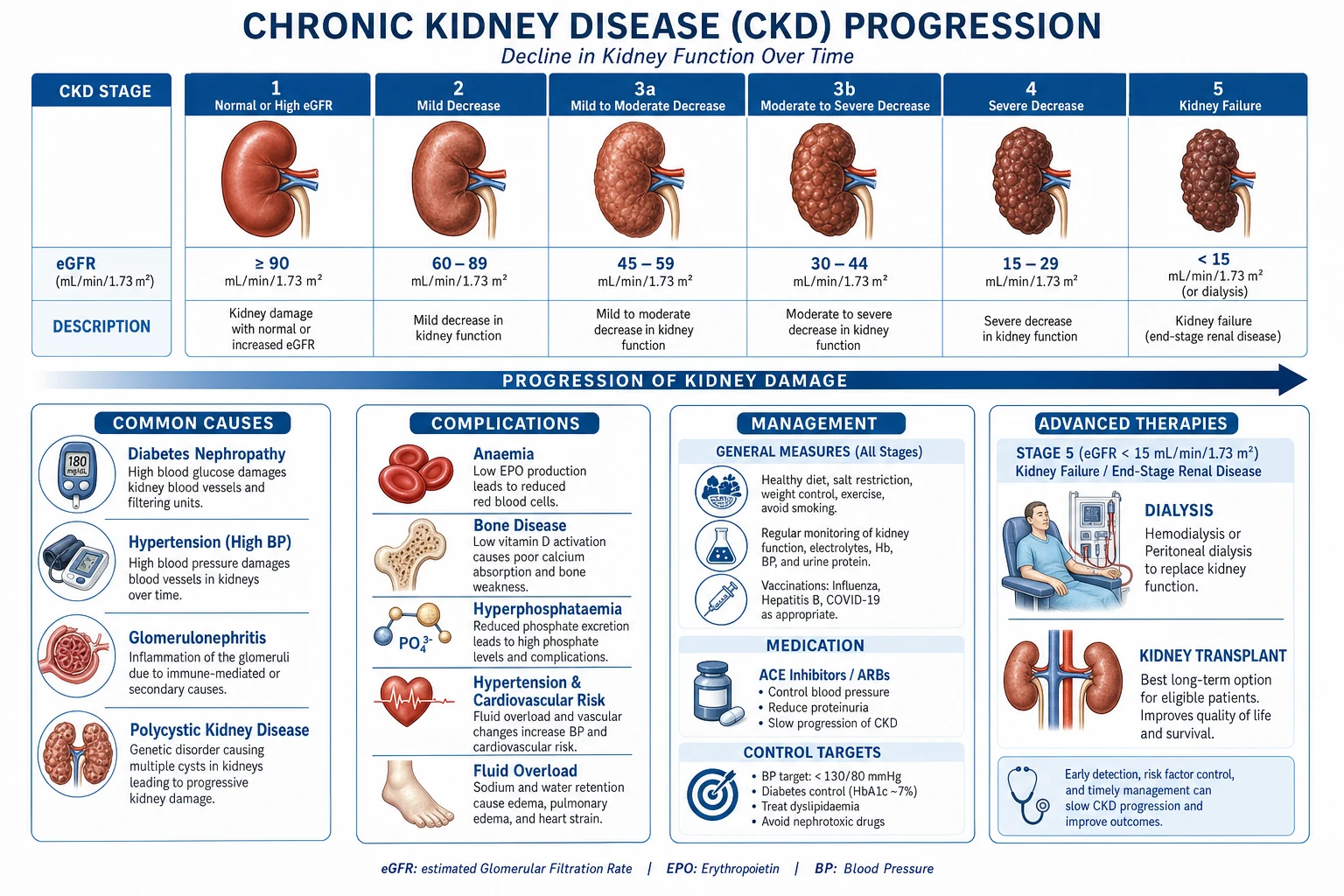
CKD is classified by the KDIGO CGA system — Cause, GFR category and Albuminuria category — because each axis independently predicts the risk of progression to end-stage kidney disease and of all-cause and cardiovascular mortality. Naming the cause (diabetic kidney disease, hypertensive nephrosclerosis, IgA nephropathy, ADPKD, lupus nephritis, obstructive uropathy, and so on) drives disease-specific therapy; the G and A categories drive referral, monitoring frequency and intensity of cardiorenal protection. [1]
KDIGO GFR categories (the G axis): [1]
G1 — at least 90
- Normal or high GFR
- CKD only if kidney-damage markers present (albuminuria, haematuria, imaging, biopsy)
G2 — 60 to 89
- Mildly decreased GFR
- A small age-related decline is physiological; CKD only if damage markers present
G3a — 45 to 59
- Mildly to moderately decreased
- Symptoms rare; start to investigate, address cardiovascular risk
G3b — 30 to 44
- Moderately to severely decreased
- Refer to nephrology; anaemia and CKD-MBD begin to appear; plan ahead
G4 — 15 to 29
- Severely decreased
- Active renal replacement therapy preparation; vascular access planning, transplant referral
G5 — under 15
- Kidney failure (end-stage kidney disease)
- Initiate dialysis or transplant when symptomatic or for AEIOU indications
KDIGO albuminuria categories (the A axis), measured by the urine albumin-to-creatinine ratio (UACR) on a random spot sample: [1]
A1 — under 30 mg per g
- Normal to mildly increased
- Low risk of progression when GFR is preserved; re-screen annually in at-risk groups
A2 — 30 to 300 mg per g
- Moderately increased (formerly microalbuminuria)
- Earliest marker of many glomerular diseases; start RAAS blockade and SGLT2 inhibitor here
A3 — over 300 mg per g
- Severely increased (formerly macroalbuminuria)
- High risk of progression; nephrotic-range over 2200 mg per g predicts a steep GFR decline
The intersection of the G and A axes gives the KDIGO heat-map risk (green, yellow, orange, red). Patients in G3b or worse combined with A3 carry the highest risk of progression to ESKD within five years and of cardiovascular death; they are referred for nephrology care. Importantly, albuminuria is a stronger predictor of progression than GFR alone: a patient in G2 A3 (mildly reduced GFR but heavy proteinuria) is at higher risk than one in G3a A1. [1]

Epidemiology & Risk Factors
CKD is one of the commonest non-communicable diseases worldwide. The global prevalence is 10 to 13 percent of adults, rising with age to over 25 percent in those older than 65, in whom a sizeable fraction of the apparent burden is the slow age-related GFR decline rather than progressive disease. CKD is a leading cause of death: it was the tenth leading cause of years of life lost globally in the most recent Global Burden of Disease assessments, with mortality continuing to rise while cardiovascular and cancer mortality fall. [1]
CKD — the headline numbers
Risk factors are divided into susceptibility, initiation and progression factors. Susceptibility factors (raise the likelihood of developing CKD but are not direct causes) include older age, family history of kidney disease, low birth weight, lower socio-economic status, and ethnic minority status — notably Black (in whom APOL1 risk variants explain much of the excess risk of FSGS and hypertension-attributed nephropathy), Hispanic, South Asian, Indigenous Australian and Native American populations. Initiation factors (the direct causes) are diabetes mellitus, hypertension, glomerulonephritis, polycystic kidney disease, obstructive uropathy, recurrent urinary tract infection, autoimmune disease (systemic lupus erythematosus, ANCA-associated vasculitis), HIV, hepatitis B and C, and nephrotoxic drugs (long-term NSAIDs, lithium, calcineurin inhibitors, tenofovir disoproxil). Progression factors (accelerate decline once CKD is established) are heavy proteinuria, uncontrolled hypertension, uncontrolled diabetes, smoking, obesity, dyslipidaemia, hyperuricaemia, dietary protein and salt excess, hypovolaemic and nephrotoxic acute kidney injury episodes, and chronic hyperphosphataemia. [1]
Pathophysiology
Regardless of the initiating insult, CKD progresses through a final common pathway of nephron loss, hyperfiltration of surviving nephrons, glomerulosclerosis, tubulointerstitial fibrosis and further nephron loss in a self-perpetuating cycle. Understanding each step — and where the modern therapies interrupt it — is exactly what the viva probes. [1]
The hyperfiltration-injury-fibrosis cascade. Initial injury destroys nephrons; the surviving nephrons increase single-nephron GFR (intraglomerular hypertension) to maintain total GFR — the Brenner hypothesis. Chronic hyperfiltration causes podocyte detachment and apoptosis, basement-membrane leak, proteinuria; filtered protein is reabsorbed by proximal tubular cells, which become activated, pro-inflammatory and pro-fibrotic, recruiting macrophages and myofibroblasts. The tubulointerstitium becomes inflamed, hypoxic and fibrotic; peritubular capillaries are lost; the kidney shrinks and scars. Proteinuria is therefore both a marker and a mediator of progression. Angiotensin II is the central effector — it constricts the efferent arteriole, raises intraglomerular pressure, activates NADPH oxidase, promotes inflammation and fibrosis, and drives aldosterone — which is why ACE inhibitors and ARBs (efferent arteriolar dilators) and non-steroidal MRAs slow progression. [1]
SGLT2 and proximal tubule physiology. The sodium-glucose co-transporter 2 in the proximal tubule reabsorbs sodium and glucose; SGLT2 inhibition restores tubuloglomerular feedback by increasing sodium delivery to the macula densa, contracting the afferent arteriole, normalising intraglomerular pressure, and reducing hypoxia and inflammation. This haemodynamic mechanism is independent of glycaemic lowering — the basis for the kidney and cardiovascular protection seen in non-diabetic CKD in DAPA-CKD and EMPA-KIDNEY. [4][5]
Phosphate-FGF-23-vitamin D axis (CKD-MBD). As GFR falls, phosphate retention stimulates osteocytes to secrete fibroblast growth factor 23 (FGF-23), which suppresses renal 1-alpha-hydroxylase, lowers 1,25-dihydroxyvitamin D, causes secondary hyperparathyroidism, and drives high-turnover renal osteodystrophy. FGF-23 itself is a powerful predictor of cardiovascular mortality and left-ventricular hypertrophy, partly through direct myocardial and vascular effects. Vascular calcification (medial and intimal) is accelerated by hyperphosphataemia and calcium loading, narrowing the arteries and increasing stiffness. [1]
Erythropoietin deficiency. Peritubular interstitial fibroblasts synthesise erythropoietin (EPO); their loss in the fibrotic kidney causes a normocytic normochromic anaemia that is compounded by uraemic inhibition of erythrocyte maturation, occult gastrointestinal blood loss, inflammation-driven hepcidin-mediated iron sequestration, and shortened red-cell survival. [1]
Other systemic consequences include metabolic acidosis (impaired bicarbonate generation and ammonia excretion — drives bone buffering, muscle catabolism and faster progression), sodium and water retention (hypertension and oedema), hyperkalaemia (impaired distal potassium secretion, worsened by RAAS blockers), hypertension (volume plus RAAS plus sympathetic activation), dyslipidaemia (high triglycerides, low HDL), insulin resistance and impaired drug excretion. [1]
Clinical Presentation
CKD is notoriously silent until G3b to G4. Most early disease is detected by routine eGFR and urine ACR screening in diabetics, hypertensives, the older population, and those with a family history of kidney disease. By the time uraemic symptoms appear, the GFR is usually below 20 to 25. The clinical picture is best understood as the earliest markers, the established uraemic syndrome, the volume and electrolyte complications, and the cause-specific features. [1]
Earliest markers (often picked up only on testing). Nocturia (loss of the normal concentrating mechanism), frothy urine (proteinuria), mild hypertension, mild fatigue, and asymptomatic anaemia on a routine blood count. A new finding of hypertension in a young person or proteinuria on a medical examination should always trigger an eGFR and UACR. [1]
Established uraemic syndrome. Anorexia, nausea, vomiting, metallic taste, weight loss, fatigue, pruritus, restless legs, sexual dysfunction, cold intolerance, easy bruising, and impaired concentration; in advanced disease asterixis, myoclonus, seizures and uraemic encephalopathy. Uraemic pericarditis (pleuritic chest pain, friction rub) and uraemic frost (white crystalline deposit on the skin) are now rare in countries with early dialysis but are classic exam stems and are absolute indications for urgent dialysis. [1]
Volume and electrolyte complications. Peripheral and pulmonary oedema, pleural effusion, ascites, hypertensive urgency or emergency, hyperkalaemic muscle weakness and ECG changes (peaked T waves, wide QRS, sine wave, cardiac arrest), metabolic acidosis with Kussmaul breathing, hyponatraemia (dilutional), and hypocalcaemia with hyperphosphataemia (tetany is rare because acidosis shifts calcium ionised). [1]
Cause-specific features. Diabetic retinopathy strongly supports diabetic kidney disease in type 1 diabetes (weaker correlation in type 2); palpable ballotable kidneys with cysts suggests ADPKD; haematuria with red-cell casts, rapidly rising creatinine suggests glomerulonephritis; prostatism, anuria, hydronephrosis on ultrasound suggests obstruction; a malar rash, arthralgia and oral ulcers suggest lupus nephritis; palpable purpura, sinusitis and ARDS suggest ANCA vasculitis. A focused system enquiry and skin, fundus, abdominal and neurological examination narrows the differential substantially. [1]
Atypical presentations examiners deliberately probe. The elderly patient may present with delirium, falls, or "failure to thrive" rather than textbook uraemia. The diabetic with autonomic neuropathy may not show the usual reflex tachycardia with volume depletion. Pregnancy in CKD presents with worsening hypertension, proteinuria and a steep GFR fall that can mimic pre-eclampsia. Single-kidney, transplant or immunocompromised patients deteriorate faster and may have atypical infections. [1]
Differential Diagnosis
The first task when a low eGFR is found is to distinguish CKD from acute kidney injury (AKI) and AKI on CKD — the management is radically different. AKI evolves over hours to days, is often reversible when the insult is removed, frequently presents with oliguria, and the kidneys are normal or enlarged on ultrasound. CKD evolves over months to years, is irreversible and progressive, is often asymptomatic early, and the kidneys are small and echogenic (except in diabetes, ADPKD, amyloidosis and HIV where they may be normal or enlarged). The single most useful discriminator is a previous blood test showing a chronically raised creatinine, but when no baseline exists the history, ultrasound, and repeat testing over weeks settle the question. [1]
Key differentials once CKD is confirmed — distinguish the cause: [1]
Diabetic kidney disease
- Long-standing diabetes with retinopathy
- Slowly progressive albuminuria then falling GFR
- Biopsy only if atypical (short duration, no retinopathy, rapid decline, haematuria)
Hypertensive nephrosclerosis
- Long-standing hypertension, bland urine
- Small kidneys symmetrically
- Arteriolar hyalinosis and global glomerulosclerosis on biopsy
Glomerulonephritis
- IgA nephropathy, membranous, FSGS, lupus, ANCA vasculitis
- Haematuria, proteinuria, red-cell casts
- Biopsy to define the lesion; immunosuppression in many
ADPKD
- Family history, bilateral palpable kidneys, hypertension
- Multiple bilateral cysts on ultrasound
- Tolvaptan for rapidly progressing disease
Obstructive or reflux nephropathy
- Prostatism, stones, retroperitoneal fibrosis, recurrent UTIs in childhood
- Hydronephrosis, asymmetric scarring on DMSA
- Relieve obstruction; prophylactic antibiotics for VUR
Tubulointerstitial and drug-induced
- Long-term NSAIDs, lithium, lead, analgesic nephropathy
- Sterile pyuria, eosinophiluria, Fanconi syndrome
- Stop the offending agent; usually slowly progressive
Myeloma kidney and amyloidosis
- Older patient, anaemia disproportionate to GFR, bone pain
- Paraprotein on serum and urine electrophoresis, free light chains
- Treat the underlying clone; consider biopsy
Alport syndrome and Fabry disease
- Family history, sensorineural hearing loss, ocular signs
- Haematuria from childhood
- Genetic testing; enzyme replacement for Fabry
Other important differentials: age-related GFR decline (no albuminuria, no damage markers — not CKD by KDIGO if no damage markers and the GFR is not below 60), prerenal azotaemia (volume depletion, BUN-to-creatinine ratio above 20, responds to fluids), postrenal obstruction (BPH, stones, pelvic tumour, hydronephrosis on ultrasound — always exclude with imaging in any new CKD diagnosis), and rapidly progressive glomerulonephritis (eGFR fall over 50 percent in under 3 months with haematuria and red-cell casts — biopsy and immunosuppress urgently). [1]
Clinical & Bedside Assessment
The focused assessment of a patient with suspected CKD aims to answer three questions in parallel: what is the cause, how fast is it progressing, and what complications are present? [1]
History. Establish the tempo — review all previous creatinine and eGFR values to distinguish CKD from AKI and to compute the annual decline. Establish the cause — diabetes duration and control, hypertension duration and control, recurrent UTIs (especially in childhood, suggesting reflux), proven urinary tract obstruction, nephrotoxic drugs (NSAIDs, lithium, tenofovir, calcineurin inhibitors, herbal and traditional remedies), autoimmune symptoms (rash, arthralgia, sinusitis, haemoptysis), recent infections (post-streptococcal, hepatitis B and C, HIV), family history of kidney disease or hearing loss (Alport). Screen for uraemic symptoms (anorexia, nausea, pruritus, restless legs), volume symptoms (dyspnoea, orthopnoea, ankle swelling), cardiovascular symptoms (chest pain, claudication), and drug history with attention to renally excreted drugs that need dose adjustment and to over-the-counter NSAID and herbal use that patients often do not volunteer. [1]
Examination. Volume status is the most important bedside finding — JVP, blood pressure (both arms, postural), peripheral and sacral oedema, basal crackles, S3 gallop. Abdominal examination for ballotable kidneys (ADPKD, hydronephrosis, tumour), palpable bladder (obstruction), renal bruits (renovascular disease), and hepatosplenomegaly (polycystic liver disease, amyloid). Skin for pallor of anaemia, excoriations and uraemic frost, half-and-half nails (Lindsay nails), yellow-brown discolouration, and palpable purpura (vasculitis). Cardiovascular for pericardial friction rub (uraemic pericarditis — a dialysis indication), murmurs (endocarditis, aortic sclerosis of vascular calcification), and signs of heart failure. Neurological for asterixis, myoclonus, peripheral neuropathy and reduced vibration sense. Fundoscopy for diabetic and hypertensive retinopathy (directly supports the cause). A urine dipstick at the bedside for protein, blood, glucose, leucocytes and nitrites — the single most useful point-of-care test in nephrology. [1]
Investigations
The investigation of CKD has three goals: confirm the diagnosis and stage, define the cause, and detect the complications. All are pursued in parallel. [1]
Bloods. Creatinine and eGFR using the CKD-EPI 2021 (race-free) equation — more accurate than MDRD, especially at higher GFR; trend over at least three months to confirm chronicity. Urea and electrolytes — sodium, potassium, bicarbonate (metabolic acidosis when bicarbonate below 22), urea, chloride. Full blood count — normocytic normochromic anaemia of CKD (typically appears at G3b); macrocytosis prompts B12 or folate checks. Iron studies (ferritin and transferrin saturation) — functional iron deficiency is the rule. Bone profile — calcium low or normal, phosphate high, alkaline phosphatase raised, PTH high in established CKD-MBD. 25-hydroxyvitamin D (nutritional deficiency is common and worsens secondary hyperparathyroidism). HbA1c, lipid profile, glucose, urate for cardiovascular risk and metabolic context. CRP and albumin for inflammation and nutrition. Arterial blood gas if acidotic or breathless. Viral serology (hepatitis B and C, HIV) — required before dialysis or transplant and biologically relevant in IgA nephropathy, membranous and lupus nephritis. Immunology when glomerulonephritis is suspected — ANA, anti-dsDNA, complement C3 and C4, ANCA (MPO and PR3), anti-GBM, serum and urine electrophoresis with free light chains (myeloma), cryoglobulins, hepatitis B and C serology. [1]
Urine. The urine albumin-to-creatinine ratio (UACR) on a random spot sample quantifies albuminuria and is the most important prognostic marker — at least 30 mg per g defines CKD if chronic. The urine protein-to-creatinine ratio (UPCR) quantifies total protein (use when tubular or overflow proteinuria is suspected). Urine microscopy is irreplaceable — dysmorphic red cells and red-cell casts indicate glomerular bleeding; white-cell casts suggest pyelonephritis or interstitial nephritis; eosinophiluria suggests interstitial nephritis; (granular, muddy-brown casts) suggest acute tubular necrosis if acute. Urine sodium and fractional excretion of sodium help when prerenal AKI is in the differential. [1]
Imaging. Renal ultrasound is mandatory in every new CKD diagnosis — kidney size, cortical thickness, echogenicity, cysts, stones, hydronephrosis and asymmetry. Small (under 9 cm) echogenic kidneys indicate established chronic irreversible disease (avoid biopsy). Normal or enlarged kidneys in CKD suggest diabetes, ADPKD, amyloidosis, HIV-associated nephropathy, or infiltrative disease. Asymmetry greater than 1.5 cm suggests renovascular disease, reflux nephropathy, or congenital dysplasia — and contraindicates ACE inhibitor or ARB without further evaluation. Doppler ultrasound for renal artery stenosis. CT or MRI for complex cysts, stones, masses and retroperitoneal fibrosis (avoid iodinated contrast in advanced CKD; prefer MRI or low-dose contrast with pre-hydration). DMSA or MAG3 renogram for differential function, reflux and suspected obstruction. Chest X-ray for pulmonary oedema, cardiomegaly and pleural effusion. Echocardiogram for left-ventricular hypertrophy, function and pulmonary artery pressure (cardiovascular disease is the leading cause of death). DEXA for osteoporosis. [1]
Renal biopsy. Indicated when the cause is unclear, the disease may be treatable, or the diagnosis changes management — heavy or increasing proteinuria, active urinary sediment, rapidly progressive glomerulonephritis, suspected lupus or vasculitis, new nephrotic syndrome, unexplained haematuria in the older patient, or unexplained CKD in a patient with two normal-sized kidneys. Contraindicated by solitary kidney (relative), uncontrolled hypertension, coagulopathy, hydronephrosis, renal neoplasm, and small scarred kidneys (no useful information and high bleeding risk). [1]
Cardiovascular risk assessment is non-negotiable — ECG, lipid profile, blood pressure, glucose, smoking status, and consideration of stress testing or angiography for symptoms. Cardiovascular disease is the leading cause of death in CKD, and its risk reduction is as important as slowing the GFR decline. [1]
Management — Resuscitation

CKD does not usually present as an acute resuscitation problem; the emergencies are its complications — chiefly hyperkalaemia and the uraemic emergencies. Recognising them and acting within minutes is the resuscitation task. [1]
Severe hyperkalaemia (K above 6.5 mmol per L, or any K with ECG changes). The ECG evolves from tall peaked T waves to loss of P waves, widening of QRS, then a sine wave and asystole or ventricular fibrillation. Stabilise the myocardium with calcium gluconate 10 percent 10 mL slow IV over 2 to 5 minutes (repeated up to 30 mL if ECG changes persist; calcium chloride through a central line if arrested). Shift potassium intracellularly with 10 units of soluble insulin IV plus 25 to 50 g of 50 percent dextrose IV over 15 minutes (monitor blood glucose), and 10 to 20 mg nebulised salbutamol (an additional 0.5 to 1 mmol per L drop). Sodium bicarbonate 50 to 100 mmol IV is added if acidotic. Remove potassium with a loop diuretic (furosemide 40 to 80 mg IV) if the patient makes urine, or a potassium binder (patiromer 8.4 g orally daily, or sodium zirconium cyclosilicate 10 g three times daily for 48 to 72 hours then daily). Refractory hyperkalaemia, especially in advanced CKD, requires haemodialysis. Stop all potassium-retaining drugs — ACE inhibitors, ARBs, potassium-sparing diuretics, non-steroidal MRAs, heparin, beta-blockers — and recheck K and ECG within 1 to 2 hours. [1]
Uraemic emergencies requiring urgent dialysis — the AEIOU mnemonic. A — severe metabolic Acidosis (pH below 7.1 or bicarbonate below 10 unresponsive to bicarbonate). E — Electrolyte derangement — refractory hyperkalaemia. I — Intoxications — dialysable poisons (lithium, salicylate, methanol, ethylene glycol, metformin). O — fluid Overload refractory to diuretics, with pulmonary oedema. U — Uraemia with uraemic pericarditis, uraemic encephalopathy, intractable nausea and vomiting, or bleeding from uraemic platelet dysfunction. A patient with any of these needs urgent dialysis via a temporary central venous catheter (internal jugular or femoral) while a definitive access (AV fistula or peritoneal dialysis catheter) is planned. The exception is intoxication with a dialysable poison, where dialysis is the treatment even in the absence of advanced CKD. [1]
Severe metabolic acidosis is treated with sodium bicarbonate (oral in chronic mild acidosis; intravenous in emergencies, with careful volume and sodium monitoring). Refractory fluid overload with pulmonary oedema is treated with high-dose loop diuretic (furosemide 80 to 250 mg IV), oxygen, morphine, nitrate infusion, and CPAP or NIV; if refractory, urgent dialysis or haemofiltration. Symptomatic severe anaemia with heart failure is treated with intravenous iron first (ferric carboxymaltose or iron isomaltoside), with transfusion reserved for haemodynamic instability (transfuse carefully because of the risk of fluid overload and sensitisation pre-transplant). [1]
Management — Definitive & Stepwise
The long game of CKD management has four aims: slow the loss of GFR, prevent and treat complications, prevent cardiovascular events, and prepare for renal replacement therapy before it is needed. The therapy is multifactorial and lifelong, deployed in parallel rather than sequentially. [1]
Slowing progression — the four-pillar combination
The single most important conceptual point for the viva is that the four pillars of disease-modifying therapy — RAAS blockade, SGLT2 inhibition, finerenone in diabetics, and multifactorial cardiovascular risk reduction — are deployed together, not in sequence, in any patient with CKD and albuminuria (A2 or A3) or an eGFR below 60. Each pill addresses a different mechanism of progression, and the trials were largely additive on top of maximised background therapy. [1]
Pillar 1 — RAAS blockade (ACE inhibitor or ARB). First-line in any CKD with albuminuria (UACR at least 30 mg per g) or hypertension. ACE inhibitors and ARBs lower intraglomerular pressure by dilating the efferent arteriole, reduce proteinuria by 30 to 50 percent, and slow progression (proven in IDNT, RENAAL and REIN). Titrate to the maximum tolerated dose. Examples — ramipril 2.5 to 10 mg once daily, enalapril 5 to 20 mg twice daily, lisinopril 10 to 40 mg once daily, losartan 50 to 100 mg once daily, irbesartan 150 to 300 mg once daily, valsartan 80 to 320 mg once daily. Do not combine an ACE inhibitor with an ARB (no extra benefit, more harm). Expect a small (up to 30 percent) reversible rise in creatinine on starting; stop and investigate for bilateral renal artery stenosis if creatinine rises more than 30 percent or potassium rises uncontrollably. Contraindicated in bilateral renal artery stenosis, pregnancy, hyperkalaemia unresponsive to measures, and previous angioedema (ACE inhibitor). [1]
Pillar 2 — SGLT2 inhibition. Recommended in any CKD with eGFR above 20, regardless of diabetes status (KDIGO 2024). Dapagliflozin 10 mg once daily (DAPA-CKD) or empagliflozin 10 mg once daily (EMPA-KIDNEY) reduce progression to ESKD, cardiovascular death and hospitalisation for heart failure by roughly 30 percent. Continue until eGFR falls to 20 then reassess; the benefit persists on dialysis for the cardiovascular indication. Canagliflozin 100 mg once daily is an alternative supported by CREDENCE in diabetic nephropathy. Monitor for the transient early GFR dip (typically 3 to 5 mL per min, stabilises by 4 weeks), genital mycotic infection, volume depletion, and (rarely, mainly in type 1) euglycaemic ketoacidosis. [3][4][5]
CREDENCE — canagliflozin in diabetic nephropathy (Perkovic, 2019)
N Engl J Med 2019
PMID 30990260
Key finding
In type 2 diabetics with eGFR 30 to 90 and UACR over 300 mg per g on maximum RAAS blockade, canagliflozin 100 mg daily reduced the composite of ESKD, doubling of creatinine, or renal or cardiovascular death by 30 percent. Stopped early for efficacy.
DAPA-CKD — dapagliflozin in CKD with and without diabetes (Heerspink, 2020)
N Engl J Med 2020
PMID 32970396
Key finding
In patients with eGFR 25 to 75 and UACR 200 to 5000 mg per g (around a third non-diabetic), dapagliflozin 10 mg daily on top of RAAS blockade reduced the composite of GFR decline, ESKD, or renal or cardiovascular death by 39 percent and all-cause mortality by 31 percent.
EMPA-KIDNEY — empagliflozin in a broad CKD population (2023)
N Engl J Med 2023
PMID 36331190
Key finding
In a large trial of over 13000 patients with eGFR 20 to 45 (or over 45 with UACR over 200), including a third without diabetes and many with glomerular disease, empagliflozin 10 mg daily reduced the composite of kidney disease progression or cardiovascular death by 28 percent.
Pillar 3 — finerenone (non-steroidal MRA). In type 2 diabetics with CKD and albuminuria already on maximum RAAS blockade, finerenone 10 to 20 mg once daily (FIDELIO-DKD and FIGARO-DKD) reduces CKD progression and cardiovascular events. Monitor potassium — the risk of hyperkalaemia is lower than with steroidal MRAs but real; hold if K rises above 5.5. Distinguish finerenone from spironolactone and eplerenone, which are not recommended for CKD progression per se and carry higher hyperkalaemia risk. [6]
FIDELIO-DKD — finerenone in diabetic CKD (Bakris, 2020)
N Engl J Med 2020
PMID 33264825
Key finding
In type 2 diabetics with CKD and albuminuria on maximum RAAS blockade, finerenone 10 to 20 mg daily reduced the composite of kidney failure, sustained doubling of creatinine, or renal death by 18 percent and reduced cardiovascular events.
Pillar 4 — multifactorial cardiovascular risk reduction. Because cardiovascular disease is the leading cause of death, this is as important as slowing GFR decline. Blood pressure target under 130 over 80 mm Hg in any CKD with albuminuria (KDIGO 2021), individualised upward in the frail elderly. Add a thiazide or thiazide-like diuretic (chlorthalidone, indapamide) if eGFR above 30, switch to a loop diuretic (furosemide, torsemide, bumetanide) if eGFR below 30; add a dihydropyridine calcium-channel blocker (amlodipine, nifedipine) and beta-blocker as needed. Statin — atorvastatin 20 to 80 mg daily or rosuvastatin 20 mg daily — in any CKD over the age of 50 (SHARP); reduce intensity on dialysis. Glycaemic control to HbA1c around 7 to 8 percent (avoid hypoglycaemia, which is dangerous in CKD); metformin is safe down to eGFR 30 (reduce dose below 45) and contraindicated below 30; prefer SGLT2 inhibitor and GLP-1 receptor agonist (semaglutide, dulaglutide) for additional cardiorenal benefit. Smoking cessation, salt restriction under 5 g per day, weight loss, exercise, dietary protein 0.6 to 0.8 g per kg per day in advanced CKD, potassium and phosphate restriction as needed. [1][2]
SHARP — simvastatin plus ezetimibe in CKD (Baigent, 2011)
Lancet 2011
PMID 21663949
Key finding
In over 9000 patients with CKD (a third on dialysis), simvastatin 20 mg plus ezetimibe 10 mg daily reduced major atherosclerotic events by 17 percent. No reduction in mortality in the dialysis subgroup.
Treating complications in parallel
Anaemia. Investigate (B12, folate, ferritin, TSAT, occult blood, haemolysis, paraprotein) before attributing anaemia to CKD. Treat iron deficiency first — oral ferrous fumarate 200 mg three times daily if tolerated, or intravenous ferric carboxymaltose 500 to 1000 mg, iron isomaltoside 1000 mg, or iron sucrose in dialysis or when oral iron fails or is not tolerated. Aim for ferritin 200 to 500 micrograms per L and transferrin saturation 30 percent or more. If anaemia persists (Hb below 100 g per L) and iron is replete, start an erythropoiesis-stimulating agent — epoetin alfa 50 to 100 units per kg subcutaneously twice weekly (or darbepoetin 0.45 micrograms per kg weekly, or methoxy polyethylene glycol-epoetin beta monthly). Target Hb 100 to 120 g per L; do not exceed 130 because of increased thromboembolic and cardiovascular events (proven by TREAT and by Drueke 2006). Hold ESA in active malignancy or recent stroke. [7][8]
TREAT — darbepoetin in diabetic CKD anaemia (Pfeffer, 2009)
N Engl J Med 2009
PMID 19880844
Key finding
In diabetics with CKD and anaemia, targeting Hb of 130 g per L with darbepoetin versus placebo (rescue only when Hb below 90) did not improve outcomes and doubled the risk of fatal or non-fatal stroke.
CHOIR / Drueke 2006 — ESA normalisation harm
N Engl J Med 2006
PMID 17108342
Key finding
In CKD patients with anaemia, targeting full Hb normalisation (around 130 to 150 g per L) with epoetin increased cardiovascular events and deaths versus a partial-correction target.
CKD-mineral and bone disorder (CKD-MBD). Restrict dietary phosphate (processed food, dairy, meat). Add a phosphate binder with meals if phosphate remains high — calcium acetate 667 mg one to two capsules per meal, sevelamer 800 mg one to three tablets three times daily with meals, lanthanum carbonate 500 to 1000 mg chewable with meals, or sucroferric oxyhydroxide 500 mg chewable daily. Non-calcium binders (sevelamer, lanthanum, sucroferric) are preferred when serum calcium is high or to limit vascular calcification. Treat secondary hyperparathyroidism with an active vitamin D analogue (calcitriol 0.25 to 0.5 micrograms daily, paricalcitol, doxercalciferol, alfacalcidol) titrated to PTH; cinacalcet 30 mg once daily titrated to 180 mg (or etelcalcetide IV in dialysis) when PTH remains high despite vitamin D — a calcimimetic directly lowers PTH and calcium. KDIGO target PTH roughly two to nine times the upper limit of normal for dialysis patients. Parathyroidectomy is reserved for refractory severe secondary hyperparathyroidism. Bisphosphonates should be avoided in advanced CKD (eGFR below 30) because of the risk of adynamic bone disease. [1]
Metabolic acidosis. Sodium bicarbonate 500 mg to 1 g three times daily (0.5 to 1 g oral TDS) titrated to maintain serum bicarbonate 22 to 26 mmol per L. Correction slows progression, reduces bone buffering and muscle catabolism, and improves nutritional status. [1]
Hyperkalaemia. Dietary potassium restriction (avoid bananas, citrus, tomatoes, potatoes, salt substitutes, dried fruit). Patiromer 8.4 g orally daily (titrate to 25.2 g) or sodium zirconium cyclosilicate 10 g three times daily for 48 to 72 hours then 10 g daily for chronic hyperkalaemia that would otherwise force RAAS blockade down-titration. Loop diuretics (furosemide 40 to 80 mg orally daily) promote kaliuresis. [1]
Fluid overload and hypertension. Loop diuretic (furosemide 40 to 240 mg orally daily in divided doses, or torsemide 10 to 100 mg) titrated to dry weight; thiazide synergy is useful in advanced CKD (metolazone 2.5 to 5 mg orally). Salt restriction, fluid restriction, daily weights. [1]
Avoiding nephrotoxins
Avoid NSAIDs (ibuprofen, naproxen, diclofenac, COX-2 inhibitors) — the commonest reversible cause of CKD decline. Avoid high-dose or long-term PPIs (associated with interstitial nephritis and CKD progression). Use tenofovir alafenamide instead of tenofovir disoproxil, monitor lithium and calcineurin inhibitor levels carefully. Use the lowest dose of iodinated contrast with pre- and post-procedure intravenous hydration; metformin is held for 48 hours after contrast. Avoid aminoglycosides when possible (or use once-daily extended-interval dosing with drug-level monitoring). Warn about herbal and traditional remedies (Aristolochia, St John's wort, Chinese herbal preparations). [1]
Renal replacement therapy preparation
Refer to nephrology early — at eGFR below 30 (G4) — to allow time for education, modality choice, vascular access and transplant evaluation. Create an arteriovenous fistula 6 months before the expected dialysis start (it takes 6 to 12 weeks to mature and frequently needs revision). Peritoneal dialysis catheter placement 2 to 4 weeks before expected start. Refer for transplant evaluation at eGFR below 20; a pre-emptive living-donor kidney transplant (before dialysis begins) is the best option for suitable candidates and gives the longest graft and patient survival. For older patients with multiple comorbidities, comprehensive conservative care (symptom-focused, no dialysis) is a legitimate and increasingly chosen pathway. [1]
Initiation of dialysis
Initiate dialysis when one of the AEIOU emergencies is present, or symptomatic uraemia, fluid overload or electrolyte derangement can no longer be managed medically. There is no absolute eGFR threshold — the IDEAL trial showed that early initiation (eGFR 10 to 14) was not superior to late initiation (eGFR 5 to 7) in asymptomatic patients. [1]
Stepwise CKD management ladder
1
Glycaemic and BP control, immunosuppression for GN, relieve obstruction, stop nephrotoxins, manage myeloma
2
Cause, GFR (G1 to G5), albuminuria (A1 to A3); use the heat-map to set monitoring frequency and referral
3
RAAS blockade, SGLT2 inhibitor, finerenone in diabetics, multifactorial CV risk reduction
4
Anaemia (iron then ESA), CKD-MBD (phosphate binders, vitamin D, calcimimetics), acidosis (sodium bicarbonate), hyperkalaemia (diet, binders), fluid and BP (loop diuretic, salt restriction)
5
NSAIDs, contrast, aminoglycosides, tenofovir, lithium, PPIs; renally dose every drug
6
Hepatitis B (pre-dialysis, test anti-HBs titre), influenza annually, pneumococcal, COVID-19, varicella if transplant candidate
7
AV fistula at G4, peritoneal dialysis catheter, transplant referral (especially living donor) at eGFR below 20
8
For AEIOU emergencies or symptomatic uraemia unresponsive to medical therapy; conservative care if unsuitable
Specific Subtypes & Scenarios
Diabetic kidney disease is the single largest cause of CKD worldwide. Screen with annual UACR and eGFR from diagnosis in type 2 and from 5 years after diagnosis in type 1. Treat with the full four-pillar regimen; tight glycaemic control (HbA1c around 7 to 8 percent), and SGLT2 inhibition is mandatory unless contraindicated. Biopsy only if atypical — short diabetes duration, no retinopathy (in type 1), rapid decline, haematuria, or active sediment. [3][6]
Hypertensive nephrosclerosis is the second commonest cause. Long-standing hypertension, bland urine, small symmetric kidneys on ultrasound. Treat to BP below 130 over 80 with RAAS blockade first-line; consider secondary causes (renovascular disease — particularly in flash pulmonary oedema, asymmetry greater than 1.5 cm, abdominal bruit, or a steep creatinine rise on starting ACE inhibitor or ARB). [1]
Autosomal dominant polycystic kidney disease (ADPKD). Family history, bilateral palpable kidneys with multiple cysts on ultrasound, hypertension, haematuria, recurrent cyst infections, intracranial aneurysms. Tolvaptan 60 to 120 mg per day in two divided doses (TEMPO 3:4 and REPRISE) slows the rise in kidney volume and GFR decline in rapidly progressive disease (Mayo class 1C to 1D). Monitor liver enzymes monthly for 18 months then three-monthly (risk of idiosyncratic hepatotoxicity), and ensure free access to water (aquaretic effect). Avoid in advanced CKD, hepatobiliary disease and pregnancy. [9]
TEMPO 3:4 — tolvaptan in ADPKD (Torres, 2012)
N Engl J Med 2012
PMID 23121377
Key finding
In 1445 patients with ADPKD and preserved GFR, tolvaptan (a vasopressin V2 antagonist) over 36 months slowed the increase in total kidney volume and the decline in kidney function versus placebo, at the cost of aquaretic adverse effects and idiosyncratic liver enzyme rises.
IgA nephropathy — the commonest primary glomerulonephritis. Treat with RAAS blockade, SGLT2 inhibition, and targeted-release budesonide (Nefecon) or systemic corticosteroids in selected high-risk patients (TESTING trial, with dose-limiting toxicity); newer agents under trial include sibeprenlimab (anti-APRIL) and atasertant (endothelin A antagonist). Membranous nephropathy — RAAS blockade plus rituximab as first-line immunosuppression in high-risk disease (GEMRITUX, MENTOR). FSGS — RAAS blockade plus corticosteroids, calcineurin inhibitors, mycophenolate or rituximab depending on subtype. [1]
Lupus nephritis and ANCA-associated vasculitis — combined nephrology and rheumatology; renal biopsy defines the lesion; induction with cyclophosphamide or rituximab plus glucocorticoids, then maintenance with mycophenolate or azathioprine. Reflux nephropathy — prophylactic low-dose antibiotics in children with high-grade VUR; surgical reimplantation only rarely. Obstructive uropathy — relieve with ureteric stent, nephrostomy, or prostate surgery; recovery depends on chronicity. Myeloma kidney — treat the clone (bortezomib-based), hydration, avoid contrast and nephrotoxins, consider plasma exchange for cast nephropathy. [1]
Complications & Pitfalls
Cardiovascular disease is the leading cause of death in CKD — 10 to 20 times the rate of the general population at any given age. Coronary artery disease, heart failure, sudden cardiac death (from arrhythmia in the fibrotic, hypertrophied uraemic heart), stroke and peripheral arterial disease all contribute. Aggressive cardiovascular risk reduction (statin, BP, glycaemia, smoking, weight, antiplatelet) is as important as slowing the GFR decline and is the single biggest determinant of survival. [1]
Anaemia (EPO deficiency, iron deficiency, inflammation) causes fatigue, left-ventricular hypertrophy and worsening heart failure. CKD-MBD — secondary hyperparathyroidism, renal osteodystrophy (high-turnover osteitis fibrosa, low-turnover adynamic bone disease, mixed, osteomalacia), vascular calcification, and accelerated arterial stiffening. Hyperkalaemia — sudden cardiac death risk. Metabolic acidosis — bone buffering, muscle catabolism, faster progression. Fluid overload — pulmonary oedema, hypertension. Uraemic complications — pericarditis, encephalopathy, peripheral and autonomic neuropathy, restless legs, pruritus, anorexia, platelet dysfunction, infertility, immunocompromise. Infection — uraemic immunoparesis plus dialysis-access infection and hospital exposure; vaccinate against hepatitis B, influenza, pneumococcus and COVID-19. Malnutrition and sarcopenia — protein-energy wasting is a powerful predictor of mortality. Cancer — modest increase in renal-cell and other cancers. Depression is common and under-treated. [1]
The classic pitfalls. Attributing a low eGFR to CKD when it is acute kidney injury on CKD — always look for a baseline. Missing bilateral renal artery stenosis before starting an ACE inhibitor or ARB (rise in creatinine over 30 percent or hyperkalaemia is the clue). Overusing calcium-based phosphate binders — drives vascular calcification. Escalating an ESA without correcting iron first — and aiming for a normal Hb (TREAT, Drueke 2006). Continuing NSAIDs or high-dose PPIs in advanced CKD. Forgetting to renally dose-adjust — metformin, NOACs, gabapentin, opioids, antibiotics. Creating an AV fistula too late — or failing to refer for transplant early. Failing to vaccinate against hepatitis B before dialysis. [1]
Prognosis & Disposition
CKD prognosis is dominated by cardiovascular mortality, which exceeds the risk of progressing to dialysis in most early-CKD cohorts. The KDIGO CGA stage is the best single predictor. G1 to G3a with normal albuminuria has minimal impact on life expectancy with treatment. G3b to G4 carry 5 to 10 percent annual mortality, mostly cardiovascular; 1 to 5 percent per year progress to ESKD. G5 / ESKD carries 10 to 20 percent annual mortality on dialysis, with 50 percent 5-year survival overall — comparable to several cancers. Transplantation (especially from a living donor) roughly doubles survival, with 90 percent 5-year graft survival and 85 percent patient survival for a living-donor kidney, versus 80 percent for a deceased-donor kidney. [1]
Disposition. Primary care manages G1 to G3a CKD with stable albuminuria and controlled cardiovascular risk, monitoring eGFR and UACR annually. Refer to nephrology at eGFR below 30 (G4), persistent albuminuria (A2 or A3) despite RAAS blockade, rapidly progressive GFR decline (over 5 mL per min per year), refractory hypertension, heavy or increasing proteinuria, active urinary sediment, anaemia with Hb below 100 g per L, refractory hyperkalaemia or acidosis, or suspected glomerulonephritis or inherited disease. Multidisciplinary nephrology care (nephrologist, dialysis nurse, dietitian, vascular access surgeon, transplant coordinator, social worker, psychologist) is the standard for G4 to G5. [1]
Special Populations
Pregnancy in CKD. High maternal and fetal risk — pre-eclampsia (often superimposed on existing hypertension), fetal growth restriction, prematurity, and accelerated maternal GFR decline, especially with baseline eGFR below 40, heavy proteinuria, or hypertension. Preconception counselling — switch from ACE inhibitor or ARB to labetalol, methyldopa or nifedipine before conception; avoid mycophenolate, cyclophosphamide, warfarin, statin, spironolactone and SGLT2 inhibitors during pregnancy (safety data lacking for the latter). Aspirin 75 to 150 mg daily from 12 weeks reduces pre-eclampsia. Tight BP control. Multidisciplinary care (obstetric medicine, nephrology, obstetrics). [1]
Elderly. The age-related GFR decline may be overestimated by creatinine-based equations; cystatin C gives a more accurate measure and identifies "shrunken pore syndrome". Be more permissive with BP targets (under 140 over 90 in the frail elderly), avoid polypharmacy and renally dose-adjust every drug. Comprehensive conservative care is often the right choice in the very elderly with multiple comorbidities. [1]
Paediatric CKD. Causes differ — congenital anomalies of the kidney and urinary tract (CAKUT, the largest single cause), hereditary nephritis (Alport), reflux nephropathy, cystinosis. Growth failure, metabolic bone disease, anaemia and psychosocial impact dominate. Paediatric nephrology and a dedicated multidisciplinary team; growth hormone for growth failure; pre-emptive transplant is the goal. [1]
CKD with HIV. Use tenofovir alafenamide instead of tenofovir disoproxil; avoid atazanavir (nephrolithiasis) and other nephrotoxic antiretrovirals; manage HIV-associated nephropathy (collapsing FSGS) with RAAS blockade and ART. [1]
Transplant recipients. Lifelong immunosuppression (calcineurin inhibitor, antiproliferative agent, glucocorticoid); infection risk (especially opportunistic in the first six months and with rejection treatment); post-transplant CKD from chronic CNI toxicity or recurrent disease; cardiovascular disease remains the leading cause of death. [1]
Evidence, Guidelines & Regional Differences
The evidence base for CKD management has been transformed over the past decade by four classes of trial: the SGLT2 inhibitor outcome trials (CREDENCE, DAPA-CKD, EMPA-KIDNEY), the non-steroidal MRA trials (FIDELIO-DKD, FIGARO-DKD, FIDELITY), the lipid-lowering trial in CKD (SHARP), and the ESA target trials (CHOIR, CREATE, TREAT, Drueke 2006), together with the long-established RAAS blockade trials (REIN, IDNT, RENAAL). The result is a four-pillar combination that was simply not available before 2010. [1][3][4][5][6][7][8][9]
Guideline deltas. KDIGO (Kidney Disease: Improving Global Outcomes) sets the international standard — the 2024 KDIGO CKD guideline is the current authoritative source and reaffirms the KDIGO 2012 definition and CGA classification. NICE (UK) NG203 endorses the CGA framework, the CKD-EPI 2021 equation, SGLT2 inhibition for CKD with albuminuria, and an eGFR threshold of 20 for SGLT2 initiation. The US (KDIGO 2024, KDOQI, ACR 2022) aligns with KDIGO, with the ACC/AHA adding statin guidance. India (ICMR / Indian Society of Nephrology) broadly follows KDIGO, with regional adaptations for endemic causes (diabetic nephropathy predominates; chronic interstitial nephritis from herbal and environmental toxins is more prominent). Dose and threshold deltas — BP target is under 130 over 80 (KDIGO 2021) but under 140 over 90 in the frail elderly; ESA target Hb is 100 to 120 g per L worldwide; SGLT2 initiation threshold is eGFR 20 (KDIGO 2024 and NICE); statin is universal in CKD over 50 (SHARP) but de-escalated on dialysis. Folic acid dose in pregnancy (1 mg in the US, 5 mg in the UK and India) and ACE inhibitor choice (ramipril commonly in the UK and India, lisinopril in the US) are minor regional deltas. [1][2]
Controversies and weak evidence. The optimal BP target in the elderly remains debated (the SPRINT trial supports lower targets but excluded diabetes). Whether SGLT2 inhibitors work below eGFR 20 (trials excluded this group; observational data suggest benefit). Whether bisphosphonates are safe in advanced CKD. The role of ketoanalogues and very-low-protein diets in delaying dialysis. Whether to continue RAAS blockade in advanced (stage 4 to 5) CKD — a balance of hyperkalaemia risk versus ongoing kidney protection. The best dialysis start time (IDEAL supports later, symptomatic initiation). Access to transplantation and dialysis in low-resource settings — a major global inequity. [1]
Exam Pearls
AEIOU
KUSMAUL
The high-yield one-liners examiners reward — CKD is kidney damage or eGFR below 60 for more than 3 months; stage by cause-GFR-albuminuria; diabetes is the commonest cause, hypertension second; albuminuria is the dominant prognostic marker; deploy the four pillars — ACE inhibitor or ARB, SGLT2 inhibitor, finerenone in diabetics, multifactorial cardiovascular risk reduction; treat anaemia with iron first then ESA targeting Hb 100 to 120; treat CKD-MBD with phosphate binders, active vitamin D and calcimimetics; correct acidosis with sodium bicarbonate; plan AV fistula at G4 and transplant pre-emptively; dialyse for the AEIOU indications; the leading cause of death is cardiovascular disease, not kidney failure. [1]
Frequently misremembered facts (correctly stated). The KDIGO definition is more than 3 months of damage or low GFR — not "chronic renal failure". Albuminuria A2 is 30 to 300 mg per g (microalbuminuria), A3 is over 300 — the cut-off is 30, not 150 or 300 for the definition. The GFR cut-off for CKD is under 60, not under 30 (which is G4). ACE inhibitor or ARB but not both combined (no benefit, more harm). SGLT2 inhibitors work in non-diabetic CKD (DAPA-CKD, EMPA-KIDNEY), not just in diabetes. ESA target Hb is 100 to 120 — over 130 increases mortality (TREAT). Metformin is safe down to eGFR 30, not 60. Avoid NSAIDs — the single commonest reversible cause of CKD decline. A small (under 9 cm) kidney is irreversibly scarred — do not biopsy. [1]
Exam application bank (NEET-PG / INICET)
One-line answer
Chronic kidney disease (CKD) is defined by KDIGO as abnormalities of kidney structure or function present for more than 3 months, with implications for health. The operational definition is kidney damage markers (albuminuria, urine sediment abnormalities, imaging or histology) OR eGFR below 60 mL/min/1.73 m squared for over 3 months. CKD is classified by cause, GFR category (G1 to G5) and albuminuria category (A1 to A3) — the CGA staging that predicts risk of progression, cardiovascular events and mortality. CKD affects 10 to 13 percent of adults worldwide; diabetic kidney disease is the single largest cause (30 to 50 percent), followed by hypertensive nephrosclerosis (around 25 percent), glomerulonephritis, ADPKD, and obstructive or reflux nephropathy. CKD is usually silent until G3b to G4: uraemic symptoms, fluid overload, hyperkalaemia, acidosis, anaemia and renal bone disease emerge [1]
Worked stems (answer without another resource)
Stem 1 — Classic presentation. Map symptoms to mechanism; name the first investigation and first treatment step with dose/route if drug therapy is standard. [1]
Stem 2 — Unstable / complicated. List red flags that force immediate resuscitation, theatre, ICU, antidote, or reperfusion — and what you do in the first 15 minutes. [1]
Stem 3 — Atypical group. Elderly, pregnancy, child, or immunocompromised: how presentation and thresholds change. [1]
Stem 4 — Differential trap. Name the three closest mimics and one discriminator for each. [1]
Stem 5 — Disposition. Who goes home with safety-netting, who is admitted, who needs HDU/ICU/theatre, and what follow-up is mandatory. [1]
Rapid viva checklist
- Definition + classification
- Pathophysiology chain
- Bedside signs / criteria
- Score with exact components (if any)
- Emergency bundle
- Definitive therapy with doses
- Complications of disease and of treatment
- Special populations
- Guideline/trial name if classic
- Three exam traps
Coverage self-check
If you cannot answer any stem above from this page alone, re-read the matching section — the page is intended to be self-sufficient for final-prof and NEET-PG/INICET questions on Chronic Kidney Disease.
References
- [1]Kidney Disease: Improving Global Outcomes (KDIGO) CKD Work Group Executive summary of the KDIGO 2024 Clinical Practice Guideline for the Evaluation and Management of Chronic Kidney Disease: known knowns and known unknowns Kidney Int, 2024.PMID 38519239
- [2]Baigent C, Landray MJ, Reith C, et al. The effects of lowering LDL cholesterol with simvastatin plus ezetimibe in patients with chronic kidney disease (Study of Heart and Renal Protection): a randomised placebo-controlled trial Lancet, 2011.PMID 21663949
- [3]Perkovic V, Jardine MJ, Neal B, et al. Canagliflozin and Renal Outcomes in Type 2 Diabetes and Nephropathy N Engl J Med, 2019.PMID 30990260
- [4]Heerspink HJL, Stefansson BV, Correa-Rotter R, et al. Dapagliflozin in Patients with Chronic Kidney Disease N Engl J Med, 2020.PMID 32970396
- [5]The EMPA-KIDNEY Collaborative Group Empagliflozin in Patients with Chronic Kidney Disease N Engl J Med, 2023.PMID 36331190
- [6]Bakris GL, Agarwal R, Anker SD, et al. Effect of Finerenone on Chronic Kidney Disease Outcomes in Type 2 Diabetes N Engl J Med, 2020.PMID 33264825
- [7]Pfeffer MA, Burdmann EA, Chen CY, et al. A trial of darbepoetin alfa in type 2 diabetes and chronic kidney disease N Engl J Med, 2009.PMID 19880844
- [8]Drueke TB, Locatelli F, Clyne N, et al. Normalization of hemoglobin level in patients with chronic kidney disease and anemia N Engl J Med, 2006.PMID 17108342
- [9]Torres VE, Chapman AB, Devuyst O, et al. Tolvaptan in patients with autosomal dominant polycystic kidney disease N Engl J Med, 2012.PMID 23121377