Nephrology · General Medicine
Contrast-Associated AKI (Contrast Nephropathy) & Acute Tubular Necrosis vs Pre-Renal AKI
Also known as Contrast-associated AKI · Contrast-induced nephropathy · CIN · CA-AKI · Acute tubular necrosis · ATN · Pre-renal AKI · Pre-renal uraemia
Contrast-associated AKI (CA-AKI, contrast nephropathy) is an intrinsic AKI developing within 48 to 72 hours of iodinated contrast, driven by renal medullary vasoconstriction and direct tubular cytotoxicity, highest-risk in CKD and diabetes. It is one cause of acute tubular necrosis (ATN), which must be distinguished from pre-renal AKI at the bedside: pre-renal (hypovolaemia, hypoperfusion with intact tubules) shows a FENa under 1 percent, urine sodium under 20, urine osmolality over 500, BUN/Cr over 20 and responds to fluids; ATN (ischaemia, nephrotoxins, contrast) shows a FENa over 2 percent, urine sodium over 40, urine osmolality under 350, muddy brown granular casts and a slower recovery over days to weeks. There is no specific treatment for established contrast AKI, so prevention dominates: risk-stratify, isotonic saline hydration, lowest contrast dose, hold nephrotoxins (metformin held if AKI develops, not because of a direct contrast-metformin interaction).
On this page & tools
Your progress
Saved locally on this device.
Exam tags
Red flags
Overview & Definition
Two linked concepts dominate the bedside reasoning of any AKI and form the spine of this chapter. First, the pre-renal vs intrinsic (ATN) distinction - the pivotal early question in any AKI, settled at the bedside by urine sodium, FENa, urine osmolality and the urine sediment, and confirmed by the response to a fluid challenge. Second, contrast-associated AKI - a common and largely preventable nephrotoxic ATN, for which prevention is the whole game, because there is no specific treatment once it occurs.[1][2]
Acute kidney injury (AKI) is defined by the KDIGO 2012 criteria as any of: a rise in serum creatinine of 0.3 mg/dL (26.5 micromol/L) or more within 48 hours, a creatinine 1.5 times the known or presumed baseline within the prior 7 days, or a urine output under 0.5 mL/kg/h for 6 hours or more.[1] AKI is classified anatomically into pre-renal (hypovolaemia/hypoperfusion with intact tubules, around 60 percent), intrinsic/renal (ATN the largest single cause at around 35 percent, plus glomerular, interstitial and vascular causes), and post-renal (obstruction, around 5 percent).[1]
Contrast-associated AKI (CA-AKI), formerly called contrast-induced nephropathy (CIN), is defined by ESUR/ACR as a rise in serum creatinine of 0.3 mg/dL (26.5 micromol/L) or more, or 1.5 times baseline or more, within 48 to 72 hours of iodinated contrast administration, in the absence of an alternative cause.[1][2] Modern literature deliberately uses "contrast-associated" rather than "contrast-induced" because causality is hard to prove in observational data (patients receiving contrast often have multiple simultaneous AKI insults - sepsis, hypotension, surgery, nephrotoxins). Mechanistically, CA-AKI is a form of acute tubular necrosis.[1]
Acute tubular necrosis (ATN) is the commonest intrinsic renal AKI, characterised by tubular epithelial cell injury and death, with sloughing of cells and cast formation that obstruct the lumen, disruption of tight junctions allowing back-leak of glomerular filtrate, and renal vasoconstriction that lowers GFR. Its hallmarks at the bedside are the muddy brown granular cast in the urine sediment and sodium-wasting urine chemistries (FENa over 2 percent).[2]
Classification
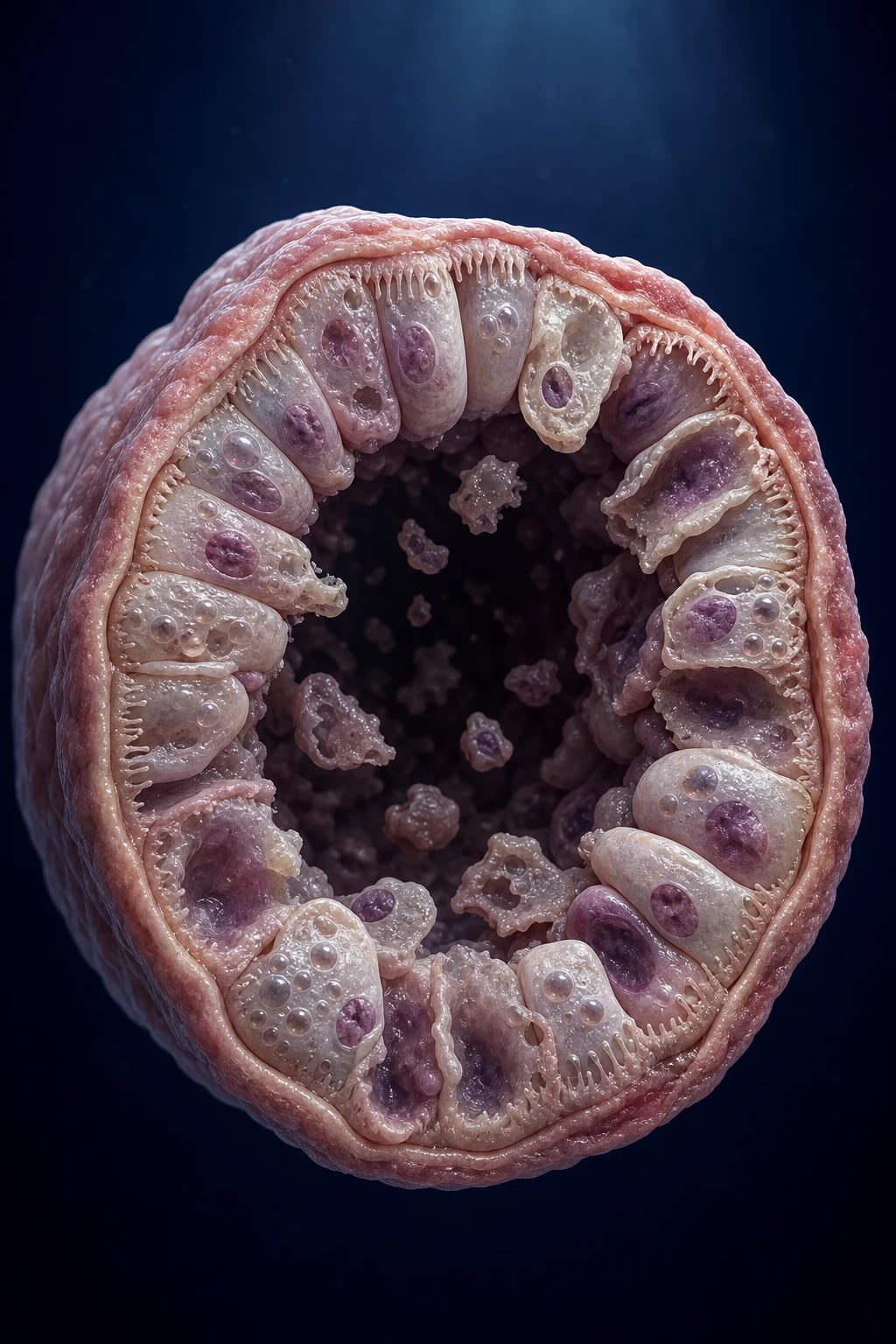
Pre-renal AKI
- Cause: hypovolaemia, hypoperfusion, sepsis, heart failure, dehydration
- Tubule: structurally INTACT; avid sodium and water retention
- Urine sodium under 20 mmol/L; FENa under 1 percent
- Urine osmolality over 500 mOsm/kg; BUN/Cr over 20
- Sediment: bland, few hyaline casts (no cells)
- Reverses within HOURS of fluid resuscitation
ATN (intrinsic)
- Cause: ischaemia, nephrotoxins (contrast, aminoglycosides, myoglobin), sepsis
- Tubule: INJURED; lost sodium-reabsorptive capacity
- Urine sodium over 40 mmol/L; FENa over 2 percent
- Urine osmolality under 350 mOsm/kg; BUN/Cr under 10-20
- Sediment: muddy brown granular casts, renal tubular epithelial cells
- Recovery takes DAYS TO WEEKS (initiation, maintenance, polyuric recovery)
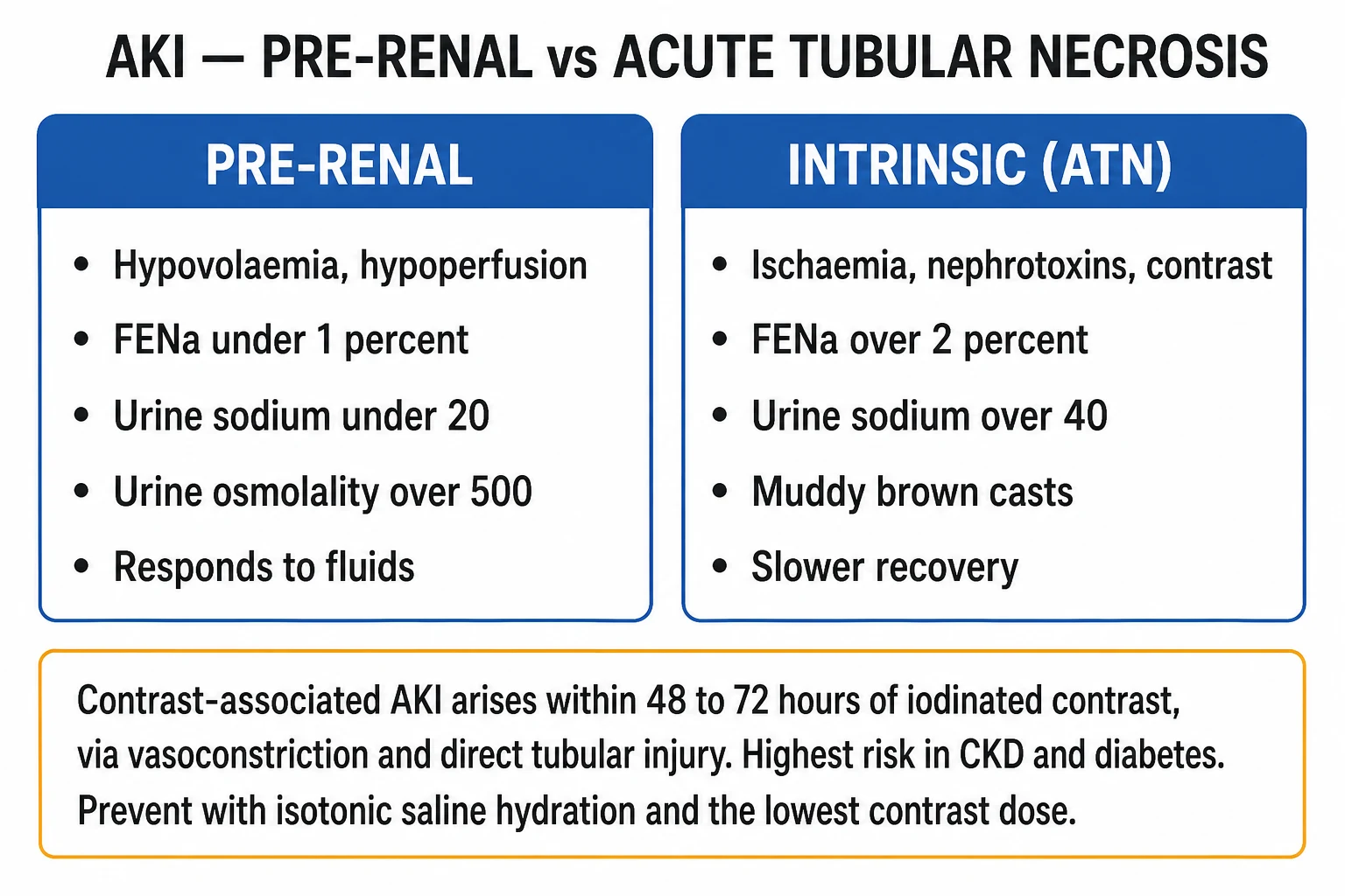
ATN is further sub-classified by aetiology into three groups, which direct the history-taking and the management: [1]
- Ischaemic ATN - prolonged hypoperfusion: haemorrhagic or septic shock, prolonged hypotension, major surgery (especially cardiac and aortic), pancreatitis, prolonged pre-renal state that has not been corrected.
- Nephrotoxic ATN - direct tubular toxins: aminoglycosides (gentamicin, tobramycin, amikacin), radiocontrast, amphotericin B, cisplatin, tenofovir, acyclovir (crystalline), vancomycin, NSAIDs (haemodynamic + interstitial), heavy metals, ethyleneglycol (oxalate crystals), pentamidine.
- Pigment nephropathy - myoglobin (rhabdomyolysis: crush, statins, prolonged immobilisation, seizures, heat stroke) and haemoglobin (intravascular haemolysis: mismatched transfusion, malaria, G6PD). Hallmark: dipstick-positive blood with no red cells on microscopy, pigmented granular casts.[2]
The KDIGO staging locates the severity of any AKI (including CA-AKI) and gates escalation: Stage 1 (Cr 1.5-1.9x baseline or a rise of at least 0.3 mg/dL; urine output under 0.5 mL/kg/h for 6-12 h); Stage 2 (Cr 2.0-2.9x baseline; UO under 0.5 for over 12 h); Stage 3 (Cr 3.0x baseline or over 4.0 mg/dL, or initiation of renal replacement therapy; UO under 0.3 mL/kg/h for over 24 h or anuria for over 12 h).[1]
Epidemiology & Risk Factors
Pre-renal AKI is the commonest form overall (around 60 percent of community AKI). ATN is the commonest cause of intrinsic AKI and of hospital-acquired AKI, which complicates around 1 in 5 adult hospital admissions and up to half of ICU admissions.[1] Sepsis, prolonged hypotension and nephrotoxic drugs are the leading hospital causes; in the community, volume depletion, infection and obstructive uropathy predominate.
Contrast-associated AKI is now the third commonest cause of hospital-acquired AKI after hypoperfusion and surgery/medications. Its incidence is strongly risk-dependent:[1][2]
- Normal renal function: under 2 percent.
- CKD stage 3 (eGFR 30-59): 5-10 percent.
- CKD stage 4-5 (eGFR under 30) and/or diabetes: 20-50 percent.
- Multiple risk factors combined (Mehran high/very-high-risk band): over 25-50 percent. [1]
CA-AKI incidence by risk band
The principal patient risk factors for CA-AKI are: pre-existing CKD (the single strongest predictor, especially eGFR under 45-60), diabetes mellitus (additive with CKD), dehydration/hypovolaemia, age over 75, heart failure (NYHA III-IV), acute myocardial infarction within 24 h, hypotension or use of an intra-aortic balloon pump, anaemia (haematocrit under 36 percent for women / 39 percent for men), multiple myeloma (paraprotein is tubulotoxic), concurrent nephrotoxic drugs (NSAIDs, aminoglycosides, cisplatin, amphotericin B, calcineurin inhibitors), high contrast volume (over 3-5x the eGFR in mL), intra-arterial (especially intra-renal or intra-cardiac) rather than intravenous administration, and repeat contrast within 72 hours.[1][7]
The Mehran risk score (2004), validated after percutaneous coronary intervention, is the standard pre-procedural risk stratification tool. It assigns weighted points to eight variables: hypotension (5 pts - SBP under 80 mmHg for over 1 h requiring inotropes/IABP); intra-aortic balloon pump use (5 pts); congestive heart failure (NYHA III-IV and/or pulmonary oedema; 5 pts); eGFR under 20 (6 pts) or 20-40 (4 pts); age over 75 (4 pts); anaemia (3 pts - baseline Hct under 36 percent women / 39 percent men); diabetes (3 pts); and contrast volume (1 pt for each 100 mL). The corresponding CIN, dialysis and mortality risk rises across four bands: low (under 6 points, CIN 7.5 percent), moderate (6-10 points, CIN 14 percent), high (11-16 points, CIN 26 percent), and very high (at least 16 points, CIN 57 percent, dialysis over 12 percent).[7]
A modern re-appraisal (AMACING 2017; post-2018 ACR/ESUR guidance; Barbaritos 2020) has revised the perceived risk downward - with modern low/iso-osmolar agents, smaller contrast volumes and iso-osmolar agents, even moderate-risk patients have a substantially lower absolute risk than historically reported, and contrast should not be withheld for clinically essential imaging.[6]
Pathophysiology
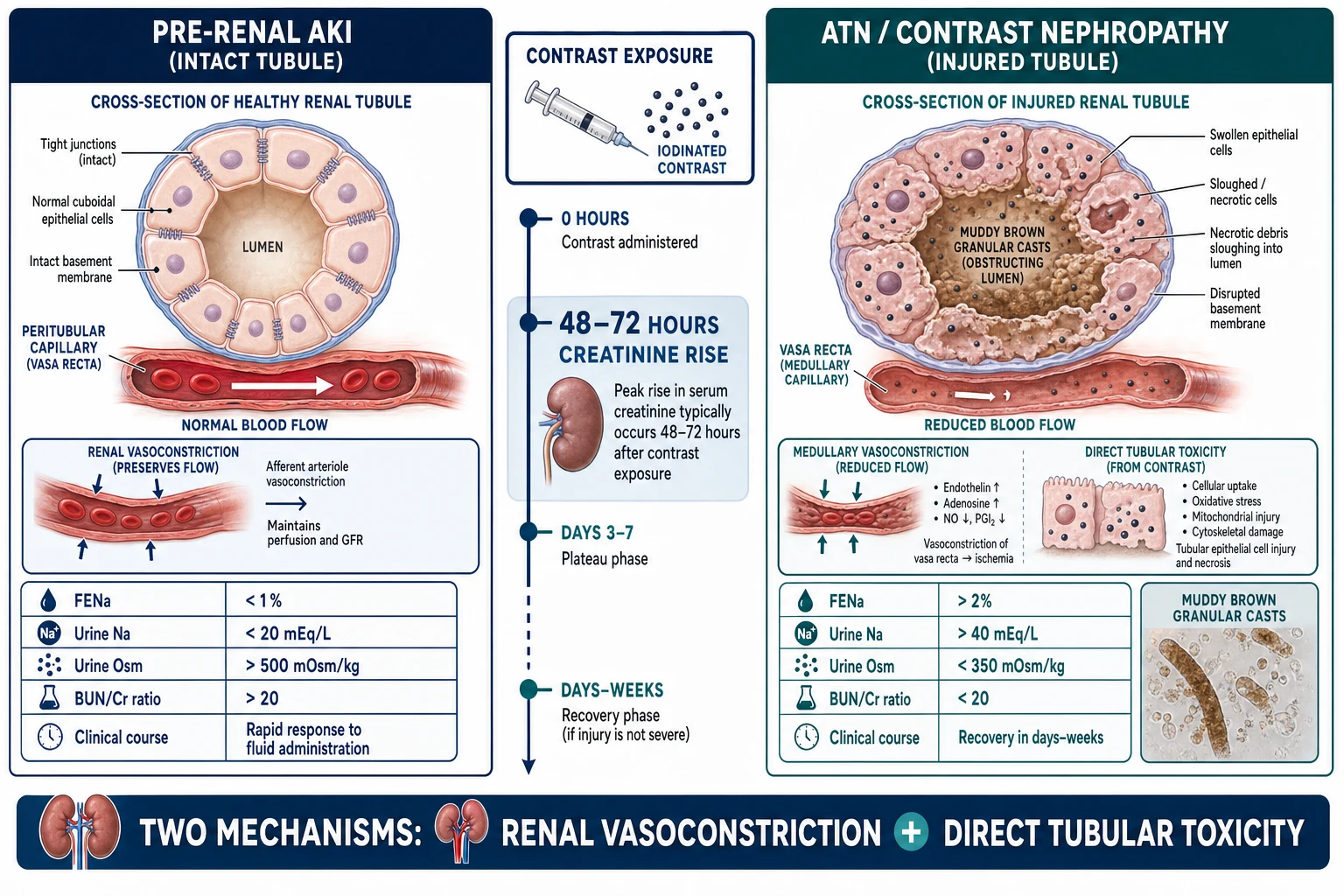
Pre-renal azotaemia - why it is rapidly reversible
In pre-renal physiology, the kidney perceives a real or effective intravascular volume deficit (haemorrhage, dehydration, sepsis with vasodilatation, heart failure with low effective circulating volume, NSAID- or ACE-inhibitor-induced haemodynamic stress). The intact kidney mounts a compensatory neurohormonal response: activation of the sympathetic nervous system and renin-angiotensin-aldosterone system (RAAS), and non-osmotic vasopressin release. Together these cause efferent arteriolar constriction (RAAS) to maintain intraglomerular pressure, afferent arteriolar constriction (sympathetic), avid sodium reabsorption in the proximal tubule and collecting duct (aldosterone), and free-water retention (vasopressin acting on V2 receptors in the collecting duct). The result is a concentrated, sodium-poor urine (FENa under 1 percent, urine osmolality over 500) and a BUN reabsorption disproportionate to creatinine (BUN/Cr over 20). Because the tubules are structurally intact, restoring perfusion reverses the picture within hours - the kidney simply resumes its normal set-point. The danger is that an uncorrected pre-renal state progresses to ATN within hours to a day or two, as prolonged ischaemia finally injures the tubule cells.[1]
Acute tubular necrosis - the four mechanisms
ATN is the phenotype that emerges when tubular cells are injured by ischaemia or a direct toxin (or both, which is why sepsis plus contrast or aminoglycoside is so damaging). Four interlinked mechanisms lower GFR:[2]
- Tubular epithelial cell injury and death. Sublethal injury causes loss of the brush border, apical membrane blebbing, cytoplasmic vacuolation and mitochondrial swelling; lethal injury produces apoptosis or necrosis with cellular sloughing into the tubular lumen. The surviving cells dedifferentiate, proliferate and re-epithelialise the basement membrane during recovery - which is why ATN can be reversible but takes time.
- Cast obstruction of the tubular lumen. Sloughed cells, Tamm-Horsfall protein and debris aggregate to form muddy brown granular casts that physically obstruct the tubule, raising intratubular pressure and opposing filtration.
- Back-leak of glomerular filtrate. Disruption of the intercellular tight junctions and the basement membrane lets filtrate leak back into the interstitium and peritubular capillaries, so the GFR that is generated does not appear as urine output.
- Afferent arteriolar vasoconstriction. Tubuloglomerular feedback is altered (macula densa sensing increased distal sodium delivery), and intra-renal mediators (adenosine, endothelin, thromboxane) drive afferent vasoconstriction, lowering glomerular capillary hydrostatic pressure. [1]
The loss of sodium-reabsorptive capacity is the biochemical signature - the injured tubule cannot reabsorb sodium normally, so urine sodium rises over 40 mmol/L and FENa exceeds 2 percent, with an isosthenuric urine (osmolality under 350, close to plasma).[2]
The dual mechanism of contrast-associated AKI
CA-AKI is driven by two synergistic mechanisms acting on the same vulnerable cell populations:[1][2]
- Renal medullary vasoconstriction and hypoxia. Iodinated contrast produces an initial transient increase in renal blood flow (osmotic load) followed by a sustained decrease, with the outer medulla - which physiologically operates on the brink of hypoxia - bearing the brunt. The S3 segment of the proximal tubule and the medullary thick ascending limb are the most oxygen-sensitive nephron segments and the first to die. Mediators include adenosine (which constricts the afferent arteriole via A1 receptors), endothelin, reactive oxygen species, and loss of prostaglandin-mediated vasodilation. The result is medullary hypoxic ATN.
- Direct cytotoxicity to tubular epithelial cells. The iodinated benzene ring of contrast media is directly toxic to tubular cells via mitochondrial injury, oxidative stress and apoptosis. Cells of the proximal tubule internalise contrast (and other toxins) and concentrate it in their cytoplasm, prolonging exposure. Osmolar load (greater with high-osmolar agents such as diatrizoate) and viscosity (greater with the iso-osmolar iodixanol) both slow tubular flow and increase contact time. High-osmolar agents increase tubular pressure and diuresis, prolonging toxic exposure; very viscous iso-osmolar agents slow tubular transit, also prolonging exposure - the trade-off explains why the choice between iso-osmolar and low-osmolar agents is a matter of patient risk and route rather than a universal winner. [1]
Pigment nephropathy and nephrotoxic mechanisms
Myoglobin and haemoglobin (molecular weight around 17 kDa) are freely filtered, taken up by proximal tubule cells, and cause cast formation (with Tamm-Horsfall protein, especially at acidic urine pH), direct cytotoxicity via ferrous iron catalysing lipid peroxidation (Fenton chemistry), and consumption of nitric oxide causing vasoconstriction. Aminoglycosides (gentamicin, tobramycin, amikacin) bind to the megalin/cubilin receptor on proximal tubule cells, are endocytosed, and accumulate in lysosomes, causing non-oliguric AKI classically on days 5-7 of therapy. Amphotericin B forms pores in tubular cell membranes (normal-anion-gap metabolic acidosis from distal RTA plus AKI). Cisplatin accumulates in proximal tubules via OCT2 and causes oxidative injury. Tenofvir causes proximal tubular toxicity with Fanconi syndrome. NSAIDs cause haemodynamic AKI by inhibiting prostaglandin-mediated afferent arteriolar vasodilation (and may also cause acute interstitial nephritis). [1]
Why creatinine is a lagging marker - the role of biomarkers
Serum creatinine is a late, lagging marker of AKI: it only rises once the body creatinine pool accumulates against a reduced GFR, which may take 24-48 hours after the actual injury. By the time creatinine rises, the damage is already done. This lag drives the search for injury biomarkers that rise within 2-6 hours of injury: Cystatin C (freely filtered, not dependent on muscle mass), NGAL (neutrophil gelatinase-associated lipocalin), KIM-1 (kidney injury molecule-1), and the TIMP-2 x IGFBP7 cell-cycle-arrest marker (NephroCheck). These are not yet routine in NEET-PG/INICET practice but inform trials and ICU prognostication.[2]
Clinical Presentation
AKI itself is often clinically silent in its early phase and is detected by biochemistry. The symptoms and signs of established AKI are those of uracmic toxicity and fluid/electrolyte disturbance:[1]
- Volume overload: dyspnoea, orthopnoea, raised JVP, bibasal crackles, S3 gallop, peripheral and sacral oedema, hypertension.
- Uraemic features: anorexia, nausea, vomiting, hiccups, metallic taste, pruritus, easy bruising (uraemic platelet dysfunction), pericardial friction rub (uraemic pericarditis), asterixis, confusion, seizures (uraemic encephalopathy).
- Electrolyte: arrhythmia from hyperkalaemia, muscle weakness, abdominal cramps.
- Volume depletion (when the cause is pre-renal): thirst, oliguria, dry mucosae, reduced skin turgor, tachycardia, postural hypotension, cool peripheries. [1]
Cause-specific clues should be actively sought: [1]
- Pre-renal: hypovolaemia (haemorrhage, vomiting, diarrhoea, burns, diuretic over-use), sepsis, heart failure, cirrhosis with ascites, nephrotic syndrome. The kidney is intact; the problem is perfusion.
- Ischaemic ATN: a clear episode of prolonged hypotension (cardiac arrest, prolonged intra-operative hypotension, septic shock, haemorrhagic shock) followed by oliguria within 24-72 hours.
- Nephrotoxic ATN: a recent drug course (gentamicin day 5-7, amphotericin B, cisplatin chemotherapy), recent iodinated contrast within 48-72 hours, tenofovir, acyclovir at high dose.
- Pigment nephropathy / rhabdomyolysis: muscle pain, swelling and tenderness after trauma, prolonged immobilisation, status epilepticus, heat stroke, or a new statin; dark cola-coloured urine; dipstick-positive for blood with no red cells on microscopy (myoglobin).
- Acute interstitial nephritis (AIN): drug (beta-lactam, PPI, NSAID, sulphonamide, rifampicin) with fever, rash, eosinophilia, arthralgia - the classic triad, though often incomplete. [1]
Timing of CA-AKI is characteristic: the creatinine rise peaks at 48 to 72 hours post-exposure, and in the uncomplicated case resolves within 7 to 10 days. Persistence or worsening beyond this, especially with eosinophilia and livedo after a catheter procedure, should raise cholesterol atheroembolisation (1-4 weeks after catheter manipulation, often irreversible) rather than ongoing contrast injury. [1]
The diuretic / polyuric recovery phase of ATN is an exam favourite: as the regenerating tubules cannot yet concentrate urine (loss of medullary gradient and aquaporin reinsertion lagging), a polyuric phase of 3-5 L/day for several days may occur, with risk of dehydration, hypernatraemia and hypokalaemia if not monitored and replaced. [1]
Atypical presentations are common and high-yield. In the elderly, the first sign may be confusion, drug toxicity (digoxin, lithium), or volume overload rather than oliguria. In diabetics, autonomic neuropathy may mask volume signs and a neuropathic bladder may coexist with obstruction. In CKD, a small insult (an NSAID, mild dehydration) tips the patient into acute-on-chronic AKI - distinguish from progression. In cirrhosis, hepatoreal syndrome must be excluded with an albumin challenge before attributing AKI to ATN.[1]
Differential Diagnosis
The pivotal bedside distinction is pre-renal vs intrinsic (ATN), made by urine chemistries and sediment, supplemented by the response to a fluid challenge:[2]
Pre-renal AKI
- FENa under 1 percent (under 1 percent sodium excretion)
- Urine sodium under 20 mmol/L (avid reabsorption)
- Urine osmolality over 500 mOsm/kg (concentrated)
- BUN/creatinine ratio over 20
- Sediment: bland, hyaline casts only
- Rapid response to isotonic crystalloid within hours
ATN (intrinsic)
- FENa over 2 percent (sodium wasting)
- Urine sodium over 40 mmol/L
- Urine osmolality under 350 mOsm/kg (isosthenuric)
- BUN/creatinine ratio under 10-20
- Sediment: muddy brown granular casts, RTE cells
- Slower recovery over days-weeks; fluid challenge does not reverse
Diuretic caveat. A loop or thiazide diuretic forces natriuresis, so a patient in genuine pre-renal state who has just received furosemide will show a spuriously high urine sodium and FENa. In this situation use the fractional excretion of urea (FEurea), which is diuretic-independent: FEurea = (urine urea x serum creatinine) / (serum urea x urine creatinine) x 100; a value under 35 percent supports pre-renal disease. The BUN/creatinine ratio is also diuretic-independent and supports pre-renal when over 20.[2]
Distinguishing the four major intrinsic AKI patterns by urine sediment and history: [1]
ATN
- Muddy brown granular casts, renal tubular epithelial (RTE) cells
- Cause: ischaemia, contrast, aminoglycoside, pigment
- FENa over 2 percent; urine sodium over 40
- Treatment: supportive, remove the insult
Acute interstitial nephritis (AIN)
- WBC casts, eosinophiluria, lymphocyturia
- Cause: beta-lactams, PPIs, NSAIDs, sulphonamides, rifampicin
- Fever, rash, eosinophilia (classic triad, often incomplete)
- Treatment: stop drug; corticosteroids if severe
Glomerulonephritis / RPGN
- RBC casts, dysmorphic RBCs, proteinuria
- Cause: post-streptococcal, IgA nephropathy, ANCA vasculitis, anti-GBM, lupus nephritis
- Rapidly progressive AKI over days-weeks (crescents on biopsy)
- Treatment: urgent immunosuppression, plasma exchange for anti-GBM
Atheroembolic disease
- Bland or few RTE cells; eosinophilia, low complement
- Cause: catheter/vascular procedure 1-4 weeks prior
- Livedo reticularis, blue toes, digital infarcts
- Treatment: supportive; often irreversible
Post-renal / obstructive AKI must be excluded in any AKI, especially with anuria, a single functioning kidney, palpable bladder, BPH, pelvic malignancy or neurogenic bladder. A renal ultrasound showing hydronephrosis confirms it; relief is by urinary catheter or nephrostomy. Beware post-obstructive diuresis after relief (polyuria with sodium/water wasting).[1]
AKI vs CKD. Distinguish by kidney size on ultrasound (small, echogenic kidneys in CKD; normal or enlarged in AKI), baseline creatinine (known chronic elevation in CKD), anaemia (normocytic in CKD from low EPO), hyperphosphataemia/hypocalcaemia, uraemic platelet dysfunction, and broad waxy casts (CKD). An acute rise on a chronic baseline is acute-on-chronic KD - treat the reversible insult. [1]
Contrast AKI vs cholesterol embolisation syndrome (a classic exam differential after catheter): CA-AKI occurs within 48-72 hours and resolves in 7-10 days; atheroembolisation occurs 1-4 weeks after catheter manipulation, with livedo reticularis, blue toes, eosinophilia, hypocomplementaemia, and is often irreversible - the two are NOT the same condition.[1]
Clinical & Bedside Assessment
A structured bedside assessment in suspected AKI has four limbs:[1]
- Volume status - the single most important bedside observation. Look for hypovolaemia: tachycardia, hypotension or postural drop (SBP fall over 20 mmHg or DBP over 10 mmHg on standing), dry mucosae, reduced skin turgor, cool peripheries, capillary refill over 2 seconds, sunken eyes, low JVP. Look for hypervolaemia: raised JVP (over 3-4 cm above sternal angle), S3 gallop, bibasal crackles, peripheral/sacral oedema, ascites, hypertension. A passive leg raise or fluid challenge (250-500 mL balanced crystalloid over 15-30 min with reassessment of JVP, lung bases, blood pressure and urine output) helps discriminate responders (pre-renal) from non-responders (ATN).
- Targeted drug and exposure history - review every drug on the chart: NSAIDs (including over-the-counter and combination analgesics), ACE inhibitors/ARBs (especially in bilateral renal artery stenosis), aminoglycosides (gentamicin levels), vancomycin (trough levels), amphotericin B, cisplatin, tenofovir, acyclovir, calcineurin inhibitors (cyclosporin, tacrolimus), recent iodinated contrast (timing and volume), herbal/AYUSH agents (aristolochic acid, lead-containing preparations). Stop all nephrotoxins on diagnosing AKI.
- Search for the cause - signs of sepsis (fever, source, warm vasodilated shock vs cold shock), abdominal exam (palpable bladder, renal mass, abdominal aortic aneurysm, ascites), skin (rash, purpura, livedo, cholesterol emboli, signs of vasculitis), signs of rhabdomyolysis (swollen tender muscle, recent trauma or prolonged immobility), heart failure, decompensated cirrhosis.
- Establish baseline - obtain the most recent pre-illness creatinine and eGFR (in India, often from a recent health-check or outpatient panel); quantify the rise and stage the AKI; measure weight, urine output (catheter if oliguric), blood pressure (target MAP over 65 mmHg), and lactate (perfusion surrogate). [1]
Drug chart review is the single highest-yield bedside manoeuvre in hospital-acquired AKI - most iatrogenic AKI is caused by a drug on the chart that is being missed.[1]
Investigations
First-line investigations in any AKI:[2][1]
- Serum biochemistry: urea, creatinine (with baseline), eGFR, sodium, potassium, bicarbonate (metabolic acidosis), chloride (anion gap), calcium, phosphate, magnesium, glucose, albumin.
- Venous blood gas for pH and bicarbonate (acidosis severity).
- Full blood count (anaemia, eosinophilia in AIN/atheroemboli), CRP, blood cultures if sepsis.
- Creatine kinase (CK) if any suspicion of rhabdomyolysis (CK over 5x upper limit of normal, classically over 5000 U/L, with myoglobinuria).
- Urinalysis and microscopy - the single most useful bedside test. Dipstick for blood, protein, leucocytes, nitrites, specific gravity, pH. Microscopy for casts (see below).
- Urine electrolytes for FENa (and FEurea if on diuretics): paired spot urine and serum sodium, creatinine and urea.
- ECG for hyperkalaemia (peaked T waves, flattened P waves, prolonged PR, widened QRS, sine wave).
- Renal ultrasound to exclude obstruction (mandatory in anuria, single kidney, no recovery within 24 h, suspected pelvic malignancy) and assess kidney size/echogenicity (CKD vs AKI).
- Chest X-ray for pulmonary oedema, sepsis source. [1]
Fractional excretion of sodium (FENa)
FENa thresholds & pitfalls
FENa = (Urine Na x Serum Cr) / (Serum Na x Urine Cr) x 100[2]
- Under 1 percent - pre-renal (intact tubule avidly reabsorbs sodium).
- Over 2 percent - intrinsic (ATN loses sodium-reabsorptive capacity).
- Pitfalls: misleadingly high in diuretic use (use FEurea under 35 percent), sepsis, bicarbonaturia (sodium dragged with bicarbonate), chronic kidney disease (baseline tubular dysfunction), pre-existing CKD (already sodium-wasting); misleadingly low in pigment nephropathy early and radiocontrast AKI early (some patients show a low FENa in the first 24 h). [1]
The urine sediment - high-yield exam table
Casts and their diseases (high-yield)
BUN/creatinine ratio
A BUN/Cr ratio over 20 (using conventional mg/dL units) supports pre-renal azotaemia (urea is passively reabsorbed with water in the proximal tubule and concentrated by RAAS-driven water retention, whereas creatinine is not reabsorbed). A ratio under 10-20 favours intrinsic AKI. The ratio is diuretic-independent and is useful when FENa is confounded. [1]
Hyperkalaemia ECG sequence
The progressive ECG changes of hyperkalaemia are an exam staple: (1) peaked (tented) T waves - earliest, at K around 5.5-6.5 mmol/L; (2) flattening or disappearance of P waves and prolongation of the PR interval - K 6.5-7.5; (3) widening of the QRS - K 7.5-8.5; (4) sine-wave pattern, ventricular fibrillation, asystole - K over 8.5. Treat any K over 6.0-6.5 mmol/L or any ECG change as an emergency with intravenous calcium gluconate 10 percent 10 mL for membrane stabilisation.[1]
Mehran risk score (reproduced)
Mehran CA-AKI risk score (8 variables, weighted)
Second-line / targeted investigations
- Serum immunoglobulins, serum protein electrophoresis, serum-free light chains - if multiple myeloma suspected (unexplained AKI in an older patient, especially with anaemia and bone pain).
- ANCA, anti-GBM, anti-nuclear antibodies, complement (C3, C4) - if glomerulonephritis / vasculitis suspected (RBC casts, rapidly progressive AKI).
- Hepatitis B/C, HIV serology - if membranous or membranoproliferative GN or before immunosuppression.
- Renal biopsy - indicated for RPGN, persistent unexplained AKI, suspected AIN (eosinophils on biopsy), suspected systemic vasculitis with renal involvement; helps plan immunosuppression.
- CT angiography or renal Doppler - if renal artery stenosis or renal vein thrombosis suspected (but contrast CT itself causes AKI - use cautiously).
- Novel biomarkers (Cystatin C, NGAL, KIM-1, NephroCheck / TIMP-2 x IGFBP7) - early injury detection, mainly research/ICU. [1]
Management - Resuscitation
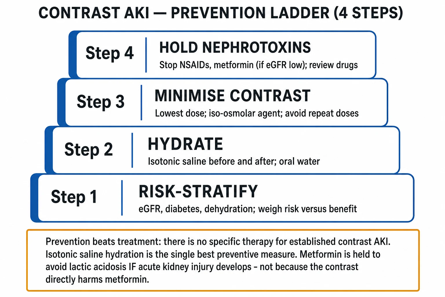
The resuscitation principles apply to all AKI (pre-renal, ATN, or CA-AKI) and are time-critical. The aim is to restore perfusion, remove ongoing insult, prevent progression to ATN, and treat life-threatening metabolic complications.[1]
Step 1 - Treat the cause (parallel, not sequential)
- Hypovolaemic / pre-renal: give a fluid challenge with balanced crystalloid (Hartmann's or PlasmaLyte; avoid 0.9 percent saline in large volumes - causes hyperchloraemic metabolic acidosis and renal vasoconstriction) - 250-500 mL (10-15 mL/kg) over 15-30 minutes, with reassessment of JVP, lung bases, blood pressure and urine output. Repeat if the patient remains volume-deplete and is not overloaded.
- Septic ATN: implement the Surviving Sepsis hour-1 bundle - oxygen, two sets of blood cultures, broad-spectrum antibiotics within 1 hour, lactate, balanced crystalloid 30 mL/kg, and noradrenaline for fluid-refractory shock (target MAP over 65 mmHg).
- Obstruction: urinary catheter (urethral or suprapubic) or percutaneous nephrostomy; watch for post-obstructive diuresis (replace urine output ml-for-ml with oral or IV fluid for 24-48 h). [1]
Step 2 - Universal AKI measures
Universal AKI measures (apply to ALL types)
- Stop all nephrotoxic drugs: NSAIDs (including over-the-counter), ACE inhibitors and ARBs (especially if hyperkalaemic or bilateral renal artery stenosis), aminoglycosides, amphotericin B, cisplatin, tenofovir, iodinated contrast; review herbal/AYUSH agents.
- Re-dose every drug for the current eGFR (loading doses usually unchanged; maintenance doses adjusted - see drug-dosing-in-kidney-disease topic).
- Optimise perfusion: target MAP over 65 mmHg (noradrenaline in vasodilatory shock; vasopressin if noradrenaline-refractory; cautious fluid boluses only if volume-responsive).
- Monitor: urine output (catheter if oliguric), serum potassium, bicarbonate, creatinine, fluid balance and weight every 6-12 hours. [1]
Step 3 - Treat hyperkalaemia as an emergency
Hyperkalaemia with ECG changes is a medical emergency - membrane stabilisation first:[1]
- Calcium gluconate 10 percent, 10 mL IV over 2-5 minutes (or calcium chloride 10 percent via central line in arrest). Onset within minutes; does NOT lower potassium - it stabilises the myocardial membrane. Repeat if ECG changes persist. [1]
Then shift potassium into cells: [1]
- Insulin 10 units IV with 25 g (50 mL of 50 percent) dextrose over 15-30 minutes (reduces K by 0.5-1.2 mmol/L for 4-6 h; monitor blood glucose for hypoglycaemia for up to 6 h).
- Salbutamol 10-20 mg nebulised (or 0.5 mg IV) - beta-2 agonist drives K into cells; add to insulin for synergy. Onset 15-30 min.
- Sodium bicarbonate is weak as a K-shift agent but useful if the patient is also acidotic (give if pH under 7.1-7.15). [1]
Then remove potassium from the body: [1]
- Potassium binders: sodium polystyrene sulfonate 15-30 g orally or rectally (onset 2-24 h; risk of intestinal necrosis, especially post-operative); newer patiromer (8.4 g daily) or sodium zirconium cyclosilicate (10 g three times daily for up to 48 h) are better tolerated.
- Renal replacement therapy (dialysis) if refractory, ongoing tissue breakdown (rhabdomyolysis, tumour lysis), or severe fluid overload. [1]
Why colloids and starches are avoided
The 6S trial (NEJM 2012), CHEST trial (NEJM 2012) and CRISTAL trial (NEJM 2013) established that hydroxyethyl starch and other synthetic colloids increase the risk of AKI and the need for RRT. Albumin is not superior to crystalloid for resuscitation in most AKI. Balanced crystalloids (Hartmann's, PlasmaLyte) are preferred over 0.9 percent saline for large-volume resuscitation (SMART trial, NEJM 2018 showed balanced crystalloids reduced Major Adverse Kidney Events). Fluid overload itself worsens AKI outcomes - via venous congestion, raised intra-abdominal pressure and interstitial oedema - so fluids are NOT a blanket prescription.[1]
Management - Definitive & Stepwise
The four pillars of contrast-associated AKI prevention
Because there is no specific treatment that reverses established CA-AKI, prevention is the entire strategy. The four pillars are:[1][3]
Pillar 1 - Risk-stratify. Measure eGFR in every patient scheduled for contrast. Stratify by risk factors (CKD, diabetes, dehydration, age over 75, heart failure, anaemia, multiple myeloma, concurrent nephrotoxins). Apply the Mehran score in PCI settings. For low-risk patients with eGFR over 60 and no risk factors, no specific prophylaxis is required. For moderate-to-high-risk patients (eGFR under 45-60 with risk factors), apply the remaining three pillars. [1]
Pillar 2 - Isotonic saline hydration. The single best-proven preventive measure. KDIGO-endorsed protocols:
- Elective: isotonic saline 1 mL/kg/h for 6-12 hours before and 6-12 hours after contrast (typically around 1 L pre and 1 L post). 0.9 percent saline is the evidence standard for hydration volume, though balanced solutions are increasingly used.
- Urgent / emergency: 3 mL/kg/h for 1 hour before and 1-3 mL/kg/h for 4-6 hours after contrast, where the longer protocol is infeasible.
- Volume-overload caveat: in heart failure, adjust the rate and monitor for pulmonary oedema; consider diuretics only if overload develops.
- Oral hydration is acceptable in lower-risk patients (POHED and other trials suggest oral water alone is adequate for moderate risk when eGFR over 30) - the AMACING trial challenged routine IV hydration in moderate-risk patients.[6]
Pillar 3 - Minimise contrast. Use the lowest possible contrast volume (a practical rule: keep contrast volume under 3-5 times the eGFR in mL), use iso-osmolar (iodixanol) or low-osmolar (iohexol, iopamidol, ioversol) agents in preference to high-osmolar (diatrizoate, metrizoate), use the intravenous rather than intra-arterial route when feasible (intra-renal arterial exposure carries the highest risk), and avoid repeat contrast within 72 hours.[1]
Pillar 4 - Hold nephrotoxins. Review the drug chart and hold NSAIDs (including over-the-counter), reconsider ACE inhibitors/ARBs (continue in stable chronic heart failure; hold if hyperkalaemic or volume-deplete), and review aminoglycosides, amphotericin B, cisplatin, tenofovir, vancomycin for ongoing necessity and dose adjustment. [1]
N-acetylcysteine, bicarbonate and other unproven agents - the PRESERVE verdict
Agents that do NOT prevent CA-AKI (PRESERVE trial)
The landmark PRESERVE trial (Weisbord et al., NEJM 2018) randomised 5177 high-risk patients undergoing angiography to sodium bicarbonate vs isotonic saline and N-acetylcysteine vs placebo in a 2x2 factorial design. Neither bicarbonate nor N-acetylcysteine reduced the rate of contrast-associated AKI, the need for dialysis, or 90-day mortality. The trial decisively ended routine use of these agents; isotonic saline hydration and contrast volume minimisation are what actually work.[3] Other agents - statins (mixed evidence, PRATO-ACS suggested benefit in ACS), ascorbic acid, theophylline (adenosine antagonist), fenoldopam - are not recommended for routine prophylaxis.
The metformin rule - precise and exam-relevant
The metformin-and-contrast rule (memorise this exactly)
The AEIOU indications for renal replacement therapy
When ATN progresses to severe AKI with life-threatening complications, renal replacement therapy (haemodialysis, sustained low-efficiency dialysis / SLED, or continuous renal replacement therapy / CRRT) is indicated. The AEIOU mnemonic reproduces the indications:[1]
AEIOU - indications for renal replacement therapy in AKI
The dialysis timing question - AKIKI and STARRT-AKI
The two pivotal randomised trials on timing of RRT initiation in critically ill AKI showed no mortality benefit of early initiation:[4][5]
- AKIKI (Gaudry et al., NEJM 2016): 620 critically ill patients with KDIGO Stage 3 AKI randomised to immediate vs delayed RRT (delayed = initiation only for AEIOU-type indications). No difference in 60-day mortality; almost half the delayed group never needed dialysis (i.e. early dialysis exposes patients unnecessarily).[4]
- STARRT-AKI (Bagshaw et al., NEJM 2020): 3019 critically ill patients with severe AKI randomised to accelerated (early) vs standard RRT. No difference in mortality; the accelerated strategy had more adverse events (mostly excess fluid overload and dialysis-associated complications).[5]
The take-home: initiate RRT for AEIOU criteria, persistent severe AKI, or worsening clinical status - not for a biochemical trigger alone.[4][5]
The hyperkalaemia treatment ladder (reproduced in full)
Hyperkalaemia ladder - agent, dose, route
Pigment nephropathy / rhabdomyolysis management
When the cause is rhabdomyolysis (CK over 5000, myoglobinuria), the management is aggressive volume expansion:[2]
- Isotonic saline at a rate to achieve urine output of 200-300 mL/hour (i.e. 4-7 L/day, often a 1-1.5 L bolus then 500-1000 mL/h, adjusted to urine output and haemodynamics). Watch for fluid overload in the elderly and cardiac patients.
- Sodium bicarbonate to alkalinise the urine to pH over 6.5 is controversial - meta-analyses show no clear benefit over saline alone; it may be considered in severe acidosis or life-threatening hyperkalaemia.
- Mannitol is no longer recommended (no proven benefit, risk of volume overload and acute kidney injury from hyperoncocity).
- Treat the cause (compartment syndrome - fasciotomy; status epilepticus - control seizures; statin - stop). [1]
Acute interstitial nephritis management
When AIN is suspected (drug, rash, fever, eosinophilia, WBC casts): stop the offending drug (the single most important step). If AKI persists or is severe, give a short course of prednisolone 1 mg/kg/day (max 60 mg) for 1-2 weeks then taper; most patients recover within weeks.[1]
Renal drug dosing and nutrition
- Loading doses are usually unchanged (depend on volume of distribution, not clearance).
- Maintenance doses are adjusted to the current eGFR (which may be much lower than baseline in AKI) and to whether the drug is dialysis-cleared (e.g. vancomycin, gentamicin, lithium, many beta-lactams need post-dialysis dosing).
- Nutrition: avoid the historical "renal diet" protein restriction (which worsens malnutrition); target 1.0-1.5 g/kg/day of protein; prefer enteral over parenteral nutrition; avoid hyperglycaemia. [1]
Specific Subtypes & Scenarios
Contrast-associated AKI (the central subtype)
Definition (ESUR/ACR): serum creatinine rise of 0.3 mg/dL or 1.5x baseline within 48-72 hours of iodinated contrast, no alternative cause. Prevention pillars above. Usually transient and self-limiting; severe AKI requiring dialysis is rare (under 1 percent) but more common in CKD + diabetes + large-volume intra-arterial contrast.[1]
Ischaemic ATN from prolonged hypotension or sepsis
The classic intensive-care AKI. Treat the cause (septic shock - Surviving Sepsis bundle; haemorrhage - transfuse and source control; cardiogenic shock - inotropes / mechanical support). Provide supportive care, optimise perfusion, watch for the polyuric recovery phase 1-3 weeks later. Recovery may be incomplete, leaving residual CKD.[2]
Nephrotoxic ATN - drug-specific mechanisms
Nephrotoxic ATN - drug and mechanism (high-yield)
Pigment nephropathy / rhabdomyolysis
CK over 5000 U/L (or a clinical picture of muscle injury with myoglobinuria) defines significant rhabdomyolysis. Hallmark: urine dipstick positive for blood with NO red cells on microscopy (myoglobin). Pigmented granular casts. Treat with aggressive isotonic saline to urine output 200-300 mL/h.[2]
Tumour lysis syndrome
In high-turnover haematological malignancy (high-grade lymphoma, leukaemia with high white-cell count), rapid tumour breakdown releases potassium, phosphate, uric acid (and causes hypocalcaemia from calcium-phosphate deposition), precipitating AKI. Prevention with aggressive isotonic hydration (3 L/m2/day) plus rasburicase (recombinant uricase, high-risk) or allopurinol (low-risk) is the standard; treat established TLS with hydration, rasburicase, and dialysis for refractory hyperkalaemia or severe hyperphosphataemia.[1]
Hepatorenal syndrome (HRS-AKI) in cirrhosis
AKI in advanced cirrhosis with ascites, caused by intense splanchnic vasodilatation producing renal hypoperfusion with avid sodium and water retention. Diagnose only after excluding other causes with an albumin challenge (1 g/kg day 1, then 20-40 g/day for 2 days, to a max 100 g/day); if creatinine does not improve, HRS-AKI is likely. Treat with terlipressin + albumin (terlipressin 1-2 mg IV every 4-6 hours, titrated; albumin 20-40 g/day); consider TIPS or liver transplant.[1]
Acute interstitial nephritis (AIN)
Most often drug-induced (beta-lactams - classically methicillin; proton pump inhibitors - increasingly recognised; NSAIDs; sulphonamides; rifampicin; 5-aminosalicylates; allopurinol). Classic triad (fever, rash, eosinophilia) is incomplete in most cases. Urine: WBC casts, eosinophiluria. Stop the drug; give prednisolone 1 mg/kg/day for 1-2 weeks if severe or persistent; biopsy if diagnostic doubt.[1]
Acute-on-chronic kidney disease
A patient with known CKD develops an acute rise - identify and treat the reversible insult (volume depletion, nephrotoxin, obstruction, infection); renal function may not return to the prior baseline, accelerating progression to end-stage renal disease. [1]
Complications & Pitfalls
Metabolic complications of ATN
Metabolic consequences of ATN (and their direction)
Life-threatening complications
Hyperkalaemic arrhythmia (VF, asystole), uraemic pericarditis with effusion and tamponade, uraemic encephalopathy (coma, seizures), severe volume overload with pulmonary oedema, uraemic bleeding (platelet dysfunction), infection (the leading cause of death in AKI).[1]
The polyuric / diuretic recovery phase
As ATN recovers, surviving tubule cells regenerate but cannot yet concentrate urine (the medullary gradient is disrupted, and aquaporin reinsertion lags). A polyuric phase of 3-5 L/day for several days may occur, with risk of dehydration, hypernatraemia, hypokalaemia and hypomagnesaemia. Replace urine output ml-for-ml (oral or IV) for 24-48 hours and monitor electrolytes every 6-12 hours. [1]
Classic pitfalls (exam-favourite)
Pitfalls that cost marks (and lives) in AKI
Long-term consequences of an AKI episode
An AKI episode is not benign even when recovered - survivors carry an increased long-term risk of developing CKD, accelerated CKD progression, and cardiovascular events (Coca et al., meta-analyses). Every effort should be made to prevent AKI and to follow up renal function after discharge.[2]
Prognosis & Disposition
Recovery pattern by type: [1]
- Pre-renal AKI reverses within hours of restoring perfusion (kidney intact).
- ATN recovers over days to weeks, classically passing through initiation (hours-days, the insult), maintenance (1-2 weeks, oliguric, peak creatinine) and recovery (days-weeks, polyuric then normalising) phases; some patients have residual CKD.
- Obstructive AKI recovers after relief (beware post-obstructive diuresis). [1]
Mortality. Hospital-acquired AKI carries around 10 percent mortality when uncomplicated, rising to over 50 percent in ICU / multi-organ failure. The mortality of CA-AKI is usually driven by the underlying condition (the reason for the contrast - sepsis, ACS, vascular emergency) rather than the AKI itself, but when CA-AKI requires dialysis the in-hospital mortality is high.[1][1]
CA-AKI prognosis. Usually transient and self-limiting when prevented well: creatinine peaks at 48-72 hours, resolves within 7-10 days. Dialysis is required in under 1 percent of unselected patients but in over 5-12 percent of Mehran very-high-risk PCI patients. Modern evidence suggests the risk has been overestimated historically.[1][6]
Disposition. Admit under the primary team for AKI Stage 1; nephrology referral for Stage 2-3, persistent AKI, suspected glomerulonephritis/AIN, or need for RRT. ICU for multi-organ failure, vasopressor requirement, or RRT need. Most CA-AKI is managed on the ward with conservative care.[1]
Special Populations
The elderly: reduced renal reserve (lower baseline GFR), polypharmacy (more nephrotoxins on the chart), atypical presentation (confusion, drug toxicity, volume overload), and lower threshold to monitor creatinine. Conservative fluid management to avoid pulmonary oedema. [1]
CKD (especially eGFR under 45): highest CA-AKI risk. Maximise prevention (full four-pillar strategy), consider alternative imaging (ultrasound, MRI without contrast, CT without contrast), use iso-osmolar contrast (iodixanol) in the highest-risk intra-arterial settings, hold ACE inhibitors/ARBs cautiously if volume-deplete. [1]
Diabetes: diabetics with CKD are at the highest risk; prevention principles identical. Do NOT withhold clinically essential contrast imaging for fear of AKI - the risk of missing the diagnosis exceeds the risk of CA-AKI in modern practice. [1]
Emergency imaging (trauma, stroke, suspected aortic dissection, acute pulmonary embolism, ACS for PCI): do NOT delay essential contrast imaging for AKI prevention. Apply pragmatic hydration and dose minimisation; deal with the primary threat first.[1]
Gadolinium (MRI contrast) in severe renal impairment: avoid in eGFR under 30 unless essential, because of nephrogenic systemic fibrosis (NSF) - a fibrosing disorder of skin and viscera. Use group II macrocyclic agents (gadoteridol, gadobutrol) which carry a much lower NSF risk (under 0.01 percent). NSF risk has fallen dramatically since group I agents (gadodiamide, gadopentetate) were restricted.[2]
Pregnancy: AKI causes include pre-eclampsia / HELLP, acute fatty liver of pregnancy (third trimester, hypoglycaemia, hepatitis), postpartum HUS (atypical HUS, complement-mediated), septic abortion (especially in resource-limited settings). Peripartum AKI may need urgent obstetric and nephrology input. [1]
Children: causes include haemolytic uraemic syndrome (post-diarrhoeal, Shiga-toxin), post-streptococcal GN, congenital anomalies. Weight-based fluid and drug dosing is essential. [1]
Patients already on dialysis: contrast can usually be given without additional prophylaxis - the kidneys are not at risk. No need to time dialysis around contrast. Gadolinium, however, still requires caution in the dialysis population (residual NSF risk; prefer group II agents). [1]
Evidence, Guidelines & Regional Differences
KDIGO 2012 - the international standard
The KDIGO 2012 Clinical Practice Guideline for Acute Kidney Injury is the international standard for AKI definition, staging and management, including contrast AKI prevention. KDIGO endorses: intravenous volume expansion with isotonic saline or balanced crystalloid in high-risk patients; the lowest possible contrast dose; low- or iso-osmolar contrast agents; and NAC and sodium bicarbonate NOT recommended for routine prophylaxis.[1]
The PRESERVE trial (Weisbord, NEJM 2018) - the definitive prevention trial
The PRESERVE trial randomised 5177 high-risk patients undergoing angiography in a 2x2 factorial design to bicarbonate vs isotonic saline and N-acetylcysteine vs placebo. Neither bicarbonate nor NAC reduced the rate of CA-AKI, the need for dialysis, or 90-day mortality. This trial decisively ended routine use of NAC and bicarbonate in CA-AKI prevention.[3]
The AMACING trial (Nijssen, Lancet 2017) - challenging routine prophylaxis
The AMACING trial randomised 660 moderate-risk patients (eGFR 30-59) undergoing elective procedures to no prophylactic hydration vs intravenous prophylactic hydration. The no-prophylaxis strategy was non-inferior for CA-AKI and dialysis, challenging the practice of routine IV hydration in moderate-risk patients - and notably avoided the costs and complications of hydration. This trial, alongside modern lower-contrast-dose practice, has driven a more permissive approach to contrast in moderate-risk patients.[6]
Dialysis timing - AKIKI and STARRT-AKI
The AKIKI (NEJM 2016) and STARRT-AKI (NEJM 2020) trials established that early initiation of RRT in critically ill AKI does not improve mortality and may increase adverse events. The current practice is to initiate RRT for AEIOU criteria, persistent severe AKI, or clinical deterioration - not for a biochemical trigger alone.[4][5]
ACR / ESUR contrast guidance (international)
The American College of Radiology (ACR) and European Society of Urogenital Radiology (ESUR) publish the contrast guidelines. They converge on: risk-stratify by eGFR, hydrate high-risk patients, use the lowest contrast volume, iso- or low-osmolar agents, and no routine NAC or bicarbonate. The ACR 2024 guidance emphasises that the absolute risk of CA-AKI with modern practice is much lower than historically reported, and essential imaging should not be withheld.[1]
Regional deltas - NICE NG148 (UK) and Indian / resource-limited practice
Modern re-appraisal of contrast risk
Modern studies (post-2018 ACR/ESUR guidance; Barbaritos et al. 2020 systematic review) show that with modern low/iso-osmolar contrast, smaller volumes, and prophylactic hydration where indicated, the absolute risk of CA-AKI is substantially lower than historical estimates, even in CKD stage 3. The message for clinicians: do not withhold clinically essential contrast imaging for fear of AKI.[1]
Exam Pearls
Exam application bank (NEET-PG / INICET)
One-line answer
Contrast-associated AKI (CA-AKI, contrast nephropathy) is an intrinsic AKI developing within 48 to 72 hours of iodinated contrast, driven by renal medullary vasoconstriction and direct tubular cytotoxicity, highest-risk in CKD and diabetes. It is one cause of acute tubular necrosis (ATN), which must be distinguished from pre-renal AKI at the bedside: pre-renal (hypovolaemia, hypoperfusion with intact tubules) shows a FENa under 1 percent, urine sodium under 20, urine osmolality over 500, BUN/Cr over 20 and responds to fluids; ATN (ischaemia, nephrotoxins, contrast) shows a FENa over 2 percent, urine sodium over 40, urine osmolality under 350, muddy brown granular casts and a slower recovery over days to weeks. There is no specific treatment for established contrast AKI, so prevention dominates: risk-stratify, isotonic saline hydration, lowest contrast dose, hold nephrotoxins (metformin h
Worked stems (answer without another resource)
Stem 1 — Classic presentation. Map symptoms to mechanism; name the first investigation and first treatment step with dose/route if drug therapy is standard. [1]
Stem 2 — Unstable / complicated. List red flags that force immediate resuscitation, theatre, ICU, antidote, or reperfusion — and what you do in the first 15 minutes. [1]
Stem 3 — Atypical group. Elderly, pregnancy, child, or immunocompromised: how presentation and thresholds change. [1]
Stem 4 — Differential trap. Name the three closest mimics and one discriminator for each. [1]
Stem 5 — Disposition. Who goes home with safety-netting, who is admitted, who needs HDU/ICU/theatre, and what follow-up is mandatory. [1]
Rapid viva checklist
- Definition + classification
- Pathophysiology chain
- Bedside signs / criteria
- Score with exact components (if any)
- Emergency bundle
- Definitive therapy with doses
- Complications of disease and of treatment
- Special populations
- Guideline/trial name if classic
- Three exam traps
Coverage self-check
If you cannot answer any stem above from this page alone, re-read the matching section — the page is intended to be self-sufficient for final-prof and NEET-PG/INICET questions on Contrast-Associated AKI (Contrast Nephropathy) & Acute Tubular Necrosis vs Pre-Renal AKI.
[1]References
- [1]Mehran R, Dangas GD, Weisbord SD. Contrast-Associated Acute Kidney Injury N Engl J Med, 2019.PMID 31141635
- [2]Colbert GB, Patel TK, Gaddy A, et al. Update and review of contrast-associated acute kidney injury Dis Mon, 2022.PMID 34304868
- [3]Weisbord SD, Gallagher M, Jneid H, et al. Outcomes after Angiography with Sodium Bicarbonate and Acetylcysteine N Engl J Med, 2018.PMID 29130810
- [4]Gaudry S, Hajage D, Schortgen F, et al. Initiation Strategies for Renal-Replacement Therapy in the Intensive Care Unit N Engl J Med, 2016.PMID 27181456
- [5]STARRT-AKI Investigators; Bagshaw SM, Wald R, et al. Timing of Initiation of Renal-Replacement Therapy in Acute Kidney Injury. Reply N Engl J Med, 2020.PMID 33113308
- [6]Nijssen EC, Rennenberg RJ, Nelemans PJ, et al. Prophylactic hydration to protect renal function from intravascular iodinated contrast material in patients at high risk of contrast-induced nephropathy (AMACING): a prospective, randomised, phase 3, controlled, open-label, non-inferiority trial Lancet, 2017.PMID 28233565
- [7]Mehran R, Aymong ED, Nikolsky E, et al. A simple risk score for prediction of contrast-induced nephropathy after percutaneous coronary intervention: development and initial validation J Am Coll Cardiol, 2004.PMID 15464318