Nephrology · General Medicine
Diabetic Kidney Disease
Also known as Diabetic kidney disease · DKD · Diabetic nephropathy · Diabetic glomerulosclerosis · Kimmelstiel-Wilson disease
Diabetic kidney disease (DKD, diabetic nephropathy) is the commonest single cause of end-stage kidney disease (ESKD) worldwide, developing over years through a predictable pathway of glomerular hyperfiltration, microalbuminuria, macroproteinuria and declining GFR. Its histological hallmark is nodular glomerulosclerosis (Kimmelstiel-Wilson nodules) with thickening of the glomerular basement membrane and mesangial expansion, driven by chronic hyperglycaemia and intraglomerular hypertension. The earliest clinical marker is albuminuria measured as the urine albumin-to-creatinine ratio (UACR): screen annually from diagnosis in type 2 diabetes and from 5 years after diagnosis in type 1 diabetes. Renal biopsy is reserved for atypical features (short diabetes duration, absence of retinopathy, rapid decline, haematuria, more than 30 percent creatinine rise on RAAS blockade). Management is multifactorial and combination-based: RAAS blockade (ACE inhibitor or ARB), an SGLT2 inhibitor (dapagliflozin, empagliflozin or canagliflozin) as a glucose-independent disease-modifying agent, the non-steroidal mineralocorticoid receptor antagonist finerenone, plus glycaemic and blood pressure control, statin and lifestyle. Combined, these slow progression and reduce cardiovascular events, the leading cause of death in this population.
On this page & tools
Your progress
Saved locally on this device.
Practise this topic
Exam tags
Red flags
Overview & Definition
Diabetic kidney disease (DKD), formerly called diabetic nephropathy, is a specific microvascular complication of diabetes mellitus characterised by persistent albuminuria, progressive decline in GFR, and the structural lesions of nodular glomerulosclerosis, ultimately culminating in ESKD in a substantial minority of patients. It is defined clinically by the co-existence of diabetes and one or more of persistent albuminuria (UACR at least 30 mg/g), progressive decline in eGFR, or characteristic biopsy findings (basement membrane thickening, mesangial expansion, Kimmelstiel-Wilson nodules).[1]
The clinical skill in DKD is not the diagnosis of diabetes, but three things examiners deliberately probe: (1) recognising the natural history so that the disease is detected at the silent, treatable microalbuminuric stage; (2) excluding the mimics that hide behind diabetes — because not every proteinuric diabetic has DKD; and (3) deploying the modern four-pillar combination therapy (RAAS blockade, SGLT2 inhibitor, finerenone, multifactorial risk reduction) that has transformed prognosis in the last decade. Historically a third of patients with type 1 diabetes developed ESKD; with intensive therapy this is now rare, and the same therapy applied early in type 2 diabetes markedly slows progression.[1][12]
The single most important conceptual shift since the 2010s is that SGLT2 inhibitors are disease-modifying in DKD independent of glucose lowering — proven across three large outcome trials (CREDENCE, DAPA-CKD, EMPA-KIDNEY) — and that finerenone (a non-steroidal mineralocorticoid receptor antagonist) adds further kidney and cardiovascular protection on top of RAAS blockade. DKD is now managed as a chronic, combination-treated disease, not as a one-drug-after-another ladder.[3][4][5][9]
Classification
DKD is classified along two axes that the examiner expects you to articulate: the clinical/morphological stages of the disease (Mogensen) and the KDIGO albuminuria/GFR heat-map that quantifies risk and drives referral. [1]
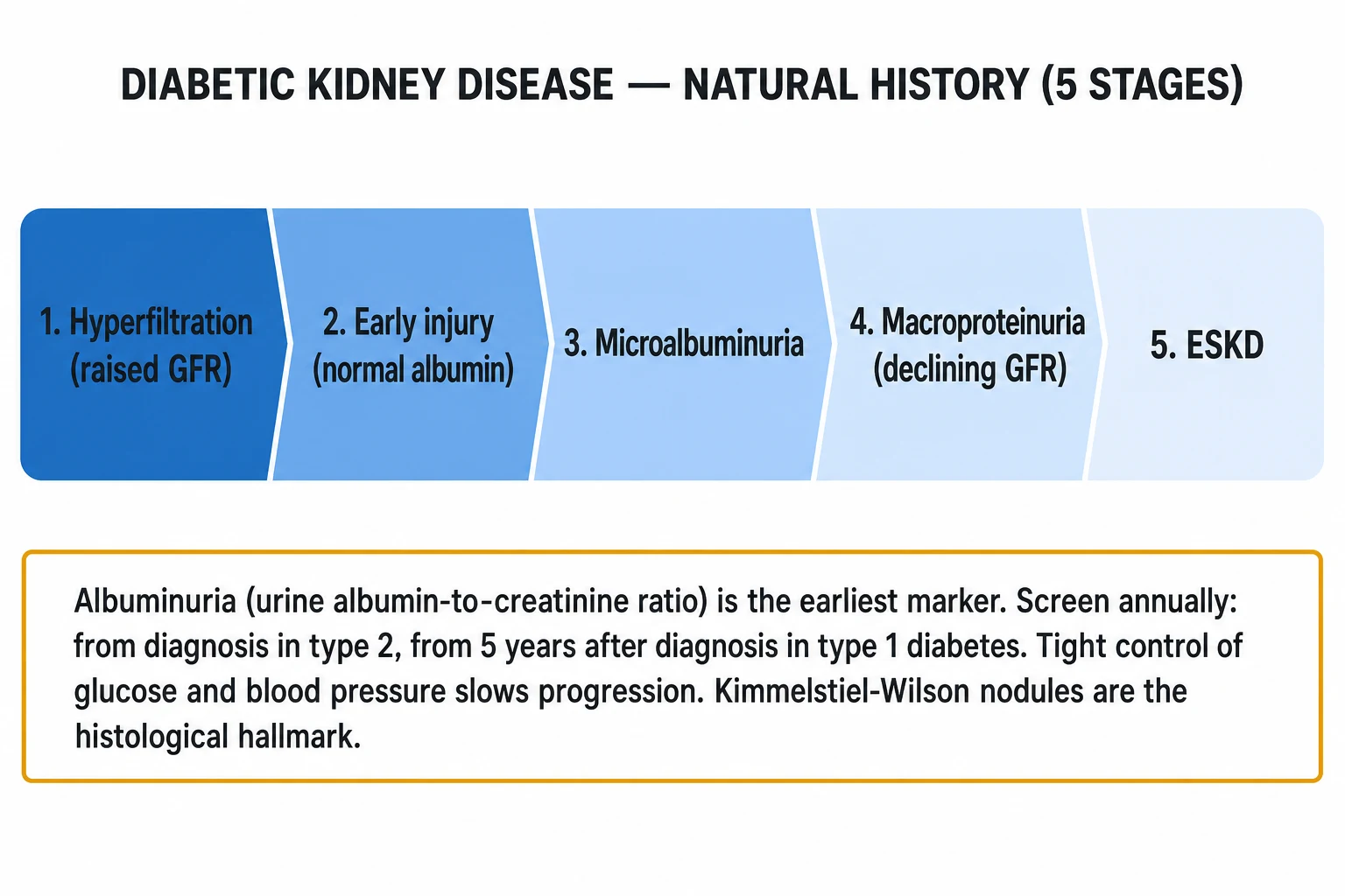
Mogensen five-stage classification of diabetic nephropathy (the classical natural history):[1]
- Stage I — glomerular hyperfiltration and hypertrophy (at diagnosis of diabetes). GFR is raised (over 120 mL/min/1.73 m²), kidney size increased; albuminuria is normal; reversible with good glycaemic control.
- Stage II — silent/latent phase (develops over years). Histological lesions (basement membrane thickening, mesangial expansion) are present; albuminuria remains in the normal range at rest but may rise with exercise or poor glycaemic control.
- Stage III — incipient diabetic nephropathy (microalbuminuria) (typical onset 5 to 15 years after diagnosis). Persistent UACR 30 to 300 mg/g (A2) on at least two of three samples over 3 to 6 months; GFR may still be normal or raised; blood pressure begins to rise. This is the earliest clinically detectable stage and represents the treatment window.
- Stage IV — overt diabetic nephropathy (macroproteinuria) (typical onset 10 to 25 years after diagnosis). UACR over 300 mg/g (A3), often nephrotic-range; GFR begins a steady decline; hypertension is universal; oedema may develop. This stage historically progressed to ESKD over 5 to 10 years without therapy.
- Stage V — end-stage kidney disease (ESKD). GFR under 15 mL/min/1.73 m² (G5); requires renal replacement therapy (dialysis or transplantation). [1]
KDIGO albuminuria categories (apply to all CKD including DKD):[2]
A1 — Normal to mildly increased
- UACR under 30 mg/g (under 3 mg/mmol)
- Risk: low if eGFR preserved
- Re-screen annually in diabetes
A2 — Moderately increased (microalbuminuria)
- UACR 30 to 300 mg/g (3 to 30 mg/mmol)
- Earliest marker of DKD
- Start RAAS blockade + SGLT2i here
A3 — Severely increased (macroproteinuria)
- UACR over 300 mg/g (over 30 mg/mmol)
- Equivalent to overt nephropathy
- Nephrotic-range over 2200 mg/g predicts rapid progression
KDIGO GFR categories (combined with the albuminuria category above into a heat-map of risk): [1]
- G1 — GFR 90 or above (normal or high)
- G2 — GFR 60 to 89 (mildly decreased)
- G3a — GFR 45 to 59 (mildly to moderately decreased)
- G3b — GFR 30 to 44 (moderately to severely decreased)
- G4 — GFR 15 to 29 (severely decreased; plan RRT)
- G5 — GFR under 15 (kidney failure; initiate RRT) [1]
The intersection of albuminuria category (A1-A3) with GFR category (G1-G5) gives the KDIGO heat-map risk (green/yellow/orange/red). Patients in G3b or worse combined with A3 are at the highest risk of progression and of cardiovascular events; they are referred for nephrology care. [1]
Epidemiology & Risk Factors
Diabetic kidney disease is the leading single cause of end-stage kidney disease worldwide, accounting for roughly 30 to 50 percent of all patients reaching dialysis or transplantation in most industrialised countries, and an even larger share of incident ESKD in India, China, the Middle East and the Pacific Islands. Globally, diabetes affects more than 500 million adults, and DKD develops in approximately 30 to 40 percent of patients with either type 1 or type 2 diabetes over their lifetime.[1]
Diabetic kidney disease — the headline numbers
Risk factors are traditionally divided into modifiable and non-modifiable. Both axes are examinable. [1]
Non-modifiable risk factors: long duration of diabetes (the dominant risk factor — DKD is rare within the first 5 years of T1DM), older age at onset, male sex, family history of DKD or hypertension, and genetic susceptibility with ethnic clustering — notably high incidence in Pima Indians, South Asians, Black Americans, Hispanic Americans and Pacific Islanders. Specific susceptibility loci (e.g., variants in UMOD, APOL1 in people of African ancestry, ACACB) modulate risk, but no gene is yet used clinically.[1]
Modifiable risk factors (the targets of therapy): [1]
- Chronic hyperglycaemia — the central driver; HbA1c correlates with risk. The DCCT (T1DM) and UKPDS (T2DM) showed that intensive glycaemic control reduces the development of microalbuminuria and overt nephropathy, with a durable metabolic memory effect that persists for years after the trial ended.
- Hypertension — accelerates glomerular injury; systolic blood pressure over 130 mmHg doubles the rate of GFR decline.
- Smoking — independent risk factor for progression and for cardiovascular death.
- Obesity and dyslipidaemia — promote inflammation and progression.
- Dietary protein and sodium intake — high intake accelerates hyperfiltration.
- Insulin resistance (in T2DM) — drives hyperfiltration independent of glucose. [1]
Microvascular context — retinopathy correlation. In type 1 diabetes, the correlation between DKD and proliferative diabetic retinopathy is strong: a patient with proteinuria almost always has retinopathy, and its absence is a red flag prompting consideration of an alternative diagnosis. In type 2 diabetes, the correlation is weaker: roughly 20 to 30 percent of patients with classical DKD have no detectable retinopathy, so absence of retinopathy is a softer pointer in T2DM.[1]
Pathophysiology
The pathogenesis of DKD is multifactorial and progressive, integrating a haemodynamic, a metabolic, and an inflammatory/fibrotic axis. Understanding each — and how the modern therapies interrupt them — is exactly what the examiner probes at viva depth. [1]
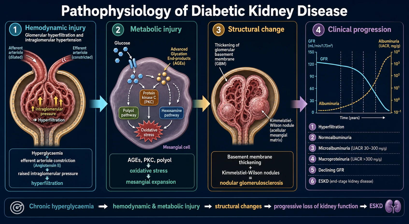
1. The haemodynamic hypothesis (glomerular hypertension). Chronic hyperglycaemia activates the intrarenal renin-angiotensin-aldosterone system (RAAS), increasing local angiotensin II. Angiotensin II preferentially constricts the efferent arteriole, raising intraglomerular capillary pressure and producing glomerular hyperfiltration (GFR over 120 mL/min/1.73 m²). The increased pressure and flow mechanically stress podocytes and the glomerular basement membrane, and shear-stress stimulates mesangial cells to lay down matrix. This is the mechanistic rationale for RAAS blockade: ACE inhibitors and ARBs dilate the efferent arteriole, lowering intraglomerular pressure and reducing proteinuria beyond what blood-pressure lowering alone achieves.[10] SGLT2 inhibitors act on the proximal tubule: by restoring tubuloglomerular feedback (more sodium delivered to the macula densa -> afferent arteriolar constriction), they lower intraglomerular pressure from the afferent side and reduce hyperfiltration — the mechanistic basis for their glucose-independent kidney protection.[3][4]
2. The metabolic hypothesis (intracellular glucose toxicity). When intracellular glucose is persistently raised, four biochemical pathways are over-activated:[1]
- Polyol pathway — aldose reductase converts glucose to sorbitol, consuming NADPH and generating osmotic stress.
- Hexosamine pathway — generates UDP-GlcNAC, which O-glycosylates transcription factors (e.g., Sp1) up-regulating TGF-beta and PAI-1.
- Protein kinase C (PKC) activation — diacylglycerol from glycolytic intermediates activates PKC isoforms, up-regulating VEGF, TGF-beta, NF-kappaB and extracellular matrix proteins.
- Advanced glycation end-products (AGEs) — non-enzymatic glycation of proteins and lipids forms irreversible cross-links that bind the RAGE receptor, driving inflammation, oxidative stress and matrix accumulation. [1]
The net result is increased reactive oxygen species (ROS), mitochondrial dysfunction, and up-regulation of profibrotic cytokines — most importantly transforming growth factor-beta (TGF-beta) and connective tissue growth factor (CTGF) — which drive mesangial matrix expansion and tubulointerstitial fibrosis.[1]
3. The structural lesions (the histology the examiner expects you to draw). Three lesions characterise DKD on light and electron microscopy: [1]
- Diffuse glomerulosclerosis — mesangial matrix expansion and glomerular basement membrane thickening, the commonest lesion (not specific to diabetes).
- Nodular glomerulosclerosis (Kimmelstiel-Wilson nodules) — rounded, acellular, eosinophilic nodules in the mesangial centre of glomerular lobules, pathognomonic of diabetes (although similar nodules occur in light-chain deposition disease and amyloidosis, distinguished by immunofluorescence and Congo red staining).
- Arteriolar hyalinosis — affecting both afferent and efferent arterioles (the latter is highly specific to diabetes, unlike hypertensive nephrosclerosis which affects only afferent). [1]
Additional lesions include exudative lesions (fibrin caps, capsular drop), tubular basement membrane thickening, and interstitial fibrosis with tubular atrophy — and it is the degree of interstitial fibrosis that best correlates with the rate of GFR decline, more so than the glomerular changes themselves.[1]
4. The natural history and why some patients deviate. Classical DKD follows the albuminuria-to-GFR-decline pathway of the Mogensen stages. However, a substantial minority of patients (especially in T2DM) show non-albuminuric DKD — a progressive fall in GFR without significant albuminuria. This is now attributed to tubulointerstitial and vascular injury (ischaemic nephropathy, hypertensive nephrosclerosis superimposed on diabetes) and to the prior use of RAAS blockade lowering albuminuria while GFR continues to fall. It is not benign — these patients still progress and still carry high cardiovascular risk.[1][2]
Clinical Presentation
Early DKD is asymptomatic. It is detected only by annual UACR and eGFR screening in a known diabetic; symptoms appear only when proteinuria becomes heavy or GFR falls substantially. The examiner expects you to describe both the silent early phase and the overt clinical picture. [1]
Classical overt DKD presents in a long-standing diabetic (typically 10 to 25 years post-diagnosis in T1DM, more variable in T2DM) with the tetrad of: [1]
- Proteinuria — initially microalbuminuria (UACR 30 to 300 mg/g), progressing to macroproteinuria (UACR over 300 mg/g), and frequently to nephrotic-range proteinuria (over 3.5 g/day) with oedema and hypoalbuminaemia. The dipstick becomes positive only once UACR exceeds about 300 mg/g — the dipstick does NOT detect microalbuminuria, a perennial exam point.
- Hypertension — develops early (Stage III) and is virtually universal by Stage IV; it is both a cause and consequence of progression.
- Progressive decline in GFR — once macroproteinuria is established, GFR falls at an average of 10 to 12 mL/min/year untreated, accelerating with poor glycaemic and blood-pressure control.
- Associated microvascular disease — diabetic retinopathy (especially in T1DM), sensory-motor peripheral neuropathy and autonomic neuropathy, plus macrovascular disease (coronary, cerebrovascular, peripheral arterial). The patient with a foot ulcer, retinopathy and proteinuria has multi-system microvascular disease requiring integrated care.[1]
Nephrotic presentation. A subset present with heavy proteinuria, oedema and hypoalbuminaemia (the nephrotic syndrome pattern). Nephrotic-range proteinuria in DKD predicts a rapidly progressive course and a high cardiovascular risk. [1]
Atypical / non-albuminuric DKD. The patient (usually T2DM, often elderly) shows a falling eGFR with a UACR persistently under 30 mg/g. This is increasingly recognised and reflects tubulointerstitial and vascular injury; it is a clinical diagnosis after other causes are excluded. [1]
Atypical presentations requiring biopsy. The features that do not fit classical DKD and should prompt renal biopsy are: [1]
- Short duration of diabetes (under 5 years in T1DM, or recently diagnosed T2DM).
- Absence of retinopathy (strong red flag in T1DM; softer in T2DM).
- Rapidly declining GFR (over 5 mL/min/year or an acute rise in creatinine).
- Active urinary sediment — haematuria (especially macroscopic, or dysmorphic red cells/red-cell casts), suggesting a glomerulonephritis.
- Sudden onset of heavy proteinuria out of keeping with prior course.
- Signs of systemic disease (rash, arthralgia, haemoptysis, constitutional symptoms) suggesting lupus, vasculitis or amyloidosis.
- A greater than 30 percent fall in eGFR after starting ACEi/ARB, suggesting underlying renal artery stenosis.[2]
Differential Diagnosis
Not every proteinuric diabetic has DKD, and missing an alternative glomerulonephritis is a classic exam and clinical pitfall. The complete differential, with distinguishing features, is:[1][2]
Classical DKD
- Long-standing diabetes (10+ yr)
- Albuminuria, progressive CKD
- Retinopathy (strong in T1DM)
- Normotensive-then-hypertensive course
- No active sediment; no systemic features
Hypertensive nephrosclerosis
- Long-standing HTN, often predating diabetes
- Mild albuminuria, slowly falling GFR
- LVH, retinopathy of HTN
- Biopsy: afferent arteriolar hyalinosis only
- UACR usually under 1000 mg/g
Primary glomerulonephritis (IgA, membranous, FSGS, MCD)
- Haematuria, active sediment
- Short diabetes duration, no retinopathy
- Sudden heavy proteinuria
- Biopsy diagnostic (immunofluorescence pattern)
Ischaemic / renovascular disease
- Vascular disease, refractory HTN
- Over 30% eGFR fall on ACEi/ARB
- Asymmetric small kidneys on US
- Recurrent flash pulmonary oedema
Amyloidosis
- Nephrotic-range proteinuria
- Chronic inflammatory disease / myeloma
- Congo-red positive biopsy
- Systemic features (hepatosplenomegaly, macroglossia)
Multiple myeloma (cast nephropathy)
- Elderly, anaemia, bone pain
- Anaemia out of proportion to GFR
- High calcium, low anion gap
- SPEP / SFLC diagnostic; biopsy: light-chain casts
The can't-miss diagnoses in a diabetic with a rising creatinine or new proteinuria are: acute kidney injury (from volume depletion, sepsis, contrast, NSAIDs or the triple whammy of NSAID + ACEi/ARB + diuretic), renal artery stenosis (a fall in eGFR greater than 30 percent on starting RAAS blockade), obstructive uropathy (especially in older men with autonomic neuropathy — check the bladder scan), contrast-induced nephropathy, and superimposed glomerulonephritis. The clinical rule the examiner rewards is: "Albuminuria plus long-standing diabetes plus retinopathy equals classical DKD; anything else mandates a renal ultrasound and consideration of biopsy."[1]
Clinical & Bedside Assessment
The focused assessment of a patient with (or at risk of) DKD integrates kidney-specific findings with systemic micro- and macrovascular disease — because DKD is the renal manifestation of a multi-system process. [1]
Kidney- and volume-specific assessment. Measure blood pressure in both arms (target under 130/80 mmHg in albuminuric DKD), assess volume status (JVP, sacral and ankle oedema, basal crackles indicating fluid overload), and inspect for the consequences of advanced CKD (pallor of anaemia, scratch marks of uraemic pruritus, asterixis of uraemia in advanced disease). Perform bedside urinalysis: a dipstick positive for protein indicates macroproteinuria (UACR over about 300 mg/g), but a negative dipstick does not exclude microalbuminuria — a spot UACR is mandatory.[2]
Integrated cardiovascular and microvascular exam. DKD rarely travels alone. Examine the fundus for diabetic retinopathy (its presence supports classical DKD; its absence in T1DM is a red flag), the heart for ischaemic heart disease, heart failure and an S4 gallop of hypertensive heart disease, the peripheral pulses and listen for carotid, abdominal and femoral bruits (atherosclerotic disease), examine the feet for neuropathy (10 g monofilament, vibration with a 128 Hz tuning fork) and ulcers, and assess for autonomic neuropathy (postural blood-pressure drop). The burden of cardiovascular disease is what determines prognosis and what most of these patients will die of.[1]
Bedside and clinical 'traps': [1]
- A proteinuric diabetic with normal fundi and short diabetes duration is not classical DKD — pursue alternative diagnoses.
- A rising creatinine after starting an ACE inhibitor/ARB: a rise of under 30 percent is expected and benign; a rise of over 30 percent, especially with new hypertension, suggests renal artery stenosis and mandates stopping the drug and imaging.
- An acute kidney injury in a diabetic on an NSAID, ACEi/ARB and diuretic is the classic 'triple whammy' — stop the offending drugs and resuscitate. [1]
Investigations
Investigations serve three purposes: screening and early detection (annual UACR and eGFR), diagnosis and staging of established DKD (KDIGO heat-map), and exclusion of alternative diagnoses (biopsy, serology). [1]
First-line investigations for every diabetic (the screening bundle):[1][2]
- Spot urine albumin-to-creatinine ratio (UACR) on a random sample — the earliest marker. Categories: A1 under 30, A2 (microalbuminuria) 30 to 300, A3 (macroproteinuria) over 300 mg/g.
- Serum creatinine with eGFR (CKD-EPI 2021 equation; do not use the MDRD equation for staging in 2026).
- HbA1c (glycaemic control, metabolic memory).
- Lipid profile (statin target).
- Blood pressure and weight/BMI. [1]
Screening schedule: annually from diagnosis in T2DM; annually from 5 years after diagnosis (or puberty, whichever is later) in T1DM. Confirm persistent albuminuria on two of three samples over 3 to 6 months, excluding the false-positive triggers: urinary tract infection (urine microscopy/culture), heavy exercise in the prior 24 hours, fever, heart failure decompensation, uncontrolled hypertension, menstruation, and marked hyperglycaemia.[2]
Confirmatory and staging tests: [1]
- eGFR (CKD-EPI 2021) with a category G1-G5; if creatinine-based eGFR is unreliable (very low or high muscle mass, amputation, pregnancy), use cystatin C-based eGFR.
- Renal ultrasound — to exclude obstruction, assess kidney size and symmetry (small symmetric kidneys suggest advanced CKD; asymmetric kidneys suggest renovascular disease). In DKD the kidneys are often normal or enlarged early and become small late.
- Urinalysis and microscopy — to exclude haematuria and active sediment (which would mandate biopsy).
- ECG and cardiac work-up for the almost-always coexisting cardiovascular disease. [1]
Investigations to exclude alternative diagnoses (when features are atypical): serum immunoglobulins, serum free light chains and serum protein electrophoresis (myeloma), ANA, anti-dsDNA, ANCA, complement (lupus, vasculitis), hepatitis B, C and HIV serology (membranous, cryoglobulinaemia), anti-PLA2R antibody (primary membranous), cryoglobulins and rheumatoid factor, and renal biopsy.[2]
Indications for renal biopsy in a patient with diabetes (know these verbatim): [1]
- Short duration of diabetes (under 5 years in T1DM).
- Absence of retinopathy (especially in T1DM).
- Rapidly declining GFR (over 5 mL/min/1.73 m²/year or an acute creatinine rise).
- Active urinary sediment — haematuria, dysmorphic red cells, red-cell casts.
- Sudden onset of heavy or nephrotic-range proteinuria out of keeping with the prior course.
- More than 30 percent fall in eGFR on starting ACEi/ARB (suggests renal artery stenosis).
- Signs of systemic disease (rash, arthralgia, haemoptysis) suggesting lupus, vasculitis or amyloidosis.[2]
Management — Resuscitation
DKD is a chronic, slowly progressive disease, but decompensation can be acute and life-threatening. The resuscitation scenarios the examiner expects you to handle: [1]
1. Hyperkalaemia in a patient with DKD. Hyperkalaemia (K over 5.5 mmol/L) is common because of reduced potassium excretion plus RAAS blockade. Manage by assessment of ECG changes (peaked T waves, wide QRS — give IV calcium gluconate 10 mL of 10 percent over 5 to 10 minutes for membrane stabilisation), insulin-dextrose (10 units of rapid-acting insulin with 25 to 50 g of IV dextrose over 15 minutes to shift potassium into cells), salbutamol 10 to 20 mg nebulised as an adjunct, sodium bicarbonate if acidotic, and potassium binders (patiromer, sodium zirconium cyclosilicate) or sodium polystyrene sulfonate for chronic control. Avoid stopping ACEi/ARB for modest hyperkalaemia (under 6.0 mmol/L) — the kidney protection outweighs the risk; address dietary potassium, acidosis and concomitant drugs first. [1]
2. Euglycaemic diabetic ketoacidosis on an SGLT2 inhibitor. SGLT2 inhibitors can precipitate euglycaemic DKA — glucose often under 14 mmol/L (250 mg/dL) — typically in the setting of fasting, surgery, acute illness, alcohol or very low carbohydrate intake. Suspect it in any SGLT2i-treated patient with nausea, vomiting, abdominal pain, malaise, ketotic breath. Management: stop the SGLT2 inhibitor, give IV fluids (normal saline), IV insulin infusion (often required even with near-normal glucose), co-infuse IV dextrose to allow insulin dosing, correct electrolytes, and treat the precipitant. The key diagnostic pitfall is missing DKA because the glucose is not high.[3]
3. Acute kidney injury superimposed on DKD. Common precipitants are volume depletion (vomiting, diuretics, sepsis), contrast media, NSAIDs (alone or as the triple whammy with ACEi/ARB + diuretic), sepsis, and hypoglycaemia. Manage ABCDE, stop nephrotoxins, resuscitate with IV fluids (balanced crystalloid), treat the precipitant, withhold ACEi/ARB, SGLT2i, metformin and NSAIDs temporarily, and recheck renal function and potassium daily. Resume disease-modifying therapy once eGFR has returned to baseline.[2]
4. Flash pulmonary oedema / accelerated hypertension. Sit the patient up, give high-flow oxygen, IV loop diuretic (furosemide 40 to 80 mg, repeated), consider IV nitrate (glyceryl trinitrate infusion) if hypertensive, and investigate for renal artery stenosis if eGFR fell on starting RAAS blockade. [1]
Management — Definitive & Stepwise
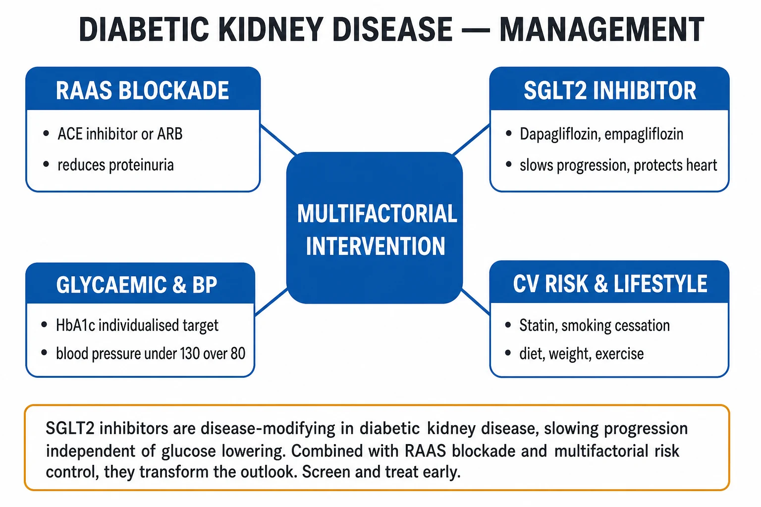
The management of DKD is multifactorial and combination-based. The modern paradigm is a four-pillar combination deployed early, supported by lifestyle and risk-factor control, with escalation to renal replacement therapy planning as eGFR falls. [1]
Four pillars of DKD therapy — 'R-S-F-M'
RSFM
ACE inhibitor or ARB at max tolerated dose — first-line in albuminuric DKD
dapagliflozin, empagliflozin or canagliflozin — disease-modifying, glucose-independent
non-steroidal MRA; kidney + CV protection on top of RAAS blockade
glycaemic + BP control, statin, smoking cessation, diet, exercise
Pillar 1 — RAAS blockade (ACE inhibitor or ARB)
RAAS blockade is first-line therapy in albuminuric DKD, with the mechanism of efferent arteriolar dilatation lowering intraglomerular pressure and reducing proteinuria. The landmark evidence: in type 1 diabetes the captopril trial (Lewis 1993) showed captopril halved the combined endpoint of death, dialysis and transplantation in proteinuric T1DM nephropathy.[6] In type 2 diabetes, the IDNT (irbesartan) and RENAAL (losartan) trials each showed that ARB therapy reduced the risk of doubling of creatinine or ESKD by about 16 to 20 percent versus placebo.[7][8]
Key points: [1]
- Use either an ACE inhibitor or an ARB — they are equivalent in efficacy; ARBs are better tolerated (less cough).
- Start only in albuminuric DKD (A2 or A3). There is no proven benefit of routine RAAS blockade in normoalbuminuric diabetes — it is not a primary-prevention drug for DKD.
- Titrate to the maximum tolerated licensed dose (e.g., ramipril 10 mg daily or lisinopril 20 to 40 mg daily; losartan 100 mg daily or irbesartan 300 mg daily).
- Monitor creatinine and potassium at 1 to 2 weeks after initiation or dose change — a creatinine rise of under 30 percent is expected and benign; a rise over 30 percent mandates drug cessation and imaging for renal artery stenosis.
- Do not combine an ACE inhibitor with an ARB — the ONTARGET trial showed dual RAAS blockade increased AKI, hyperkalaemia and hypotension with no kidney-outcome benefit.
- Continue RAAS blockade in advanced CKD unless hyperkalaemia or hypotension prevent it; stopping in stable advanced DKD does not improve outcomes.[10]
Pillar 2 — SGLT2 inhibitor
Sodium-glucose co-transporter-2 inhibitors are the most important recent change in DKD management — they slow progression and reduce cardiovascular events independent of glucose lowering, and are recommended in all patients with DKD and an eGFR over 20 mL/min/1.73 m² (continue down to eGFR 20). The mechanism is restoration of tubuloglomerular feedback — by inhibiting proximal sodium-glucose reabsorption, more sodium reaches the macula densa, afferent arteriolar tone rises and intraglomerular pressure falls.[3][4][5]
The three pivotal trials (reproduce the headlines at viva): [1]
- CREDENCE (2019) — canagliflozin in T2DM with albuminuric CKD (eGFR 30 to 90, UACR over 300). Reduced the primary composite of ESKD, doubling of creatinine, or renal/CV death by 30 percent; stopped early for efficacy.[3]
- DAPA-CKD (2020) — dapagliflozin in CKD with and without diabetes (eGFR 25 to 75, UACR over 200). Reduced the composite of kidney failure, sustained eGFR decline or renal/CV death by 39 percent; benefit was identical in diabetics and non-diabetics.[4]
- EMPA-KIDNEY (2023) — empagliflozin in a broad CKD population including eGFR down to 20 and many non-diabetic causes. Reduced progressive kidney disease or CV death by 28 percent, extending the benefit to lower eGFR and less albuminuric phenotypes.[5]
Practical use: [1]
- Agent and dose — dapagliflozin 10 mg once daily (eGFR down to 25, and now authorised to 20 for CKD), empagliflozin 10 mg once daily (eGFR down to 20), or canagliflozin 100 mg daily (titrate to 300 mg).
- Start in all DKD patients with eGFR over 20; the kidney-protective effect is independent of HbA1c.
- Monitor: volume status (mild diuresis; the drug is not a diuretic but increases natriuresis), genital candidiasis, and euglycaemic DKA risk — stop in acute illness, fasting, or before surgery.
- Contraindications: T1DM (off-label and causes DKA), current DKA, volume depletion, recurrent UTI or genital infection; caution with loop diuretics in the elderly. [1]
Pillar 3 — Finerenone (non-steroidal MRA)
Finerenone is a non-steroidal mineralocorticoid receptor antagonist that adds kidney and cardiovascular protection on top of ACE inhibitor or ARB therapy. Compared with spironolactone and eplerenone, it has less hyperkalaemia and a more favourable kidney distribution. The landmark evidence: FIDELIO-DKD (2020) showed finerenone reduced the composite kidney failure outcome by 18 percent in T2DM with CKD already on ACEi/ARB.[9]
Practical use: [1]
- Indication — T2DM with CKD (eGFR 25 to 60, UACR over 30) on max-tolerated ACEi/ARB.
- Dose — 10 mg once daily if eGFR 25 to 60; 20 mg once daily if eGFR over 60; titrate to 20 mg daily at 4 weeks if potassium is under 4.8 mmol/L.
- Monitoring — serum potassium at 4 weeks and periodically; reduce dose or hold if potassium over 5.5 mmol/L.
- Do not combine with steroidal MRAs (spironolactone, eplerenone) or with potassium supplements.[9][11]
Pillar 4 — Multifactorial risk reduction
The Steno-2 study proved that intensive, multifactorial intervention (RAAS blockade, blood pressure and glycaemic control, statins, aspirin, lifestyle) in T2DM with microalbuminuria reduced cardiovascular events, nephropathy, retinopathy and autonomic neuropathy — and at 21 years of follow-up the cohort gained an average of 8 years of life.[12] Multifactorial care is the foundation of all four-pillar therapy.
Glycaemic control. Target an individualised HbA1c of around 7 percent (53 mmol/mol); looser (7.5 to 8 percent) in advanced CKD, the frail elderly, or those with frequent hypoglycaemia. As eGFR falls, the diabetes drug choice changes:[2]
- Metformin — first-line; dose-reduce to 1000 mg/day at eGFR 30 to 45 and stop below 30 (lactic acidosis risk, though rare).
- Sulfonylureas — avoid in advanced CKD (hypoglycaemia risk); glipizide is the safest if one must be used.
- SGLT2 inhibitor — preferred second-line (kidney-protective).
- GLP-1 receptor agonist (liraglutide, semaglutide, dulaglutide) — preferred second-line when an SGLT2i is not tolerated; kidney-protective and CV-protective, no dose adjustment in CKD.
- DPP-4 inhibitor — safe in CKD; avoid saxagliptin and alogliptin (heart-failure risk); linagliptin needs no dose adjustment.
- Insulin — reduce doses by 25 to 50 percent as eGFR falls (insulin clearance drops); use basal-bolus or premixed. [1]
Blood pressure control. Target under 130/80 mmHg in albuminuric DKD (KDIGO); under 130 systolic reduces progression. Use ACE inhibitor or ARB as first-line; add a dihydropyridine calcium channel blocker (amlodipine 5 to 10 mg) or a thiazide/thiazide-like diuretic (chlorthalidone, indapamide) as second and third agents; loop diuretics replace thiazides once eGFR is under 30. [1]
Lipid management. A statin (atorvastatin 20 to 80 mg, rosuvastatin 10 to 40 mg) is indicated in all diabetics with CKD aged 40 to 75 (KDIGO); start at diagnosis of DKD — cardiovascular disease is the leading cause of death. [1]
Lifestyle. Smoking cessation (single biggest modifiable CV risk factor), dietary protein 0.8 g/kg/day (avoid high-protein diets that worsen hyperfiltration), sodium restriction under 2 g/day, weight management, regular aerobic and resistance exercise, and alcohol moderation. [1]
Aspirin. Consider aspirin 75 to 100 mg daily for secondary prevention in established CVD; routine primary prevention is individualised. [1]
Escalation triggers and referral
Refer to nephrology when:[2]
- eGFR under 30 mL/min/1.73 m² (plan for RRT).
- A3 albuminuria (over 300 mg/g) — confirm and refer.
- Rapidly declining eGFR (over 5 mL/min/year).
- Refractory hypertension or hyperkalaemia.
- Atypical features requiring biopsy.
- Nephrotic-range proteinuria. [1]
Plan vascular access (arteriovenous fistula) when eGFR is under 20; consider transplant assessment in suitable candidates when eGFR is under 20; initiate dialysis when eGFR is under 10 with symptoms or complications. [1]
Specific Subtypes & Scenarios
Type 1 vs type 2 diabetes. The pathophysiology and histology are essentially identical, but the clinical course and screening differ. In T1DM the latency from diagnosis to microalbuminuria is 5 to 15 years, screening starts at 5 years post-diagnosis (or puberty), the correlation with retinopathy is strong, and the cumulative incidence of ESKD is markedly reduced by intensive insulin therapy (DCCT/EDIC — the metabolic memory effect). In T2DM DKD can be present at diagnosis, screening is annual from diagnosis, the retinopathy correlation is weaker (20 to 30 percent have no retinopathy), and patients carry a much heavier burden of macrovascular disease (the usual cause of death).[1]
Non-albuminuric DKD (GFR-decline phenotype). Increasingly recognised in T2DM: a progressive fall in eGFR with UACR persistently under 30 mg/g. Attributed to tubulointerstitial and vascular injury (ischaemic nephropathy, hypertensive nephrosclerosis, ageing) and partly to prior RAAS blockade lowering albuminuria. These patients still progress and still benefit from SGLT2 inhibitors (EMPA-KIDNEY included patients with low albuminuria).[5]
Acute kidney injury superimposed on DKD. The diabetic kidney is vulnerable — precipitants include volume depletion, contrast, NSAIDs, sepsis, hypoglycaemia and the triple whammy. Manage by stopping nephrotoxins, resuscitating, treating the precipitant and resuming disease-modifying therapy once the eGFR returns. Every AKI accelerates the long-term trajectory of CKD.[2]
Refractory oedema and nephrotic-range proteinuria. Heavy proteinuria in DKD causes oedema; treat with loop diuretics (furosemide 40 to 250 mg daily; add a thiazide for synergism in resistant cases), salt restriction, and consider that adding finerenone further reduces proteinuria. Nephrotic-range proteinuria predicts rapid progression and high CV risk.[9]
Cardiorenal / Cardiovascular-Kidney-Metabolic (CKM) syndrome. DKD is the renal manifestation of a systemic cardiometabolic disorder in which diabetes, hypertension, obesity, atherosclerosis and heart failure interact. The SGLT2 inhibitor and finerenone benefit heart failure as well as kidney, supporting a unified cardiorenal therapy.[3][9]
Complications & Pitfalls
Kidney-related complications: progression to ESKD (the historical endpoint; now substantially deferred by combination therapy), nephrotic syndrome (heavy proteinuria, oedema, hypoalbuminaemia, hypercoagulability, hyperlipidaemia), AKI superimposed on CKD (NSAIDs, contrast, sepsis, triple whammy), hyperkalaemia, anaemia of CKD (which develops earlier in DKD than in non-diabetic CKD because of combined EPO deficiency and chronic inflammation), renal bone disease (CKD-MBD: high phosphate, low calcitriol, secondary hyperparathyroidism, vascular calcification), metabolic acidosis, and increased infection risk.[1]
Systemic complications: the dominant one is accelerated atherosclerosis — coronary artery disease, stroke and peripheral arterial disease are the leading causes of death in patients with DKD. Other: progression of diabetic retinopathy (often concurrent), neuropathy (sensory, autonomic, gastroparesis), foot ulcers and amputation, erectile dysfunction, and hypoglycaemia unawareness as autonomic neuropathy develops.[1]
Drug-related pitfalls (high-yield): [1]
- The triple whammy — NSAID + ACEi/ARB + diuretic -> AKI. Avoid the combination; stop NSAIDs in any CKD patient.
- Hypoglycaemia with insulin and sulfonylureas as eGFR falls — reduce doses, prefer glipizide if a sulfonylurea is needed.
- Metformin lactic acidosis — stop below eGFR 30 or in acute illness; rare but real.
- SGLT2i — euglycaemic DKA, volume depletion, genital mycotic infection, rare Fournier gangrene, stop perioperatively and in acute illness.[3]
- RAAS blockade — hyperkalaemia, fall in eGFR (under 30 percent acceptable), contraindicated in bilateral renal artery stenosis.
- Statins — start at diagnosis in all diabetics with CKD aged 40 to 75; stop in advanced CKD if not already on (low benefit) but continue if already established.[2]
Classic diagnostic pitfalls: [1]
- Missing an alternative glomerulonephritis because "it is just diabetes" — always check for haematuria, short diabetes duration, absent retinopathy.
- Not screening with UACR — the dipstick misses microalbuminuria.
- Under-dosing RAAS blockade — the benefit is dose-dependent; titrate to max tolerated.
- Forgetting finerenone — a third pillar of kidney protection.
- Failing to plan dialysis access early — arteriovenous fistula needs months to mature.
- Stopping ACEi/ARB for a modest creatinine rise — under 30 percent is expected and benign. [1]
Prognosis & Disposition
Prognostic markers. The strongest predictors of progression are: albuminuria level (higher UACR = faster decline), rate of eGFR decline (over 5 mL/min/year is rapid), blood pressure (uncontrolled is the dominant modifiable risk), glycaemic control (HbA1c), smoking, age, presence of cardiovascular disease, and histological interstitial fibrosis (better than glomerular lesions). Combining the KDIGO heat-map category with these factors estimates the trajectory.[1][2]
Mortality. Patients with DKD have a two- to four-fold higher mortality than non-diabetic CKD, and cardiovascular disease is the leading cause of death — more patients die with DKD than ever reach dialysis. This is why statins, blood-pressure control and SGLT2i (which reduce CV events) are central, not adjunctive. [1]
Disposition. Primary care manages stable early DKD (A1/G1-2, A2/G1-2 with stable eGFR). Refer to nephrology for atypical features, eGFR under 30, A3 albuminuria, rapidly declining GFR, refractory hypertension, or biopsy. Coordinate integrated care with ophthalmology (retinal screening), podiatry (foot care), cardiology (CV risk), and diabetes/endocrinology.[2]
Impact of modern therapy. With early detection and the four-pillar combination, DKD progression can be markedly slowed and CV events reduced. The Steno-2 21-year follow-up demonstrated a 8-year gain in median lifespan with intensive multifactorial intervention.[12] Historically, a third of patients with T1DM developed ESKD; with DCCT-era intensive control and modern combination therapy this is now well under 10 percent. The same approach applied early in T2DM is the single most effective way to reduce incident ESKD globally.
Special Populations
Pregnancy. Pre-conception counselling is essential. Stop SGLT2 inhibitors (insufficient safety data, theoretical risk of intrauterine growth restriction and ketoacidosis), stop finerenone (no pregnancy safety data), and stop ACE inhibitors and ARBs (teratogenic — cause fetal renal agenesis, oligohydramnios, pulmonary hypoplasia). Switch hypertension control to labetalol, nifedipine modified-release, or methyldopa. Target HbA1c under 6.5 percent pre-conception (with effective contraception until achieved). Pregnancy increases the risk of DKD progression and of pre-eclampsia — give low-dose aspirin 75 to 150 mg from 12 weeks in those with CKD or long-standing diabetes.[2]
The elderly. Adopt a less tight glycaemic target (HbA1c 7.5 to 8.5 percent) to avoid hypoglycaemia and falls; a less tight blood pressure target (under 140/90 is reasonable in the frail) to avoid orthostatic hypotension. The SGLT2i benefit persists in the elderly, but watch volume status carefully (especially with loop diuretics) and stop in acute illness or with poor oral intake. Continue RAAS blockade unless hyperkalaemia or hypotension intervene. [1]
Children and adolescents. In T1DM screen from puberty plus 5 years after diagnosis (whichever is first). Puberty accelerates microvascular complications. There are no licensed SGLT2i indications in children for DKD at present; management focuses on intensive glycaemic control and early RAAS blockade for albuminuria. [1]
The immunocompromised and post-transplant patient. Calcineurin inhibitors (ciclosporin, tacrolimus) and steroids worsen hypertension, glucose intolerance and proteinuria, accelerating the course. Diabetic nephropathy can recur in a transplanted kidney, typically 10 to 15 years post-transplant, and is managed with the same four-pillar approach. Adjust immunosuppression and prefer SGLT2i where appropriate.[2]
Evidence, Guidelines & Regional Differences
Landmark trials and what they changed. [1]
- Captopril trial, Lewis 1993 (NEJM) — captopril halved the risk of death, dialysis or transplantation in T1DM nephropathy. Established ACE inhibitor as standard of care in T1DM DKD.[6]
- IDNT (2001) — irbesartan and RENAAL (2001) — losartan — ARBs reduced the risk of doubling creatinine or ESKD by 16 to 20 percent in T2DM nephropathy. Established ARB as standard of care in T2DM DKD.[7][8]
- CREDENCE (2019) — canagliflozin — the first trial to show an SGLT2i reduced ESKD in T2DM nephropathy by 30 percent; stopped early. Transformed the field.[3]
- DAPA-CKD (2020) — dapagliflozin — extended the benefit to non-diabetic CKD and to lower eGFR; 39 percent risk reduction.[4]
- EMPA-KIDNEY (2023) — empagliflozin — extended the benefit to eGFR as low as 20 and to less albuminuric phenotypes; 28 percent risk reduction.[5]
- FIDELIO-DKD (2020) — finerenone — added a fourth pillar: 18 percent reduction in kidney outcomes on top of RAAS blockade.[9]
- Steno-2 (1993-2016, 21-year follow-up) — intensive multifactorial intervention (RAAS, BP, glycaemic, statin, lifestyle) gained 8 years of life in T2DM with microalbuminuria.[12]
The metabolic memory / legacy effect. The DCCT/EDIC follow-up in T1DM and UKPDS post-trial follow-up in T2DM both showed that early intensive glycaemic control produces benefits that persist for decades, even if later control worsens — a phenomenon called metabolic memory. This is why early, intensive therapy in the first years of diabetes is disproportionately important for preventing DKD.[1]
[1] [1]KDIGO 2024 CKD Guideline (global). The international reference. Recommends four-pillar combination therapy (RAAS blockade, SGLT2i, finerenone where indicated, multifactorial risk reduction); eGFR thresholds for SGLT2i down to 20 mL/min/1.73 m²; finerenone for T2DM CKD (eGFR over 25, potassium under 4.8); statin for all CKD aged 50 or older; metformin dose-reduce below 45 and stop below 30; blood-pressure under 130 systolic (target 120 to 129 systolic where tolerated in albuminuric CKD).
India (RSSDI-ESI 2023 / ICMR). Adopts the four-pillar approach. Empirical considerations: high prevalence of T2DM at low BMI; cost barriers to SGLT2i and finerenone in rural care; metformin widely used and dose-reduced as eGFR falls; ACEi/ARB first-line in albuminuria; SGLT2i recommended where accessible; statin in all aged 40 to 75; free public-sector dialysis programmes (e.g., PMNDP) for ESKD.
Controversies. [1]
- ACEi vs ARB equivalence — they are equivalent; choice driven by tolerability and cost. Combination (ACEi + ARB) is harmful (ONTARGET).
- Dual SGLT2i + GLP-1 RA — increasingly used, additive cardiorenal benefit, but cost and long-term safety data evolving.
- Low-protein diet (0.6 to 0.8 g/kg/day) — modest slowing of progression; hard to adhere; reserved for selected patients.
- Glycaemic targets in advanced CKD — relax to avoid hypoglycaemia, but the legacy effect argues for earlier tight control.
- SGLT2i in T1DM — not licensed; risk of euglycaemic DKA.
- Metformin in advanced CKD — historical conservatism relaxing; dose-reduce, not stop, in moderate CKD. [1]
Exam Pearls
- "Kimmelstiel-Wilson nodules = nodular glomerulosclerosis = diabetic nephropathy" — the single most testable histology one-liner.
- "Diabetic kidney disease is the commonest single cause of ESKD worldwide."
- "Microalbuminuria = UACR 30 to 300 mg/g (A2); the dipstick does NOT detect it."
- "Screen T2DM from diagnosis, T1DM from 5 years post-diagnosis (or puberty, whichever first), then annually."
- "Biopsy if: short diabetes duration, no retinopathy, rapid decline, haematuria, over 30 percent eGFR fall on RAAS blockade, or systemic features."
- "The four-pillar therapy: RAAS blockade (ACEi/ARB), SGLT2 inhibitor, finerenone, multifactorial risk reduction."
- "SGLT2 inhibitors are disease-modifying, independent of glucose lowering (CREDENCE, DAPA-CKD, EMPA-KIDNEY)."
- "ACEi/ARB are first-line only in albuminuric DKD — no benefit (and some harm) in routine normoalbuminuric use; never combine ACEi and ARB."
- "A rise in creatinine under 30 percent after starting ACEi/ARB is expected and benign; over 30 percent mandates stopping and imaging for renal artery stenosis."
- "The triple whammy: NSAID + ACEi/ARB + diuretic -> AKI."
- "Cardiovascular disease is the leading cause of death in DKD — give a statin to every diabetic with CKD aged 40 to 75."
- "SGLT2i euglycaemic DKA — glucose often under 14 mmol/L; stop in acute illness and perioperatively."
- "In T1DM, retinopathy correlates strongly with DKD; its absence is a red flag. In T2DM the correlation is weaker."
- "Arteriolar hyalinosis affecting BOTH afferent and efferent arterioles is highly specific to diabetes (vs only afferent in hypertension)."
- "Steno-2: 21-year follow-up showed 8 years of life gained with multifactorial intervention — metabolic memory / legacy effect." [1]
Exam application bank (NEET-PG / INICET)
One-line answer
Diabetic kidney disease (DKD, diabetic nephropathy) is the commonest single cause of end-stage kidney disease (ESKD) worldwide, developing over years through a predictable pathway of glomerular hyperfiltration, microalbuminuria, macroproteinuria and declining GFR. Its histological hallmark is nodular glomerulosclerosis (Kimmelstiel-Wilson nodules) with thickening of the glomerular basement membrane and mesangial expansion, driven by chronic hyperglycaemia and intraglomerular hypertension. The earliest clinical marker is albuminuria measured as the urine albumin-to-creatinine ratio (UACR): screen annually from diagnosis in type 2 diabetes and from 5 years after diagnosis in type 1 diabetes. Renal biopsy is reserved for atypical features (short diabetes duration, absence of retinopathy, rapid decline, haematuria, more than 30 percent creatinine rise on RAAS blockade). Management is multifa
Worked stems (answer without another resource)
Stem 1 — Classic presentation. Map symptoms to mechanism; name the first investigation and first treatment step with dose/route if drug therapy is standard. [1]
Stem 2 — Unstable / complicated. List red flags that force immediate resuscitation, theatre, ICU, antidote, or reperfusion — and what you do in the first 15 minutes. [1]
Stem 3 — Atypical group. Elderly, pregnancy, child, or immunocompromised: how presentation and thresholds change. [1]
Stem 4 — Differential trap. Name the three closest mimics and one discriminator for each. [1]
Stem 5 — Disposition. Who goes home with safety-netting, who is admitted, who needs HDU/ICU/theatre, and what follow-up is mandatory. [1]
Rapid viva checklist
- Definition + classification
- Pathophysiology chain
- Bedside signs / criteria
- Score with exact components (if any)
- Emergency bundle
- Definitive therapy with doses
- Complications of disease and of treatment
- Special populations
- Guideline/trial name if classic
- Three exam traps
Coverage self-check
If you cannot answer any stem above from this page alone, re-read the matching section — the page is intended to be self-sufficient for final-prof and NEET-PG/INICET questions on Diabetic Kidney Disease.
References
- [1]Thomas MC, Brownlee M, Susztak K, et al. Diabetic kidney disease Nat Rev Dis Primers, 2015.PMID 27188921
- [2]Ambalavanan J, Caramori ML. Management of Diabetes in Patients with Chronic Kidney Disease Endocr Res, 2025.PMID 40119502
- [3]Perkovic V, Jardine MJ, Neal B, et al. Canagliflozin and Renal Outcomes in Type 2 Diabetes and Nephropathy N Engl J Med, 2019.PMID 30990260
- [4]Heerspink HJL, Stefánsson BV, Correa-Rotter R, et al. Dapagliflozin in Patients with Chronic Kidney Disease N Engl J Med, 2020.PMID 32970396
- [5]The EMPA-KIDNEY Collaborative Group. Empagliflozin in Patients with Chronic Kidney Disease N Engl J Med, 2023.PMID 36331190
- [6]Lewis EJ, Hunsicker LG, Bain RP, Rohde RD. The effect of angiotensin-converting-enzyme inhibition on diabetic nephropathy. The Collaborative Study Group N Engl J Med, 1993.PMID 8413456
- [7]Lewis EJ, Hunsicker LG, Clarke WR, et al. Renoprotective effect of the angiotensin-receptor antagonist irbesartan in patients with nephropathy due to type 2 diabetes N Engl J Med, 2001.PMID 11565517
- [8]Brenner BM, Cooper ME, de Zeeuw D, et al. Effects of losartan on renal and cardiovascular outcomes in patients with type 2 diabetes and nephropathy N Engl J Med, 2001.PMID 11565518
- [9]Bakris GL, Agarwal R, Anker SD, et al. Effect of Finerenone on Chronic Kidney Disease Outcomes in Type 2 Diabetes N Engl J Med, 2020.PMID 33264825
- [10]Ruggenenti P, Cravedi P, Remuzzi G. The RAAS in the pathogenesis and treatment of diabetic nephropathy Nat Rev Nephrol, 2010.PMID 20440277
- [11]Bakris GL, Agarwal R, Chan JC, et al. Effect of Finerenone on Albuminuria in Patients With Diabetic Nephropathy: A Randomized Clinical Trial JAMA, 2015.PMID 26325557
- [12]Gæde P, Oellgaard J, Carstensen B, et al. Years of life gained by multifactorial intervention in patients with type 2 diabetes mellitus and microalbuminuria: 21 years follow-up on the Steno-2 randomised trial Diabetologia, 2016.PMID 27531506