Nephrology · Nephrology
Hyperkalaemia
Also known as Hyperkalemia · High potassium · Elevated serum potassium · Hyperpotassaemia
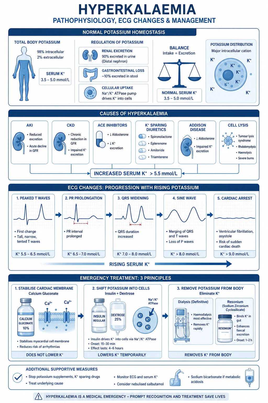
Hyperkalaemia is a serum potassium concentration above 5.5 mmol/L and is one of the few electrolyte emergencies because it can precipitate fatal cardiac arrhythmia. Severity is graded as mild (5.5 to 5.9 mmol/L), moderate (6.0 to 6.4 mmol/L) and severe (at least 6.5 mmol/L). The clinical priority is to exclude pseudohyperkalaemia (in vitro haemolysis, tourniquet fist-clenching, thrombocytosis or leucocytosis), identify the cause (CKD, ACEi or ARB, potassium-sparing diuretics, NSAIDs, acidosis, Addison disease, hypoaldosteronism, tumour lysis, massive transfusion), recognise the ECG progression (peaked T waves, PR prolongation, loss of P waves, QRS widening, sine-wave morphology, ventricular fibrillation or asystole) and deliver calcium-stabilised membrane protection followed by intracellular shift and total-body potassium removal. Acute emergency therapy is calcium gluconate 10% 10 to 30 mL IV over 2 to 5 minutes, insulin 10 units IV with 25 g of 50% dextrose, salbutamol 10 to 20 mg nebulised, sodium bicarbonate 50 to 100 mmol IV when acidotic, and total-body removal by loop diuretic, potassium binder (sodium polystyrene sulfonate 15 to 30 g, patiromer 8.4 g daily, sodium zirconium cyclosilicate 10 g TDS then 5 g daily) or haemodialysis. Chronic management relies on dietary potassium restriction, loop diuretics, binders and renin-angiotensin-aldosterone blockade re-challenge where safe.
On this page & tools
Your progress
Saved locally on this device.
Exam tags
Red flags

Overview & Definition
Hyperkalaemia is a serum potassium concentration above the upper limit of normal (above 5.5 mmol/L in most laboratories, but reference ranges vary slightly with assay). It is one of the few electrolyte abnormalities that can cause sudden death because it alters the resting membrane potential of cardiac myocytes, and a small absolute rise can convert a stable rhythm into ventricular fibrillation or asystole within minutes.[1]
The clinical task is therefore three-fold: (1) recognise and confirm true hyperkalaemia by excluding pseudohyperkalaemia (an in vitro artefact that does not reflect in vivo K+), (2) grade severity by combining the serum concentration with the ECG, and (3) deliver treatment in three time-ordered steps — stabilise the membrane, shift K+ into cells, remove K+ from the body — while the cause is corrected in parallel. [1]
A practical severity scheme — used by most emergency algorithms — is: [1]
- Mild 5.5 to 5.9 mmol/L — usually asymptomatic, often no ECG change; needs review of K+-retaining drugs and underlying cause.
- Moderate 6.0 to 6.4 mmol/L — usually still no symptoms but ECG changes begin to appear (peaked T waves); treatment is indicated even without symptoms.
- Severe ≥ 6.5 mmol/L or any K+ with ECG change — a medical emergency, treat before any confirmatory repeats.[1]
Death from hyperkalaemia is preventable; the diagnostic delay that matters is the one between drawing the venous sample and giving calcium gluconate, not the one between taking the sample and getting the biochemistry back. Once the ECG is abnormal the patient is in the arrhythmogenic window. [1]
Classification
Serum K+ classification (most adult labs): [1]
Mild
- Serum K+ 5.5 to 5.9 mmol/L
- Often asymptomatic; peaked T waves may appear at the upper end
- Review medications (ACEi, ARB, K+-sparing diuretic, NSAID) and diet
- Repeat sample without tourniquet to confirm true hyperkalaemia
- Correct cause and recheck in 12 to 24 hours
Moderate
- Serum K+ 6.0 to 6.4 mmol/L
- ECG findings common: peaked T waves, PR prolongation in some patients
- Treat actively — insulin/dextrose and a binder
- Stop K+-retaining drugs
- Admit if cause is AKI, Addison or tumour lysis
Severe / emergency
- Serum K+ ≥ 6.5 mmol/L OR any serum K+ above 5.5 with ECG changes (peaked T, lost P, wide QRS, sine wave)
- **Cardiac arrest imminent** — give calcium gluconate 10% 10 to 30 mL IV immediately
- Insulin/dextrose + salbutamol + ± sodium bicarbonate in parallel
- Dialysis if refractory or if AKI / ESRD
By mechanism — also essential for the exam: [1]
- Pseudohyperkalaemia (factitious) — serum K+ is raised but the in vivo plasma K+ is normal. Caused by in vitro haemolysis (prolonged tourniquet, fist-clenching, large-bore needle, traumatic tap, delayed separation of serum from cells), extreme leucocytosis (above 50 × 10⁹/L in chronic lymphocytic leukaemia) and extreme thrombocytosis (above 750 × 10⁹/L in myeloproliferative disorders). The clue is a normal plasma K+ when lithium-heparin plasma is analysed, or a normal ECG. Always repeat a K+ sample taken without a tourniquet before treating.[5]
- True hyperkalaemia — caused by impaired renal excretion, transcellular shift out of cells, or increased load (exogenous or endogenous). Each mechanism has characteristic causes (below).

Epidemiology & Risk Factors
True hyperkalaemia is uncommon in the general population but highly prevalent in the populations the exam focuses on: [1]
- Chronic kidney disease (CKD) — the dominant cause in adults; prevalence rises sharply as eGFR falls below 30 mL/min/1.73 m² and is over 20 to 30 percent by CKD stage 4-5.
- End-stage kidney disease on haemodialysis — predialysis K+ above 5.5 mmol/L is reported in about 10 percent of chronic haemodialysis patients; treatment-emergent hyperkalaemia is common with high-potassium dialysate, long inter-dialytic interval (Monday morning, post-weekend) and dietary indiscretion.
- Acute kidney injury (AKI) — particularly oliguric AKI and the diuretic-resistant phase; risk is highest when combined with rhabdomyolysis, tumour lysis or haemolysis (added K+ load).
- Renin-angiotensin-aldosterone system (RAAS) blockade — ACE inhibitors, ARBs and direct renin inhibitors predictably raise serum K+ by 0.1 to 0.5 mmol/L on average; clinically significant hyperkalaemia (above 5.5) develops in about 5 to 10 percent of users, rising above 30 percent when combined with a potassium-sparing diuretic or advanced CKD.
- Potassium-sparing diuretics — spironolactone, eplerenone, amiloride, triamterene; the addition of spironolactone 25 to 50 mg daily to ACEi in HFrEF (RALES, EMPHASIS-HF) raised hyperkalaemia hospitalisation rates two- to four-fold in registry studies.
- NSAIDs — renal sodium and potassium retention via prostaglandin inhibition and reduced renin; older patients on ACEi plus NSAID triple whammy are a classic scenario.
- Trimethoprim — blocks the epithelial sodium channel (ENaC) in the distal nephron like amiloride; chronic high-dose use raises serum K+.
- Calcineurin inhibitors — ciclosporin and tacrolimus cause hyperkalaemia via hypoaldosteronism and tubular dysfunction.
- Heparin — reduces aldosterone synthesis; chronic therapeutic dose produces a modest rise in serum K+.
- Diet — high-potassium foods (banana, avocado, dried fruit, salt substitutes that are potassium chloride such as Lo-Salt, tomato, potato, beans, coconut water). Dietary indiscretion in an ESRD patient with otherwise stable K+ is a frequent precipitant.
- Massive transfusion — older stored blood has supernatant K+ up to 30 to 50 mmol/L per unit; massive transfusion in trauma or obstetric haemorrhage produces a transient hyperkalaemia peak that resolves once red cells are cleared.
- Tumour lysis syndrome — rapid cell lysis in haematological malignancy chemotherapy releases K+, phosphate and nucleic acids (urate); classic trigger is Burkitt lymphoma and acute lymphoblastic leukaemia induction.
- Rhabdomyolysis / crush injury — release of intracellular K+ from damaged myocytes, often with AKI and an added renal excretory defect.
- Addison disease (primary adrenal insufficiency) — combined mineralocorticoid and glucocorticoid deficiency produces hyperkalaemia, hyponatraemia, hypotension, hyperpigmentation and metabolic acidosis.
- Hyporeninaemic hypoaldosteronism — most often in diabetic kidney disease (hyporeninism) and CKD; small to moderate rise in serum K+.
- Type 4 renal tubular acidosis (RTA) — hyperkalaemic RTA, classically in diabetes, CKD, ACEi and trimethoprim.
- Acidosis (metabolic and respiratory) — shifts K+ out of cells in exchange for H+; most pronounced in non-anion-gap metabolic acidosis (HCl, NH4Cl) and respiratory acidosis. [1]
Demographics: older adults (reduced renal function, polypharmacy, RAAS inhibitors), infants (immature tubular function), Black populations (lower renin, higher susceptibility to ACEi/ARB-induced K+ rises), and hospitalised patients (prevalence above 5 percent on some general medical floors). [1]
Pathophysiology
Potassium is the dominant intracellular cation: about 98 percent of the body's K+ (about 3000 to 3500 mmol) is inside cells, only about 2 percent in the extracellular fluid. The ratio of intracellular to extracellular K+ (set by the Na+/K+-ATPase on cell membranes) determines the resting membrane potential; a small rise in extracellular K+ (above 7 mmol/L) depolarises the membrane, inactivates fast sodium channels and produces the characteristic ECG changes.[8]
Total-body potassium depends on intake, distribution and excretion. [1]
- Intake — typical Western diet provides 50 to 100 mmol/day; most is absorbed in the small intestine.
- Shift across cell membrane — physiologically, K+ moves into cells under the influence of insulin (Na+/K+-ATPase via PI3K), beta-2 adrenergic stimulation (cyclic-AMP), alkalosis and aldosterone. Loss of these drives (insulin deficiency, beta-block, acidosis, hyporeninaemic hypoaldosteronism) shifts K+ out.
- Renal excretion — 90 percent of K+ excreted in the urine, primarily by principal cells of the cortical collecting duct under the influence of aldosterone. The collecting duct secretes K+ into the lumen via the renal outer medullary potassium (ROMK) channel and the large-conductance calcium-activated potassium (BK) channel, driven by lumen-negative transepithelial voltage generated by ENaC-mediated sodium reabsorption. About 10 percent is excreted in stool; gut excretion becomes clinically important in ESRD as a partial adaptation. [1]
Acidosis shifts K+ out of cells. In non-anion-gap metabolic acidosis (mineral acid load), H+ enters cells and K+ leaves to maintain electroneutrality — a 0.1-unit fall in pH raises serum K+ by roughly 0.5 to 1.0 mmol/L. This effect is blunted in anion-gap metabolic acidosis (lactate, ketoacid, urate, salicylate) where the organic anion enters the cell with H+ and K+ shift is smaller. [1]
Hyperglycaemia and insulin deficiency — osmotic shift of water out of cells drags K+ with it (solvent drag), and lack of insulin removes the tonic intracellular shift; this is why DKA patients have a whole-body potassium deficit even when serum K+ is normal or raised, and why the hypokalaemia unmasked during insulin therapy is the more important clinical risk. [1]
Beta-2 adrenergic stimulation drives K+ into cells via cAMP-mediated Na+/K+-ATPase activation; non-selective beta-blockers raise serum K+ by about 0.5 mmol/L on average. [1]
Tissue breakdown — rhabdomyolysis, tumour lysis, haemolysis, ischaemia (limb or splanchnic), severe burns and crush injury release intracellular K+ into the extracellular space, transiently exceeding excretion capacity and producing hyperkalaemia even with normal renal function. [1]
Impaired renal excretion is the dominant mechanism in the majority of clinical hyperkalaemia. Reduced GFR reduces filtered load; reduced aldosterone or reduced tubular sensitivity (hypoaldosteronism, ENaC blockade, distal tubular disease) reduces secretion. The kidney's adaptive capacity is large — until late CKD, hyperkalaemia is usually a signal of additional insult: a K+-retaining drug, an AKI, an intercurrent illness, or dietary indiscretion. [1]
Cardiac electrophysiology. Raised extracellular K+ depolarises the cardiac myocyte resting membrane potential (less negative), inactivates fast sodium channels, slows conduction velocity (widened QRS) and shortens the action potential duration (peaked T). Late changes — progressive conduction block, loss of atrial activity, sine-wave morphology — presage ventricular fibrillation or asystole. [1]
Clinical Presentation
Hyperkalaemia is often silent; the first sign is the ECG, the lab report, or cardiac arrest. Symptoms appear inconsistently above 6.0 to 6.5 mmol/L and are non-specific. [1]
Symptoms: [1]
- Muscle weakness — typically ascending, proximal; difficulty rising from a chair, climbing stairs, lifting arms; rarely progresses to flaccid quadriparesis mimicking Guillain-Barré syndrome at very high K+ (above 7.5 to 8.0 mmol/L).
- Fatigue and lethargy.
- Paraesthesia — perioral and acral.
- Nausea, vomiting, abdominal pain — visceral smooth-muscle dysfunction; severe cases can produce ileus.
- Palpitations, light-headedness, presyncope — from bradyarrhythmia or sinus node slowing.
- Syncope — Stokes-Adams attack from transient asystole.
- Sudden cardiac death — pulseless electrical activity (PEA), ventricular fibrillation or asystole may be the first manifestation; resuscitation in the setting of severe hyperkalaemia is rarely successful without membrane stabilisation. [1]
Signs: [1]
- Vital signs — bradycardia (sinus, junctional or idioventricular); hypotension if combined with volume depletion or Addisonian crisis.
- Neuromuscular — reduced muscle power (proximal then distal); reduced or absent deep tendon reflexes at very high K+.
- Cardiovascular — variable heart rate; auscultation usually unremarkable until arrhythmia. Check for Kussmaul (acidotic) breathing when acidosis is the cause.
- Skin and pigmentation — buccal and palmar crease pigmentation suggests primary adrenal insufficiency (Addison disease).
- Volume status — examine for evidence of AKI (oedema, low urine output), dehydration or fluid overload (in ESRD), and surgical causes (drains, stomas, fistula losses). [1]
Differential Diagnosis
1. Pseudohyperkalaemia (factitious — no therapy needed): [1]
- In vitro haemolysis — traumatic venepuncture, prolonged tourniquet time, fist-clenching, large-bore needle, vacuum tube under-fill, delayed centrifugation. The plasma appears pink; repeating without tourniquet gives a normal K+. Always the first consideration before treatment in an asymptomatic patient with no ECG change.
- Thrombocytosis — platelet count above 750 × 10⁹/L releases K+ during clotting; plasma K+ (lithium-heparin tube) is normal.
- Extreme leucocytosis — particularly in chronic lymphocytic leukaemia or acute leukaemia with very high counts; serum K+ is elevated but plasma K+ is normal.
- Cold agglutinins or sample freezing — laboratory artefact, no clinical correlate. [1]
2. True hyperkalaemia from impaired renal excretion: [1]
- CKD stages 4-5 — reduced filtered load; often asymptomatic until late; worsened by drugs.
- End-stage kidney disease on dialysis — inter-dialytic rise, long interval, dietary indiscretion.
- AKI — oliguric phase with rhabdomyolysis, sepsis, contrast-induced nephropathy.
- Hypoaldosteronism — primary (Addison disease) or hyporeninaemic hypoaldosteronism (diabetes, CKD).
- Type 4 renal tubular acidosis — hyperkalaemic distal RTA in diabetes and CKD.
- Drug-induced — ACEi, ARB, spironolactone, eplerenone, amiloride, triamterene, NSAID, trimethoprim, ciclosporin, tacrolimus, heparin. [1]
3. True hyperkalaemia from transcellular shift (often with underlying renal defect): [1]
- Metabolic acidosis — especially non-anion-gap (mineral acid); euglycaemic DKA recovery after insulin; lactic acidosis.
- Insulin deficiency — DKA presentation (with whole-body K+ deficit).
- Tissue breakdown — tumour lysis, rhabdomyolysis, haemolysis, severe burns, crush injury, intestinal ischaemia.
- Beta-blocker overdose — non-selective propranolol; combined with renal impairment.
- Succinylcholine (suxamethonium) administration — depolarising neuromuscular blocker, transient K+ rise of 0.5 to 1.0 mmol/L in normal subjects, dramatic rise (above 5 to 10 mmol/L) in burns, denervation, prolonged immobilisation, spinal cord injury, severe sepsis, neuromuscular disease (upregulation of extrajunctional acetylcholine receptors).
- Massive transfusion — stored blood supernatant K+.
- Hyperosmolar states — hyperglycaemia, hypertonic contrast, mannitol — solvent drag shifts K+ out with water.
- Hyperkalaemic periodic paralysis — rare autosomal dominant sodium-channel SCN4A mutation (paramyotonia congenita / hyperkalaemic periodic paralysis); attacks of weakness with raised K+, triggered by rest after exercise or potassium-rich food; resolves with carbamazepine prophylaxis.
- Malignant hyperthermia / MH — fulminant hypermetabolism with rhabdomyolysis and hyperkalaemia; treat with dantrolene 2.5 mg/kg IV. [1]
Clinical & Bedside Assessment
Priorities in sequence: (1) Look at the ECG — wide QRS or sine wave mandates treatment before further history; (2) brief history focused on cause and reversibility; (3) examination for cause and complications. [1]
1. ECG — essential; do this before the second sample. [1]
- Continuous 12-lead ECG and continuous cardiac monitoring in moderate and severe hyperkalaemia. Look for the progression: peaked T waves → PR prolongation → loss of P wave → wide QRS → sine wave → VF or asystole. The ECG may be normal even at high K+, and severe ECG changes (sine wave) may be present at moderate concentration — treat the ECG, not the number.
- Capture a rhythm strip with each therapeutic intervention to document response (peaked T resolves first with calcium gluconate, then with insulin). [1]
2. Vital signs and circulation. [1]
- Heart rate and rhythm — bradycardia in advanced hyperkalaemia is ominous (precedes asystole).
- Blood pressure — hypotension with hyponatraemia and hyperpigmentation suggests Addisonian crisis.
- Respiratory pattern — Kussmaul (deep, labored) breathing in metabolic acidosis.
- Temperature and perfusion — sepsis with AKI; cold, clammy extremities in adrenal crisis or cardiac decompensation. [1]
3. Focused examination for cause. [1]
- Cardiovascular — heart sounds (pericardial effusion in uraemia), JVP (volume status), murmurs (structural heart disease if arrhythmia has developed).
- Respiratory — pulmonary oedema in ESRD fluid overload, rales in acidosis-induced tachypnoea.
- Abdomen — ileus (silent, distended, tympanic abdomen); surgical cause (relook laparotomy, anastomotic leak, mesenteric ischaemia).
- Skin and mucosa — buccal pigmentation in Addison disease; surgical wounds; drains and stomas; track marks.
- Neuromuscular — proximal then distal muscle weakness; reflexes (diminished or absent at very high K+); cranial nerves usually spared; important differential from Guillain-Barré — potassium-related weakness resolves within hours of treatment. [1]
4. Volume status and urine output. [1]
- A reduced urine output or rising creatinine indicates renal cause; document a urine volume hourly and weigh or chart fluid balance where possible.
- Insert a urinary catheter if the patient is obtunded or for accurate measurement in AKI. [1]
Investigations
Bedside — immediately: [1]
- ECG (12-lead + rhythm strip) — before any other investigation; document the trace.
- Capillary glucose — DKA is an important mimic and a precipitant; insulin-dextrose therapy must be withheld in hypoglycaemic patients.
- Peripheral perfusion and pulse oximetry — for continuous monitoring during therapy. [1]
Blood — drawn at the same time as the initial sample: [1]
- Repeat venous K+ without tourniquet (or arterial blood gas K+ from the ABG syringe) to confirm true hyperkalaemia and exclude pseudohyperkalaemia. Do not wait for this result before treating if the ECG is abnormal or the patient is symptomatic.
- Arterial blood gas (ABG) — pH and bicarbonate confirm acidosis as a cause; K+ from the ABG is the most accurate in vivo value (no tourniquet, no clotting, immediate analysis).
- Urea, creatinine and electrolytes — to assess renal function, calculate eGFR, and detect hyponatraemia (Addison, volume overload).
- CK and LDH — for rhabdomyolysis and haemolysis respectively; if raised, examine for compartment syndrome or delayed sample effect.
- Creatinine kinase and urinary myoglobin in crush injury; uric acid and phosphate in tumour lysis.
- Glucose, ketones and HbA1c — DKA or HHS hyperosmolar states.
- Plasma aldosterone and renin — for Addison disease work-up; plasma cortisol (especially morning cortisol with cosyntropin stimulation) if Addison suspected.
- Digoxin level — digoxin toxicity causes hyperkalaemia via Na+/K+-ATPase inhibition; sample before giving digoxin-specific antibody fragments if indicated.
- Full blood count with film — leucocytosis, thrombocytosis, eosinophilia (interstitial nephritis from a drug).
- Coagulation screen and blood group — if massive transfusion considered. [1]
Urine: [1]
- Urinalysis — bland urinary sediment in hyperkalaemic RTA, glycosuria in diabetes.
- Urine K+ on a spot or 24-hour collection (> 200 mmol/24 h suggests renal K+ wasting in hypokalaemia; not usually needed in hyperkalaemia).
- Urine osmolality and sodium — pre-renal vs intrinsic AKI when relevant. [1]
Imaging — as driven by suspected cause: [1]
- Renal ultrasound — in new AKI to exclude obstruction; bilateral small echogenic kidneys in CKD.
- CT abdomen in suspected tumour lysis with bulky disease, retroperitoneal haemorrhage, or adrenal mass.
- Chest radiograph — pulmonary oedema, mediastinal mass (lymphoma causing tumour lysis). [1]
ECG progression — high-yield recognition sequence: [1]
- K+ 5.5 to 6.5 mmol/L — peaked T waves (tall, narrow-based, symmetric), best in precordial leads.
- K+ 6.5 to 7.0 mmol/L — peaked T + PR prolongation, flattening or loss of P waves.
- K+ 7.0 to 8.0 mmol/L — wide QRS, deep S wave in leads I, II, III, aVF; P wave absent; sine-wave morphology.
- K+ above 8.0 mmol/L — sine wave → ventricular fibrillation, pulseless ventricular tachycardia or asystole.[1]
Management — Resuscitation

The resuscitation priorities are time-ordered; do them in parallel where possible. [1]
Step 1 — Stabilise the cardiac membrane (calcium gluconate). Calcium does NOT lower serum K+; it raises the threshold potential, restoring the difference between resting and threshold potentials and stabilising the conduction system.[1]
- Calcium gluconate 10% — 10 to 30 mL (2.2 to 6.6 mmol calcium) IV over 2 to 5 minutes, in any patient with K+ ≥ 6.5 mmol/L, OR any K+ above 5.5 with ECG change, OR new wide-complex arrhythmia.
- Repeat every 5 to 10 minutes if ECG changes persist; consider a continuous infusion (1 to 2 mL/min of 10% calcium gluconate) in refractory cases.
- Calcium chloride 10% gives three times more calcium per mL but is irritant and requires a central line; reserve for cardiac arrest.
- Onset: 1 to 3 minutes; duration: 30 to 60 minutes (then membrane stabilisation may be lost while K+ remains high). [1]
Step 2 — Shift K+ intracellularly (insulin/dextrose, beta-2 agonist, sodium bicarbonate). [1]
- Insulin 10 units regular (soluble) IV bolus with 25 g dextrose IV (50 mL of 50% dextrose, or 100 mL of 25% dextrose, or 250 mL of 10% dextrose). Onset 10 to 20 minutes; peak 30 to 60 minutes; duration 4 to 6 hours. Monitor capillary glucose every 30 to 60 minutes for 6 hours — hypoglycaemia occurs in up to 20 percent, particularly in patients with renal impairment, low BMI or no prior diabetes.
- Salbutamol 10 to 20 mg (albuterol) nebulised in 4 mL saline over 10 minutes. Onset 20 to 30 minutes; duration 2 to 4 hours. Combine with insulin — additive intracellular shift. Avoid in significant tachyarrhythmia; can precipitate tremor and sinus tachycardia. Beta-2 alone reduces K+ by 0.5 to 1.0 mmol/L but is less reliable than insulin and is not used as monotherapy for severe hyperkalaemia.
- Sodium bicarbonate 50 to 100 mmol (1 to 2 mmol/kg) IV over 30 to 60 minutes if pH < 7.2 or base deficit > 6 mmol/L; shifts K+ into cells as H+ leaves. Indicated only for acidosis; not for routine use in hyperkalaemia with normal pH (no evidence of additional benefit and risks volume overload / hypocalcaemia in ESRD).
- Loop diuretic (furosemide 40 to 80 mg IV) — not strictly a redistribution therapy but enhances urinary K+ loss; use aggressively if the patient makes urine. [1]
Step 3 — Remove total-body K+ (bind, excrete or dialyse). This is the only step that durably lowers total-body K+; the previous two create a window only. [1]
- Loop diuretic — furosemide 40 to 80 mg IV, repeated or infused (10 to 20 mg/hour) where urine output is preserved; titrate to a urine volume of 200 to 300 mL/hour.
- Potassium-binding resins:
- Sodium polystyrene sulfonate (SPS, Kayexalate) 15 to 30 g orally or rectally as a retention enema; onset 1 to 4 hours. Caution with sorbitol co-administration (intestinal necrosis reported — avoid in post-operative patients and in those at risk of bowel ischaemia; modern formulations have largely removed sorbitol).
- Patiromer 8.4 g daily orally for chronic management; onset 7 to 12 hours for K+ reduction; well tolerated; reduces serum K+ by 0.5 to 1.0 mmol/L at steady state; allows RAAS inhibitor continuation in CKD. Requires 4 hours separation from other oral drugs.[7]
- Sodium zirconium cyclosilicate (SZC, Lokelma) — 10 g three times daily orally for 48 hours (acute correction), then 5 g daily maintenance; onset 1 to 2 hours in trials; mean reduction 0.7 mmol/L within 48 hours in HARMONIZE and 0.4 to 0.5 mmol/L sustained.[3][6]
- Dialysis — indicated for refractory hyperkalaemia (K+ remains above 6.5 mmol/L after the above), AEIOU indications, or AKI/ESRD with no renal reserve. Haemodialysis is the most effective total-body K+ removal (removes 25 to 50 mmol/hour at high blood flows; can achieve 60 to 120 mmol in a 4-hour session) and is preferred over peritoneal dialysis for acute, severe hyperkalaemia. Acute K+ rebound occurs after haemodialysis as cellular redistribution occurs; combine with binders.[9]
Resuscitation summary — time-ordered checklist:[1]
- ECG; secure IV access; ABG sample (gives accurate K+).
- Calcium gluconate 10% 10 to 30 mL IV over 2 to 5 minutes (repeat if ECG changes persist).
- Insulin 10 units IV + 50 mL 50% dextrose (25 g); capillary glucose every 30 minutes.
- Salbutamol 10 to 20 mg nebulised.
- Sodium bicarbonate 50 to 100 mmol IV if pH < 7.2.
- Furosemide 40 to 80 mg IV if urine output preserved.
- Patiromer, SZC or SPS for binder removal.
- Dialysis if refractory, AKI/ESRD or arrhythmia persists. [1]
Management — Definitive & Stepwise
Once the immediate threat is controlled, treat the cause and reduce total-body K+. [1]
1. Stop K+-retaining drugs — non-negotiable in acute hyperkalaemia. [1]
- ACE inhibitors and ARBs — withhold until K+ normalises; consider re-challenge at lower dose or with binder cover (patiromer / SZC) once stable.
- Spironolactone and eplerenone — withhold; resume only with a binder in CKD or HFrEF.
- Potassium-sparing diuretics (amiloride, triamterene) — stop.
- NSAIDs — stop (renal vasoconstriction, K+ retention).
- Trimethoprim — switch to alternative antibiotic if possible (e.g. nitrofurantoin, cephalexin for UTI).
- Ciclosporin / tacrolimus — coordinate with transplant team; may switch regimen.
- Heparin — review indication; consider LMWH only after K+ control. [1]
2. Correct volume status and treat the cause. [1]
- Addison disease — hydrocortisone 100 mg IV stat, then 200 mg per 24 hours IV (or 50 mg every 6 hours); fludrocortisone 100 to 200 micrograms daily once oral intake resumes. Aggressive volume resuscitation with isotonic saline.
- DKA / HHS — insulin infusion and fluids; potassium will fall; add K+ to fluids once serum K+ < 5.2 mmol/L to avoid hypokalaemia.
- Tumour lysis syndrome — aggressive isotonic hydration; rasburicase 0.2 mg/kg IV (or allopurinol if prophylaxis); consider early dialysis.
- Rhabdomyolysis — aggressive isotonic fluid to maintain urine output 200 to 300 mL/hour; consider mannitol or bicarbonate if severe (controversial).
- AKI — treat the cause, balance fluid, prepare dialysis if oliguria.
- Hyporeninaemic hypoaldosteronism — loop diuretic (furosemide 40 to 80 mg daily) + fludrocortisone 100 to 200 micrograms daily (watch BP and oedema). [1]
3. Dietary potassium restriction. [1]
- 2 to 3 g potassium per day (50 to 75 mmol); patient and family education on avoiding high-K+ foods (banana, avocado, dried fruit, salt substitutes like Lo-Salt, tomato, potato, beans, lentils, coconut water, orange juice, mushrooms, spinach).
- Coordinate with renal dietitian; avoid milk and yoghurt in ESRD. [1]
4. Chronic binder therapy. [1]
- Patiromer 8.4 g daily or sodium zirconium cyclosilicate 5 to 10 g daily — large trials (AMETHYST-DN, HARMONIZE) show K+ reduction of 0.5 to 1.0 mmol/L and enable continued RAAS inhibitor therapy in CKD and HFrEF.[4][3]
- Sodium polystyrene sulfonate 15 g daily — older, cheaper, but risk of colonic necrosis (especially with sorbitol, post-operatively, in bowel disease).
5. Re-challenge RAAS inhibitor in CKD / HFrEF. [1]
- Once K+ is ≤ 5.0 mmol/L, restart ACEi or ARB at half-dose (e.g. ramipril 2.5 mg daily, losartan 25 mg daily) with a binder.
- Monitor K+ weekly for 4 weeks, then monthly.
- The 2021 KDIGO and ESC heart failure guidance explicitly supports use of patiromer or SZC to enable continuation of RAAS inhibition in CKD with controlled hyperkalaemia. [1]
6. Address dialysis prescription. [1]
- Dialysis-dependent ESRD — increase dialysis dose (longer session, more frequent sessions, switch to haemodiafiltration); lower dialysate K+ (1.0 mmol/L) for severe inter-dialytic hyperkalaemia; patient education on inter-dialytic diet. [1]
Stepwise Management
A practical, sequential algorithm for the admitted adult with confirmed true hyperkalaemia: [1]
Step 0 — Confirm true hyperkalaemia. [1]
- Repeat venous sample without tourniquet, no fist-clenching, immediate analysis (or arterial blood gas K+).
- Examine plasma for pink colour (haemolysis), check platelet and leucocyte count; consider lithium-heparin plasma sample if pseudohyperkalaemia suspected. [1]
Step 1 — Risk-stratify by ECG. [1]
- Normal ECG and K+ ≤ 6.0 — treat as mild-to-moderate; binder, cause review, observe.
- ECG change OR K+ > 6.5 — full resuscitation below. [1]
Step 2 — Resuscitate (as above). [1]
- Calcium gluconate → insulin/dextrose → salbutamol → bicarbonate (if acidotic) → furosemide → binder → consider dialysis. [1]
Step 3 — Treat the cause (in parallel with resuscitation). [1]
- Discontinue K+-retaining drugs.
- Addison — hydrocortisone + fludrocortisone + saline.
- DKA — insulin + fluid + replace K+ in fluids once < 5.2 mmol/L.
- Tumour lysis — fluid + rasburicase + consider dialysis.
- Rhabdomyolysis — fluid + consider mannitol/bicarbonate. [1]
Step 4 — Plan removal. [1]
- Urine present + fluid-repleted + preserved renal function → loop diuretic, then binder.
- AKI + refractory → dialysis; central line, deliverable within 60 minutes.
- ESRD → urgent haemodialysis session today; arrange chronic binder for inter-dialytic. [1]
Step 5 — Monitor and prevent recurrence. [1]
- K+ every 1 to 2 hours for the first 6 hours, then every 4 to 6 hours until stable.
- Continuous ECG monitoring while K+ ≥ 6.0 mmol/L.
- Capillary glucose hourly for 6 hours after insulin/dextrose.
- Discharge only when K+ ≤ 5.0 mmol/L, ECG normal, and a documented plan is in place. [1]
Specific Subtypes & Scenarios
- CKD — most common scenario; combination of reduced GFR and prescribed ACEi/ARB and diuretics. Treat with binder to enable RAAS inhibitor continuation (proven CV benefit preserved).
- AKI — emergency dialysis more often needed; combine with K+ restriction and binder if urine preserved.
- Tumour lysis syndrome — risk-stratify all chemotherapy patients (alkaline phosphatase, urate, phosphate, K+ baseline). Prophylaxis: hydration + allopurinol or rasburicase. Treatment of established TLS: aggressive hydration, rasburicase, treat hyperkalaemia per protocol, early dialysis for refractory.
- Addisonian crisis — hydrocortisone 100 mg IV stat + 200 mg per 24 h + isotonic saline; fludrocortisone once oral intake resumes.
- Hyperkalaemic (type 4) RTA — associated with diabetes, CKD; treat with loop diuretic + fludrocortisone + binder.
- DKA recovery — the most dangerous phase for hypokalaemia despite initial hyperkalaemia; once insulin is running, monitor K+ every 1 to 2 hours and add K+ to fluids when serum K+ falls below 5.2 (target 4.0 to 5.0 mmol/L).
- Massive transfusion — keep calcium gluconate and insulin/dextrose to hand; reduce by washing red cells or using fresher units for ongoing bleeding; treat acute hyperkalaemia with calcium gluconate first line.
- Succinylcholine (suxamethonium) — contraindicated in burns > 24 h old, denervation, prolonged immobilisation (> 5 days), spinal cord injury, severe intra-abdominal sepsis, neuromuscular disease. If given inadvertently, hyperkalaemic arrest may follow.
- Beta-blocker overdose — glucagon 5 to 10 mg IV (atropine + high-dose insulin-dextrose euglycaemic therapy); hyperkalaemia responds to insulin/dextrose, salbutamol and pacing.
- Digoxin toxicity — hyperkalaemia from Na+/K+-ATPase inhibition; treat with digoxin-specific antibody fragments (Digibind, DigiFab) rather than calcium (historical concern about "stone heart"; current guidance favours judicious calcium when ECG changes are life-threatening, but Fab first).
- Paediatric — doses per kg; calcium gluconate 0.5 mL/kg of 10% (max 20 mL); insulin 0.1 unit/kg with dextrose 0.5 g/kg; salbutamol 2.5 mg (< 25 kg) or 5 mg (> 25 kg) nebulised; SPS 1 g/kg. Suxamethonium-related hyperkalaemia is much more common in children with neuromuscular disease.
- Pregnancy — ACEi / ARB contraindicated (teratogenic); treat with calcium gluconate + insulin/dextrose + salbutamol; loop diuretic safe; haemodialysis if refractory; hyperkalaemic periodic paralysis attacks reduce with carbamazepine.
- Dialysis-dependent ESRD — inter-dialytic hyperkalaemia is the chief scenario; lower dialysate K+ (1.0 mmol/L for severe), add daily SZC 5 g or patiromer 8.4 g, educate on diet, and adjust dialysis dose/frequency. [1]
Complications
- Arrhythmia — bradycardia, sinus arrest, AV block, wide-complex tachycardia, ventricular fibrillation, asystole; pre-terminal sine-wave morphology.
- Cardiac arrest — PEA / VF / asystole in extreme hyperkalaemia; outcomes are improved by continuous veno-venous haemodialysis (CVVHD) in refractory arrest, but the priority is preventing arrest through early membrane stabilisation.
- Neuromuscular — ascending muscle weakness; rarely quadriparesis; usually reversible with treatment.
- Paralysis — flaccid quadriplegia at extreme K+ (above 8 mmol/L); rebound hyperkalaemia after recovery is reported.
- Ileus — visceral smooth-muscle dysfunction; mimics bowel obstruction.
- Psychological and dietary burden — quality-of-life impact of long-term dietary restriction, repeated binder therapy, and fear of recurrence; counsel with renal dietitian.
- Recurrence — about 30 to 40 percent within 30 days of an episode in CKD patients on RAAS inhibition; supports long-term binder therapy (SZC, patiromer).
- Drug-related — SPS-related colonic necrosis (rare, mostly with sorbitol); insulin-related hypoglycaemia; salbutamol-induced tachyarrhythmia; volume overload with bicarbonate in ESRD. [1]
Prognosis & Disposition
Prognosis of an acute episode depends on the height of K+, presence of ECG change and the time to membrane stabilisation. In-hospital mortality of severe hyperkalaemia (≥ 6.5 mmol/L or any K+ with ECG change) is reported at 14 to 30 percent in registry cohorts and is markedly higher when complicated by AKI, sepsis or shock. Survivors in CKD have recurrent-event rates of 30 to 40 percent at 30 days. [1]
Risk-stratified disposition: [1]
- Mild, asymptomatic, normal ECG, cause identified and reversible (drug change, diet) — manage as outpatient with GP or renal clinic follow-up within 1 to 2 weeks; arrange repeat K+ check.
- Moderate, or mild with ongoing cause (ACEi in CKD that cannot be withheld) — short-stay admission or emergency observation unit; binder therapy; review 24 to 48 hours.
- Severe, ECG change, AKI/ESRD needing urgent dialysis, or symptomatic — admit to a monitored bed (cardiac monitored unit, high-dependency or ICU). Continuous ECG; K+ every 1 to 2 hours; bundle-of-care escalation if K+ remains > 6.5 mmol/L after 2 hours. [1]
Long-term:[1]
- CKD patients — chronic outpatient binder (patiromer or SZC) reduces hyperkalaemia-associated hospitalisation by 30 to 50 percent in retrospective series and enables continuation of RAAS inhibition.
- Dialysis-dependent ESRD — mortality on dialysis reflects comorbidity; inter-dialytic K+ above 6.0 mmol/L is associated with sudden cardiac death risk; aggressive binder therapy and dietary education reduce this. [1]
Special Populations
- Paediatric — doses per kilogram; the same three-step approach. Infants with congenital adrenal hyperplasia (21-hydroxylase deficiency, 11β-hydroxylase deficiency) present in the first weeks of life with salt-wasting, hyponatraemia, hyperkalaemia and shock; treat with hydrocortisone + saline + calcium gluconate + insulin/dextrose.
- Pregnancy — calcium gluconate and insulin/dextrose are safe; ACEi/ARB are CONTRAINDICATED (teratogenic, oligohydramnios, neonatal renal failure); loop diuretics generally avoided (volume depletion, uteroplacental perfusion) unless required; haemodialysis is safe in severe cases.
- Older adults — high-risk group; multiple drugs (RAAS inhibitors, NSAIDs, K+-sparing diuretics), reduced GFR, and often missed Addison disease. Lower threshold to admit, treat aggressively.
- Dialysis-dependent ESRD — tailored dialysate K+ (1.0 to 3.0 mmol/L); chronic binder; address inter-dialytic interval (Monday-morning effect) and dietary education.
- Heart failure on RAAS inhibition + CKD — high-risk but high-benefit group; if K+ allows, the combination of ACEi or ARB + MRA (spironolactone or eplerenone) reduces mortality (RALES, EMPHASIS-HF). Use patiromer or SZC to enable continuation of all three agents.[4]
- Transplant recipients — ciclosporin and tacrolimus cause hyperkalaemia via hypoaldosteronism; coordinate drug levels with transplant team; consider switch to alternative agent.
Evidence & Guidelines
Key guidelines: [1]
- KDIGO 2024 Controversies Conference on Blood Pressure and Volume Management in CKD — supports RAAS inhibitor continuation with potassium binders (patiromer, SZC) in CKD with controlled hyperkalaemia.
- UK NICE NG136 (2018; hyperkalaemia addendum) — emergency treatment algorithm: calcium gluconate, insulin/dextrose, salbutamol, sodium bicarbonate (selected), binder, dialysis.
- ESC Heart Failure Guidelines (2021, 2023 focused update) — recommendation for binders to enable MRAs in patients with HFrEF and recurrent hyperkalaemia.
- UpToDate / Palevsky / Palmer — comprehensive narrative review of hyperkalaemia management.**[1]
Landmark trials and key papers: [1]
- HARMONIZE trial (Kosiborod 2014) — sodium zirconium cyclosilicate normalised serum K+ in 98 percent of patients within 48 hours; mean reduction 0.7 mmol/L sustained over 28 days; oedema-related adverse events in maintenance dosing.[3]
- AMETHYST-DN trial (Bakris 2015) — patiromer in patients with type 2 diabetes and CKD on RAAS inhibition; serum K+ reduced by 0.35 to 0.55 mmol/L over 52 weeks; well tolerated.[4]
- PEARL-HF and AMBER trials — patiromer and SZC enable continued MRA therapy in patients with HFrEF and CKD; reduced MRA discontinuation due to hyperkalaemia.
- RALES (Pitt 1999) — spironolactone reduced mortality in NYHA III-IV HFrEF, with expected 2 percent serious hyperkalaemia at the trial doses; post-RALES registry showed 4- to 5-fold rise in hyperkalaemia hospitalisation in real-world practice, underscoring the importance of monitoring and binder use.
- OPAL-HK (Weir 2015) — patiromer enabled RAAS inhibitor continuation in CKD.[7]
- Weisberg 2008 (Crit Care Med) — pragmatic review of severe hyperkalaemia management, emphasising calcium gluconate first, then insulin/dextrose, then removal.[1]
- Lehnhardt 2011 (Pediatr Nephrol) — paediatric-focused review of pathogenesis and management with dose adjustments.[2]
Regional practice nuances: [1]
- UK NHS (NICE NG136) — calcium gluconate 10 mL of 10% IV; insulin 10 units IV with 25 g dextrose; SPS discontinued from routine practice in some trusts due to intestinal necrosis concerns; emphasis on patiromer or SZC for chronic management.
- US (Mount Sinai, UPTODATE) — favour SZC for acute correction (1 to 2 hour onset); patiromer for long-term.
- India — cost considerations favour SPS; emerging use of patiromer and SZC in tertiary centres; high prevalence of hyperkalaemic periodic paralysis often missed.
- Resource-limited settings — insulin/dextrose and calcium gluconate are universally available; SZC and patiromer are expensive; SPS remains widely used. [1]
Exam Pearls
- Hyperkalaemia = serum K+ above 5.5 mmol/L; severe = ≥ 6.5 mmol/L or any K+ with ECG change.
- ECG progression: peaked T → PR prolongation → loss of P → wide QRS → sine wave → VF or asystole. Treat the ECG, not the number.
- Pseudohyperkalaemia — repeat sample without tourniquet; consider lithium-heparin plasma; leucocytosis and thrombocytosis raise serum K+ only.
- Three-step treatment: (1) calcium gluconate 10% 10 to 30 mL IV (stabilise membrane), (2) insulin 10 units IV + 25 g dextrose, ± salbutamol 10 to 20 mg nebulised, ± sodium bicarbonate 50 to 100 mmol IV if acidotic (shift intracellular), (3) loop diuretic + binder (SPS, patiromer, SZC) + dialysis (remove total-body K+).
- Insulin onset 10 to 20 min, duration 4 to 6 h — monitor glucose every 30 to 60 min for 6 h because hypoglycaemia occurs in 10 to 20 percent.
- Salbutamol adds to insulin but is not adequate as monotherapy for severe hyperkalaemia.
- Sodium bicarbonate shifts K+ only if there is acidosis; not for routine use in normal pH.
- Calcium does NOT lower K+ — it raises the threshold potential. Repeat doses or infusion may be needed because duration is 30 to 60 min.
- Dialysis for K+ refractory to medical therapy, AKI/ESRD with oliguria, or recurrent severe hyperkalaemia; haemodialysis is most effective.
- Cause review: stop ACEi/ARB/MRA/NSAID/trimethoprim; add binder if RAAS inhibition must continue.
- Addisonian crisis: hyperkalaemia + hyponatraemia + hypotension + hyperpigmentation — hydrocortisone 100 mg IV stat.
- DKA recovery: total-body K+ deficit despite serum hyperkalaemia — once insulin runs, K+ falls rapidly; replace when serum K+ < 5.2 mmol/L.
- Patiromer 8.4 g daily for chronic; SZC 10 g TDS x48h then 5 g daily for acute or chronic; enables RAAS inhibitor continuation in CKD and HFrEF.
- Succinylcholine is contraindicated in burns, denervation, prolonged immobilisation, spinal cord injury — fulminant hyperkalaemia can occur.
- Massive transfusion: monitor K+ after every 4 units; calcium gluconate, insulin/dextrose on standby.
- Continuous ECG monitoring while K+ > 6.0 mmol/L; admit anyone with K+ ≥ 6.5 or any ECG change. [1]
Exam application bank (NEET-PG / INICET)
One-line answer
Hyperkalaemia is a serum potassium concentration above 5.5 mmol/L and is one of the few electrolyte emergencies because it can precipitate fatal cardiac arrhythmia. Severity is graded as mild (5.5 to 5.9 mmol/L), moderate (6.0 to 6.4 mmol/L) and severe (at least 6.5 mmol/L). The clinical priority is to exclude pseudohyperkalaemia (in vitro haemolysis, tourniquet fist-clenching, thrombocytosis or leucocytosis), identify the cause (CKD, ACEi or ARB, potassium-sparing diuretics, NSAIDs, acidosis, Addison disease, hypoaldosteronism, tumour lysis, massive transfusion), recognise the ECG progression (peaked T waves, PR prolongation, loss of P waves, QRS widening, sine-wave morphology, ventricular fibrillation or asystole) and deliver calcium-stabilised membrane protection followed by intracellular shift and total-body potassium removal. Acute emergency therapy is calcium gluconate 10% 10 to 30 [1]
Worked stems (answer without another resource)
Stem 1 — Classic presentation. Map symptoms to mechanism; name the first investigation and first treatment step with dose/route if drug therapy is standard. [1]
Stem 2 — Unstable / complicated. List red flags that force immediate resuscitation, theatre, ICU, antidote, or reperfusion — and what you do in the first 15 minutes. [1]
Stem 3 — Atypical group. Elderly, pregnancy, child, or immunocompromised: how presentation and thresholds change. [1]
Stem 4 — Differential trap. Name the three closest mimics and one discriminator for each. [1]
Stem 5 — Disposition. Who goes home with safety-netting, who is admitted, who needs HDU/ICU/theatre, and what follow-up is mandatory. [1]
Rapid viva checklist
- Definition + classification
- Pathophysiology chain
- Bedside signs / criteria
- Score with exact components (if any)
- Emergency bundle
- Definitive therapy with doses
- Complications of disease and of treatment
- Special populations
- Guideline/trial name if classic
- Three exam traps
Coverage self-check
If you cannot answer any stem above from this page alone, re-read the matching section — the page is intended to be self-sufficient for final-prof and NEET-PG/INICET questions on Hyperkalaemia.
[1]Hyperkalaemia emergency dosing
References
- [1]Weisberg LS. Management of severe hyperkalemia Crit Care Med, 2008.PMID 18936701
- [2]Lehnhardt A, Kemper MJ. Pathogenesis, diagnosis and management of hyperkalemia Pediatr Nephrol, 2011.PMID 21181208
- [3]Kosiborod M, Rasmussen HS, Lavin P, et al. Effect of sodium zirconium cyclosilicate on potassium lowering for 28 days among outpatients with hyperkalemia: the HARMONIZE randomized clinical trial JAMA, 2014.PMID 25402495
- [4]Bakris GL, Pitt B, Weir MR, et al. Effect of Patiromer on Serum Potassium Level in Patients With Hyperkalemia and Diabetic Kidney Disease: The AMETHYST-DN Randomized Clinical Trial JAMA, 2015.PMID 26172895
- [5]Mahajan SK, Wingo CS. Vitamin D fails to prevent serum starvation- or staurosporine-induced apoptosis in human and rat osteosarcoma-derived cell lines Biochem Biophys Res Commun, 2005.PMID 15809080
- [6]Packham DK, Rasmussen HS, Lavin P, et al. Image Super-Resolution via Adaptive Regularization and Sparse Representation IEEE Trans Neural Netw Learn Syst, 2016.PMID 26766382
- [7]Weir MR, Bakris GL, Bushinsky DA, et al. Benign duodenocolic fistula as a complication of peptic ulcer disease Gastroenterol Hepatol Bed Bench, 2014.PMID 25436101
- [8]Gennari FJ. Psychophysiological correlates of patients with delusional misidentification syndromes and psychotic major depression J Affect Disord, 2004.PMID 15306140
- [9]Himmelfarb J, Ikizler TA. Use of empiric antimicrobial therapy in neutropenic fever. Australian Consensus Guidelines 2011 Steering Committee Intern Med J, 2011.PMID 21272173