Nephrology · General Medicine
Hypertensive Nephrosclerosis
Also known as Hypertensive nephrosclerosis · Hypertensive kidney disease · Benign nephrosclerosis · Malignant-phase hypertension · Nephroangiosclerosis
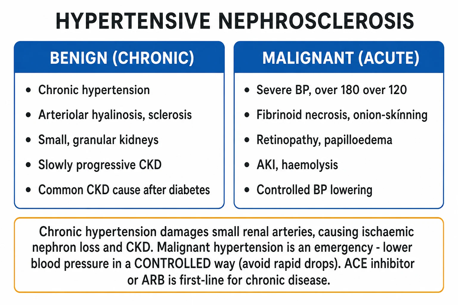
Hypertensive nephrosclerosis is chronic kidney injury caused by long-standing systemic hypertension — after diabetic kidney disease, the commonest cause of CKD and ESKD worldwide. Benign (chronic) nephrosclerosis produces hyaline arteriolosclerosis of the afferent arteriole, small granular kidneys and slowly progressive CKD with sub-nephrotic proteinuria. Malignant-phase (accelerated) hypertension — severe BP (typically over 180 over 120 mmHg) with acute target-organ damage (retinopathy grade III-IV, encephalopathy, AKI, microangiopathic haemolysis) — is a medical emergency whose histology is fibrinoid necrosis of arterioles and onion-skinning of interlobular arteries. Chronic management is strict BP control with an ACE inhibitor or ARB (BP target under 130 over 80, or SBP under 120 if tolerated — KDIGO 2021), an SGLT2 inhibitor added to CKD (DAPA-CKD, EMPA-KIDNEY), and aggressive cardiovascular risk reduction. Malignant hypertension requires CONTROLLED IV BP lowering (labetalol, nicardipine) — about a 25 percent reduction in mean arterial pressure in the first hour — because a rapid or excessive fall precipitates ischaemic stroke, MI or AKI. Resistant or early-onset hypertension mandates screening for a secondary cause (renal artery stenosis, primary aldosteronism).
On this page & tools
Your progress
Saved locally on this device.
Exam tags
Red flags
Overview & Definition
Hypertension is the most common chronic disease of adults worldwide, affecting more than one billion people, and the kidney is one of its prime target organs. Hypertensive nephrosclerosis is the chronic renal injury that results from long-standing systemic hypertension, characterised structurally by hyaline arteriolosclerosis of the afferent arterioles with downstream ischaemic glomerulosclerosis, tubular atrophy and interstitial fibrosis, and clinically by slowly progressive chronic kidney disease (CKD) with sub-nephrotic proteinuria and a bland urinary sediment.[1][2]
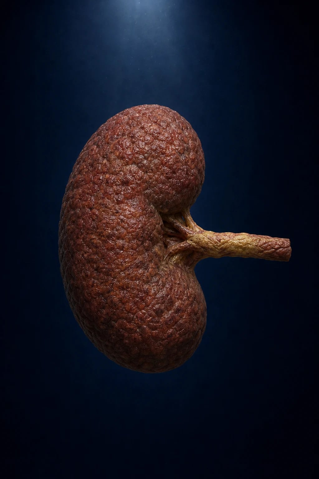
The term nephrosclerosis (literally, "hardening of the kidney") reflects the gross appearance — the kidney becomes small, contracted, and granular with a thinned cortex, a finding already familiar to pathologists of the 19th century. The traditional label "benign nephrosclerosis" is misleading: although slow, the process is the second commonest cause of end-stage kidney disease (ESKD) worldwide (after diabetic kidney disease), and the cardiovascular disease it accompanies is the leading cause of death in this population.[1]
The clinical skill the topic demands is twofold. First, control chronic hypertension tightly to prevent and slow CKD progression — using RAAS blockade (an ACE inhibitor or ARB), an SGLT2 inhibitor in established CKD, and cardiovascular risk reduction. Second, recognise malignant-phase (accelerated) hypertension — the severe, acute form — and lower the blood pressure fast enough to stop ongoing organ damage but controlled enough to avoid precipitating ischaemic stroke, MI or AKI.[2]
Hypertensive nephrosclerosis — the numbers that matter
Classification
Hypertensive nephrosclerosis is classified by tempo, severity and histology into the slow benign (chronic) form and the acute malignant (accelerated) phase. The two are ends of a spectrum — a patient with long-standing benign nephrosclerosis can decompensate into a malignant phase — but they differ sharply in urgency and management. [1]
Benign (chronic) nephrosclerosis
- **Long-standing** (years–decades) hypertension; BP often **moderately** elevated
- **Hyaline arteriolosclerosis** of the afferent arteriole (plasma protein + lipid insudation under endothelium)
- **Small, contracted, granular kidney** with thinned cortex; **ischaemic glomerulosclerosis**, tubular atrophy, interstitial fibrosis
- **Slowly progressive CKD**; sub-nephrotic proteinuria (UACR usually 30–300 mg/g); **bland sediment**
- Commonest renal lesion of ageing and the dominant non-diabetic ESKD cause; often coexisting cardiovascular disease
- Managed as **outpatient**: ACEi/ARB, BP under 130/80, SGLT2i, statin, lifestyle
Malignant-phase (accelerated) hypertension
- **Severe** BP, typically **over 180/120 mmHg**, WITH acute target-organ damage
- **Fibrinoid necrosis** of arterioles + **'onion-skin' hyperplastic arteriolitis** of interlobular arteries
- **Retinopathy grade III–IV** (flame haemorrhages, cotton-wool spots, **papilloedema**); **encephalopathy**, acute pulmonary oedema, **AKI**, **microangiopathic haemolytic anaemia** (schistocytes)
- **Medical emergency** — historically near-fatal within months; high rate of underlying **secondary** hypertension
- Managed as **inpatient (HDU/ICU)**: **controlled IV BP lowering** (labetalol, nicardipine), no more than 25 percent MAP reduction in the first hour

A second, clinically vital classification is the distinction between a hypertensive emergency and a hypertensive urgency. Both feature severe BP; what separates them is the presence or absence of acute target-organ damage, and this single distinction dictates the speed and setting of treatment.[3]
Hypertensive emergency
- Severe BP **+ acute target-organ damage**
- **Encephalopathy / confusion, retinopathy with papilloedema (grade III–IV)**
- **Acute pulmonary oedema**, ACS, **aortic dissection**, **AKI**, eclampsia
- **IV titratable** therapy (labetalol, nicardipine, clevidipine); **controlled** lowering — 25% MAP in hour 1
- **Admit (HDU/ICU)** with arterial-line monitoring
Hypertensive urgency
- Severe BP **WITHOUT** acute target-organ damage
- Patient may have **headache, epistaxis, anxiety** — but no end-organ injury
- **Oral** agents over **24–48 hours** with close follow-up
- Examples: amlodipine, captopril, labetalol PO, clonidine
- **Avoid short-acting nifedipine** — precipitous fall can precipitate stroke/MI
Epidemiology & Risk Factors
Hypertension is the leading modifiable risk factor for global mortality and affects more than one billion adults. As a cause of ESKD, hypertensive nephrosclerosis accounts for roughly 25–30 percent of patients reaching dialysis or transplantation in most registries — second only to diabetic kidney disease.[1][2] The burden is unevenly distributed: in people of recent West-African ancestry (broadly, Black/African populations), hypertensive nephrosclerosis is the dominant non-diabetic cause of ESKD, with earlier onset, more severe disease and faster progression than in European-ancestry populations.[6]
The risk factors for developing and progressing hypertensive nephrosclerosis are: [1]
- Age — prevalence of both hypertension and arteriolar thickening rises steeply with age; the kidney of the elderly hypertensive is the archetype.
- Black / African ancestry and APOL1 risk variants — the G1 and G2 variants of the APOL1 gene explain much of the markedly higher ESKD risk in this population. APOL1 is a component of the high-density lipoprotein particle; the risk variants arose through positive selection because they confer resistance to Trypanosoma brucei rhodesiense (sleeping sickness), but they also cause a markedly increased risk of FSGS, hypertensive nephrosclerosis and HIV-associated nephropathy. This genetic explanation has reframed what was historically attributed to "race".[6]
- Family history and low birth weight — reflecting both genetic and developmental (reduced nephron number) contributions.
- Obesity, high dietary sodium, low potassium intake, physical inactivity, excess alcohol — the lifestyle drivers of hypertension generally.
- Coexisting diabetes, dyslipidaemia, smoking — accelerate vascular injury and produce mixed ("hypertensive–diabetic") nephropathy.
- Socioeconomic deprivation and therapeutic non-adherence — major drivers of severe and malignant-phase presentation.
Malignant-phase hypertension is rare (incidence approximately 1–2 per 100,000 per year in Europe) but disproportionately affects Black and South-Asian populations, untreated or non-adherent patients, and those with an underlying secondary cause (renal artery stenosis, renovascular disease, phaeochromocytoma, scleroderma renal crisis).[2]
Pathophysiology
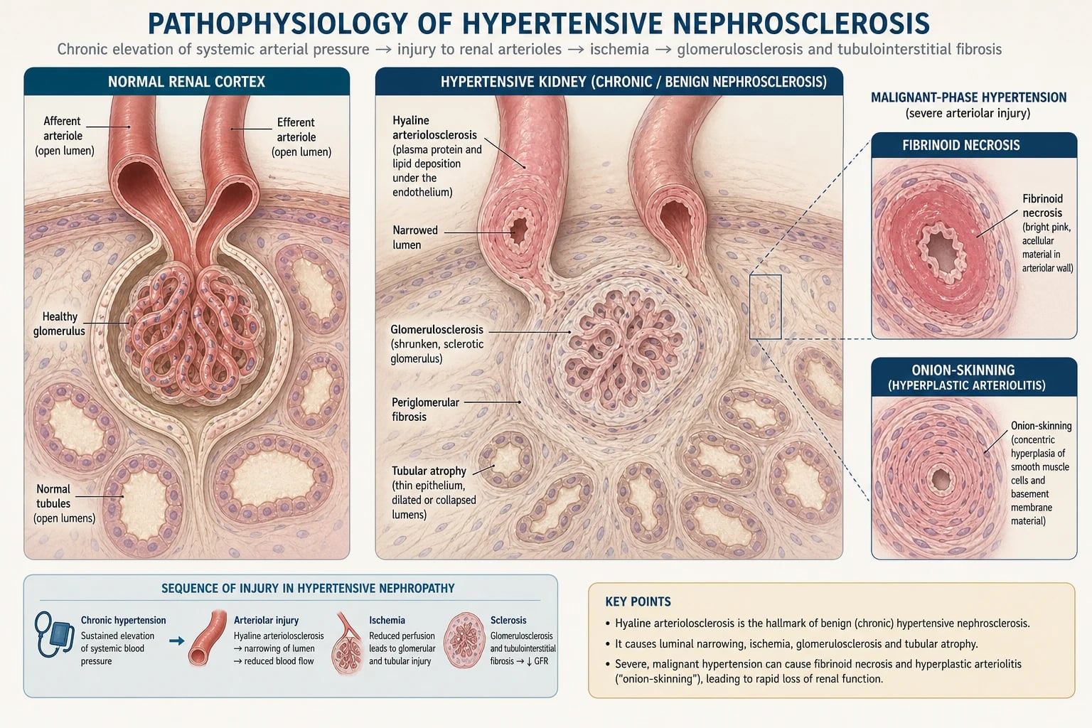
The central lesion of hypertensive nephrosclerosis is hyaline arteriolosclerosis of the afferent (preglomerular) arteriole. Understanding how this single lesion produces the whole syndrome requires following the haemodynamic, structural and humoral consequences of chronic pressure overload. [1]
Step 1 — Pressure transmission to the afferent arteriole
The kidney autoregulates its blood flow and glomerular filtration rate (GFR) across a wide range of systemic pressures by adjusting the tone of the afferent and efferent arterioles. When systemic BP rises, the myogenic response of the afferent arteriole constricts to shield the delicate glomerular capillary tuft. In chronic hypertension, this protective mechanism is overwhelmed: sustained high pressure transmits mechanical stress to the afferent endothelium and smooth muscle, injuring them and triggering the insudation of plasma proteins and lipids into the arteriolar wall — the hyaline change seen on light microscopy. The vessel wall thickens, the lumen narrows, and — critically — the arteriole becomes rigid, losing its ability to constrict or dilate.[1][2]
Step 2 — Loss of autoregulation: glomerular hypertension AND ischaemia
Once the afferent arteriole is rigid, autoregulation fails, and two opposing injuries follow from the same lesion: [1]
- When systemic pressure is high, the stiff afferent cannot buffer it, and the pressure is transmitted directly to the glomerulus, producing intraglomerular hypertension. This drives podocyte stress and effacement, mesangial expansion, and a focal segmental glomerulosclerosis (FSGS)-like scarring — the hyperfiltration injury pathway.
- When systemic pressure falls (volume depletion, sleep, antihypertensive therapy), the stiff afferent cannot dilate to maintain flow, and the glomerulus becomes ischaemic, undergoing ischaemic glomerulosclerosis — solidification and global sclerosis with surrounding periglomerular fibrosis, tubular atrophy and interstitial fibrosis. [1]
Both pathways converge on the same endpoint — nephron loss and interstitial fibrosis — the structural correlate of progressive CKD. The histological hallmarks are therefore: hyaline arteriolosclerosis, ischaemic (solidified) glomeruli, periglomerular fibrosis, tubular atrophy, and a fibrotic interstitium, producing the gross picture of a small, contracted, granular kidney.[2]
Step 3 — The RAAS amplifies injury
Ischaemic juxtaglomerular cells release renin, which cleaves angiotensinogen to angiotensin I, converted by angiotensin-converting enzyme (ACE) to angiotensin II. Angiotensin II has three renal effects that worsen the disease: [1]
- Preferential efferent arteriolar constriction, which raises intraglomerular pressure and worsens the pressure transmission and protein leak.
- Direct pro-fibrotic signalling on mesangial cells, podocytes and tubular cells (TGF-β, NF-κB), accelerating scarring.
- Aldosterone release, which drives sodium retention (worsening hypertension) and has direct profibrotic effects on the heart and kidney. [1]
This is the mechanistic rationale for why ACE inhibitors and ARBs are uniquely renoprotective beyond their blood-pressure effect — they dilate the efferent arteriole, lower intraglomerular pressure, and interrupt the pro-fibrotic signalling cascade.[6]
Step 4 — Malignant phase: vessel rupture and thrombotic microangiopathy
When the BP rise is severe and abrupt, the vessel wall fails outright. Endothelial injury allows plasma proteins and fibrin to flood the intimal and medial layers, producing fibrinoid necrosis of the arterioles — a pink, acellular, amorphous material on light microscopy. In the larger interlobular arteries, a concentric proliferation of myofibroblasts produces the "onion-skin" hyperplastic arteriolitis. Endothelial injury activates platelets and the coagulation cascade, and red cells are sheared against the damaged vessel wall — producing a thrombotic microangiopathy with schistocytes on the blood film, a falling platelet count, rising LDH, and acute kidney injury.[2]
The governing equation
Glomerular filtration obeys Starling forces across the glomerular capillary: [1]
GFR = Kf × (P_GC − P_BS − π_GC) [1]
where P_GC is the glomerular capillary hydrostatic pressure (set by the balance of afferent versus efferent arteriolar tone), P_BS is the Bowman's space hydrostatic pressure, π_GC is the glomerular oncotic pressure, and Kf is the ultrafiltration coefficient (the product of surface area and permeability, reduced by mesangial contraction and podocyte loss). In hypertensive nephrosclerosis, P_GC rises (afferolular pressure transmission, efferent angiotensin-II constriction), then falls as afferolular occlusion and Kf loss dominate — explaining the early hyperfiltration and late progressive decline. [1]
Why an ACE inhibitor/ARB is uniquely renoprotective — HOT-RAAS
Efferent
Angiotensin II preferentially constricts the efferent arteriole, raising P_GC and protein leak; ACEi/ARB vasodilates it, lowering intraglomerular pressure
Angiotensin II drives TGF-β and mesangial/podocyte scarring; RAAS blockade interrupts pro-fibrotic signalling
Expect an early creatinine rise — up to 30% is acceptable and haemodynamic; over 30% → stop and screen for bilateral renal artery stenosis
Watch serum K+ — ACEi/ARB raise potassium by lowering aldosterone
Lowering P_GC reduces hyperfiltration injury and preserves surviving nephrons over years
ACEi and ARB are fetopathic (renal agenesis, oligohydramnios) — never in pregnancy
Never combine an ACEi with an ARB (ONTARGET: more harm, no extra benefit)
AASK (ramipril > amlodipine/metoprolol in hypertensive CKD) and IDNT/RENAAL established the class in CKD
Clinical Presentation
Chronic (benign) hypertensive nephrosclerosis
The presentation is insidious. A typical patient is middle-aged or older, with long-standing — often poorly controlled — hypertension, frequently with coexisting ischaemic heart disease, heart failure, stroke, peripheral vascular disease, or diabetes. The renal disease declares itself through: [1]
- Slowly rising serum creatinine and a falling eGFR detected on routine testing.
- Mild, sub-nephrotic proteinuria (UACR typically 30–300 mg/g, occasionally higher) — heavy proteinuria suggests a coexisting glomerular disease (especially diabetic kidney disease).
- A bland urinary sediment — no red-cell casts, no dysmorphic red cells, no fatty casts.
- Small, echogenic kidneys on ultrasound, often symmetric; cortical thinning.
- Signs of hypertensive target-organ damage elsewhere: left ventricular hypertrophy (displaced, heaving apex; S4 gallop; ECG voltage criteria), retinopathy (arteriolar narrowing, AV nicking — Keith-Wagener-Barker grades I–II), carotid or abdominal bruits, and reduced peripheral pulses. [1]
The patient is often asymptomatic until the CKD is advanced, when uraemic symptoms (fatigue, anorexia, pruritus, nocturia) or fluid overload appear. [1]
Malignant-phase (accelerated) hypertension
The presentation is acute and dramatic. The patient has severe BP, typically over 180/120 mmHg, with one or more of: [1]
- Headache, visual disturbance, confusion, seizures, or focal deficit — hypertensive encephalopathy.
- Chest pain or dyspnoea — acute coronary syndrome, acute pulmonary oedema, or aortic dissection.
- Oliguria, haematuria, or rapidly rising creatinine — acute kidney injury.
- Pallor, jaundice, fatigue — microangiopathic haemolytic anaemia with schistocytes on blood film.
- Fundoscopic findings of grade III–IV retinopathy: flame-shaped haemorrhages, cotton-wool spots (ischaemic nerve-fibre-layer infarcts), hard exudates, and papilloedema. [1]
Atypical presentations examiners probe deliberately
- The elderly with isolated systolic hypertension and a wide pulse pressure — stiff, atherosclerotic vasculature; check standing BP for orthostatic hypotension before intensifying therapy.
- The patient presenting for the first time with advanced CKD and small kidneys — the kidney disease is already irreversible; the goal is to slow further decline and manage cardiovascular risk.
- The "asymptomatic hypertensive" discovered on screening with proteinuria or reduced eGFR — do NOT dismiss as benign; assess end-organ damage fully.
- In diabetic patients — overlap with diabetic kidney disease: heavier proteinuria, Kimmelstiel–Wilson nodules on biopsy, and diabetic retinopathy (which favours the diabetic component); arteriolar hyalinosis, however, is shared.
- Clues to a secondary cause rather than primary hypertension: age under 30, abrupt onset, severe or resistant hypertension, abdominal bruit (renovascular), spontaneous hypokalaemia (primary aldosteronism), the triad of episodic headache, palpitations and sweating (phaeochromocytoma), unequal kidney size (renal artery stenosis). [1]
Differential Diagnosis
The diagnosis of hypertensive nephrosclerosis is one of exclusion — chronic hypertension with sub-nephrotic proteinuria and small kidneys is consistent with it, but several other causes of CKD must be actively considered and excluded.[1][2]
Diabetic kidney disease
- **Heavier, often nephrotic-range proteinuria** (UACR frequently over 300 mg/g)
- **Kimmelstiel–Wilson nodules** on biopsy (nodular glomerulosclerosis)
- **Diabetic retinopathy** present in most type-1 and many type-2 cases — its absence questions the diagnosis
- Long-standing diabetes; arteriolar hyalinosis is shared with HTN — mixed disease is common
- ACEi/ARB first-line; SGLT2i strongly indicated
Chronic glomerulonephritis (e.g. IgA nephropathy)
- **Haematuria** (macro- or microscopic), **dysmorphic red cells** and **red-cell casts** in the sediment
- **Larger kidneys** (not contracted) early; younger age group; episodic synpharyngitic haematuria (IgA)
- Proteinuria may be nephrotic-range in membranous/FSGS; complement and serology guide the cause
- Renal biopsy diagnostic; immunosuppression may be indicated
Renovascular disease / renal artery stenosis
- **Flash pulmonary oedema**, **refractory hypertension**, **abdominal or flank bruit**
- **Asymmetric kidneys** (over 1.5 cm size difference); **rapidly declining eGFR on starting an ACEi/ARB**
- **Atherosclerotic** (older, vascular disease) vs **fibromuscular dysplasia** (young woman, 'string-of-beads' on angiography)
- Confirm with **renal Doppler, CT or MR angiography**; medical therapy first; revascularisation (stent/angioplasty) reserved for refractory cases, flash oedema or rapidly declining function
Primary aldosteronism
- **Hypokalaemia** (spontaneous or diuretic-induced), **metabolic alkalosis**, mild hypernatraemia
- **Resistant hypertension**; absence of peripheral oedema early
- Screen with **aldosterone-to-renin ratio** (ARR over 30 with aldo over 15 ng/dL); confirm with saline or captopril suppression
- **Adrenal CT** then **adrenal vein sampling**; treat adenoma with surgery, hyperplasia with spironolactone/eplerenone
Two further differentials: [1]
- Atheroembolic renal disease — follows recent catheterisation, angiography or anticoagulation; showers of cholesterol emboli produce AKI (rather than slow CKD), eosinophilia, livedo reticularis, blue toes, and hypocomplementaemia.
- Hypertensive emergency vs urgency — already discussed under Classification; the distinction rests entirely on acute target-organ damage, not the BP number itself. [1]
Clinical & Bedside Assessment
The bedside assessment of a hypertensive patient serves two purposes — to quantify the blood pressure correctly and to screen for end-organ damage and secondary causes. [1]
Correct blood-pressure measurement
Errors of technique are the commonest source of misdiagnosis. Measure correctly: [1]
- Rest for 5 minutes, seated, feet flat on the floor, arm supported at heart level, correct cuff size (bladder over 80% of arm circumference — too small over-reads).
- Take two to three readings, 1 minute apart, both arms at the first visit (a difference over 15–20 mmHg suggests subclavian/renovascular disease). Use the higher arm thereafter.
- Confirm with ambulatory (ABPM) or home monitoring to exclude white-coat and masked hypertension; ABPM is the gold standard.
- Check standing BP after 1–3 minutes in older patients and those on therapy to detect orthostatic hypotension (drop over 20/10 mmHg). [1]
End-organ damage assessment
- Fundoscopy — grade the hypertensive retinopathy using Keith–Wagener–Barker: [1]
KW-B Grade I
- **Generalised arteriolar narrowing** (AV ratio under 2:3)
- Mild, chronic HTN; not target-organ *damage* per se
KW-B Grade II
- **Focal/segmental arteriolar narrowing + AV nicking** ('copper'/'silver' wiring)
- Chronic HTN; correlates with LVH and cardiovascular risk
KW-B Grade III
- **Haemorrhages, cotton-wool spots, hard exudates** (flame haemorrhages)
- **Accelerated** hypertension — search for AKI, encephalopathy
KW-B Grade IV
- **Papilloedema** (in addition to grade III changes)
- **Malignant** hypertension — a hypertensive emergency
- Cardiovascular exam — displaced/heaving apex and S4 gallop of LVH, murmurs, peripheral pulses (radio-femoral delay of coarctation; absent/diminished pulses of vascular disease), and bruits (carotid, abdominal over the renal artery, femoral).
- Chest — basal crackles of pulmonary oedema; pleural effusions.
- Abdomen — palpable bladder (obstruction), palpable kidneys (polycystic kidney disease), abdominal bruit (renal artery stenosis).
- Neurology — focal deficits, encephalopathy, signs of prior stroke. [1]
Volume status and a focused review
In malignant-phase hypertension, examine for fluid overload (JVP, crackles, oedema) versus dehydration (which can drive RAAS activation), and review the drug chart for non-adherence, sympathomimetics, NSAIDs, oral contraceptives, and herbal/licorice (glycyrrhizin-induced apparent mineralocorticoid excess). [1]
Investigations
Investigations serve four goals: confirm CKD and stage it, quantify proteinuria, exclude other causes, and screen for secondary hypertension when indicated. [1]
First-line (all patients)
| Test | Finding in hypertensive nephrosclerosis |
|---|---|
| U&E, creatinine + eGFR (CKD-EPI 2021 creatinine equation) | Slowly rising creatinine, falling eGFR; hyperkalaemia and acidosis in advanced CKD |
| Urine albumin-to-creatinine ratio (UACR) | Sub-nephrotic — usually 30–300 mg/g; over 300 mg/g suggests coexisting diabetic or glomerular disease |
| Urinalysis + microscopy | Bland — no red-cell casts, no dysmorphic red cells, no fatty casts; haematuria/proteinuria in malignant phase |
| Full blood count | Anaemia of CKD (normocytic); schistocytes + thrombocytopenia in malignant phase (TMA) |
| Fasting lipids, glucose/HbA1c | Cardiovascular risk stratification; exclude coexisting diabetes |
| Serum bicarbonate, calcium, phosphate, PTH | Metabolic acidosis and CKD–MBD in advanced disease |
| ECG | LVH voltage criteria + strain (Sokolow-Lyon, Cornell) |
| Renal ultrasound | Bilateral small (under 9–10 cm) echogenic kidneys with reduced cortical thickness, symmetric |
CKD definition and staging — KDIGO
CKD is defined as eGFR under 60 mL/min/1.73 m² for more than 3 months, OR markers of kidney damage (UACR over 30 mg/g, haematuria, or structural abnormality).[1] Staging is by GFR category:
| GFR category | eGFR (mL/min/1.73 m²) | Term |
|---|---|---|
| G1 | 90 or more | Normal or high (with kidney damage markers) |
| G2 | 60–89 | Mildly decreased |
| G3a | 45–59 | Mildly–moderately decreased |
| G3b | 30–44 | Moderately–severely decreased |
| G4 | 15–29 | Severely decreased |
| G5 | Under 15 | Kidney failure (uraemia; prepare for RRT) |
ECG criteria for left ventricular hypertrophy
- Sokolow-Lyon: SV1 + (RV5 or RV6) over 35 mm.
- Cornell voltage: R in aVL + S in V3 — over 28 mm in men, over 20 mm in women.
- Cornell product: Cornell voltage × QRS duration — over 2436 mm·ms.
- Strain pattern: ST-segment depression and T-wave inversion in the lateral leads (V5–V6, I, aVL). [1]
Renal ultrasound
The classic finding is bilateral, symmetric, small (under 9–10 cm) echogenic kidneys with cortical thinning. Findings that argue against pure hypertensive nephrosclerosis and demand further work-up: [1]
- Asymmetry (over 1.5 cm size difference) → renovascular disease.
- Large kidneys → diabetic kidney disease, polycystic kidneys, infiltrative disease, or acute inflammation superimposed on CKD.
- Hydronephrosis → obstructive uropathy. [1]
Screening for secondary causes (indicated by resistant, early-onset, severe, or malignant hypertension)
- Primary aldosteronism — aldosterone-to-renin ratio (ARR) (screen; positive if ARR over 30 with aldo over 15 ng/dL), confirmed by saline or captopril suppression testing; adrenal CT then adrenal vein sampling.
- Renal artery stenosis — renal Doppler ultrasound (resistive indices, peak systolic velocity), CT angiography or MR angiography (gold-standard imaging; catheter angiography reserved for intervention).
- Phaeochromocytoma — 24-hour fractionated urine metanephrines or plasma free metanephrines.
- Cushing syndrome — 1 mg overnight dexamethasone suppression test, 24-hour urine cortisol, late-night salivary cortisol.
- Coarctation of the aorta (younger patients, radio-femoral delay, bruits) — CT/MR angiography or echocardiography.
- Obstructive sleep apnoea — sleep study (polysomnography).
- Renal parenchymal disease — consider biopsy if presentation atypical (heavy proteinuria, haematuria, rapid decline, large kidneys). [1]
Investigations specific to suspected malignant hypertension
Add blood film and LDH/haptoglobin (microangiopathic haemolysis with schistocytes), platelet count and coagulation, troponin and repeat ECG, and urgent renal function; fundoscopy is essential and diagnostic when grade III–IV changes are present. [1]
[1]Management — Resuscitation
This section concerns malignant-phase hypertension and other hypertensive emergencies — the time-critical state. Chronic management is covered in the next section. [1]
The governing principle: lower controlled, never precipitous
In a hypertensive emergency the aim is NOT to normalise the blood pressure quickly. Chronic hypertension shifts the cerebral autoregulation curve to the right — the brain, heart and kidneys have adapted to the higher pressures and rely on them for perfusion. A precipitous fall drops cerebral, coronary and renal perfusion and causes ischaemic stroke, myocardial infarction, and acute kidney injury — the central, classic pitfall of treating a hypertensive emergency.[2][3]
[1]Setting, monitoring, and agents
Admit to a monitored bed (HDU/ICU) with a continuous arterial line and hourly neurology and urine-output checks. Use a titratable intravenous agent with a rapid onset and short half-life: [1]
- Labetalol (combined α- and β-blocker) — 20–40 mg IV over 2 minutes, repeated every 10 minutes up to 80 mg, max 300 mg; or infusion 0.5–2 mg/min. First-line in most settings, safe in pregnancy.
- Nicardipine (dihydropyridine CCB) — 5 mg/h infusion, titrate up by 2.5 mg/h every 5 minutes to max 15 mg/h. Smooth, titratable; avoid in advanced heart failure.
- Clevidipine (ultra-short-acting DHP CCB) — infusion starting 1–2 mg/h, doubling every 90 seconds; very rapid titration.
- Nitroprusside — potent arterial and venous vasodilator; effective but requires continuous monitoring and carries a cyanide/thiocyanate toxicity risk in renal failure — use only short-term.
- Esmolol (ultra-short β1-blocker), fenoldopam (dopamine-1 agonist, useful in renal impairment), hydralazine (used in pregnancy). [1]
Scenario-specific targets
Different emergencies have different targets — learn the contrasts: [1]
| Scenario | BP target / approach |
|---|---|
| Hypertensive encephalopathy | 25% MAP reduction in hour 1, then gradual |
| Acute pulmonary oedema | Preload reduction (nitrates, loop diuretic) plus BP lowering; labetalol/nicardipine |
| Acute coronary syndrome | Nitroglycerin + β-blocker; SBP under 140 |
| Aortic dissection | Rapid — SBP to under 120 within 20 minutes; β-blocker first (to lower dp/dt), then vasodilator |
| Ischaemic stroke (not thrombolysis) | Permissive — treat only if over 220/120 |
| Ischaemic stroke (for thrombolysis) | Lower to under 185/110 before alteplase |
| Intracerebral haemorrhage | SBP target under 140 (INTERACT2/ATACH2) |
| Pre-eclampsia / eclampsia | Magnesium sulphate for seizure prophylaxis + BP control; delivery is definitive |
Fluid and electrolyte management
- Avoid volume overload — if pulmonary oedema coexists, treat with loop diuretic and preload reduction alongside BP control.
- Watch for and treat hyperkalaemia and acute kidney injury; in the acute emergency, do NOT start an ACE inhibitor/ARB — the acute fall in efferent tone can precipitate AKI, especially if bilateral renovascular disease is present (often unrecognised). [1]
Management — Definitive & Stepwise
Chronic management has three pillars: (1) tight blood-pressure control, (2) RAAS blockade plus an SGLT2 inhibitor, and (3) cardiovascular risk reduction. Each is supported by landmark trials. [1]
Pillar 1 — Blood-pressure target: tight, individualised
The blood-pressure target has fallen over successive guidelines and trials: [1]
- SPRINT (NEJM 2015) — in over 9,000 high-risk patients, an intensive standardised SBP target under 120 mmHg (versus under 140) reduced cardiovascular events and all-cause mortality by roughly 25 percent, without excess in serious adverse events — including in the elderly subgroup. The trial excluded patients with diabetes, prior stroke, heart failure, recent MI, and those in nursing homes.[5]
- KDIGO 2021 (BP in CKD) — recommends SBP under 120 mmHg (standardised, office) in adults with CKD and hypertension, when tolerated.[1]
- 2017 ACC/AHA (US) — BP target under 130/80 for most adults, including those with CKD.[3]
- 2018 ESC/ESH (Europe) — initial target under 140/90, then under 130/80 if tolerated, especially in CKD; under 140 in older/frail patients.[4]
ACC/AHA 2017 / KDIGO 2021 (US, international): target SBP under 120 mmHg standardised (KDIGO 2021) or under 130/80 (ACC/AHA) in CKD with hypertension, individualised to tolerance.
The shared principle is unambiguous: tight control, with RAAS blockade, individualised to the patient's tolerance and comorbidity. [1]
Pillar 2 — RAAS blockade is first-line and uniquely renoprotective
Begin with an ACE inhibitor OR an ARB (never both together — ONTARGET showed harm with no benefit). ACE inhibitors and ARBs dilate the efferent arteriole, lower intraglomerular pressure, reduce proteinuria, and interrupt the pro-fibrotic actions of angiotensin II. They are uniquely renoprotective beyond their blood-pressure effect.[6]
Common agents and oral doses: [1]
| Drug class | Agent | Oral dose |
|---|---|---|
| ACE inhibitor | Ramipril | 2.5–10 mg once daily |
| Enalapril | 5–20 mg twice daily | |
| Lisinopril | 10–40 mg once daily | |
| Perindopril | 2–8 mg once daily | |
| ARB | Losartan | 50–100 mg once daily |
| Valsartan | 80–320 mg once daily | |
| Telmisartan | 20–80 mg once daily | |
| Irbesartan | 150–300 mg once daily |
The expected creatinine rise on starting an ACE inhibitor/ARB — a fall in eGFR of up to 30 percent (or creatinine rise up to 30 percent) within 2–4 weeks is haemodynamic and acceptable; it reflects the desired drop in intraglomerular pressure. A fall over 30 percent, or a potassium over 5.6 mmol/L, should prompt stopping the drug and screening for bilateral renal artery stenosis (a feared, often-unrecognised cause of AKI on RAAS blockade). [1]
[1]Pillar 3 — Step-up therapy to reach target
If the BP target is not met on an ACE inhibitor/ARB alone, step up: [1]
- Add a dihydropyridine calcium-channel blocker — amlodipine 5–10 mg OD, nifedipine LA 30–90 mg OD, or felodipine 2.5–10 mg OD. The ACEi/ARB + CCB combination is the most evidence-based two-drug regimen (ACCOMPLISH: benazepril + amlodipine superior to benazepril + hydrochlorothiazide for cardiovascular events).
- Add a thiazide-like diuretic — chlorthalidone 12.5–25 mg OD or indapamide 1.25–2.5 mg OD (thiazide-like preferred to hydrochlorothiazide for duration and outcome data). Switch to a loop diuretic (furosemide 20–80 mg OD, or bumetanide 1–4 mg OD) when eGFR is under 30 mL/min/1.73 m², as thiazides lose efficacy in advanced CKD.
- Add further agents as needed (β-blocker, α-blocker, centrally acting) — but at this stage suspect and exclude a secondary cause (resistant hypertension). [1]
Resistant hypertension and the fourth agent
Resistant hypertension is defined as BP above target despite three antihypertensive agents at optimal doses, including a diuretic, or requiring four or more agents. After confirming adherence (witnessed dosing, pill counts) and excluding secondary causes, the most effective fourth agent is spironolactone 25–50 mg OD (PATHWAY-2: superior to bisoprolol or doxazosin as the fourth-line agent). Watch for hyperkalaemia (especially in CKD) and gynaecomastia in men (use eplerenone 25–50 mg OD if it occurs). [1]
Pillar 4 — Add an SGLT2 inhibitor to CKD
The SGLT2 inhibitors reduce intraglomerular pressure (tubuloglomerular feedback restoration) and are now foundational CKD therapy, on top of RAAS blockade, regardless of glycaemic status: [1]
- Dapagliflozin 10 mg OD — DAPA-CKD (NEJM 2020) reduced the composite of sustained eGFR decline, ESKD, or renal/cardiovascular death by 39 percent, with benefit seen regardless of diabetes.[9]
- Empagliflozin 10 mg OD — EMPA-KIDNEY (NEJM 2023) confirmed a 28 percent relative risk reduction in kidney-disease progression or cardiovascular death across a broad CKD population including non-diabetic and lower-eGFR patients.[10]
- Canagliflozin — CREDENCE (NEJM 2019) in diabetic kidney disease.[8]
Current indication: an SGLT2 inhibitor is recommended in all patients with CKD (eGFR down to 20 mL/min/1.73 m² for dapagliflozin and empagliflozin), on top of maximum-tolerated RAAS blockade, with or without diabetes.[9][10]
Pillar 5 — Cardiovascular risk reduction
Because cardiovascular disease is the leading cause of death in patients with hypertensive nephrosclerosis (more common than ESKD), do not stop at blood pressure: [1]
- Statin (atorvastatin 20–80 mg OD or rosuvastatin 10–40 mg OD) — for all CKD patients at cardiovascular risk; reduce dose in severe CKD.
- Antiplatelet therapy (aspirin 75–100 mg OD) for secondary prevention; weigh bleeding risk in primary prevention.
- Smoking cessation, glycaemic control (if diabetic, HbA1c target individualised), weight management, moderate alcohol.
- Sodium and dietary modification — sodium under 5 g/day (under 2 g sodium); the DASH diet (rich in fruit, vegetables, low-fat dairy, reduced saturated fat); adequate dietary potassium unless contraindicated. [1]
Non-pharmacological measures (lifestyle)
| Measure | Target |
|---|---|
| Sodium restriction | Under 5 g salt/day (under 2 g sodium) |
| Weight reduction | BMI 18.5–24.9; loss of 1 kg ≈ 1 mmHg SBP fall |
| DASH diet | Fruits, vegetables, low-fat dairy, low saturated fat |
| Physical activity | 150 min/week moderate aerobic, plus resistance |
| Moderate alcohol | ≤ 2 drinks/day (men), ≤ 1 (women) |
| Smoking cessation | Cessation (does not lower BP but cuts CV risk) |
Hypertensive urgency (no acute end-organ damage)
Lower the BP over 24–48 hours with oral agents — amlodipine 5–10 mg, captopril 25 mg (onset 15–30 min, can repeat), labetalol 200 mg, clonidine 0.1–0.2 mg (caution in heart failure/elderly), felodipine. Arrange close outpatient follow-up within 24–72 hours. Avoid short-acting nifedipine capsules — they cause a precipitous, uncontrolled fall that can precipitate stroke or MI.[3]
Specific Subtypes & Scenarios
Malignant-phase (accelerated) hypertension
A medical emergency. Histology: fibrinoid necrosis of arterioles + onion-skinning (hyperplastic arteriolitis) of interlobular arteries. Clinical triad: severe BP + retinopathy grade III–IV + AKI/MHA. Management: controlled IV BP lowering (labetalol, nicardipine) with the 25 percent MAP rule, admission to HDU/ICU, treatment of complications (pulmonary oedema, encephalopathy, AKI), and active search for an underlying secondary cause (high yield). After stabilisation, transition to an oral regimen including a RAAS blocker (with the expected early creatinine rise monitored).[2]
Hypertensive urgency
Severe BP without acute target-organ damage; oral agents over 24–48 hours. The trap is mistaking an emergency (e.g., with subtle encephalopathy) for an urgency — when in doubt, treat as an emergency. [1]
Renovascular disease / renal artery stenosis
Two mechanisms: atherosclerotic (older patients with vascular disease; ostial lesions) and fibromuscular dysplasia (younger women; mid-artery 'string-of-beads'). Clues: flash pulmonary oedema, asymmetrical kidneys (over 1.5 cm difference), refractory hypertension, rising creatinine on an ACEi/ARB, abdominal bruit. Confirm with renal Doppler, CT or MR angiography. Medical therapy is first-line (RAAS blockade — paradoxically protective if unilateral disease; with caution if bilateral). Revascularisation (percutaneous angioplasty ± stent for atherosclerosis; angioplasty alone for FMD) is reserved for refractory hypertension, recurrent flash pulmonary oedema, rapidly declining renal function, or cardiac destabilisation — ASTRAL, STAR, and CORAL showed no routine benefit over medical therapy.[1]
APOL1-mediated nephropathy
In people of recent West-African ancestry, the APOL1 G1/G2 risk variants confer a markedly increased risk of FSGS-like and hypertensive nephrosclerosis lesions with rapid progression to ESKD at a young age. There is no specific therapy yet (APOL1 inhibitors are in trials); management is maximal BP control with RAAS blockade, SGLT2 inhibition, and cardiovascular risk reduction.[6]
Ischaemic nephropathy in the elderly
Diffuse atherosclerosis compromises renal perfusion; the kidney is an end-organ of systemic vascular disease. Manage with statin, antiplatelet, BP control, and avoid volume depletion and nephrotoxins. [1]
Complications & Pitfalls
Renal complications
- Progressive CKD culminating in ESKD requiring dialysis or transplantation.
- Acute kidney injury superimposed on chronic disease (acute-on-chronic) — triggered by volume depletion, NSAIDs, iodinated contrast, or starting an ACE inhibitor/ARB (especially with undiagnosed bilateral renovascular disease).
- Hyperkalaemia, metabolic acidosis, anaemia, and CKD–mineral and bone disorder (CKD-MBD) of advanced CKD. [1]
Cardiovascular complications — the leading cause of death
- Left ventricular hypertrophy, heart failure with preserved (HFpEF) or reduced (HFrEF) ejection fraction, ischaemic heart disease, atrial fibrillation, aortic dissection, and stroke. [1]
Complications specific to malignant-phase hypertension
- Hypertensive encephalopathy, intracerebral haemorrhage, acute pulmonary oedema, acute kidney injury, thrombotic microangiopathy with schistocytes and platelet consumption, retinal damage and blindness, and aortic dissection. [1]
Classic pitfalls (examiner favourites)
- Lowering BP too rapidly in malignant-phase hypertension → ischaemic stroke, MI or AKI. Apply the 25 percent rule.
- Missing a secondary cause in resistant or early-onset hypertension (renovascular disease, primary aldosteronism, phaeochromocytoma, OSA, coarctation).
- Starting an ACE inhibitor/ARB in undiagnosed bilateral renal artery stenosis → precipitous AKI. Check creatinine and potassium at 1–2 weeks; a rise over 30 percent mandates stopping and investigating.
- Using short-acting nifedipine capsules for hypertensive urgency → precipitous, uncontrolled BP fall, stroke/MI.
- Attributing CKD to "just hypertension" when a glomerulonephritis or other treatable cause is present — always exclude active sediment and heavy proteinuria.
- Failing to add an SGLT2 inhibitor in CKD — a now-standard, high-yield, evidence-based therapy.
- Combining an ACE inhibitor with an ARB (ONTARGET: more renal harm, hyperkalaemia, hypotension, with no added benefit).
- Prescribing an ACE inhibitor or ARB in pregnancy — fetopathy (renal agenesis, oligohydramnios, limb contractures, hypoplastic lungs). [1]
Prognosis & Disposition
Chronic hypertensive nephrosclerosis
Generally slowly progressive over years to decades with good blood-pressure control. The rate of eGFR decline is steepest in patients with proteinuria, poor BP control, coexisting diabetes, and APOL1 risk variants.[6] Cardiovascular events, not ESKD, are the commonest cause of death — cardiovascular risk reduction is therefore as important as renal protection. With tight control (SBP under 120 per KDIGO 2021, RAAS blockade, SGLT2 inhibition) the slope of decline can be substantially slowed, though existing scarring is irreversible.
Malignant-phase hypertension
Untreated, historically near-universal mortality within months from uraemia, heart failure or stroke. With prompt, controlled BP lowering and management of complications, 5-year survival exceeds 70–80 percent, though many patients retain some degree of CKD and remain at high cardiovascular risk.[2]
Disposition and follow-up
- Chronic disease: managed in primary care for uncomplicated cases, with nephrology referral when eGFR is under 30, proteinuria is heavy (UACR over 300), BP is resistant, or progression is rapid. Monitor BP, eGFR, UACR, and electrolytes every 3–12 months depending on CKD stage.
- Malignant-phase hypertension: admit to HDU/ICU; follow-up in a combined nephrology/hypertension clinic after discharge; address adherence and provide structured patient education.
- Safety-net and ESKD planning: when eGFR is under 20–25, begin renal replacement therapy education (transplantation, dialysis modalities, conservative care), vascular-access planning, and anaemia/CKD-MBD management. [1]
Special Populations
The elderly
Isolated systolic hypertension with a wide pulse pressure reflects stiff, atherosclerotic vasculature. Higher risk of orthostatic hypotension — always measure standing BP before intensifying therapy. SPRINT showed benefit of intensive control even in the over-75 subgroup, but exclude frailty and weigh falls risk. Prefer longer-acting, well-tolerated agents (amlodipine, an ACEi/ARB, a thiazide-like diuretic). [1]
Diabetes mellitus (the common coexisting disease)
Target BP under 130/80; begin with an ACE inhibitor/ARB if albuminuria is present (UACR over 30). Add an SGLT2 inhibitor (which also provides glycaemic and cardiovascular benefit — empagliflozin reduced cardiovascular death in EMPA-REG OUTCOME). Screen for diabetic retinopathy to help distinguish diabetic kidney disease from a pure hypertensive process. [1]
Pregnancy
Chronic hypertension in pregnancy increases the risks of pre-eclampsia, fetal growth restriction, placental abruption, and preterm birth. ACE inhibitors and ARBs are CONTRAINDICATED at all stages of pregnancy (fetopathy — renal agenesis, oligohydramnios, limb contractures, hypoplastic lungs) and should be stopped before conception. Use labetalol (200–1200 mg/day in divided doses), nifedipine (modified-release 20–60 mg OD), or methyldopa (250–500 mg TDS, limited to second and third trimester). If pre-eclampsia with severe features develops, magnesium sulphate (4 g IV loading over 20 min, then 1–2 g/h infusion for 24 h, or IM regimen) for seizure prophylaxis and delivery is the definitive treatment. [1]
Black / African ancestry
Higher hypertension prevalence, earlier onset, greater salt sensitivity, and APOL1-driven nephropathy. Emphasise early aggressive control, RAAS-based regimens (often combined with a CCB and/or thiazide), and dietary sodium reduction. ALLHAT showed that thiazide-type diuretics (chlorthalidone) and dihydropyridine CCBs are particularly effective initial agents in Black patients, but the AASK trial established that an ACE inhibitor (ramipril) slowed GFR decline better than amlodipine or metoprolol in African-Americans with hypertensive CKD — so an ACEi/ARB remains first-line in those with CKD or proteinuria, often with a CCB added.[6][7]
Advanced chronic kidney disease
Switch the thiazide to a loop diuretic when eGFR is under 30; titrate doses of renally cleared drugs; continue SGLT2 inhibition down to eGFR 20; prepare for renal replacement therapy as eGFR declines; manage anaemia (iron, erythropoiesis-stimulating agents) and CKD-MBD. [1]
Evidence, Guidelines & Regional Differences
Landmark trials
SPRINT (NEJM 2015)
- Over 9,000 high-risk patients; **intensive SBP under 120** vs **under 140** (standardised)
- Reduced CV events and all-cause mortality by ~**25%**
- **Excluded** diabetes, prior stroke, HF, recent MI, nursing-home elderly
- Drove **KDIGO 2021 and ACC/AHA 2017** lower targets
AASK (JAMA 2002)
- 1,094 African-Americans with **hypertensive CKD** (GFR 20–65)
- **Ramipril (ACEi)** slowed GFR decline and reduced ESKD/proteinuria vs **amlodipine (CCB)** and **metoprolol (β-blocker)**
- Established ACEi as first-line in hypertensive nephrosclerosis
- Lower BP target (MAP under 92) slowed decline only in proteinuric patients
ALLHAT (JAMA 2002)
- Over 33,000 high-risk hypertensives; **chlorthalidone** vs **lisinopril** vs **amlodipine** vs **doxazosin**
- Thiazide was **as good as or better** than alternatives for CV outcomes and **HF prevention**
- Better **stroke/HF prevention in Black patients** with thiazide
- Established thiazides as excellent first-line
SGLT2i CKD trials
- **CREDENCE** (canagliflozin, T2D + CKD, NEJM 2019); **DAPA-CKD** (dapagliflozin, CKD ± diabetes, NEJM 2020); **EMPA-KIDNEY** (empagliflozin, broad CKD, NEJM 2023)
- **30–40% reduction** in kidney failure / CV death across CKD populations, including non-diabetic
- **Now foundational CKD therapy** on top of RAAS blockade, eGFR down to ~20
Guidelines and regional differences
| Guideline | Body | BP target |
|---|---|---|
| KDIGO 2021 | International (CKD) | SBP under 120 mmHg (standardised office), tolerated |
| 2017 ACC/AHA | US | Under 130/80 for most, including CKD |
| 2018 ESC/ESH | Europe | Under 140/90 initial, to under 130/80 if tolerated |
| NICE NG136 (2019) | UK | Under 140/90 (under 80 y); CKD with proteinuria under 130/80 |
| ICMR / Indian consensus | India | Under 140/90 in most; under 130/80 with CKD/diabetes; emphasises cost-effective therapy and lower SGLT2i access |
Controversies
- How low is too low? The very tight KDIGO target (SBP under 120) is not universally accepted; some bodies caution that orthostatic hypotension and AKI risk rise with very intensive lowering, especially in older or frail patients.
- APOL1-directed therapy — the first APOL1 inhibitors (e.g., inaxaplin) are in trials and may transform the management of APOL1-mediated disease.
- Renal artery stenosis — CORAL (NEJM 2014) confirmed no routine benefit of stenting over medical therapy, but selected subgroups (recurrent flash oedema, refractory hypertension, rapidly declining function) still warrant revascularisation.
- Renal denervation — modest, contested benefit in resistant hypertension; not routine. [1]
Exam Pearls
The 10 pearls that decide a hypertensive-nephrosclerosis answer
BP-KIDNEY
After diabetic kidney disease, hypertensive nephrosclerosis is the commonest cause of ESKD
Benign = hyaline arteriolosclerosis + small granular kidney; Malignant = fibrinoid necrosis + onion-skinning
Retinopathy grade III (haemorrhages/cotton-wool) and grade IV (papilloedema) define malignant hypertension
Loss of afferent autoregulation → glomerular hypertension (injury) AND ischaemia — both scar the kidney
ACEi OR ARB (renoprotective) — never combine; expect up to 30% eGFR drop, over 30% → stop + screen RAS
Add an SGLT2 inhibitor (dapagliflozin/empagliflozin 10 mg OD) to CKD regardless of diabetes
Malignant HTN → controlled IV lowering, ≤25% MAP in 1st hour; rapid fall → stroke/MI/AKI
Resistant or young HTN → screen for secondary cause (renovascular, aldosteronism, phaeo, OSA)
Exam application bank (NEET-PG / INICET)
One-line answer
Hypertensive nephrosclerosis is chronic kidney injury caused by long-standing systemic hypertension — after diabetic kidney disease, the commonest cause of CKD and ESKD worldwide. Benign (chronic) nephrosclerosis produces hyaline arteriolosclerosis of the afferent arteriole, small granular kidneys and slowly progressive CKD with sub-nephrotic proteinuria. Malignant-phase (accelerated) hypertension — severe BP (typically over 180 over 120 mmHg) with acute target-organ damage (retinopathy grade III-IV, encephalopathy, AKI, microangiopathic haemolysis) — is a medical emergency whose histology is fibrinoid necrosis of arterioles and onion-skinning of interlobular arteries. Chronic management is strict BP control with an ACE inhibitor or ARB (BP target under 130 over 80, or SBP under 120 if tolerated — KDIGO 2021), an SGLT2 inhibitor added to CKD (DAPA-CKD, EMPA-KIDNEY), and aggressive cardio
Worked stems (answer without another resource)
Stem 1 — Classic presentation. Map symptoms to mechanism; name the first investigation and first treatment step with dose/route if drug therapy is standard. [1]
Stem 2 — Unstable / complicated. List red flags that force immediate resuscitation, theatre, ICU, antidote, or reperfusion — and what you do in the first 15 minutes. [1]
Stem 3 — Atypical group. Elderly, pregnancy, child, or immunocompromised: how presentation and thresholds change. [1]
Stem 4 — Differential trap. Name the three closest mimics and one discriminator for each. [1]
Stem 5 — Disposition. Who goes home with safety-netting, who is admitted, who needs HDU/ICU/theatre, and what follow-up is mandatory. [1]
Rapid viva checklist
- Definition + classification
- Pathophysiology chain
- Bedside signs / criteria
- Score with exact components (if any)
- Emergency bundle
- Definitive therapy with doses
- Complications of disease and of treatment
- Special populations
- Guideline/trial name if classic
- Three exam traps
Coverage self-check
If you cannot answer any stem above from this page alone, re-read the matching section — the page is intended to be self-sufficient for final-prof and NEET-PG/INICET questions on Hypertensive Nephrosclerosis.
[1]References
- [1]Chen TK, Knicely DH, Grams ME. Chronic Kidney Disease Diagnosis and Management: A Review JAMA, 2019.PMID 31573641
- [2]Romagnani P, Remuzzi G, Glassock R, et al. Chronic kidney disease Nat Rev Dis Primers, 2017.PMID 29168475
- [3]Whelton PK, Carey RM, Aronow WS, et al. 2017 ACC/AHA/AAPA/ABC/ACPM/AGS/APhA/ASH/ASPC/NMA/PCNA Guideline for the Prevention, Detection, Evaluation, and Management of High Blood Pressure in Adults: A Report of the American College of Cardiology/American Heart Association Task Force on Clinical Practice Guidelines Hypertension, 2018.PMID 29133356
- [4]Williams B, Mancia G, Spiering W, et al. 2018 Practice Guidelines for the management of arterial hypertension of the European Society of Hypertension and the European Society of Cardiology: ESH/ESC Task Force for the Management of Arterial Hypertension J Hypertens, 2018.PMID 30379783
- [5]SPRINT Research Group (Wright JT Jr, Williamson JD, Whelton PK, et al.). A Randomized Trial of Intensive versus Standard Blood-Pressure Control N Engl J Med, 2015.PMID 26551272
- [6]Wright JT Jr, Bakris G, Greene T, et al. (AASK Study Group). Effect of blood pressure lowering and antihypertensive drug class on progression of hypertensive kidney disease: results from the AASK trial JAMA, 2002.PMID 12435255
- [7]ALLHAT Officers and Coordinators for the ALLHAT Collaborative Research Group. Major outcomes in high-risk hypertensive patients randomized to angiotensin-converting enzyme inhibitor or calcium channel blocker vs diuretic: The Antihypertensive and Lipid-Lowering Treatment to Prevent Heart Attack Trial (ALLHAT) JAMA, 2002.PMID 12479763
- [8]Perkovic V, Jardine MJ, Neal B, et al. (CREDENCE Trial). Canagliflozin and Renal Outcomes in Type 2 Diabetes and Nephropathy N Engl J Med, 2019.PMID 30990260
- [9]Heerspink HJL, Stefánsson BV, Correa-Rotter R, et al. (DAPA-CKD Trial). Dapagliflozin in Patients with Chronic Kidney Disease N Engl J Med, 2020.PMID 32970396
- [10]The EMPA-KIDNEY Collaborative Group. Empagliflozin in Patients with Chronic Kidney Disease N Engl J Med, 2023.PMID 36331190
- [11]Levey AS, Bosch JP, Lewis JB, Greene T, Rogers N, Roth D. (MDRD Study Group). A more accurate method to estimate glomerular filtration rate from serum creatinine: a new prediction equation. Modification of Diet in Renal Disease Study Group Ann Intern Med, 1999.PMID 10075613