nephrology · nephrology
Hyponatraemia
Also known as Hyponatremia · Low serum sodium · SIADH · Syndrome of inappropriate antidiuresis · Dilutional hyponatraemia · Osmotic demyelination syndrome
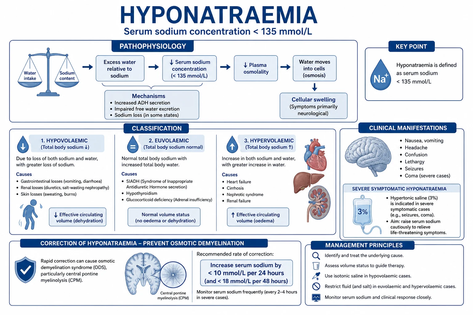
Hyponatraemia (serum Na under 135 mmol/L) is the commonest inpatient electrolyte disorder and reflects an excess of total body water relative to sodium, almost always mediated by non-osmotic vasopressin (ADH). The clinician's job is to classify by volume status (hypo-, eu-, hypervolaemic) and by onset and symptoms (acute severe vs chronic), because these two axes decide treatment. Severe symptomatic (seizure/coma) is a time-critical emergency treated with 3% hypertonic saline 100 mL bolus to raise Na 4 to 6 mmol/L in the first hour and relieve cerebral oedema. In all other cases correct slowly — under 10 mmol/L in 24 h and under 18 mmol/L in 48 h — to prevent osmotic demyelination syndrome (central pontine myelinolysis). The diagnostic cornerstone is serum osmolality (true hypo-osmolar vs pseudohypo-/translocational), urine osmolality (over 100 mOsm/kg means ADH is acting) and urine sodium (with clinical volume status) to localise the cause. SIADH — eu-Volaemic, inappropriately concentrated urine, low uric acid — is the prototype euvolaemic cause. Treat the cause, never the number alone.
On this page & tools
Your progress
Saved locally on this device.
Exam tags
Red flags

Overview & Definition
Hyponatraemia is defined as a serum sodium concentration below 135 mmol/L. It is the commonest electrolyte disorder encountered in hospital practice, complicating 15 to 30 percent of all inpatient admissions and approaching 40 to 50 percent in critical care, post-operative and elderly-care settings.[1][9]
Sodium is the principal extracellular osmole. Because water moves freely across cell membranes to equalise osmolality, a fall in serum sodium is — in nearly every case — a fall in serum tonicity, and therefore a movement of water into cells. The brain, confined in a rigid cranium, is the organ least able to accommodate this swelling, so the clinical syndrome of hyponatraemia is essentially a central nervous system syndrome of cerebral oedema in acute cases, and of subtle neuro-cognitive impairment, falls and gait instability in chronic cases.[4]
The central dogma the examiner wants you to internalise: hyponatraemia = relative excess of total body water to total body sodium, mediated — with the sole exception of primary polydipsia and a few renal salt-wasting states — by arginine vasopressin (ADH) acting on the V2 receptor. In other words, the kidney cannot excrete the ingested water because ADH is forcing it to retain water. Even the hypovolaemic patient (who has lost both Na and water) becomes hyponatraemic because the non-osmotic, volume-driven ADH response overrides osmotic suppression, and the kidney continues to reabsorb free water.[1]
The clinical task decomposes into two orthogonal questions that together decide every management decision: [1]
- What is the volume status? (hypovolaemic, euvolaemic, or hypervolaemic) — localises the cause and the definitive therapy.
- What is the onset and severity? (acute vs chronic; mild, moderate, severe/profound) — sets the speed and aggressiveness of correction and the risk of complications. [1]
Get these two right and the rest follows. Get them wrong and the patient dies — of cerebral herniation if you under-treat an acute case, or of osmotic demyelination if you over-treat a chronic one.[2][7]
Classification
Hyponatraemia is classified along three independent axes. Every examiner expects you to hold all three simultaneously. [1]
Axis 1 — by serum osmolality (the FIRST step, do not skip)
Hypo-osmolar (true)
over 95 percent of cases
- Serum osmolality **under 275 mOsm/kg** — the only form that reflects true water excess
- Sub-classified by **volume status** into hypo-, eu- and hypervolaemic (see below)
- ADH is acting inappropriately in all subtypes — even hypervolaemic states have non-osmotic ADH from low effective arterial volume
- This is the form that causes cerebral oedema and needs sodium correction
Iso-osmolar (pseudo)
artefact
- Normal serum osmolality (275 to 295) with a spuriously low measured Na
- **Pseudohyponatraemia** — extreme hyperlipidaemia (chylomicrons, over 10 g/L triglyceride) or hyperproteinaemia (Waldenstrom over 100 g/L, myeloma) dilutes the aqueous phase of serum
- Na per litre of **plasma water** is normal; ion-selective electrode on diluted sample is falsely low
- Do NOT treat — treat the hyperlipidaemia/hyperproteinaemia. Measured osmolality is normal; no symptoms of cerebral oedema
Hyperosmolar (translocational)
osmotic water shift
- Serum osmolality **over 295 mOsm/kg**
- **Hyperglycaemia** is the classic cause — glucose draws intracellular water into the extracellular space, diluting Na. Corrected Na rises **2 mmol/L for every 5.5 mmol/L (100 mg/dL) glucose above 5.5**
- Other causes: **mannitol**, glycine irrigation (TURP), sorbitol, maltose (IVIG)
- Treat the cause (insulin for glucose, stop mannitol); the sodium corrects itself as glucose falls
Axis 2 — by volume status (only meaningful once hypo-osmolar is confirmed)
Hypovolaemic
Na and water loss, more Na than water
- **Signs of dehydration** — dry mucosae, reduced skin turgor, tachycardia, orthostatic drop, low JVP
- **Urine Na under 20** if extrarenal loss (vomiting, diarrhoea, burns, third-space, pancreatitis); **urine Na over 30** if renal loss (thiazides, loop diuretics, mineralocorticoid deficiency, salt-losing nephropathy, cerebral salt wasting)
- **ADH is appropriately elevated** in response to hypovolaemia — the kidney retains water, worsening dilution
- **Treatment: 0.9% saline** IV — restores volume, switches off ADH, Na corrects
Euvolaemic
water excess, normal total Na
- **No oedema, no dehydration** — clinically eu-Volaemic on exam
- **SIADH is the prototype** — others: glucocorticoid deficiency (cortisol is needed to suppress ADH), hypothyroidism (reduced cardiac output and GFR), primary polydipsia (water intake exceeds excretory capacity — urine Osm under 100), low solute intake (beer potomania, tea-and-toast)
- **Urine Osm over 100 mOsm/kg** (inappropriate concentration); **urine Na over 30** (euvolaemia maintains natriuresis)
- **Treatment: fluid restriction** 800 to 1000 mL/day first-line; urea, loop diuretic + saline, vaptans, demeclocycline (historical)
Hypervolaemic
Na and water excess, more water than Na
- **Oedema** — peripheral, sacral, pulmonary; raised JVP, ascites
- **Effective arterial volume is low** despite total body Na overload — heart failure, cirrhosis, nephrotic syndrome, advanced renal failure — drives non-osmotic ADH
- **Urine Na under 20** unless on diuretics or in renal failure (urine Na may be over 30 in CKD)
- **Treatment: fluid restrict + loop diuretic (furosemide) + treat the underlying cause**; vaptans in selected cases
Axis 3 — by onset and severity (sets the speed and risk of correction)
Acute
under 48 hours
- Brain has **not yet adapted** — no time to extrude osmolyles (idiogenic osmoles), so cerebral oedema develops rapidly
- **Causes:** post-operative hyponatraemia (especially premenopausal women after gynaecological surgery — most feared), TURP/hysteroscopic surgery (glycine/sorbitol absorption), **polydipsia / ecstasy (MDMA)**, endurance exercise (marathon, ultramarathon), recent diuretic start, colonoscopy preparation
- **High risk of cerebral oedema, seizures, herniation** — correct **4 to 6 mmol/L in the first hour** with 3% saline if symptomatic
- ODS risk is LOW after rapid correction of acute hyponatraemia — brain has not accumulated osmolyles
Chronic
over 48 hours or indeterminate
- Brain has **partially adapted** by extruding osmolyles (potassium, then organic osmoles like glutamate, myo-inositol, taurine) — so cerebral oedema is mild but correction can dehydrate the brain
- **Assume chronic if onset unknown** — the safe default
- **Low risk of cerebral oedema** but **high risk of osmotic demyelination syndrome (ODS/CPM)** if corrected faster than 10 mmol/L in 24 h
- **Correct slowly** — under 8 to 10 mmol/L in 24 h, under 18 mmol/L in 48 h; treat only if symptomatic
Severity bands by absolute sodium (a guide only — symptoms and onset matter more than the number):[2]
- Mild — 130 to 134 mmol/L: usually asymptomatic or subtle (poor concentration, fatigue).
- Moderate — 125 to 129 mmol/L: headache, nausea, confusion, gait instability.
- Severe / profound — under 125 mmol/L: vomiting, seizures, coma; under 120 carries significant mortality. Profound (under 105 to 110) has the highest risk of ODS if over-corrected.[10]
Epidemiology & Risk Factors
Hyponatraemia is the commonest inpatient electrolyte derangement. Hospital prevalence figures worth remembering:[1][9]
Headline numbers
Risk factors and the subtype they favour (a high-yield table): [1]
| Risk factor / host | Subtype / cause to consider |
|---|---|
| Post-operative patient (esp. young women, gynaecological/genitourinary surgery) | Acute SIADH from pain, nausea, opiates; TURP/hysteroscopy glycine syndrome |
| Elderly on polypharmacy | Drug-induced (thiazides, SSRIs, carbamazepine, NSAIDs, desmopressin); chronic |
| Heart failure / cirrhosis / nephrotic syndrome | Hypervolaemic dilutional hyponatraemia (poor prognosis) |
| Endurance athletes (marathon, ultramarathon, triathlon) | Exercise-associated hyponatraemia — excessive water + non-osmotic ADH |
| Psychiatric / MDMA users | Psychogenic polydipsia; MDMA-induced SIADH + polydipsia (acute, severe) |
| Beer drinker ("beer potomania") | Low solute intake limits free-water excretion; urine Osm under 100 |
| Diuretics (esp. thiazides) | Hypovolaemic; commonest drug cause; check 2 weeks after start |
| Adrenal insufficiency / hypopituitarism | Glucocorticoid-deficient SIADH (cortisol normally suppresses ADH) |
| Hypothyroidism | Reduced cardiac output + GFR; low distal delivery of solute |
| Cancer (small-cell lung, head & neck, brain, lymphoma) | SIADH from ectopic ADH or tumour |
| CNS disease (meningitis, encephalitis, SAH, TBI, brain tumour) | SIADH or cerebral salt wasting |
| Pneumonia / mechanical ventilation | SIADH; positive-pressure ventilation reduces venous return |
| Burns, pancreatitis, bowel obstruction | Third-space loss — hypovolaemic |
| Acute kidney injury / advanced CKD | Impaired dilution; hypervolaemic |
| Children with rotavirus | Severe dehydration + sodium loss |
Mortality signal: even mild "asymptomatic" hyponatraemia is independently associated with a roughly 2-fold increase in in-hospital mortality, increased length of stay, falls, fractures, and osteoporosis — this is why it cannot be dismissed as a laboratory curiosity.[9]
Pathophysiology
Normal water homeostasis — the osmostat and ADH
Body water is held within narrow osmolality (275 to 295 mOsm/kg) by a negative-feedback loop:[1]
- Osmoreceptors in the organum vasculosum of the lamina terminalis (OVLT) sense a 1 to 2 percent rise in plasma osmolality and stimulate magnocellular neurons in the supraoptic and paraventricular nuclei of the hypothalamus.
- These neurons release arginine vasopressin (ADH) from the posterior pituitary into the systemic circulation.
- ADH binds V2 receptors on the basolateral membrane of principal cells in the collecting duct, activating a Gs-cAMP cascade.
- cAMP drives aquaporin-2 (AQP2) water channels to insert into the apical (luminal) membrane, making the collecting duct water-permeable.
- Water is reabsorbed passively down the cortico-medullary osmotic gradient established by the countercurrent multiplier (loop of Henle) and urea recycling, producing concentrated urine (up to 1000 to 1200 mOsm/kg) and dilute plasma.
- The reverse — water ingestion lowering plasma osmolality — suppresses ADH, AQP2 is internalised, the collecting duct becomes impermeable, and maximally dilute urine (50 to 80 mOsm/kg) is produced. A normal adult can excrete up to 15 to 20 L of free water per day if ADH is fully suppressed. [1]
A healthy kidney therefore protects against hyponatraemia by excrecing free water — hyponatraemia cannot develop unless the kidney cannot excrete the ingested water (ADH present, or diluting capacity exceeded by overwhelming intake / very low solute).[1][4]
The two prerequisites for hypo-osmolar hyponatraemia
True hypo-osmolar hyponatraemia requires both of:[4]
- A source of free water — ingested (oral intake, IV D5W, irrigation fluid), or generated (release of intracellular water in catabolic states).
- Impaired ability to excrete that water — almost always because ADH is present and acting on the collecting duct. The sole exceptions are primary polydipsia (intake of over 10 to 15 L/day overwhelms even the maximally diluting kidney) and advanced renal failure (GFR under 10 mL/min, loss of diluting capacity). [1]
This is why ADH is central to every form — in hypovolaemia ADH is high because of volume depletion (the body prioritises volume over osmolality); in SIADH ADH is high inappropriately; in heart failure and cirrhosis the effective arterial blood volume is low (despite total body Na overload) so the baroreceptors drive ADH. Even the oedematous patient behaves, from the kidney's point of view, as if hypovolaemic.[1]
The brain's response — adaptation and maladaptation
The brain sits inside a rigid skull and cannot accommodate volume change. A fall in extracellular tonicity moves water into astrocytes (the glial cells that constitute the blood-brain barrier interface). To prevent swelling, astrocytes mount a volume-regulatory response:[7]
- Within minutes to hours — astrocytes extrude inorganic osmolytes (potassium, chloride) via ion channels, including the AQP4 channel and volume-regulated anion channels. Astrocytes also express AQP4 which is the major route of osmotic water movement; loss of AQP4 protects against cerebral oedema in hyponatraemia.
- Within 24 to 48 hours — astrocytes extrude organic osmolyles (formerly called idiogenic osmoles): myo-inositol, glutamate, glutamine, taurine, betaine. This is the dominant adaptive mechanism that allows the chronic hyponatraemic brain to avoid fatal herniation. [1]
This adaptation explains the clinical picture: [1]
- Acute hyponatraemia (under 48 h) — no time for osmolyte extrusion; brain swells; cerebral oedema, raised intracranial pressure, seizures, herniation. Mortality may exceed 50 percent if untreated.
- Chronic hyponatraemia (over 48 h) — brain has adapted by shrinking (osmolyles lost); minimal symptoms; but if sodium is corrected rapidly, the now-hypo-osmolar brain loses water faster than it can re-accumulate osmolyles, leading to osmotic demyelination syndrome (ODS) — demyelination of the basis pontis (central pontine myelinolysis, CPM) and, in 10 percent, extrapontine sites (basal ganglia, cerebellum, thalamus).[7]
Risk factors that amplify ODS (lower the threshold for safe correction): hypokalaemia, hypoxia or hypoxaemia, hepatic failure, alcohol use disorder, malnutrition, anorexia, advanced age, female sex, premenopausal, profound hyponatraemia (under 105), burns, and use of SSRIs/antipsychotics.[2][7]
Clinical Presentation
Symptoms reflect cerebral oedema and raised intracranial pressure in acute disease, and subtle neuro-cognitive and gait disturbance in chronic disease. Severity correlates more with the rate of fall than the absolute value — a patient whose sodium has fallen to 120 over 12 hours is gravely ill; one who has lived at 120 for weeks may be walking and talking.[1]
Symptom severity bands (the European guideline grading):[2]
- Moderately severe / severe symptoms — vomiting, cardiovascular collapse, abnormal/somnolent, seizure, coma (GCS under 8). Treat as emergency.
- Mildly symptomatic — nausea without vomiting, confusion, headache.
- Asymptomatic — no symptoms attributable to hyponatraemia. [1]
Acute severe hyponatraemia (under 48 h, cerebral oedema)
The presentation is that of acute rise in intracranial pressure:[4]
- Headache, nausea, vomiting (early — vomiting in a hyponatraemic patient is a red flag).
- Confusion, disorientation, agitation, lethargy, progressing to obtundation.
- Generalised tonic-clonic seizure, often self-limiting.
- Coma, decorticate/decerebrate posturing, fixed dilated pupils, respiratory arrest — signs of tentorial herniation (mortality over 50 percent).
- Signs of the precipitant: post-operative pain/incision, recent MDMA, marathon finisher, recent diuretic, recent uterine surgery. [1]
A premenopausal woman 24 to 48 hours after elective gynaecological or pelvic surgery who develops headache, nausea and seizure has acute post-operative hyponatraemia from SIADH — the classic, feared exam scenario with high mortality from cerebral oedema, because the female brain is less able to adapt (oestrogen inhibits Na-K ATPase and astrocyte volume regulation).[7]
Chronic hyponatraemia (over 48 h)
Symptoms are subtle and easily missed — the patient (often elderly) is "just a bit off":[9]
- Fatigue, lethargy, apathy, poor concentration.
- Cognitive impairment, forgetfulness, depression-like picture.
- Gait instability and falls (a 3-fold increased fall risk even at Na 130 to 134, due to mild cerebral oedema disrupting cerebellar-vestibular function) — a major cause of hip fracture in the elderly.
- Anorexia, nausea, muscle cramps.
- Osteoporosis — chronic hyponatraemia directly stimulates osteoclastogenesis (ADH effects on bone) and is associated with a 2- to 3-fold increased fracture risk. [1]
Symptoms of the cause (do not miss these)
- SIADH — small-cell lung cancer (cough, haemoptysis, weight loss, smoker), CNS disease (headache, focal signs, meningism), pneumonia (cough, fever).
- Hypovolaemia — thirst, oliguria, orthostatic dizziness, dry mucosae; check for diuretic / vomiting / diarrhoea history.
- Hypervolaemia — dyspnoea (heart failure), jaundice and ascites (cirrhosis), periorbital and dependent oedema (nephrotic).
- Adrenal insufficiency — fatigue, weight loss, hyperpigmentation, abdominal pain, hypotension — easily mislabelled as SIADH; cortisol deficiency unmasks ADH action and the picture is of SIADH-like euvolaemic hyponatraemia.
- Hypothyroidism — cold intolerance, bradycardia, constipation, dry skin. [1]
Atypical presentations
- Elderly — fall, delirium, hip fracture as the presenting complaint; sodium 128 found incidentally. The "off-legs" elderly patient needs sodium on the panel.
- Premenopausal women — acute post-operative hyponatraemia; high mortality; vomiting in this group is a red flag.
- Pregnant — gestational hyponatraemia (Na falls 5 mmol/L physiologically from relaxin/ADH-like effects), and oxytocin infusion (structurally similar to ADH) during labour causes iatrogenic hyponatraemia — restrict free water in labour.
- Endurance athlete — collapse, seizure, pulmonary oedema at the finish line of a marathon; weight gain during the race (from over-drinking) is a clue.
- Beer potomania (chronic alcohol user) — chronic low-solute diet, sudden confusion; rapid correction is particularly dangerous (high ODS risk).
- Diabetic on thiazide — the commonest outpatient drug-induced cause; check within 14 days of initiation. [1]
Differential Diagnosis
The differential is structured by the volume status (the second axis). Within each, the specific cause is localised by urine sodium, urine osmolality, and clinical context.[1][2]
Hypovolaemic hyponatraemia (clinical dehydration)
| Cause | Distinguishing features |
|---|---|
| GI loss (vomiting, diarrhoea) | History; urine Na under 20 (kidney conserving Na); metabolic alkalosis (vomiting) or acidosis (diarrhoea) |
| Burns, pancreatitis, third-space | Obvious cause; urine Na under 20; oedema at the site |
| Diuretics (thiazide > loop) | Commonest drug cause; urine Na over 30; hypokalaemia, metabolic alkalosis; check 2 weeks after start; risk factors female, elderly, low body mass |
| Mineralocorticoid deficiency (Addison's) | Hyperpigmentation, hypotension, hyperkalaemia, hypoglycaemia, metabolic acidosis; urine Na over 30 (kidney wastes Na); a cortisol level is mandatory in any unexplained hyponatraemia |
| Cerebral salt wasting | Subarachnoid haemorrhage, TBI; hypovolaemic with high urine Na, high urine output; responds to saline and fludrocortisone (contrast with SIADH which is euvolaemic) |
| Salt-losing nephropathy | Polycystic kidney disease, chronic interstitial nephritis, recovering ATN; urine Na over 30, renal impairment |
Euvolaemic hyponatraemia (no oedema, no dehydration)
| Cause | Distinguishing features |
|---|---|
| SIADH | See dedicated section — the prototype. Euvolaemic, urine Osm over 100, urine Na over 30, low uric acid under 0.30 mmol/L, normal renal/adrenal/thyroid function, no diuretics |
| Glucocorticoid deficiency (secondary adrenal insufficiency, hypopituitarism) | Cortisol is required to suppress ADH; presents as SIADH-identical picture but cortisol low, ACTH low (secondary) or high (primary Addison's, but Addison's is hypovolaemic) |
| Hypothyroidism | Reduced cardiac output and GFR, low distal solute delivery; TSH high, free T4 low; responds to thyroxine |
| Primary polydipsia | Psychiatric history, intake over 10 L/day; urine Osm under 100 (appropriately dilute), uric acid normal, slight fall in Na; treat water restriction |
| Low solute ("tea and toast", beer potomania) | Elderly or alcohol-dependent; very low protein/salt intake limits free-water excretion (need solute to excrete water); urine Osm under 100, urine Na under 30 |
| Post-operative pain/nausea/opiates | Acute SIADH from stress response; high-risk in premenopausal women |
| MDMA / ecstasy | Acute SIADH + polydipsia (MDMA releases ADH and makes the patient thirsty); severe, rapid onset |
Hypervolaemic hyponatraemia (oedema)
| Cause | Distinguishing features |
|---|---|
| Heart failure | Dyspnoea, raised JVP, basal crackles, S3, peripheral oedema; echo; urine Na under 20 unless diuretics |
| Cirrhosis | Jaundice, ascites, spider naevi, asterixis; low albumin, deranged LFTs; urine Na under 10 (severe avidity) — prognostically ominous |
| Nephrotic syndrome | Periorbital and dependent oedema, heavy proteinuria over 3.5 g/day, low albumin; urine Na variable |
| Advanced renal failure / AKI | Raised urea/creatinine; impaired dilution; urine Na over 30 in CKD |
Distinguishing SIADH from cerebral salt wasting (CSW) — both occur in CNS disease with high urine Na, but CSW is hypovolaemic (negative fluid balance, falling weight, raised haematocrit/urea, responds to saline) whereas SIADH is euvolaemic. CSW is treated with saline + fludrocortisone 0.1 to 0.4 mg/day; SIADH with fluid restriction. The distinction matters because restricting fluid in CSW (mistakenly diagnosing SIADH) is dangerous.[1]
Clinical & Bedside Assessment
Begin with ABCDE, vital signs, and a deliberate volume-status examination — the volume status decides everything.[3]
Volume-status assessment — look for: [1]
- Hypovolaemia — dry axillae and mucosae, reduced skin turgor (over 2 s tenting), cool peripheries, orthostatic drop (systolic fall over 20 or diastolic over 10 mmHg on standing), resting tachycardia, low JVP, oliguria.
- Hypervolaemia — raised JVP (over 3 cm above sternal angle), pulmonary crackles, S3 gallop, dependent oedema (sacral in bed-bound, ankle in ambulant), ascites, hepatomegaly.
- Euvolaemia (SIADH) — neither set: normal JVP, no oedema, no dryness — the absence of findings is the finding. [1]
Named signs and manoeuvres: [1]
- Orthostatic vital signs — measure lying and standing BP and HR; a postural drop indicates at least 10 to 15 percent volume depletion.
- Jugular venous pressure — under 3 cm in hypovolaemia, raised in hypervolaemia.
- Capillary refill — over 2 s in hypovolaemia.
- Skin turgor — tenting in hypovolaemia (unreliable in the elderly).
- Tendon reflexes — slowed in hypothyroidism and hyponatraemia itself.
- Cerebellar gait — tandem gait impairment is an early sign of even mild chronic hyponatraemia (predicts falls). [1]
Drug chart audit — every hyponatraemia assessment must review the drug chart for: thiazide diuretics (the commonest cause), SSRIs (especially in the elderly), carbamazepine/oxcarbazepine (very common, dose-dependent), NSAIDs (potentiate ADH), vincristine, cyclophosphamide, desmopressin (nocturia, haemophilia, von Willebrand), MDMA, oxytocin, antipsychotics, TCAs, morphine/opiates.[8]
Recognise the patient who needs escalation now (these warrant HDU/ICU and 3% saline): seizure, coma (GCS under 8), respiratory arrest, signs of herniation, severe symptoms with acute onset, Na falling rapidly. Always assess and correct hypoxia (which worsens cerebral oedema) and hypokalaemia (which worsens ODS risk) at the bedside.[2]
Investigations
The investigation strategy is stepwise — never give 3% saline or treat empirically without first confirming the type.[1][4]
Step 1 — Confirm true hypo-osmolar hyponatraemia
Serum osmolality is the gating test:[4]
- Under 275 mOsm/kg — true (hypo-osmolar) hyponatraemia — proceed to step 2.
- 275 to 295 — pseudohyponatraemia — check triglycerides and total protein/IgM (Waldenstrom). Treat the hyperlipidaemia/hyperproteinaemia; do not touch the sodium.
- Over 295 — translocational hyponatraemia — check glucose. Corrected Na = measured Na + 2 × (glucose − 5.5) / 5.5 (or simpler: Na rises 2 mmol/L for every 5.5 mmol/L glucose above 5.5). Also consider mannitol (check for osmolar gap and a patient receiving mannitol for raised ICP). [1]
Step 2 — Determine volume status clinically (bedside, not laboratory)
The clinical exam (above) classifies the patient into hypo-, eu-, or hypervolaemic. Then use the urine indices to localise within the category. [1]
Step 3 — Urine osmolality and urine sodium
Urine osmolality (Uosm): [1]
- Under 100 mOsm/kg — ADH is fully suppressed; an appropriately dilute urine. Causes: primary polydipsia, low solute intake (beer potomania, tea-and-toast), or recent water load. The kidney is doing the right thing; the patient has overwhelmed it.
- Over 100 mOsm/kg — ADH is acting (inappropriately); covers all other causes (SIADH, hypovolaemia, hypervolaemia). A Uosm over 100 essentially rules out primary polydipsia. [1]
Urine sodium (UNa) — interpreted with volume status, never in isolation: [1]
- Hypovolaemic with UNa under 20 — extrarenal loss (GI, burns, third-space).
- Hypovolaemic with UNa over 30 — renal loss (diuretics, Addison's, salt-losing nephropathy, CSW).
- Euvolaemic (SIADH) with UNa over 30 — euvolaemia maintains natriuresis (the patient is eating salt and excreting it).
- Hypervolaemic with UNa under 20 — heart failure, cirrhosis (avid Na retention from low effective arterial volume).
- Hypervolaemic with UNa over 30 — advanced CKD/AKI, or on diuretics. [1]
Step 4 — Confirmatory / cause-finding tests
Essential panel: [1]
- Serum osmolality, urine osmolality, urine sodium (the diagnostic triad).
- Serum potassium — hypokalaemia amplifies ODS risk; correct before sodium.
- Glucose — for translocational correction.
- Renal function (urea, creatinine) — to exclude renal failure; high urea suggests hypovolaemia, low urea supports SIADH.
- Liver function and albumin — cirrhosis; also corrects anion gap interpretation.
- Thyroid function (TSH, free T4) — exclude hypothyroidism.
- 9 am cortisol / short Synacthen test — exclude adrenal insufficiency; mandatory in every unexplained case, as cortisol deficiency mimics SIADH.
- Serum uric acid — under 0.30 mmol/L supports SIADH (also low in SIADH with low fractional excretion of urate); raised in hypovolaemia and hypervolaemia.
- Lipid profile and total protein / immunoglobulins — if pseudohyponatraemia suspected.
- ECG — to detect arrhythmia and assess for hyperkalaemia (in adrenal insufficiency).
- Chest X-ray / CT — small-cell lung cancer, pneumonia, heart failure.
- CT/MRI brain — if CNS cause of SIADH suspected; MRI changes of ODS may not appear for 1 to 2 weeks after over-correction.
- Fractional excretion of urate — under 4 percent is highly supportive of SIADH (and differentiates from cerebral salt wasting where it is normal).
- Cosyntropin (Synacthen) stimulation test — for primary adrenal insufficiency. [1]
Distinguishing the difficult cases
Hypovolaemic vs euvolaemic with urine Na over 30 — the confounder is diuretics: a thiazide makes a hypovolaemic patient look like SIADH (high urine Na). Stop diuretics for 2 to 3 days and re-evaluate. The fractional excretion of uric acid (over 9 percent in SIADH) and the response to 0.9% saline (Na rises in hypovolaemia, no change or worsens in SIADH) help.[1]
SIADH vs cerebral salt wasting (CSW):[1]
| Feature | SIADH | CSW |
|---|---|---|
| Volume status | Euvolaemic | Hypovolaemic |
| Weight | Stable | Falling |
| Fluid balance | Slight positive | Negative |
| Haematocrit, serum urea | Normal/low | Raised |
| CVP | Normal | Low |
| Urine sodium | Over 30 | Over 30 |
| Urine volume | Normal | High (polyuria) |
| Fe-urate | Low (under 9 percent) | Normal |
| Treatment | Fluid restriction | Normal saline + fludrocortisone |
Management — Resuscitation

Time-critical hyponatraemia is severe symptomatic or acute (under 48 h) — the goal of resuscitation is to raise Na 4 to 6 mmol/L in the first hour to relieve cerebral oedema; this small rise is enough to reverse impending herniation. Do NOT aim to normalise.[2][7]
The resuscitation bundle (the European/Sterns-Adrogue consensus):[2][3]
- ABCDE — secure airway, give oxygen to target SpO2 over 94 percent (hypoxia worsens cerebral oedema and ODS risk); IV access; cardiac monitor (seizure, arrhythmia risk).
- Treat seizures — IV lorazepam 4 mg (or diazepam 10 mg IV / midazolam 10 mg IM) — but recognise that the definitive anticonvulsant is 3% saline, not benzodiazepines alone; seizures stop as Na rises.
- 3% hypertonic saline — give a 100 mL IV bolus over 10 minutes (the European guideline bolus regimen; the alternative is 150 mL over 20 minutes per the US expert panel). Repeat up to 3 doses (total 300 mL) until either symptoms resolve or Na has risen 4 to 6 mmol/L. Each 100 mL bolus typically raises Na by 1 to 2 mmol/L transiently.[2]
- Check serum sodium every 2 to 4 hours during active correction; more frequently (hourly) after each bolus in the ICU.
- Stop the offending agent — diuretics, SSRIs, desmopressin, opiates; treat the precipitant (pneumonia, pain, nausea).
- Correct hypokalaemia and hypoxia aggressively — both are independent ODS risk factors; potassium repletion itself raises sodium (cellular Na-K exchange).
- Reassess — once symptoms resolve and Na has risen 4 to 6 mmol/L in the first few hours, switch to slower correction (under 10 mmol/L in 24 h) and definitive cause-specific therapy.
The maximum correction in 24 h is 8 to 10 mmol/L (lower, 4 to 6, in high-ODS-risk patients). If correction overshoots, relower with desmopressin 1 to 2 mcg IV/SC every 8 h plus free water (D5W) to bring it back into the safe band — this is the modern approach to over-correction.[7]
Cardinal rules of resuscitation:[2]
- Symptoms and onset, not the number, decide the urgency. A Na of 120 with seizure is an emergency; a Na of 120 found incidentally in a stable outpatient is not.
- 4 to 6 mmol/L rise in the first hour is the target for severe symptomatic — not normalisation. A small rise reverses cerebral oedema.
- Cap correction at 8 to 10 mmol/L in 24 h in chronic or unknown-onset disease; assume chronic if uncertain. [1]
Management — Definitive & Stepwise
Once the patient is stabilised, definitive therapy is cause-specific and the speed of correction is onset-specific.[1][3]
The correction-rate rules (reproduced verbatim)
The Adrogue-Madias formula estimates the rise in Na per litre of infusate:[1]
Change in Na = (Na-infusate − Na-serum) / (TBW + 1), where TBW = total body water = 0.6 × body weight (men) or 0.5 (women), 0.7 (children). For 3% saline (Na 513 mmol/L), a 70 kg woman at Na 110: ΔNa per L = (513 − 110) / (35 + 1) ≈ 11 mmol/L per litre infused — i.e. 100 mL raises Na ~1.1 mmol/L. This formula is a guide only; measure the actual sodium, do not rely on it. [1]
Hypovolaemic hyponatraemia
0.9% saline (normal saline, Na 154 mmol/L) — the treatment. Give 1 L bolus (or 10 to 20 mL/kg) over the first hour in hypovolaemia/shock, then reassess with Na every 2 to 4 h. Restoring volume switches off non-osmotic ADH; sodium typically rises briskly and water-driven dilution is excreted as dilute urine. Beware over-correction: in chronic severe hypovolaemic hyponatraemia the kidney can "auto-correct" rapidly once ADH is suppressed — check Na frequently and be ready to add D5W and desmopressin if the rise exceeds 10 mmol/L in 24 h. Add potassium chloride if hypokalaemic.[1]
- Addison's disease — hydrocortisone 100 mg IV stat then 200 mg/24 h (or 50 mg IM every 6 h), 0.9% saline resuscitation, identify and treat the precipitant (infection, " Addisonian crisis"). Mineralocorticoid (fludrocortisone) is not needed acutely (hydrocortisone has mineralocorticoid activity at stress doses).
- Cerebral salt wasting — 0.9% saline or balanced crystalloid to maintain euvolaemia; fludrocortisone 0.1 to 0.4 mg/day orally; treat the underlying CNS insult.
- Diuretic-induced — stop the diuretic; replete volume and potassium; consider an alternative antihypertensive. [1]
Euvolaemic hyponatraemia — SIADH
SIADH is managed in a stepwise fashion, escalating only if fluid restriction fails.[2][3]
- Fluid restriction to 800 to 1000 mL/day — first-line. The patient's intake must be less than their urine output; if urine output exceeds intake the Na will rise. A useful bedside test: a urine-to-plasma electrolyte ratio (UNa + UK) / PNa over 1 means fluid restriction will fail (the kidney is excreting more electrolyte than is in plasma, so even restricted intake will be retained). Restrict all sources — oral, IV, and "hidden" (medications, ice chips).
- Enhance solute intake — oral urea 15 to 30 g/day (in divided doses, with orange juice to mask the taste) is the European guideline's preferred second-line agent; it provides an osmotic load that drives free-water excretion. Alternative: high-protein/salt diet.
- Loop diuretic + normal saline — furosemide 20 to 40 mg orally once or twice daily plus 0.9% saline to match the urine output (to prevent hypovolaemia). Useful when fluid restriction fails or the patient cannot tolerate it.
- Vasopressin receptor antagonists (vaptans) — tolvaptan 15 mg orally daily (titrate up to 60 mg), or demeclocycline (historical, 600 to 1200 mg/day, causes nephrogenic DI but is slow and nephrotoxic). Vaptans block the V2 receptor and promote aquaresis (free-water excretion without sodium loss). Use cautiously — monitor Na every 6 h for the first 24 h (risk of over-correction), restrict fluid after the first dose, do not combine with hypertonic saline. The SALT-1 and SALT-2 trials showed tolvaptan raises Na in euvolaemic and hypervolaemic hyponatraemia.[5]
- Treat/remove the underlying cause — resect the small-cell tumour; change the SSRI; treat the pneumonia; stop the offending drug.
Glucocorticoid deficiency — hydrocortisone (stress doses) restores ADH suppression; sodium corrects. Hypothyroidism — levothyroxine 1.6 mcg/kg/day orally; sodium corrects over days. Primary polydipsia — water restriction and psychiatric treatment of the underlying disorder. Beer potomania — modest solute re-feeding and slow correction (very high ODS risk from rapid spontaneous correction); do NOT fluid restrict severely; small amounts of isotonic saline with close monitoring.[3]
Hypervolaemic hyponatraemia
Fluid restriction to 1000 to 1500 mL/day + loop diuretic (furosemide 20 to 40 mg/day orally) + treat the underlying cause. Sodium correction is slow and partial — the priority is haemodynamic.[1]
- Heart failure — optimise ACE inhibitor/ARB, beta-blocker, mineralocorticoid antagonist, SGLT2 inhibitor, loop diuretic (the GDMT regimen); tolvaptan is licensed in some countries for HF-related hyponatraemia (short-term).
- Cirrhosis — albumin, treat ascites (spironolactone + furosemide in a 100:40 ratio), avoid NSAIDs, consider liver transplant; vaptans are contraindicated in cirrhosis (sennoside, terlipressin alternatives for hepatorenal syndrome).
- Nephrotic syndrome — treat the cause; protein repletion, loop diuretic ± albumin.
- Advanced CKD/AKI — dialysis if severe; correct sodium slowly via the dialysate sodium. [1]
Specific Subtypes & Scenarios
SIADH in detail
Malignancy (ectopic ADH)
- **Small-cell lung cancer** is the classic cause (70 percent of malignancy-related SIADH); also pancreatic, prostate, lymphoma, nasopharyngeal, thymoma
- ADH or ADH-like peptide is ectopically secreted by the tumour
- **Exclude with chest X-ray/CT in every SIADH workup**, especially smoker over 50
- Treat the tumour; fluid restriction, urea, vaptans while awaiting response
CNS disease
- Stroke, subarachnoid haemorrhage, traumatic brain injury, meningitis, encephalitis, brain tumour, abscess, Guillain-Barre, acute intermittent porphyria
- **Mechanism:** direct hypothalamic-pituitary irritation or altered ADH regulation
- **Distinguish from cerebral salt wasting (CSW)** — CSW is hypovolaemic and needs saline; SIADH is euvolaemic and needs restriction
Pulmonary disease
- Pneumonia (especially Legionella), tuberculosis, asthma, COPD, mechanical ventilation
- **Mechanism:** hypoxia and intrathoracic pressure changes stimulate ADH; positive-pressure ventilation reduces venous return
- Treat the pneumonia/infection; SIADH resolves as the patient recovers
Drugs
- **Carbamazepine** (over 25 percent develop some Na fall), **SSRIs** (especially in elderly, over 30 percent incidence), **vincristine**, **cyclophosphamide**, **desmopressin**, **MDMA/ecstasy**, **chlorpropamide**, **antipsychotics**, **TCAs**, **opiates**, **NSAIDs** (potentiate ADH)
- **Mechanism:** enhanced ADH release or action, or direct V2 agonism (desmopressin, oxytocin)
- Stop the drug; check Na within 2 weeks of starting any new agent in the elderly
Acute severe scenarios
- Post-operative hyponatraemia — young women 24 to 48 h after gynaecological/genitourinary surgery; SIADH from pain, nausea, opiates, plus hypotonic fluids. Stop hypotonic fluids, treat pain and nausea, give 3% saline if symptomatic. Mortality is high; medico-legally notorious.[7]
- TURP / hysteroscopy syndrome — absorption of glycine 1.5% or sorbitol irrigation fluid through open venous sinuses causes acute dilutional hyponatraemia with transient blindness (glycine is a neurotransmitter in the retina), bradycardia, and QT prolongation. Stop the procedure, drain the bladder, supportive care; 3% saline if seizures.
- Exercise-associated hyponatraemia (EAH) — endurance athletes who over-drink; mechanism is non-osmotic ADH plus excessive hypotonic intake. Acute, can be fatal (cerebral oedema, pulmonary oedema at the finish line). Weight gain during the race is a clue. Treatment: 3% saline 100 mL bolus on-site (symptomatic); avoid NSAIDs; prevent by drinking to thirst, not to a schedule.[2]
- MDMA / ecstasy — acute SIADH plus polydipsia; severe rapid fall in Na, seizures. Treat with 3% saline.
- Beer potomania — chronic alcoholic with very low solute intake; cannot excrete free water (need solute to excrete water). Dangerous because re-feeding solute causes rapid spontaneous correction (urine becomes suddenly dilute, Na rises fast) — high ODS risk. Treat with cautious solute repletion, modest saline, measure Na every 2 to 4 h, and pre-empt with desmopressin + D5W if correcting too fast.
- Diuretic-induced — thiazide is the commonest drug cause; female, elderly, low body mass, hyponatraemia within 14 days of initiation; stop the diuretic, replete K+, monitor for over-correction.[8]
Osmotic demyelination syndrome (ODS / CPM)
The feared complication of over-rapid correction of chronic hyponatraemia:[7]
- Mechanism: when Na is corrected faster than the brain can re-accumulate organic osmolyles (myo-inositol, glutamate, taurine), astrocytes shrink, the blood-brain barrier breaks down, and oligodendrocytes undergo apoptosis → demyelination concentrated in the basis pontis (CPM) and in 10 percent extrapontine (basal ganglia, thalamus, cerebellum, external capsule).
- Risk factors: chronic Na under 105 to 110 mmol/L, hypokalaemia, hypoxia/anoxia, hepatic failure, alcohol use disorder, malnutrition/anorexia, advanced age, female, premenopausal, burns.
- Clinical course: the patient improves initially as Na rises, then 2 to 7 days later develops dysarthria, dysphagia, flaccid then spastic quadriparesis, "locked-in" syndrome (paralysed but conscious), seizures, coma, and often death or permanent disability. Mortality 20 to 40 percent; significant residual morbidity in survivors.
- Diagnosis: MRI brain (T2/FLAIR hyperintensity in the central pons) — but changes lag the clinical picture by 1 to 2 weeks; treat on clinical grounds if correction was too fast.
- Management: prevent (correct slowly); if over-corrected, relower Na immediately with D5W + desmopressin 1 to 2 mcg every 8 h to bring it back to the safe band — some evidence this reduces ODS incidence. [1]
Complications & Pitfalls
Complications of the disease:[9]
- Cerebral oedema and herniation — acute severe disease; mortality 50 percent if untreated.
- Seizures — acute disease; resolve with sodium rise.
- Falls, fractures, osteoporosis — chronic disease; ADH directly stimulates osteoclasts.
- Cognitive impairment and gait instability — chronic disease; reversible with correction.
- Increased mortality — 2-fold even with mild asymptomatic hyponatraemia. [1]
Complications of treatment:[7]
- Osmotic demyelination syndrome — over-rapid correction of chronic disease.
- Volume overload and pulmonary oedema — hypertonic saline in heart failure; monitor.
- Hypokalaemia and metabolic acidosis — large-volume saline.
- Hypernatraemia — over-correction or vaptans; monitor every 2 to 4 h.
- Thrombosis and liver injury — tolvaptan at high doses (used in PKD); hepatic monitoring required. [1]
The classic pitfalls (do not ship these):[1][2]
- Treating a normal osmolality or translocational hyponatraemia with 3% saline — always check osmolality and glucose first.
- Restricting fluid in cerebral salt wasting (mistakenly labelled SIADH) — CSW needs saline.
- Missing Addison's disease — every unexplained SIADH needs a cortisol; "secondary adrenal insufficiency looks exactly like SIADH."
- Not measuring Na during correction — auto-correction in hypovolaemia and beer potomania is unpredictable.
- Over-correcting — the single most preventable cause of ODS.
- Under-treating acute symptomatic disease out of fear of ODS — acute disease has low ODS risk and high herniation risk; give 3% saline.
- Combining vaptans with hypertonic saline — dangerous synergy; risk of over-correction.
- Giving D5W or 0.45% saline to a hyponatraemic patient — both are hypotonic and worsen Na. [1]
Prognosis & Disposition
Prognosis tracks the cause, the onset, and the absolute Na.[1][10]
- Mild chronic hyponatraemia — usually resolves with cause management; increased falls/fracture risk persists until corrected.
- Acute severe (under 120, seizure/coma) — mortality 50 percent without treatment; near-normal with prompt 3% saline.
- Hypervolaemic hyponatraemia in heart failure or cirrhosis — a marker of advanced disease; mortality 30 to 50 percent at 1 year (the sodium itself is a prognostic marker, not the cause of death).
- Profound chronic hyponatraemia (under 105) — high ODS risk; correction is a tightrope; specialist nephrology/endocrinology input.
- Drug-induced — excellent with drug cessation and repletion.
- Post-operative acute — mortality 20 to 50 percent even with treatment; medico-legal minefield. [1]
Disposition:[2]
- ICU — severe symptomatic (seizure, coma), 3% saline infusion, Na under 120 with high ODS risk, need for frequent monitoring.
- HDU / step-down — moderate symptoms, active correction, frequent Na monitoring.
- Ward — mild-moderate chronic, asymptomatic, fluid-restriction management.
- Outpatient — chronic stable SIADH on long-term fluid restriction / vaptan; endocrine/oncology follow-up for cause. [1]
Special Populations
Pregnancy — Na falls physiologically by about 5 mmol/L in pregnancy (relaxin and an ADH-like effect lower the osmostat). Hyponatraemia down to 130 is normal in pregnancy and needs no treatment unless symptomatic. Oxytocin (structurally similar to ADH) given in prolonged labour with hypotonic fluids causes iatrogenic acute hyponatraemia — restrict free water in women receiving oxytocin. Severe symptomatic hyponatraemia in pregnancy is treated with 3% saline as in non-pregnant patients (the fetus benefits from maternal stabilisation).[3]
Elderly — high prevalence (polypharmacy, decreased diluting capacity, SSRI/thiazide use); atypical presentation (delirium, falls, hip fracture); lower threshold to admit and monitor; correct slowly (high ODS risk from malnutrition and comorbidity).[9]
Children — sodium is weight-based; cerebral oedema risk is higher (larger brain-to-skull ratio). Common causes: gastroenteritis with dehydration (hypovolaemic), meningitis, diuretic use, cystic fibrosis (salt loss in sweat), posterior pituitary tumours, neonatal (mother's oxytocin, dilute formula). Use 0.9% saline for hypovolaemia; 3% saline 2 mL/kg boluses for severe symptomatic (4 to 6 mmol/L rise in first hour); correct at under 8 mmol/L in 24 h.[2]
Cirrhosis — hyponatraemia (Na under 130) is a marker of advanced disease and a poor prognostic indicator (incorporated in MELD-Na); restrict fluid, treat ascites with spironolactone + furosemide 100:40, albumin; vaptans are contraindicated in cirrhosis (sennoside or terlipressin alternatives); consider transplant.[1]
Heart failure — hyponatraemia is a marker of advanced HF and a poor prognostic indicator; optimise GDMT (ACE inhibitor, beta-blocker, MRA, SGLT2 inhibitor); tolvaptan for short-term Na improvement (no mortality benefit, SALT trial extension).[5]
Endurance athletes — prevent by drinking to thirst, not to schedule; symptomatic athlete at finish line gets 3% saline 100 mL IV bolus; NSAIDs potentiate EAH.[2]
Chronic stable / outpatient SIADH — long-term fluid restriction is the mainstay; oral urea 15 to 30 g/day is well tolerated and effective; tolvaptan for selected cases with hepatic monitoring. [1]
Evidence, Guidelines & Regional Differences
Landmark trials and what they changed: [1]
- SALT-1 and SALT-2 (Schrier et al., NEJM 2006) — tolvaptan raised Na in euvolaemic and hypervolaemic hyponatraemia over 30 days. Established vaptans as a therapy; subsequent extension studies showed no mortality benefit and the need for cautious use.[5]
- European Clinical Practice Guideline (Spasovski et al., Eur J Endocrinol 2014) — the international consensus; introduced the 100 mL 3% saline bolus regimen for severe symptomatic disease, the 10 mmol/L in 24 h limit, and the classification by symptom severity.[2]
- Verbalis et al. Expert Panel Guidelines (Am J Med 2013) — the American counterpart; emphasises symptom-based rather than absolute-Na-based management and fluid restriction first-line.[3]
- Sterns (NEJM 2015) — modern synthesis of evaluation; the Adrogue-Madias formula; the desmopressin-D5W relowering strategy for over-correction.[4][7]
Guidelines — the European 2014 guideline and American expert panel 2013 together form the international reference framework. The Adrogue & Madias NEJM 2000 review remains a definitive mechanism reference.[1]
Regional deltas — US/UK practice (NICE, ESPE, ESE) emphasises the European 2014 symptom-based classification, the 100 mL 3% saline bolus for severe symptomatic, and tolvaptan for selected euvolaemic/hypervolaemic cases with strict monitoring. The correction limits (10 mmol/L in 24 h, 18 in 48 h) are universal. In India and resource-limited settings, the principles are identical but 3% saline must sometimes be compounded locally (mix 3 parts of 5% saline with 1 part sterile water, or use locally available 3% saline ampoules — 30 mL ampoules are widely available), and demeclocycline retains a role where vaptans are unaffordable. The NMC/Indian MBBS emphasis is on the systematic volume-status classification, the correction-rate rule, and the high-yield causes (SIADH from small-cell lung cancer, thiazide-induced, gastroenteritis, Addison's) — the same framework reproduced here.
Controversies — whether tolvaptan has any role outside short-term inpatient care (no mortality benefit, cost, over-correction risk); whether profound chronic hyponatraemia (under 110) should ever be aggressively corrected or only symptomatically; whether hypertonic saline bolus vs continuous infusion is safer (bolus has gained favour for symptom control, infusion for sustained correction); and whether permissive over-correction in low-ODS-risk acute disease can be safely permitted. The conservative consensus — symptom-based therapy, 10 mmol/L in 24 h cap, relower if over-corrected — is stable.[2][7]
Exam Pearls
SIADH causes — mnemonic
SIADH
ectopic ADH; the classic cause; smoker over 50
stroke, SAH, TBI, meningitis, tumour, abscess
pneumonia (Legionella), TB, asthma, mechanical ventilation
carbamazepine, SSRIs, vincristine, cyclophosphamide, MDMA, desmopressin, NSAIDs
pancreatic, thymoma, lymphoma, idiopathic, porphyria, pain, nausea
Drugs causing hyponatraemia — mnemonic
DRIPS
direct V2 agonist; nocturia, haemophilia, von Willebrand
especially in elderly; over 30 percent incidence
potentiate ADH action
enhance ADH release or action
commonest drug cause; check within 14 days of start
- Hyponatraemia is a water problem, not a sodium problem — ADH is almost always the culprit.
- First test: serum osmolality — excludes pseudohyponatraemia and translocational (corrected Na = measured Na + 2 × (glucose − 5.5) / 5.5).
- Urine Osm over 100 = ADH acting; under 100 = polydipsia/low solute.
- Urine Na with volume status localises the cause — UNa over 30 + euvolaemic = SIADH; UNa under 20 + hypervolaemic = HF/cirrhosis; UNa under 20 + hypovolaemic = GI loss.
- Severe symptomatic (seizure/coma) = 3% saline 100 mL IV bolus, repeat to 3 doses, target Na rise 4 to 6 mmol/L in first hour.
- Correction cap: 10 mmol/L in 24 h, 18 in 48 h; use 8 in high-ODS-risk (hypokalaemia, hypoxia, alcohol, malnutrition, Na under 105).
- Assume chronic if onset unknown — the safe default.
- ODS (CPM) presents 2 to 7 days after over-correction — dysarthria, dysphagia, locked-in; MRI lags by 1 to 2 weeks.
- Over-corrected? D5W + desmopressin 1 to 2 mcg every 8 h to relower.
- Every unexplained SIADH needs a cortisol — secondary adrenal insufficiency looks identical.
- Beer potomania corrects dangerously fast — the trickiest; pre-empt with desmopressin + D5W if rising too fast.
- Premenopausal woman post-gynaecological surgery — acute SIADH, high mortality; medico-legal.
- Restricting fluid in cerebral salt wasting is dangerous — CSW is hypovolaemic, needs saline.
- Never combine vaptans with hypertonic saline — over-correction.
- Vaptans are contraindicated in cirrhosis — risk of rapid correction and hepatotoxicity. [1]
Exam application bank (NEET-PG / INICET)
One-line answer
Hyponatraemia (serum Na under 135 mmol/L) is the commonest inpatient electrolyte disorder and reflects an excess of total body water relative to sodium, almost always mediated by non-osmotic vasopressin (ADH). The clinician's job is to classify by volume status (hypo-, eu-, hypervolaemic) and by onset and symptoms (acute severe vs chronic), because these two axes decide treatment. Severe symptomatic (seizure/coma) is a time-critical emergency treated with 3% hypertonic saline 100 mL bolus to raise Na 4 to 6 mmol/L in the first hour and relieve cerebral oedema. In all other cases correct slowly — under 10 mmol/L in 24 h and under 18 mmol/L in 48 h — to prevent osmotic demyelination syndrome (central pontine myelinolysis). The diagnostic cornerstone is serum osmolality (true hypo-osmolar vs pseudohypo-/translocational), urine osmolality (over 100 mOsm/kg means ADH is acting) and urine sodi [1]
Worked stems (answer without another resource)
Stem 1 — Classic presentation. Map symptoms to mechanism; name the first investigation and first treatment step with dose/route if drug therapy is standard. [1]
Stem 2 — Unstable / complicated. List red flags that force immediate resuscitation, theatre, ICU, antidote, or reperfusion — and what you do in the first 15 minutes. [1]
Stem 3 — Atypical group. Elderly, pregnancy, child, or immunocompromised: how presentation and thresholds change. [1]
Stem 4 — Differential trap. Name the three closest mimics and one discriminator for each. [1]
Stem 5 — Disposition. Who goes home with safety-netting, who is admitted, who needs HDU/ICU/theatre, and what follow-up is mandatory. [1]
Rapid viva checklist
- Definition + classification
- Pathophysiology chain
- Bedside signs / criteria
- Score with exact components (if any)
- Emergency bundle
- Definitive therapy with doses
- Complications of disease and of treatment
- Special populations
- Guideline/trial name if classic
- Three exam traps
Coverage self-check
If you cannot answer any stem above from this page alone, re-read the matching section — the page is intended to be self-sufficient for final-prof and NEET-PG/INICET questions on Hyponatraemia.
References
- [1]Adrogue HJ, Madias NE. Characterization of the Leptospira interrogans S10-spc-alpha operon FEMS Microbiol Lett, 2000.PMID 10620683
- [2]Spasovski G, Vanholder R, Allolio B, et al. Sensitization of cutaneous neuronal purinergic receptors contributes to endothelin-1-induced mechanical hypersensitivity Pain, 2014.PMID 24569146
- [3]Verbalis JG, Goldsmith SR, Greenberg A, Schrier RW, Sterns RH. A partial deletion in non-structural protein 3A can attenuate foot-and-mouth disease virus in cattle Virology, 2013.PMID 24074589
- [4]Sterns RH. The effect of malpractice reform on emergency department care N Engl J Med, 2015.PMID 25564912
- [5]Schrier RW, Gross P, Gheorghiade M, et al. The SCORE risk model applied to recent population surveys in Norway compared to observed mortality in the general population Eur J Cardiovasc Prev Rehabil, 2006.PMID 17001212
- [6]Refardt J, Winzeler B, Christ-Crain M. Artificial sweeteners in non-alcoholic beverages: Occurrence and exposure estimation of the Portuguese population Food Addit Contam Part A Chem Anal Control Expo Risk Assess, 2020.PMID 32910867
- [7]Sterns RH, Silver SM. Upper Eyelid Splitting to Facilitate the Insertion of Glaucoma Drainage Devices J Glaucoma, 2017.PMID 28930886
- [8]Liamis G, Milionis H, Elisaf M. Tyrosine sulfation in a Gram-negative bacterium Nat Commun, 2012.PMID 23093190
- [9]Buffington MA, Abreo K. [Guideline-conform treatment of sepsis] Anaesthesiologie, 2024.PMID 37950017
- [10]Winzeler B, Jeanloz N, Nigro N, et al. Role of flavours in vaping uptake and cessation among New Zealand smokers and non-smokers: a cross-sectional study Tob Control, 2021.PMID 32060072