Nephrology · General Medicine
Membranous Nephropathy & FSGS
Also known as Membranous nephropathy · Membranous glomerulonephritis · Focal segmental glomerulosclerosis · FSGS · Podocytopathy
Membranous nephropathy and focal segmental glomerulosclerosis (FSGS) are the two commonest primary glomerular causes of nephrotic syndrome in adults. Membranous nephropathy is an antibody-mediated subepithelial immune-complex disease: in 70 to 80% it is primary, driven by IgG4 autoantibodies against the podocyte M-type phospholipase A2 receptor (PLA2R) (and rarely THSD7A); secondary forms arise from solid-organ malignancy, hepatitis B and C, lupus class V, NSAIDs/gold/penicillamine, syphilis and thyroid disease. It carries the highest thrombotic risk of any nephrotic cause (renal vein thrombosis), and about a third remit spontaneously (the rule of thirds). FSGS is a podocytopathy defined histologically by focal, segmental glomerular scarring with diffuse foot-process effacement; causes are primary (circulating permeability factor), genetic (APOL1, NPHS1/2, TRPC6, INF2), virus-associated (HIV - collapsing variant), drug-induced (heroin, pamidronate, interferon) and adaptive (hyperfiltration from obesity, reduced renal mass, reflux). It is often steroid-resistant, progresses frequently to ESKD, and recurs in 30 to 50% of renal transplants. Both present as nephrotic syndrome and require renal biopsy. Management combines shared nephrotic care (RAAS blockade, anticoagulation when albumin is low, vaccination, lipid and oedema control) with disease-specific immunosuppression risk-stratified by proteinuria and renal function: modified Ponticelli (steroid plus cyclophosphamide) or rituximab for high-risk membranous (MENTOR, NEJM 2019); a prolonged high-dose steroid trial, then calcineurin inhibitor for primary FSGS.
On this page & tools
Your progress
Saved locally on this device.
Exam tags
Red flags
Overview & Definition
Membranous nephropathy and focal segmental glomerulosclerosis (FSGS) are the two commonest primary glomerular causes of nephrotic syndrome in adults. They differ fundamentally in mechanism — membranous is an antibody-mediated subepithelial immune-complex disease; FSGS is a podocytopathy defined by segmental glomerular scarring — yet they share a nephrotic presentation and the need for biopsy, RAAS blockade, and judicious, risk-stratified immunosuppression.[1][2]
The clinician's central tasks are three. First, recognise the nephrotic syndrome and biopsy every adult (children with classic steroid-sensitive nephrotic syndrome may be treated empirically). Second, distinguish primary from secondary disease — this drives the entire workup: a search for malignancy, hepatitis B and C, lupus, drugs, HIV, obesity and genetic predisposition (APOL1). Third, stratify risk for progression using proteinuria magnitude, renal function, histology and (in membranous) the anti-PLA2R titre, because a substantial fraction of membranous patients remit spontaneously and need no immunosuppression, while most primary FSGS needs an early, prolonged steroid trial.[1][1]
A note on teaching these together: minimal change disease, primary FSGS and collapsing FSGS are now regarded as a spectrum of podocytopathies sharing foot-process effacement, distinguished by the severity and pattern of podocyte injury. Membranous nephropathy sits apart as an immune-complex disease, but both diseases produce the nephrotic syndrome and share the complications of urinary protein loss (oedema, hypercoagulability, infection, hyperlipidaemia) that dominate the supportive-care agenda.[2]
Classification
The classification that changes management is aetiological — primary versus secondary — because secondary disease is treated by removing the cause (cancer resection, antivirals, drug cessation), not by immunosuppression.[1][1]
Membranous nephropathy — aetiological classification: [1]
- Primary (idiopathic, ~70 to 80%): antibody-mediated. Anti-PLA2R (M-type phospholipase A2 receptor) positive in ~70 to 80% (IgG4 subclass, Beck 2009 discovery); anti-THSD7A (thrombospondin type-1 domain-containing 7A) positive in ~2 to 5% (Tomas 2014, strong cancer association). Associated with HLA-DQA1 and PLA2R1 risk alleles.[4][6]
- Secondary (~20 to 30%): solid-organ malignancy (lung, colon, stomach, breast, prostate — highest yield in patients over 50), hepatitis B and C, lupus nephritis class V (and rarely mixed connective-tissue disease), drugs (NSAIDs, penicillamine, gold, bucillamine, tiopronin, captopril), syphilis, thyroid disease (autoimmune thyroiditis), sarcoidosis, graft-versus-host disease, and de novo after transplant. Anti-PLA2R is typically negative.[1]
FSGS — aetiological classification (the Columbia/Vanderbilt framework):[2][5]
- Primary (idiopathic, adaptive immune-mediated): a circulating permeability factor (candidate suPAR, CLCF-1, anti-CD40, anti-nephrin) injures podocytes. Diffuse foot-process effacement on EM. Steroid-responsive subset exists; recurs in transplant.
- Genetic: APOL1 high-risk genotypes (G1, G2 — people of recent West-African ancestry), NPHS1 (nephrin), NPHS2 (podocin), TRPC6, INF2, ACTN4, WT1, COL4A3/4/5. Genetic FSGS is steroid-resistant and does not recur after transplant.
- Virus-associated: HIV-associated nephropathy (HIVAN) — collapsing variant, predominantly Black/APOL1 background, rapid AKI. Also parvovirus B19, CMV.
- Drug-induced: heroin, pamidronate, interferon, lithium, anabolic steroids, sirolimus.
- Adaptive/structural (hyperfiltration injury): obesity, reduced renal mass (unilateral agenesis, nephrectomy, reflux nephropathy, sickle cell disease), cyanotic congenital heart disease. Often focal (not diffuse) foot-process effacement. [1]

Membranous nephropathy
Antibody-mediated subepithelial immune-complex disease
- **Mechanism:** circulating **IgG4 anti-PLA2R** (70 to 80%) or anti-THSD7A forms **subepithelial immune deposits** → **C5b-9** attacks the podocyte
- **Histology:** thickened GBM with **silver-stain spikes** (Ehrenreich-Churg stages I to IV); **granular IgG4/C3** capillary-wall on IF; subepithelial deposits on EM
- **Demographics:** commonest primary nephrotic in **Caucasian adults**, peak 40 to 60 years, **male 2:1**
- **Secondary:** cancer, HBV/HCV, lupus class V, NSAIDs/gold/penicillamine, syphilis, thyroid
- **Hallmark complication:** **renal vein thrombosis** (highest-risk nephrotic cause)
- **Course:** **rule of thirds** — ⅓ remit, ⅓ stable, ⅓ progress; anti-PLA2R titre guides activity
- **Treatment:** risk-stratified — **rituximab 1 g IV x2** or **modified Ponticelli (steroid + cyclophosphamide)** for high-risk
FSGS
Podocytopathy — focal, segmental sclerosis
- **Mechanism:** circulating **permeability factor** (primary) or genetic/structural **podocyte injury** → podocyte detachment → segmental sclerosis
- **Histology:** **focal, segmental sclerosis** with hyalinosis/synechiae; **IgM/C3** in scars on IF; **diffuse foot-process effacement** on EM (primary)
- **Demographics:** commonest primary nephrotic in **Black adults**; rising overall; bimodal (children and adults)
- **Causes:** primary (permeability factor), **genetic (APOL1, NPHS1/2, TRPC6, INF2)**, **HIV** (collapsing), **heroin, pamidronate**, **adaptive (obesity, reduced renal mass)**
- **Hallmark complication:** **steroid-resistance**, rapid progression to ESKD, **transplant recurrence (30 to 50%)**
- **Course:** steroid-responsive = MCD-like (good); steroid-resistant/collapsing = high ESKD risk
- **Treatment:** **prolonged steroid trial (4 to 6 months)**, then **calcineurin inhibitor**; treat secondary/adaptive causes
Minimal change disease (contrast)
Steroid-responsive podocytopathy of childhood
- **Mechanism:** immune (T-cell cytokine) podocyte injury; no immune deposits
- **Histology:** **normal light microscopy**, **diffuse foot-process effacement** on EM only
- **Demographics:** commonest nephrotic in **children 2 to 6 years**; ~10% of adult nephrotic
- **Associations:** **Hodgkin lymphoma**, NSAIDs, allergy/atopy
- **Hallmark:** **steroid-responsive** (~90%)
- **Course:** frequent relapses but normal life expectancy; rare ESKD
- **Treatment:** **prednisolone 60 mg/m²/day x 4 to 6 weeks**; CNI/rituximab if frequently-relapsing
FSGS — the Columbia histopathological variant classification (D'Agati 2004) is examinable because each variant carries a distinct prognosis:[5]
Collapsing variant
Worst prognosis
- **Collapse** of the glomerular tuft with **proliferation/epithelialisation** of parietal epithelial cells over the collapsed segment
- Strongest association with **HIV (HIVAN)** and **APOL1**; also parvovirus B19, autoimmune, drug (pamidronate, interferon)
- **Rapid progression to ESKD** often within 2 to 3 years; presents with **AKI on nephrotic syndrome**
- Treat HIV aggressively with **antiretrovirals**; high-dose steroid in HIV-negative collapsing FSGS
Tip variant
Best prognosis
- Sclerosis at the **tubular pole** of the glomerulus (tip of the tuft)
- Often **steroid-responsive**, behaves like minimal change disease
- Best renal survival of all variants
Cellular variant
Intermediate
- **Endocapillary hypercellularity** occluding the tuft in at least one segment
- Often steroid-responsive, intermediate prognosis
Perihilar variant
Usually secondary/adaptive
- Sclerosis around the **vascular pole (hilum)**
- Common in **adaptive/secondary FSGS** (reduced renal mass, reflux, obesity)
NOS (not otherwise specified)
Most common, intermediate
- **Default pattern** when no other variant criteria are met
- Most common variant; intermediate prognosis; included to avoid diagnostic drift
Epidemiology & Risk Factors
Membranous nephropathy is the commonest primary glomerular cause of nephrotic syndrome in Caucasian adults, with a peak incidence at 40 to 60 years and a 2:1 male predominance. The overall incidence is ~1 per 100,000 per year, rising with age. Roughly 70 to 80% are primary (anti-PLA2R or anti-THSD7A positive) and 20 to 30% secondary. The discovery of anti-PLA2R (Beck, NEJM 2009) revolutionised diagnosis and monitoring; the titre tracks immunological activity and predicts relapse.[1][4]
FSGS is the commonest primary nephrotic syndrome in Black adults and its incidence is rising in all populations (linked to rising obesity, heroin/opioid use, and recognition of APOL1). It accounts for ~20 to 25% of adult nephrotic syndrome and is a leading cause of ESKD from glomerular disease. The collapsing variant is markedly over-represented in people of recent West-African ancestry (APOL1 G1/G2 risk alleles), as is hypertension-attributed nephropathy.[2]
Key epidemiology
Risk factors and the cause they favour (high-yield — screen actively): [1]
| Risk factor / clue | Favouring disease |
|---|---|
| Age over 50, smoking, weight loss | Secondary membranous (solid-organ malignancy — lung, colon, stomach, prostate, breast) |
| Chronic hepatitis B surface antigen or hepatitis C | Secondary membranous, cryoglobulinaemia; membranoproliferative pattern |
| Lupus (ANA, anti-dsDNA), low complement | Lupus class V (membranous); full-house IF |
| Chronic NSAID use, gold, penicillamine, captopril | Drug-induced secondary membranous (cessation may induce remission) |
| Recent West-African ancestry, hypertension, family history | APOL1-associated FSGS / hypertension-attributed nephropathy |
| HIV seropositivity, rapid AKI | HIV-associated nephropathy (collapsing FSGS) |
| Heroin, pamidronate, interferon, anabolic steroids | Drug-induced FSGS |
| Morbid obesity, reduced renal mass (reflux, nephrectomy, sickle cell) | Adaptive (hyperfiltration) FSGS |
| Childhood steroid-resistant nephrotic syndrome, parental consanguinity | Genetic FSGS (NPHS1, NPHS2, WT1) |
Pathophysiology
Both diseases ultimately injure the glomerular filtration barrier — the fenestrated endothelium, the glomerular basement membrane (GBM), and the podocyte foot processes with their slit diaphragm (nephrin–nephrin/NPHS1 and nephrin–podocin). Disruption of any layer — but especially the slit diaphragm and foot processes — abolishes the charge and size selectivity that normally keeps albumin in the circulation, producing non-selective proteinuria.[2]
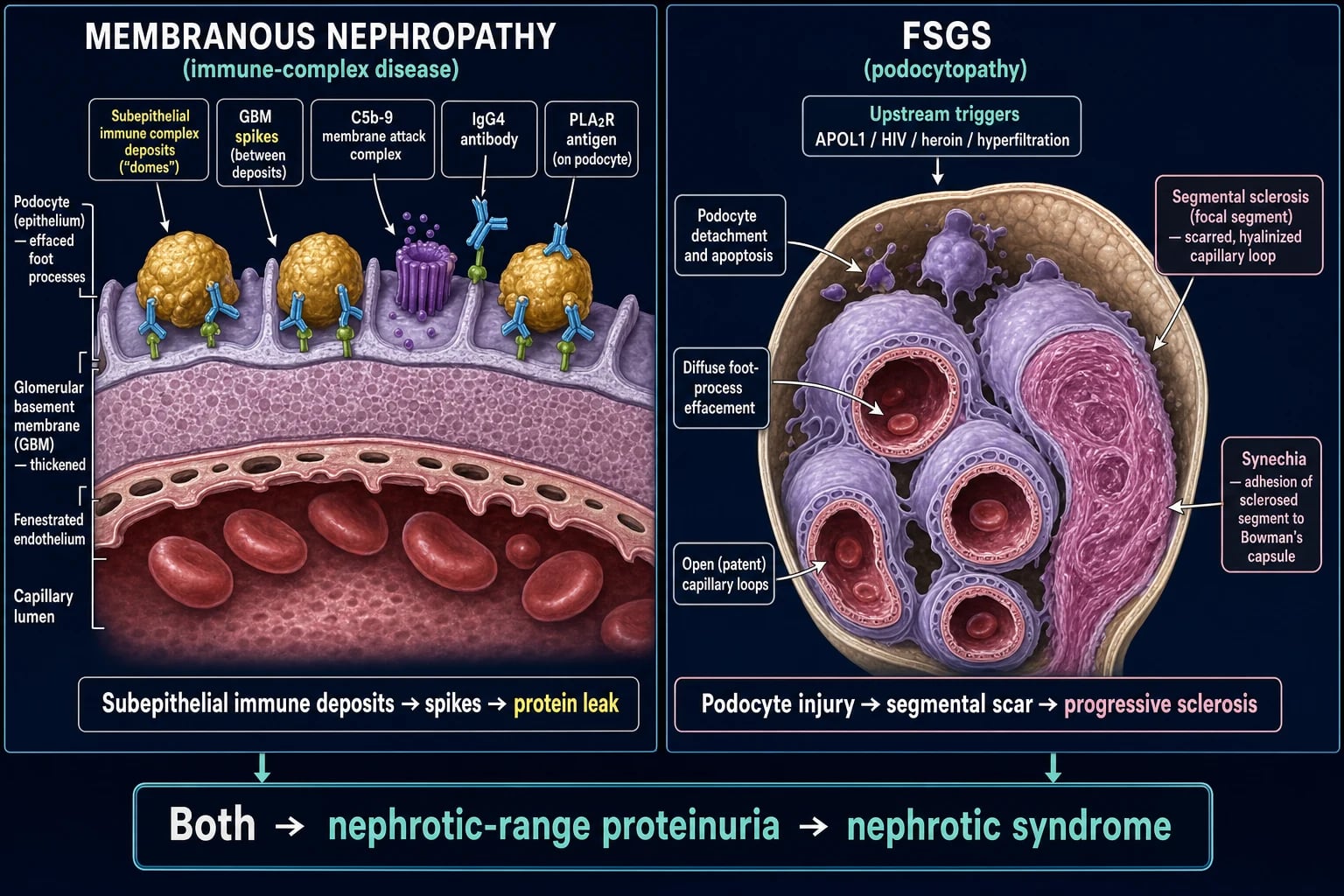
Membranous nephropathy — molecular mechanism (full detail):[1][4]
- Antigen exposure and autoantibody formation. In genetically susceptible individuals (HLA-DQA1 and PLA2R1 risk alleles), the M-type phospholipase A2 receptor (PLA2R) on the podocyte surface becomes the target of IgG4 autoantibodies. PLA2R is normally a transmembrane receptor; its conformational epitope is exposed on the podocyte foot process. THSD7A is a second, much rarer antigen (~2 to 5%), with a strong solid-malignancy association. The IgG subclass is IgG4 (a Th2-skewed, non-complement-fixing-by-classical-pathway subclass — relevant because complement is activated by the lectin and alternative pathways, not classical).
- Immune-complex formation in situ. The antibody crosses the GBM and binds its antigen on the subepithelial (podocyte) side, where the complexes form in situ. Because they sit outside the GBM, on the urinary side, they cannot be removed by circulating cells — they persist and grow.
- Complement activation and podocyte injury. Complement is activated via the lectin pathway (mannan-binding lectin recognising IgG4) and the alternative pathway, generating the C5b-9 membrane attack complex on the podocyte. Sublytic C5b-9 injures the podocyte without lysing it, triggering reactive oxygen species, proteases (cathepsins), TGF-beta, and cytoskeletal rearrangement → foot-process effacement and damage to the slit diaphragm.
- Basement-membrane reaction — spike formation (Ehrenreich-Churg stages I to IV). New GBM material is laid down between and around the deposits, producing the classic spikes (stage II) that progressively incorporate (stage III) and ultimately resolve or leave sclerosis (stage IV). Silver (methenamine) stain shows the spikes best.
- Result: non-selective proteinuria, the nephrotic syndrome, and (because the GBM is architecturally remodelled) slow progression in a subset. [1]
FSGS — molecular mechanism (full detail):[2][5]
- Podocyte injury (the unifying event). In primary FSGS, a circulating permeability factor (candidates: soluble urokinase-type plasminogen activator receptor suPAR, cardiotrophin-like cytokine CLCF-1, anti-CD40, anti-nephrin antibodies) crosses the GBM and injures the podocyte directly — analogous to how anti-PLA2R injures the podocyte in membranous, but the antigen/factor is different and elusive. In genetic FSGS, the slit diaphragm or cytoskeleton is defective from birth (NPHS1 nephrin, NPHS2 podocin, TRPC6 calcium channel, INF2 formin, ACTN4 alpha-actinin-4). In viral and drug FSGS the podocyte is injured directly (HIV genes nef and vpr in podocytes; pamidronate toxicity). In adaptive FSGS, chronic glomerular hyperfiltration and hypertension mechanically stress podocytes in residual nephrons (obesity, reduced renal mass, reflux, sickle cell).
- Podocyte detachment and apoptosis. Injured podocytes detach into the urine (podocyturia) and undergo apoptosis. The podocyte does not regenerate (it is terminally differentiated), so denuded segments of GBM are exposed.
- Synechia and segmental sclerosis. The exposed GBM adheres to Bowman's capsule (synechia). Plasma proteins leak into the segment, hyalin (PAS-positive) accumulates, and the segment scarifies. Because the lesion is initially focal (some glomeruli) and segmental (part of each tuft), early disease can be missed on biopsy — a reason to obtain an adequate sample (adequate biopsy = at least 10 to 20 glomeruli, and light + IF + EM).
- Progression and collapse. Sclerosis spreads to neighbouring loops; in the collapsing variant, dysregulated proliferation of parietal/podocyte cells collapses the entire tuft, producing rapid AKI on nephrotic syndrome. Tubulointerstitial damage (from filtered protein and ischaemia) drives GFR loss and progressive CKD.
- Transplant recurrence strongly supports the permeability-factor hypothesis: the nephrotic syndrome can recur within hours to days of transplant, and the disease can be transferred experimentally — pointing to a circulating, dialysable factor. [1]
Shared nephrotic-complication mechanisms (both diseases):[2]
- Oedema — hypoalbuminaemia lowers plasma oncotic pressure (underfill hypothesis), but the dominant mechanism in many patients is primary renal sodium retention: filtered proteases activate the epithelial sodium channel (ENaC) in the collecting duct, and RAAS is up-regulated. Both mechanisms contribute.
- Hyperlipidaemia — the liver upregulates VLDL and lipoprotein synthesis in response to low oncotic pressure (and reduced clearance via loss of lipoprotein lipase cofactors); raised cholesterol/LDL and lipiduria (oval fat bodies, Maltese cross under polarised light).
- Hypercoagulability (the highest-yield exam mechanism) — urinary loss of antithrombin III (the key thrombin inhibitor), increased hepatic fibrinogen and factor VIII, platelet activation, hemoconcentration/hyperviscosity, and loss of plasminogen. Risk is highest when albumin is under 25 g/L, and highest of all in membranous nephropathy (renal vein thrombosis).
- Infection susceptibility — urinary loss of IgG and alternative-pathway complement factors B and D impairs opsonisation of encapsulated organisms (Streptococcus pneumoniae, Haemophilus influenzae, E. coli); spontaneous bacterial peritonitis, cellulitis, pneumococcal sepsis. [1]
Clinical Presentation
Typical presentation (shared): insidious onset of dependent oedema (ankle, sacral), periorbital oedema (often worse in the morning), frothy urine, and weight gain from salt and water retention. The nephrotic syndrome is the tetrad of proteinuria over 3.5 g/day, albumin under 30 g/L, oedema, and hyperlipidaemia/lipiduria. Blood pressure is often normal in membranous; hypertension and microscopic haematuria are commoner in FSGS. GFR is typically preserved or only mildly reduced at presentation in membranous; FSGS more often presents with reduced GFR.[1][2]
Membranous-specific features: a purely nephrotic, slowly-evolving picture with little haematuria and preserved GFR is typical. Renal vein thrombosis may be the presenting event — sudden unilateral flank or loin pain with macroscopic haematuria, a rising creatinine, and (left-sided) left testicular varicocele; left renal vein thrombosis is commoner because the left renal vein is longer and traverses between the aorta and superior mesenteric artery (the nutcracker anatomy). Pulmonary embolism may be silent or symptomatic. Membranous is the nephrotic disease in which thrombosis is most common (up to a third develop a thromboembolic event).[1]
FSGS-specific features: may present as frank nephrotic syndrome (especially primary) OR as asymptomatic proteinuria with haematuria, hypertension and reduced GFR (more often in secondary/adaptive). The collapsing variant presents with AKI on nephrotic syndrome and rapid progression, classically in a Black patient with HIV (HIVAN) or APOL1 risk alleles. In children, FSGS often masquerades as steroid-resistant nephrotic syndrome after an initial minimal-change-like course.[2]
Complication-level presenting features to actively seek: sudden flank pain and haematuria (renal vein thrombosis); dyspnoea, pleuritic chest pain, haemoptysis (pulmonary embolism); fever with abdominal peritonism (spontaneous bacterial peritonitis in nephrotic ascites); cellulitis and pneumonia; xanthomata (hyperlipidaemia); frothy urine.[2]
Cause-specific clues pointing to secondary disease: chronic liver disease stigmata (HBV/HCV); malar rash, arthritis, oral ulcers (lupus); chronic NSAID use, gold/penicillamine for rheumatoid disease; weight loss, smoking, change in bowel habit, haemoptysis (malignancy-membranous); HIV with rapid AKI (HIVAN); morbid obesity, single kidney, reflux nephropathy, sickle cell disease (adaptive FSGS); family history of ESKD (genetic FSGS, APOL1).[1][2]
Atypical presentations: Elderly patient with new membranous nephropathy — a mandatory malignancy screen (often within 1 to 3 years of diagnosis; lung, colon, stomach, prostate, breast). HIV-positive patient with collapsing FSGS — HIVAN, urgent antiretrovirals. Pregnant patient with new nephrotic syndrome — distinguish from pre-eclampsia (hypertension, uric acid, low platelets; proteinuria pre-dating pregnancy favours glomerular disease). Transplant recipient with new nephrotic proteinuria — recurrent FSGS (early) or de novo membranous (late, often HCV-related).[1][1]
Differential Diagnosis
The nephrotic syndrome has a finite, examinable differential. Distinguish by bedside features, serology and biopsy:[1][2]
- Minimal change disease — commonest nephrotic in children (2 to 6 years), abrupt oedema, normal GFR, steroid-responsive ~90%; normal light microscopy with foot-process effacement only; associations — Hodgkin lymphoma, NSAIDs, atopy. Distinguished from FSGS by absence of sclerosis and steroid responsiveness (though FSGS may evolve from apparent MCD).
- Diabetic nephropathy — the commonest secondary nephrotic cause overall. Long-standing diabetes, diabetic retinopathy (highly predictive), gradual onset, often with proteinuria progression over years; no biopsy needed if typical (retinopathy + long diabetes). ACE inhibitor/ARB and SGLT2 inhibitor are cornerstone.
- Lupus nephritis class V (membranous) — ANA, anti-dsDNA, low complement (C3/C4), other lupus stigmata; biopsy shows full-house immunofluorescence (IgG, IgA, IgM, C3, C1q) and may show mixed mesangial/proliferative features.
- Amyloidosis (AL or AA) — Congo-red positive apple-green birefringence, macroglossia, periorbital purpura, chronic inflammatory disease (AA) or plasma-cell dyscrasia (AL); serum free light chains, serum/urine electrophoresis.
- Membranoproliferative GN / immune-complex disease — low complement, mixed nephrotic-nephritic picture, endocapillary proliferation and "tram-track" double contour on biopsy; HCV, cryoglobulinaemia, HBV.
- Pre-eclampsia — pregnancy over 20 weeks, hypertension, proteinuria, raised uric acid, low platelets, abnormal LFTs; resolves post-partum.
- Crescentic transformation of membranous (anti-PLA2R with superimposed anti-GBM or ANCA vasculitis) — rapidly declining GFR, active sediment; re-biopsy shows crescents. [1]
When is renal biopsy indicated? All adults with nephrotic syndrome. Children with classic steroid-sensitive nephrotic syndrome are treated empirically (no biopsy); biopsy only if atypical (age under 1 or over 10, hypertension, haematuria, low complement, renal impairment) or steroid-resistant (often FSGS).[1]
Clinical & Bedside Assessment
Vital signs and fluid status drive the initial assessment: blood pressure (often normal in membranous, raised in FSGS), weight and daily weights, fluid balance, oxygen saturation, temperature (infection). JVP, basal crackles, sacral and pedal oedema grading, and abdominal examination (ascites, renal angle tenderness) establish volume status and complication clues.[1]
Focused examination for a secondary cause: [1]
- Blood pressure and fundoscopy (diabetic/hypertensive retinopathy, hypertensive urgency).
- Skin — malar rash, discoid lesions, purpura, palpable purpura (vasculitis), xanthomata (hyperlipidaemia), thin skin/striae/easy bruising (prior chronic steroid), needle-track marks (heroin).
- Macroglossia and periorbital purpura (amyloidosis), joints (lupus, rheumatoid for NSAID/gold/pencillamine exposure).
- Abdomen — hepatosplenomegaly (chronic liver disease, HBV/HCV), masses, ascites; lymphadenopathy (malignancy, Hodgkin).
- Chest — signs of effusion, consolidation, pulmonary embolism.
- Legs — unilateral calf swelling/tenderness (DVT), which often heralds renal vein thrombosis.
- Renal angle — tenderness (renal vein thrombosis, infarction).
- Neurology — peripheral neuropathy (diabetes, amyloid, myeloma). [1]
Targeted history for secondary causes: drugs (NSAIDs, gold, penicillamine, heroin, pamidronate, interferon, lithium), infections (HBV, HCV, HIV, syphilis), malignancy symptoms (weight loss, haemoptysis, change in bowel habit, bone pain), autoimmune (lupus, thyroid), and family history (genetic FSGS, APOL1, Alport/COL4A3/4/5), as well as the kidney/transplant history (recurrent FSGS). [1]
Investigations
First-line — confirm and quantify the nephrotic syndrome:[1]
- Urinalysis and microscopy — dipstick 3 to 4+ protein; few cells/casts (distinguishes from nephritic); oval fat bodies and Maltese-cross appearance under polarised light (lipiduria).
- Quantify proteinuria — 24-hour urine protein over 3.5 g/day (or over 50 mg/kg/day), or spot urine protein-to-creatinine ratio over 350 mg/mmol (over 3.5 g/g). Serial UPCR monitors disease activity and response.
- Serum albumin (under 30 g/L, often under 25 g/L in severe disease), renal function (creatinine, eGFR, urea), electrolytes, lipid profile (raised cholesterol/LDL), full blood count, CRP/ESR.
- Renal ultrasound — kidney size (small kidneys preclude aggressive immunosuppression), exclude obstruction; Doppler for suspected renal vein thrombosis. [1]
Serological workup to classify the cause (high-yield):[1][4]
- Anti-PLA2R antibody — primary membranous (~70 to 80%, IgG4); a titre (high vs low) guides diagnosis, immunological activity, treatment response and relapse, and transplant timing.
- Anti-THSD7A antibody — rare (~2 to 5%); strong cancer association — thorough malignancy screen if positive.
- ANA, anti-dsDNA, complement C3/C4 — lupus class V (low complement, full-house IF).
- HBsAg, anti-HBs, anti-HCV, HIV serology — secondary membranous / HIVAN.
- Serum electrophoresis and serum free light chains, urine Bence-Jones — myeloma / AL amyloid.
- Anti-GBM and ANCA — if crescents or rapidly declining GFR (superimposed vasculitis/anti-GBM).
- TSH — autoimmune thyroiditis; rheumatoid factor / anti-CCP — rheumatoid arthritis (NSAID/gold/pencillamine exposure); cryoglobulins — HCV-related mixed cryoglobulinaemia. [1]
Malignancy screen in membranous (especially age over 50 or anti-THSD7A positive):[1] CT chest, abdomen and pelvis, age-appropriate colonoscopy, PSA in men, mammography in women, and a careful symptom-directed review over the first 1 to 3 years of diagnosis (the period of highest malignancy yield).
Renal biopsy — the defining investigation:[1][5][1]
- Membranous nephropathy: thickened GBM with spikes on silver/methenamine stain (light microscopy); granular capillary-wall IgG4 and C3 (immunofluorescence); subepithelial deposits with spike-and-dome and foot-process effacement (electron microscopy). Ehrenreich-Churg stages I to IV describe the evolution of deposits.
- FSGS: focal (some glomeruli) and segmental (part of the tuft) sclerosis with hyalinosis and adhesions (synechiae) to Bowman capsule; collapse/proliferation defines the collapsing variant (light microscopy); nonspecific IgM and C3 in sclerotic segments (immunofluorescence); diffuse foot-process effacement in primary FSGS vs focal effacement in adaptive/secondary (electron microscopy). An adequate sample (at least 10 to 20 glomeruli) is required because FSGS is focal and can be missed. [1]
Baseline workup before immunosuppression: glucose/HbA1c, hepatitis B/C and HIV, TB screen (interferon-gamma release assay or chest X-ray), bone density (DEXA), vaccination status, pregnancy test in women of childbearing age. [1]
Membranous diagnosis — pathology triad
Management — Resuscitation & General Nephrotic Care (Shared)

The supportive/nephrotic-care agenda is shared by membranous and FSGS and must be started at diagnosis, in parallel with disease-specific treatment.[1][2][1]
Fluid, sodium and oedema control: [1]
- Salt restriction under 2 g/day, modest fluid restriction if hyponatraemic; daily weights and fluid balance.
- Loop diuretic — oral furosemide 40 mg daily, titrated to weight; combine with amilide 5 to 10 mg daily or spironolactone 25 to 50 mg daily for resistant oedema and to antagonise secondary hyperaldosteronism.
- Refractory anasarca — IV furosemide (with 20 to 25% albumin 25 to 50 g) to mobilise fluid; nephrology input. Avoid over-diuresis (precipitates AKI). [1]
Antiproteinuric cornerstone — for ALL causes:[1]
- ACE inhibitor (e.g. ramipril 2.5 to 10 mg daily) or ARB (e.g. losartan 50 to 100 mg daily, valsartan 80 to 320 mg daily) — reduces intraglomerular pressure and proteinuria; titrate to blood pressure under 130/80 (under 125/75 with proteinuria) and tolerated potassium/creatinine (a rise in creatinine up to 30% is acceptable). Continue long-term. [1]
Lipid management:[1] atorvastatin 20 to 80 mg daily or rosuvastatin 10 to 40 mg daily for persistent hyperlipidaemia in adults with nephrotic syndrome — reduces cardiovascular risk.
Anticoagulation (the highest-yield exam point for membranous):[1][1] The thrombotic risk (DVT, PE, renal vein thrombosis) rises sharply as albumin falls and is highest of all in membranous nephropathy. Prophylactic anticoagulation is recommended when serum albumin is under 25 g/L (and considered at 25 to 30 g/L) in membranous, weighing bleeding risk; use LMWH-bridged warfarin or a direct oral anticoagulant (apixaban, rivaroxaban). Treat any thrombotic event fully — image (renal Doppler ultrasound or CT venography) and anticoagulate for at least the duration of nephrotic-range proteinuria. A left-sided renal vein thrombosis may present with flank pain, haematuria and a left varicocele.
Vaccination:[1] pneumococcal conjugate (PCV) then polysaccharide (PPSV23), annual influenza, and hepatitis B — ideally before immunosuppression; avoid live vaccines (MMR, yellow fever, live attenuated influenza, BCG) while on immunosuppression. Pneumococcal and influenza vaccination reduce the encapsulated-organism infection risk driven by IgG/complement loss.
Treat suspected infection promptly — encapsulated organisms and spontaneous bacterial peritonitis; empirical broad-spectrum antibiotics (e.g. cefotaxime or ceftriaxone 2 g IV) without waiting for cultures in the septic nephrotic patient. [1]
Management — Definitive & Stepwise (Disease-Specific)
Membranous nephropathy — risk-stratified immunosuppression
The KDIGO 2021 framework stratifies by risk because a third remit spontaneously and immunosuppression carries real toxicity:[1][3][1]
Low risk
Conservative only
- Normal renal function (eGFR normal), **proteinuria under 4 g/day** (or under 3.5 g/day average over 6 months)
- **Conservative therapy only** — ACE inhibitor/ARB, anticoagulation if low albumin, lipid, vaccination, oedema
- Monitor proteinuria and renal function every 6 to 12 weeks for 6 months
- Many will remit spontaneously (rule of thirds)
Moderate risk
Watchful then treat
- **Proteinuria 4 to 8 g/day**, normal renal function, over 6 months
- **6 months of maximal conservative therapy** (ACE inhibitor/ARB, blood pressure control); reassess
- Immunosuppress if proteinuria persists over 8 g/day, or if renal function declines, or life-threatening thrombosis occurs
- Anti-PLA2R titre trajectory helps — a falling titre predicts spontaneous remission
High risk
Immunosuppress
- **Proteinuria over 8 g/day for over 6 months**, OR **declining renal function**, OR **life-threatening complications** (renal vein thrombosis, severe hypoalbuminaemia, AKI)
- **First-line: rituximab 1 g IV at days 1 and 15** (repeat at 6 months if needed) OR **modified Ponticelli**
- Anti-PLA2R titre guides response — titre falls precede proteinuria remission
First-line immunosuppressive regimens (KDIGO 2021):[1][3][1]
- Rituximab (B-cell depleting anti-CD20) — 1 g IV at days 1 and 15, repeat at 6 months depending on response and anti-PLA2R titre. The MENTOR trial (Fervenza, NEJM 2019; PMID 31269364) showed rituximab was non-inferior to cyclosporine in inducing remission, with a significantly lower relapse rate and better renal preservation. Rituximab is now first-line or preferred in many centres for high-risk membranous, and is increasingly used in moderate-risk disease. Monitor CD19/CD20 counts; screen for hepatitis B (reactivation risk — give antiviral prophylaxis if core-antibody positive) and tuberculosis.[3]
- Modified Ponticelli regimen (alternating steroid + alkylating agent over 6 months) — month 1: IV methylprednisolone 1 g daily for 3 days then oral prednisolone 0.5 mg/kg/day for 27 days; month 2: oral cyclophosphamide 2 to 2.5 mg/kg/day for 30 days; repeat the alternating steroid (month 3) and cyclophosphamide (month 4) cycles, and a final steroid cycle (month 5), for a total of 6 months. (The original Ponticelli used chlorambucil instead of cyclophosphamide — cyclophosphamide is now preferred for a better safety profile.) Monitor full blood counts weekly (cyclophosphamide leucopenia), consider MESNA for bladder protection and Pneumocystis prophylaxis (co-trimoxazole 480 mg daily or 960 mg three times weekly).
- Calcineurin inhibitor (ciclosporin 3 to 5 mg/kg/day in two divided doses, or tacrolimus 0.05 to 0.1 mg/kg/day) plus low-dose prednisolone — an alternative when rituximab/Ponticelli are contraindicated; risk of nephrotoxicity, hypertension, diabetes (tacrolimus), gingival hyperplasia and hirsutism (ciclosporin); monitor trough levels and renal function. Relapse is common on stopping.
- Mycophenolate mofetil (1 to 2 g/day) — less-proven monotherapy; used in combination or when other agents are unsuitable.
Secondary membranous nephropathy is treated by removing the cause — resect the tumour, treat HBV/HCV with antivirals, stop the offending drug, immunosuppress the lupus. Anti-PLA2R is usually negative; immunosuppression for the membranous itself is generally not required unless severe and persistent.[1]
FSGS — prolonged steroid trial, then CNI
Primary FSGS requires a prolonged, genuinely high-dose steroid trial before declaring steroid-resistance:[2][1]
- Maximal conservative therapy — ACE inhibitor/ARB titrated to proteinuria/blood pressure, blood pressure control, salt restriction, lipid management, anticoagulation and vaccination as above.
- Prolonged high-dose steroid trial — oral prednisolone 1 mg/kg/day (maximum 80 mg/day) for at least 12 to 16 weeks (often 4 to 6 months total), then taper over several weeks/months. Steroid responsiveness is the single best prognostic factor. Confirm adherence and a long-enough trial before declaring resistance.
- Steroid-resistant primary FSGS — calcineurin inhibitor: ciclosporin 3 to 5 mg/kg/day or tacrolimus 0.05 to 0.1 mg/kg/day for at least 6 months (often 12); monitor trough levels and renal function (nephrotoxicity). Add low-dose prednisolone. Mycophenolate mofetil and rituximab are alternatives for frequently-relapsing or steroid-dependent disease.
- Genetic FSGS — does not respond to immunosuppression; manage conservatively, plan for transplant (does not recur), and offer genetic counselling.
- Adaptive/secondary FSGS — treat the underlying cause (weight loss in obesity, surgical correction of reflux, sickle cell management, manage reduced renal mass); these do not usually need immunosuppression.
- Collapsing FSGS / HIVAN — aggressive course; treat HIV with antiretrovirals plus ACE inhibitor (this combination transformed HIVAN prognosis); high-dose steroid may be added in HIV-negative collapsing FSGS; many progress to dialysis. Consider APOL1 status and counsel on transplant. [1]
Childhood steroid-resistant nephrotic syndrome (often FSGS)[1]
- Confirm adherence and a full 4 to 8 week steroid trial before resistance is declared.
- Biopsy to confirm FSGS (and exclude MCD); genetic testing (NPHS1, NPHS2, WT1) — genetic disease is steroid-resistant and does not recur post-transplant.
- Calcineurin inhibitor (ciclosporin or tacrolimus); rituximab for steroid-dependent/frequently-relapsing.
- Counsel the family on the risk of progression to ESKD and on transplant (FSGS may recur). [1]
Specific Subtypes & Scenarios
- Primary (anti-PLA2R) membranous nephropathy — the anti-PLA2R titre is a dynamic biomarker: a high titre predicts active disease and relapse; a falling titre precedes clinical remission; seronegativisation is a treatment target. Anti-PLA2R also guides transplant timing (defer transplant until immunological remission to reduce recurrence).[1][4]
- Anti-THSD7A membranous nephropathy (~2 to 5%) — strong solid-malignancy association; thorough malignancy screen mandatory. Mechanistically similar to PLA2R (IgG4, subepithelial).[6]
- Secondary membranous nephropathy — treat the cause. Cancer-related often remits after tumour resection; HBV-related with antivirals (entecavir/tenofovir); lupus class V per lupus-nephritis protocol (mycophenolate + steroid, or calcineurin inhibitor, or rituximab for refractory); drug-related with cessation.
- Collapsing FSGS — worst prognosis, rapid AKI; HIV-associated nephropathy (HIVAN) is the paradigm — treat with antiretrovirals + ACE inhibitor; high-dose steroid in HIV-negative collapsing FSGS. APOL1 risk alleles common.[2]
- Tip-lesion FSGS — best prognosis, often steroid-responsive (behaves like MCD); treat with a steroid trial first.
- HIV-associated nephropathy (HIVAN) — collapsing FSGS, predominantly Black patients (APOL1), presents with nephrotic syndrome + rapid AKI; antiretroviral therapy + ACE inhibitor (the combination transformed the prognosis, formerly near-universal ESKD). Steroid may be added.[2]
- APOL1-associated nephropathy — spectrum from FSGS to hypertension-attributed nephropathy to non-diabetic CKD progression in people of recent West-African ancestry; no specific therapy yet (APOL1 inhibitors are in trials); counsel on transplant and recurrence (APOL1 genetic disease does not recur, but a primary permeability-factor component may).[2]
- FSGS recurrence after transplant — occurs in 30 to 50%, often within hours to days of transplant, presenting with immediate massive proteinuria. Treat with plasmapheresis (removes the permeability factor), rituximab, and intensified immunosuppression (e.g. higher CNI, or switch to proteasome inhibitor). Genetic FSGS does not recur.[2]
- Membranous after transplant — may recur (anti-PLA2R positive, usually within the first years) or arise de novo (often HCV-related, late).
- Crescentic transformation of membranous — membranous nephropathy with superimposed anti-GBM or ANCA vasculitis produces crescents and rapidly declining GFR; re-biopsy; treat with plasma exchange, high-dose steroid, cyclophosphamide/rituximab.[1]
Complications & Pitfalls
Complications of heavy proteinuria (both diseases):[1][2]
- Thromboembolism — renal vein thrombosis (sudden flank/loin pain, haematuria, rising creatinine, left-sided commoner), DVT, pulmonary embolism. Risk highest in membranous (up to a third); risk rises sharply when albumin is under 25 g/L. Prophylactic anticoagulation indicated at this threshold; image (Doppler/CT venography) and treat any event.
- Infection — encapsulated organisms (S. pneumoniae, H. influenzae, E. coli); spontaneous bacterial peritonitis, cellulitis, pneumococcal sepsis; mechanism — urinary loss of IgG and alternative-complement factors B and D. Vaccinate; treat promptly.
- Hyperlipidaemia and accelerated atherosclerosis — statin in adults with persistent nephrotic syndrome.
- Acute kidney injury — from over-diuresis, hypovolaemia, superimposed ATN, bilateral renal vein thrombosis, or collapsing transformation.
- Hypocalcaemia and vitamin D deficiency — loss of cholecalciferol-binding protein (vitamin D-binding protein) in the urine; supplement vitamin D and calcium.
- Malnutrition, anaemia, muscle wasting — chronic protein loss. [1]
Immunosuppression-related complications:[1][3]
- Corticosteroid — diabetes, hypertension, infection, osteoporosis (consider bisphosphonate/calcium-vitamin D, DEXA), cataracts, peptic ulcer, mood/psychosis, growth retardation and delayed puberty in children, avascular necrosis.
- Cyclophosphamide — leucopenia and infection (weekly FBC), haemorrhagic cystitis and bladder cancer (use MESNA, generous hydration), infertility (especially cumulative dose; counsel on sperm/egg storage), secondary malignancy, nausea, alopecia.
- Calcineurin inhibitor — nephrotoxicity (afferent arteriolar vasoconstriction, chronic interstitial fibrosis), hypertension, diabetes (tacrolimus), gingival hyperplasia and hirsutism (ciclosporin), hyperkalaemia, tremor; monitor trough levels and renal function.
- Rituximab — infusion reactions (premedicate with paracetamol, antihistamine, steroid), infection (including hepatitis B reactivation — screen and prophylaxe; PJP; rarely progressive multifocal leucoencephalopathy), late-onset neutropenia, hypogammaglobulinaemia. [1]
Classic pitfalls (high-yield):[1][2]
- Not anticoagulating a severely hypoalbuminaemic (under 25 g/L) membranous patient — the highest-yield miss.
- Missing renal vein thrombosis (flank pain + haematuria in a nephrotic patient = renal vein thrombosis until proven otherwise).
- Declaring FSGS steroid-resistant before a full 4 to 6 month trial — confirm adherence and duration first.
- Missing secondary causes — malignancy in elderly membranous (screen within 1 to 3 years); HBV/HCV; adaptive FSGS (obesity, reflux); HIV in collapsing FSGS.
- Treating genetic or adaptive FSGS with immunosuppression — these do not respond; manage the cause and counsel on transplant.
- Failing to monitor cyclophosphamide FBC or calcineurin inhibitor levels.
- Missing FSGS recurrence after transplant (immediate proteinuria = recurrent FSGS — act fast with plasmapheresis/rituximab).
- Forgetting the ACE inhibitor/ARB in all causes, and vaccination before immunosuppression.
- Biopsy sampling error in FSGS — an inadequate sample (few glomeruli) may miss a focal lesion and mislabel FSGS as MCD. [1]
Prognosis & Disposition
Membranous nephropathy — the rule of thirds: about one-third remit spontaneously (complete or partial), one-third remain stable with persistent proteinuria, and one-third progress to ESKD over 10 to 15 years. Spontaneous remission is more likely with lower baseline proteinuria, female sex, and asymptomatic presentation. Modern anti-PLA2R-guided and rituximab-based therapy has improved remission rates and reduced relapse (MENTOR).[1][3]
Predictors of progression in membranous: sustained proteinuria over 8 g/day, declining renal function, male sex, older age, hypertension, tubulointerstitial fibrosis on biopsy, and a persistent high anti-PLA2R titre.[1]
FSGS prognosis: steroid-responsive FSGS behaves like minimal change disease and does well (frequent relapses but normal life expectancy); steroid-resistant primary FSGS progresses to ESKD in 50% or more over 5 to 10 years; the collapsing variant progresses fastest (ESKD often within 2 to 3 years); secondary/adaptive FSGS prognosis depends on the underlying cause and may stabilise if the cause is treated (e.g. weight loss, reflux correction). Genetic FSGS often progresses to ESKD but does not recur after transplant.[2]
Disposition: nephrology referral for all adults with nephrotic syndrome and biopsy confirmation. Plan transplant evaluation with recurrence counselling (FSGS 30 to 50% recurrence; anti-PLA2R-positive membranous — defer transplant until immunological remission to reduce recurrence). Plan dialysis for progressive disease; supportive care (fluid, BP, lipids, bone, vaccination, anticoagulation) throughout.[1]
Special Populations
- Children — membranous nephropathy is rare in children and more often secondary (HBV, lupus, congenital/maternal anti-PLA2R); biopsy and screen. Childhood steroid-resistant nephrotic syndrome is often FSGS — genetic testing is important (NPHS1, NPHS2, WT1): genetic FSGS does not respond to immunosuppression and does not recur after transplant. Counsel the family on transplant and recurrence. Weight-based dosing (prednisolone 60 mg/m²/day).[1]
- Elderly — membranous nephropathy frequently secondary (mandatory malignancy screen within 1 to 3 years); higher thrombotic risk; weigh immunosuppression toxicity against benefit (prefer rituximab over prolonged cyclophosphamide when feasible). Amyloidosis enters the differential in the elderly with chronic inflammation.
- Pregnancy — distinguish nephrotic syndrome from pre-eclampsia (proteinuria pre-dating pregnancy, low complement, or biopsy-proven GN favours glomerular disease). Stop ACE inhibitor/ARB (teratogenic, especially 2nd/3rd trimester — fetopathy); switch antiproteinuric/blood-pressure agent to labetalol, nifedipine, or methyldopa. Anticoagulate severely hypoalbuminaemic pregnant patients (LMWH, not warfarin — teratogenic). Anti-PLA2R antibodies cross the placenta and can cause transient neonatal proteinuria/nephrotic syndrome — monitor the neonate. FSGS may flare with pregnancy-related hyperfiltration in a single kidney.[1]
- Patients of recent West-African ancestry — screen for APOL1 high-risk genotypes (G1, G2) when FSGS or hypertension-attributed nephropathy presents; counsel on transplant (APOL1 disease does not recur, though a primary component may) and on genetic implications.
- Transplant recipients — FSGS recurs in 30 to 50% (often early, with massive proteinuria); treat with plasmapheresis + rituximab. Membranous recurs (anti-PLA2R positive) or arises de novo (often HCV-related, late). Anti-PLA2R-guided timing of transplant reduces recurrence.
- Immunocompromised / HIV — HIV-associated nephropathy (collapsing FSGS); screen and treat with antiretrovirals + ACE inhibitor; prognosis dramatically improved with combination therapy. Hepatitis B and C drive secondary membranous and membranoproliferative patterns — screen and treat.
Evidence, Guidelines & Regional Differences
- KDIGO 2021 Glomerular Diseases Guideline — the international standard: anti-PLA2R-based diagnosis; risk-stratified (conservative vs immunosuppression) approach to membranous; rituximab or modified Ponticelli as first-line for high-risk membranous; conservative plus prolonged steroid trial for primary FSGS; calcineurin inhibitor for steroid-resistant FSGS.[1]
- MENTOR trial (Fervenza FC et al., NEJM 2019; PMID 31269364) — landmark evidence: rituximab non-inferior to cyclosporine in membranous nephropathy, with a significantly lower relapse rate and better renal preservation at 24 months — establishing rituximab as a preferred option, including in moderate-risk disease.[3]
- Beck 2009 (NEJM; PMID 19571279) — the foundation: M-type phospholipase A2 receptor (PLA2R) is the target antigen in ~70% of idiopathic membranous nephropathy (IgG4 subclass). This transformed membranous from an idiopathic immune-complex disease to a defined autoimmune podocytopathy, enabling serological diagnosis and monitoring.[4]
- Tomas 2014 (NEJM; PMID 25394321) — identified THSD7A as a second antigen in ~2 to 5% of membranous; strong cancer association; reinforced the paradigm that membranous is an antigen-specific autoimmune disease.[6]
- Columbia FSGS classification (D'Agati VD et al., JASN 2004; PMID 14750104) — the standard morphological classification of FSGS variants (collapsing, tip, cellular, perihilar, NOS), each with distinct prognosis — the framework used worldwide.[5]
- Well-resourced settings (US, UK, ANZ) — early biopsy and rituximab availability for membranous; APOL1 genotyping in people of African ancestry; MENTOR-influenced preference for rituximab.
- India / resource-limited settings (Indian Society of Nephrology consensus) — prolonged steroid trial and Ponticelli with cyclophosphamide remain the backbone (cheaper, available; rituximab often cost-prohibitive); later/limited rituximab for resistant disease; limited access to biopsy in some regions; HBV endemicity increases secondary membranous; APOL1 screening increasingly relevant in South Asian and African-diaspora populations.
- UK (NICE NG203 / Renal Association) — anti-PLA2R titre is a commissioned test and part of the standard workup; APOL1 genotyping offered selectively. [1]
Exam Pearls
- Membranous = commonest primary nephrotic in Caucasian adults; anti-PLA2R (IgG4); thick GBM with spikes (silver stain); secondary = cancer (over 50), HBV, lupus class V, NSAIDs/gold/penicillamine, syphilis; rule of thirds; renal vein thrombosis is the highest-risk complication.[1]
- FSGS = focal, segmental sclerosis; podocytopathy; diffuse foot-process effacement; often steroid-resistant; collapsing (HIV, APOL1, worst), tip (best); recurs in 30 to 50% of transplants; APOL1 in West-African ancestry.[2]
- Both: biopsy all adults; RAAS blockade reduces proteinuria in all causes; anticoagulate membranous when albumin under 25 g/L; vaccinate (pneumococcal, influenza, hepatitis B).[1]
- Modified Ponticelli = alternating methylprednisolone/prednisolone + cyclophosphamide (replaced chlorambucil) over 6 months; rituximab = 1 g IV at days 1 and 15 (MENTOR, NEJM 2019).[3]
- Primary membranous workup: anti-PLA2R (primary, IgG4), anti-THSD7A (rare, cancer-associated); ANA/dsDNA/complement (lupus class V); HBV/HCV/HIV; malignancy screen if over 50.[4][6]
- Histology: membranous = subepithelial IgG4 deposits with spike-and-dome on EM; FSGS = segmental sclerosis with diffuse foot-process effacement (primary), IgM/C3 in scars.[5]
- FSGS Columbia variants in order of prognosis (best to worst): tip > cellular > NOS > perihilar > collapsing.[5]
- Anti-PLA2R titre trajectory: a falling titre precedes clinical remission; seronegativisation is a treatment target and guides transplant timing.[1]
- Adaptive FSGS often has only focal foot-process effacement (vs diffuse in primary) — a useful biopsy clue.
FSGS aetiology
H-A-V-E-S
Drug (heroin, pamidronate, interferon) and virus (HIV — collapsing variant, HIVAN)
Genetic (APOL1, NPHS1/2, TRPC6, INF2) and adaptive/hyperfiltration (obesity, reduced renal mass, reflux, sickle cell)
HIV (collapsing, APOL1 background); parvovirus B19, CMV
Pamidronate, interferon, lithium, anabolic steroids
Reduced renal mass, reflux nephropathy, single kidney (adaptive hyperfiltration)
Membranous secondary causes
M-E-M-B-R-A-N-E
Solid-organ carcinoma — lung, colon, stomach, breast, prostate (highest yield over age 50)
Lupus nephritis class V; also mixed connective-tissue disease
NSAIDs, gold, penicillamine, captopril, tiopronin, bucillamine
Hepatitis B (especially endemic regions) and hepatitis C
Rheumatoid arthritis drugs (gold, penicillamine) — overlaps with Medications
Hashimoto/autoimmune thyroid disease
NSAIDs (also cause MCD and acute interstitial nephritis)
Syphilis, sarcoidosis, graft-versus-host disease
Exam application bank (NEET-PG / INICET)
One-line answer
Membranous nephropathy and focal segmental glomerulosclerosis (FSGS) are the two commonest primary glomerular causes of nephrotic syndrome in adults. Membranous nephropathy is an antibody-mediated subepithelial immune-complex disease: in 70 to 80% it is primary, driven by IgG4 autoantibodies against the podocyte M-type phospholipase A2 receptor (PLA2R) (and rarely THSD7A); secondary forms arise from solid-organ malignancy, hepatitis B and C, lupus class V, NSAIDs/gold/penicillamine, syphilis and thyroid disease. It carries the highest thrombotic risk of any nephrotic cause (renal vein thrombosis), and about a third remit spontaneously (the rule of thirds). FSGS is a podocytopathy defined histologically by focal, segmental glomerular scarring with diffuse foot-process effacement; causes are primary (circulating permeability factor), genetic (APOL1, NPHS1/2, TRPC6, INF2), virus-associated
Worked stems (answer without another resource)
Stem 1 — Classic presentation. Map symptoms to mechanism; name the first investigation and first treatment step with dose/route if drug therapy is standard. [1]
Stem 2 — Unstable / complicated. List red flags that force immediate resuscitation, theatre, ICU, antidote, or reperfusion — and what you do in the first 15 minutes. [1]
Stem 3 — Atypical group. Elderly, pregnancy, child, or immunocompromised: how presentation and thresholds change. [1]
Stem 4 — Differential trap. Name the three closest mimics and one discriminator for each. [1]
Stem 5 — Disposition. Who goes home with safety-netting, who is admitted, who needs HDU/ICU/theatre, and what follow-up is mandatory. [1]
Rapid viva checklist
- Definition + classification
- Pathophysiology chain
- Bedside signs / criteria
- Score with exact components (if any)
- Emergency bundle
- Definitive therapy with doses
- Complications of disease and of treatment
- Special populations
- Guideline/trial name if classic
- Three exam traps
Coverage self-check
If you cannot answer any stem above from this page alone, re-read the matching section — the page is intended to be self-sufficient for final-prof and NEET-PG/INICET questions on Membranous Nephropathy & FSGS.
References
- [1]Hoxha E, Reinhard L, Stahl RAK. Membranous nephropathy: new pathogenic mechanisms and their clinical implications Nat Rev Nephrol, 2022.PMID 35484394
- [2]Kopp JB, Anders HJ, Susztak K. Podocytopathies Nat Rev Dis Primers, 2020.PMID 32792490
- [3]Fervenza FC, Appel GB, Barbour SJ, et al. Rituximab or Cyclosporine in the Treatment of Membranous Nephropathy N Engl J Med, 2019.PMID 31269364
- [4]Beck LH Jr, Bonegio RG, Lambeau G, et al. M-type phospholipase A2 receptor as target antigen in idiopathic membranous nephropathy N Engl J Med, 2009.PMID 19571279
- [5]D'Agati VD, Fogo AB, Bruijn JA, Jennette JC. Pathologic classification of focal segmental glomerulosclerosis: a working proposal Am J Kidney Dis, 2004.PMID 14750104
- [6]Tomas NM, Beck LH Jr, Meyer-Schwesinger C, et al. Thrombospondin type-1 domain-containing 7A in idiopathic membranous nephropathy N Engl J Med, 2014.PMID 25394321