Nephrology · Nephrology
Minimal Change Disease
Also known as MCD · Nil disease · Lipoid nephrosis · Steroid-sensitive nephrotic syndrome
Minimal change disease (MCD) is the leading cause of nephrotic syndrome in children and the prototype of steroid-responsive podocytopathy. It accounts for roughly 90 percent of childhood and 10-15 percent of adult nephrotic presentations, with selective albuminuria, normal light microscopy, no immune deposits on immunofluorescence, and diffuse foot process effacement on electron microscopy. High-dose corticosteroids induce remission in over 80 percent of cases, with cyclophosphamide, calcineurin inhibitors, mycophenolate, and rituximab reserved for frequent relapsers, steroid-dependent, and steroid-resistant disease.
On this page & tools
Your progress
Saved locally on this device.
Exam tags
Red flags

Overview & Definition
Minimal change disease (MCD), historically called nil disease or lipoid nephrosis, is a clinicopathological syndrome characterised by sudden-onset heavy proteinuria, hypoalbuminaemia, hyperlipidaemia, and oedema in a patient whose renal biopsy shows essentially normal glomeruli on light microscopy, no immune deposits on immunofluorescence (IF), and diffuse effacement of podocyte foot processes on electron microscopy (EM). It is the most common cause of nephrotic syndrome in children and the prototype of steroid-responsive glomerular disease. [3]
The disorder is classified within the spectrum of podocytopathies — primary diseases of the visceral epithelial cell whose slit diaphragm governs size- and charge-selective filtration. Like focal segmental glomerulosclerosis (FSGS) and membranous nephropathy (MN), it produces massive proteinuria, but unlike them it preserves glomerular architecture (no sclerosis, no immune deposits) and is markedly reversible with immunosuppression. The clinical hallmark of MCD is selectivity: urinary losses are dominated by albumin and smaller proteins, with sparing of larger immunoglobulins in the classic paediatric case — a feature exploited in historical work-up via the IgG/IgG4 selectivity index but rarely required today. [3][1]
The disease is idiopathic in the vast majority of cases but can be secondary to drugs (NSAIDs, lithium, gold, pamidronate, bisphosphonates, sirolimus, interferon), malignancies (Hodgkin and other lymphomas), atopy, and recent infection or immunisation. Recognising secondary forms is clinically important because treatment must address the underlying trigger as well as the renal lesion. [3][1]
Classification
Primary (idiopathic) MCD
- Classical steroid-responsive disease
- Children 2-6 years, adults 30-40 years
- Normal LM, no IF deposits, FPE on EM
- Cytokine-driven (IL-13, T-cell permeability factor)
- Remission in 80-90 percent with steroids
Secondary MCD
- Drug-induced: NSAIDs, lithium, gold, pamidronate, bisphosphonates, sirolimus, interferon
- Malignancy: Hodgkin lymphoma (classic), non-Hodgkin, CLL, thymoma
- Recent infection or immunisation (MMR, influenza)
- Atopic disease and systemic conditions (sle, sarcoidosis)
- Treat underlying cause; steroids if persistent
FSGS, collapse variant
- Widespread foot process effacement with collapse
- Often steroid-resistant, presents with AKI
- Differential considered when biopsy shows segmental collapse
- May co-exist or evolve from MCD — re-biopsy
Mesangioproliferative GN
- Mild mesangial hypercellularity on LM
- Often steroid-responsive but slower than MCD
- Some classify as IgM nephropathy or C1q nephropathy
C1q Nephropathy
A rare entity in which dominant or co-dominant C1q deposition is seen on IF, with mesangial proliferation on LM and FPE on EM. It presents with nephrotic or nephritic-nephrotic features and is now treated like MCD if steroid-responsive. Historically subsumed under mesangioproliferative glomerulonephritis, it is increasingly recognised by IF pattern. [1]
IgM Nephropathy
Defined by diffuse mesangial IgM deposition on IF with mild mesangial proliferation. It is debated whether this is a discrete entity or a heterogeneous bucket containing some steroid-responsive cases indistinguishable from MCD, and a steroid-resistant group resembling FSGS. Prognosis lies between MCD and FSGS. [1]

Epidemiology & Risk Factors
Minimal change disease is the single most common cause of nephrotic syndrome in children worldwide, accounting for roughly 70-90 percent of cases under age 10 and approximately 90 percent of cases in pre-school-aged children (peak age 2-6 years, median 3 years). After puberty the prevalence falls steeply, with a second smaller peak in adults aged 30-40 years. Adult-onset disease represents about 10-15 percent of primary nephrotic syndrome, of which MCD is one-third to one-half of the remainder after membranous nephropathy and FSGS. [3][1][1]
Sex and Race
There is a slight male predominance in children (approximately 2:1) that diminishes in adolescence. Adults show a more even sex ratio, with some series showing mild female predominance after the fifth decade. The condition is reported across all racial groups, although series suggest higher incidence in South Asian and East Asian children than in African-ancestry children, where FSGS is the predominant nephrotic histology. [1]
Genetic and Familial Predisposition
Familial clustering occurs in a minority of cases. Rare monogenic forms involve mutations in NPHS2 (podocin), NPHS1 (nephrin), WT1, and COQ6, which clinically mimic MCD but are typically steroid-resistant — these are screened in syndromic or familial childhood nephrotic syndrome, not in classic sporadic MCD. [3][1]
Triggering Factors
Triggers of secondary MCD
MEDIC
Hodgkin lymphoma (paraneoplastic), non-Hodgkin, CLL, thymoma
Recent URTI, immunisation, bee sting
NSAIDs, lithium, gold, pamidronate, sirolimus, interferon, rifampicin
Atopy, asthma, eczema, food allergy, eosinophilia
SLE, sarcoidosis, HIV (rarely), celiac disease
- Recent upper respiratory tract infection precedes the first attack in many children — historically termed post-infectious nephrotic syndrome when onset is within 5 days of a viral illness. [3]
- Atopy and asthma are over-represented in children with relapsing MCD.
- Drug exposure: NSAIDs (especially propionic acid derivatives), lithium, gold salts, pamidronate and other bisphosphonates, sirolimus, and interferon-α can produce a histologically identical lesion; remission usually follows withdrawal.
- Malignancy: Hodgkin lymphoma is the classic association (particularly in the uncommon adult-onset case). The renal lesion may remit with chemotherapy of the lymphoma.
- Post-immunisation: rare case reports of MCD following MMR, yellow fever, influenza, and pneumococcal vaccination; recurrence on re-exposure is described.
Pathophysiology
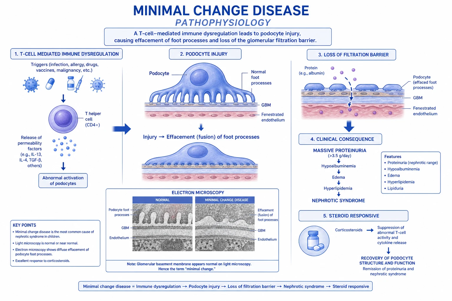
[1]MCD is fundamentally a disease of the podocyte — the visceral epithelial cell whose interdigitating foot processes and slit diaphragms form the final size- and charge-selective barrier of the glomerular filtration apparatus. Light and immunofluorescence microscopy show essentially normal glomeruli, but electron microscopy reveals the diagnostic lesion: diffuse effacement (flattening and retraction) of foot processes with microvillous transformation of podocyte cell bodies. This effacement reflects disassembly of the actin cytoskeleton and disruption of the slit-diaphragm complex (nephrin, podocin, CD2AP, NEPH1, FAT1). [3]
Circulating Permeability Factors
A large body of evidence supports the existence of one or more circulating permeability factors released by activated T lymphocytes, which render the glomerular filtration barrier leaky to albumin. Historical observations include: [1]
- Savin and Goodman's serum permeability assays demonstrated that plasma from patients with active MCD increases albumin permeability of isolated glomeruli, and that this activity falls during steroid-induced remission. [3]
- T-cell hybridomas from MCD patients produce a circulating factor of approximately 100 kDa that mimics the lesion when injected into rats.
- Cytokine profile: MCD is associated with a Th2-skewed immune response and elevated levels of interleukin-13 (IL-13), which directly injures podocytes and induces expression of CD80 (B7-1). Other implicated cytokines include IL-4, vascular permeability factor, and haemopexin.
- T-cell dysregulation is suggested by the absence of immune deposits (ruling out classical antibody-mediated injury), the response to T-cell–directed immunosuppression (steroids, calcineurin inhibitors, cyclophosphamide), and the association with Hodgkin lymphoma and atopy.
Charge-Selective Barrier Disruption
Podocytes normally produce a negatively charged glycocalyx rich in heparan sulphate and podocalyxin, which repels albumin (anionic) electrostatically. In MCD, loss of fixed negative charge — foot-process-dependent and cytokine-induced — produces selective albuminuria (predominant loss of albumin over IgG). This charge-selective leakage is reversible with steroid therapy, correlating with restoration of foot-process architecture. [3]
Why No Immune Deposits?
A defining feature of MCD is the absence of immunoglobulin or complement deposits on immunofluorescence, distinguishing it from membranous nephropathy (IgG4 granular subepithelial), lupus nephritis ("full-house"), IgA nephropathy (mesangial IgA), and post-infectious GN (C3-dominant "starry sky"). The injury is therefore a non-immune-complex podocytopathy. [1]
Reversibility with Corticosteroids
Steroids suppress cytokine-producing lymphocytes, restore the charge barrier, and allow foot processes to re-extend within 1-2 weeks — the basis for the bedside steroid trial in young children with first-presentation nephrotic syndrome without biopsy. [1]
Clinical Presentation
MCD classically presents with sudden-onset nephrotic syndrome — a tetrad of heavy proteinuria, hypoalbuminaemia, hyperlipidaemia, and oedema — often after a recent upper respiratory infection or allergic trigger. [3][1]
Childhood Presentation
- Periorbital oedema noted first by parents on waking, often misattributed to allergy or conjunctivitis.
- Ankle and pretibial oedema progressing through the day.
- Scrotal or labial oedema, sometimes the presenting complaint in boys.
- Ascites with abdominal distension and umbilical herniation in chronic relapsers.
- Pitting oedema extending to thighs, sacrum, and sometimes pleural effusions.
- Weight gain from fluid retention — paradoxically, anorexia from gut oedema and fatigue may coexist.
- Frothy urine from heavy proteinuria is the cardinal symptom reported by parents. [1]
Adult Presentation
Adults tend to present with a more indolent picture and more complications at diagnosis: [1]
- Anasarca (generalised oedema including pleural effusions and ascites) in roughly 60-70 percent.
- Shortness of breath from pleural effusions, laryngeal oedema (rare), or rarely pulmonary oedema if acute kidney injury supervenes.
- AKI in roughly 10-25 percent of adults at presentation, often from hypovolaemia, acute tubular necrosis, or renal vein thrombosis.
- Hypertension is uncommon (unlike FSGS); when present, suspect a secondary cause or adult-onset atypical MCD.
- Thromboembolic events in roughly 5-10 percent of adults — renal vein thrombosis, deep-vein thrombosis, pulmonary embolism.
- Anorexia, malaise, muscle wasting from chronic inflammation and protein loss. [1]
When to Suspect Secondary MCD
- Adult over 60 years with first episode — screen for Hodgkin lymphoma, solid tumours, drug-induced.
- Persistent eosinophilia, rash, recent drug exposure — drug-induced MCD.
- B-symptoms, lymphadenopathy, splenomegaly — Hodgkin lymphoma until proven otherwise. [1]
Differential Diagnosis
The differential diagnosis of MCD covers the entire spectrum of nephrotic syndrome in children and adults, plus oedema from non-renal causes when proteinuria is modest. [3][1]
Focal Segmental Glomerulosclerosis (FSGS)
- Segmental sclerosis on LM, more severe FPE
- Often steroid-resistant; black race and APOL1 risk
- Higher rates of AKI and CKD progression
- Distinguish by biopsy — clinical picture overlaps
- Recurs post-transplant (permeability factor)
Membranous Nephropathy
- Adult-onset, subepithelial IgG4 deposits on IF
- Subnephrotic proteinuria more often than nephrotic
- Anti-PLA2R antibodies positive in 70-80 percent
- Slow progression, immunosuppressive or conservative
Mesangioproliferative GN / IgM / C1q
- Mesangial hypercellularity with IgM or C1q deposits
- Variable steroid response between MCD and FSGS
- Often treated empirically as MCD until relapse
Amyloidosis
- Congo-red positive amyloid deposits, apple-green birefringence
- Multisystem involvement — heart, nerves, tongue
- Underlying chronic inflammation (AA) or plasma cell dyscrasia (AL)
Diabetic Nephropathy
- Long-standing diabetes with retinopathy typically present
- Kimmelstiel-Wilson nodules on LM
- Slowly progressive proteinuria, often with CKD
Lupus Nephritis (Class V)
- Full-house IF (IgG, IgA, IgM, C3, C1q)
- Low complement levels, ANA and dsDNA positive
- Treated with steroids plus or minus MMF or cyclophosphamide
Other Considerations
- Drug-induced nephrotic syndrome: NSAIDs, gold, lithium, bisphosphonates, sirolimus, interferon; remits on withdrawal.
- Hodgkin lymphoma: paraneoplastic MCD, especially in young adult males.
- Membranous-like nephropathy: anti-PLA2R-positive, particularly in older adults.
- IgA nephropathy with nephrotic-range proteinuria: mesangial IgA on IF.
- Post-infectious GN: usually nephritic, low C3, hump-like subepithelial deposits on EM.
- Congestive cardiac failure, hepatic cirrhosis, hypothyroidism for oedema with minimal proteinuria. [1]
Clinical & Bedside Assessment
A child with first-presentation nephrotic syndrome is assessed clinically and biochemically before initiating a steroid trial without biopsy — but in adults, biopsy is generally performed before treatment because the chance of non-MCD pathology is higher. [1][1]
History
- Onset and tempo of oedema (periorbital first? ankle swelling? scrotal involvement?).
- Recent URTI, immunisation, illness in the 1-2 weeks preceding onset.
- Drug exposure — particularly NSAIDs, lithium, gold, pamidronate, interferon.
- Atopic background — asthma, eczema, hayfever, food allergy.
- Urine appearance — frothy (protein), red-brown (haematuria — unusual in MCD and prompts work-up).
- B symptoms, lymphadenopathy, weight loss, night sweats — Hodgkin lymphoma screen in adults.
- Pregnancy, contraceptive, menstrual history in women.
- Family history of nephrotic syndrome or renal disease. [1]
Examination
[1]- General: ill, oedematous, may be febrile if intercurrent infection.
- Vitals: BP usually normal; tachycardia if hypovolaemic; tachypnoea if pleural effusion or pulmonary oedema; SpO2 may be low in pulmonary oedema or PE.
- Oedema: periorbital, ankle (Bilateral, pitting, grade), sacral, scrotal/labial, ascites, anasarca.
- Cardiorespiratory: JVP, heart sounds (pericardial effusion rarely), pleural effusion signs.
- Abdomen: distension, ascites, hepatomegaly (mild if any), splenomegaly (Hodgkin).
- Lymph nodes: cervical, axillary, inguinal — Hodgkin lymphoma screen.
- Joints: swelling or rash — lupus, drug reaction.
- Urine dipstick: 3+ or 4+ protein, microscopic haematuria in 10-30 percent (does not exclude MCD). [1]
Investigations
Investigations — first-line
Urine Studies
- Urine dipstick: 3+ to 4+ protein. Microscopy usually bland; red cells raise concern for FSGS or membranous, although mild haematuria can occur.
- Urine protein:creatinine ratio (UPCR or Up:Uc): greater than 3.5 g/g (or 300+ mg/mmol) confirms nephrotic-range proteinuria. First-morning sample acceptable in children.
- 24-hour urine protein: greater than 3.5 g/day in adults; less rigorously required in children.
- Selectivity index (historical): IgG:transferrin clearance ratio less than 0.20 supports MCD in children; rarely performed today. [1]
Blood Studies
- Serum albumin: typically below 25 g/L, often below 20 g/L; oedema roughly parallels the depth of hypoalbuminaemia.
- Serum cholesterol and LDL: elevated; HDL may be normal or low.
- FBC: mild haemoconcentration if hypovolaemic; leucocytosis suggests infection; eosinophilia raises suspicion of Hodgkin or drug-induced MCD.
- Urea and electrolytes / eGFR: AKI in 10-25 percent of adults; hyponatraemia from diuretic use or water retention.
- LFTs: hypoalbuminaemia with otherwise preserved hepatic synthesis.
- Complement C3 and C4: normal — a low C3 should prompt work-up for lupus, post-infectious GN, MPGN, or cryoglobulinaemia.
- Immunoglobulins: IgG often low (urinary loss); IgM relatively preserved.
- HIV, hepatitis B, hepatitis C serology in adults before immunosuppression.
- ANA and dsDNA in adults or in any patient with multisystem features.
- HbA1c: baseline before steroid therapy. [1]
Imaging
- Renal ultrasound: usually normal-sized kidneys with preserved corticomedullary differentiation; not diagnostic but excludes obstruction, mass, or chronic parenchymal disease. [1]
Biopsy
The gold standard. In children aged 1-10 years with first-presentation typical nephrotic syndrome, biopsy is not required before empirical steroid therapy, provided there are no atypical features. [1][1][1]
- Indications for biopsy:
- Age under 1 year or over 12 years at first presentation.
- Persistent haematuria, hypertension, renal impairment, or low complement.
- Steroid resistance after 4-6 weeks of therapy.
- Frequent relapsers being considered for cyclophosphamide.
- Adults with primary nephrotic syndrome (universally biopsied in centres with prompt pathology services). [1]
Biopsy Findings
- Light microscopy: normal glomeruli or at most minimal mesangial prominence; tubules may show protein resorption droplets; interstitium normal. Congo red negative. [3]
- Immunofluorescence (IF): no immune deposits (negative for IgG, IgA, IgM, C3, C1q, kappa, lambda). If IgM or C1q is positive, the diagnosis shifts toward IgM nephropathy or C1q nephropathy respectively.
- Electron microscopy (EM): diffuse effacement of podocyte foot processes, microvillous transformation, and no immune-complex electron-dense deposits. This is the diagnostic finding.
When EM Is Unavailable
Most centres without routine EM still perform LM and IF. Clinical correlates — heavy proteinuria, normal complement, abrupt onset, dramatic steroid response — strongly support MCD in classic cases. [1]
Management — Resuscitation
Initial resuscitation addresses the emergent complications of severe nephrotic syndrome that may accompany or precede the diagnosis. [1][4]
Massive Anasarca
- Hospital admission for severe generalised oedema, ascites, pleural effusions.
- Fluid restriction only if hyponatraemic; gentle oral rehydration if hypovolaemic.
- Albumin infusion (1 g/kg 20% albumin over 2-4 h) followed by furosemide 1 mg/kg IV for tense oedema with intravascular depletion — used selectively to avoid pulmonary oedema from rapid intravascular expansion.
- Furosemide 40-80 mg daily PO/IV (or 1 mg/kg in children), titrated to urine output and weight; add thiazide or metolazone if diuretic resistance from gut oedema.
- Monitor weight, urine output, electrolytes daily. [1]
Acute Kidney Injury
- Hypovolaemic AKI: volume expansion with 0.9 percent saline 10-20 mL/kg bolus, then reassess.
- Acute tubular necrosis: supportive care, hold diuretics temporarily, avoid nephrotoxins.
- Renal vein thrombosis: anticoagulation with enoxaparin 1 mg/kg SC twice daily (creatinine-adjusted) and consideration of catheter-directed thrombolysis in severe cases. [1]
Pulmonary Oedema and Pleural Effusions
- Oxygen and upright positioning.
- Furosemide 40-80 mg IV with cautious rate.
- Therapeutic paracentesis or thoracentesis for tense ascites or large pleural effusions causing respiratory compromise. [1]
Infection
- Septic screen — blood cultures, urinalysis, ascitic fluid if ascites (cell count, culture). Empirical antibiotics covering pneumococcus (e.g. ceftriaxone 2 g IV daily) and gram-negatives.
- Pneumococcal vaccination (PPSV23 and PCV13/PCV15/PCV20) once proteinuria controlled.
- Varicella-zoster immune globulin if exposed and non-immune on steroids. [1]
Thromboembolism
- Prophylactic anticoagulation with enoxaparin 40 mg SC daily in admitted adults with serum albumin under 25 g/L plus additional risk factors (immobility, recent surgery, prior VTE, malignancy). [1]
- Treatment of established VTE/PE: full anticoagulation as per standard protocols.
- Avoid NSAIDs and COX-2 inhibitors; limit lines and immobility.
See the initial management decision tree in 
Management — Definitive & Stepwise
The cornerstone of MCD treatment is high-dose corticosteroids, which induce remission in approximately 80-90 percent of children and 70-80 percent of adults. Steroid resistance is the major fork in the treatment pathway. [1][2][4]
First-Line — Empirical Steroid Trial
Indications for empirical steroid therapy without biopsy:
- Children aged 1-12 years with typical first presentation (periorbital/leg oedema, no hypertension, no macroscopic haematuria, normal complement, normal renal function). [1]
Adults are generally biopsied first before immunosuppression, except in selected centres where empirical therapy may be considered for highly suggestive features. [1]
Steroid Regimen — Children
- Prednisone 2 mg/kg/day (maximum 60 mg/day) orally for 4-6 weeks as induction.
- Taper to 1.5 mg/kg alternate days for a further 4-6 weeks.
- Many paediatric protocols extend low-dose alternate-day therapy for 4-6 months to reduce relapse frequency, although evidence varies. [1][1]
Steroid Regimen — Adults
- Prednisolone 1 mg/kg/day (maximum 80 mg/day) for 8-16 weeks, then taper over 6 months. Meyrier's protocol: 1 mg/kg/day for at least 8 weeks, taper by 5 mg/week to alternate-day, then 0.5 mg/kg alternate-day for several months. [2][4]
- Time-to-response: most adults remit within 8-12 weeks; non-response at 16 weeks prompts biopsy review and second-line therapy.
Definitions of Disease Course
Second-Line — Steroid-Resistant MCD
- Calcineurin inhibitor: tacrolimus 0.05-0.1 mg/kg/day (target trough 5-10 ng/mL) or cyclosporine 3-5 mg/kg/day for 6-12 months, often combined with low-dose prednisone. [1][4][1]
- Mycophenolate mofetil 1-1.5 g twice daily as steroid-sparing alternative.
- Rituximab 375 mg/m² IV weekly for 1-4 doses — increasingly used in steroid-dependent and frequently relapsing disease; the MENTOR and RITUXPEAK series support efficacy with durable remissions.
Third-Line — Refractory Disease
- Cyclophosphamide (oral 2 mg/kg/day for 8-12 weeks or IV pulses) historically used in frequently relapsing MCD; risks of gonadal toxicity and malignancy reserve it for selected cases.
- Levamisole (paediatric, 2.5 mg/kg alternate days) as adjunct in steroid-dependent children.
- Ofatumumab and other anti-CD20 agents in rituximab failure. [1]
Supportive Care Throughout
- Diuretics for symptomatic oedema — furosemide 20-80 mg daily, titrated.
- Prophylactic anticoagulation with enoxaparin 40 mg SC daily if albumin below 25 g/L plus risk factors.
- Statin — atorvastatin 20-40 mg daily for hyperlipidaemia and cardiovascular risk in adults with persistent nephrotic syndrome.
- ACE inhibitor or ARB (e.g. ramipril 5-10 mg daily or losartan 50-100 mg daily) once stable on diuretics, to reduce proteinuria and slow progression, with caution during hypovolaemia.
- Diet: low-sodium, adequate-protein (not high-protein) intake. [1]
Stepwise Management
MCD treatment ladder — RAINBOW
RAINBOW
Low-sodium diet under 2 g/day; gentle fluid restriction if hyponatraemia
Ramipril or losartan once stable for proteinuria reduction
Prednisolone 1 mg/kg/day x8-16 wk; paediatric 2 mg/kg/day x4-6 wk
Loop diuretic for oedema; BP and volume status review
Enoxaparin 40 mg SC if Alb under 25 plus risk; statin for lipids
CNI / MMF / rituximab for steroid-resistant or dependent
Pneumococcal vaccine, VZIG if exposed, isolate if active infection
Algorithm
- Diagnosis confirmed (biopsy in adults; empirical trial in children).
- Start high-dose steroids — prednisolone 1 mg/kg/day (max 80 mg).
- Reassess at 4-6 weeks (children) or 8-16 weeks (adults).
- Remission achieved: slow taper over 4-6 months.
- Relapse on/off taper: reinstate full dose, then taper; consider second-line.
- Steroid-resistant: re-biopsy if not done; add CNI (tacrolimus or cyclosporine) plus or minus MMF; consider rituximab.
- Frequently relapsing / steroid-dependent: cyclophosphamide, MMF, or rituximab as steroid-sparing.
- Refractory: trial of rituximab, ofatumumab, or supportive care if CKD established. [1]
Specific Subtypes & Scenarios
Adult-Onset MCD
- Biopsy before treatment in all cases.
- Slower response to steroids than children — extend induction to 16 weeks. [2][1]
- Higher rates of AKI, thrombosis, and steroid toxicity; lower rates of complete remission.
- Long-term outcome: partial remission in many, complete remission in 50-70 percent, with 10-20 percent progressing to CKD over 10 years.
Paediatric MCD
- Empirical steroid trial without biopsy.
- 80-90 percent respond within 2 weeks; 50-70 percent will have at least one relapse.
- Frequently relapsing or steroid-dependent disease in 30-50 percent of children requires cyclophosphamide, levamisole, MMF, or rituximab. [1][1]
Steroid-Dependent MCD
- Relapse during or within 2 weeks of stopping steroids.
- Treated with cyclophosphamide 2 mg/kg/day x8-12 weeks, MMF 1-1.5 g BD, or rituximab 375 mg/m² weekly for 1-4 doses. [1]
Frequently Relapsing MCD
- Two or more relapses within 6 months.
- Same agents as steroid-dependent; rituximab increasingly preferred for sustained remission. [1]
Steroid-Resistant MCD
- Failure of remission despite 4-6 weeks (children) or 8-16 weeks (adults) of full-dose steroids.
- Triumvirate work-up: re-biopsy to exclude FSGS (especially collapse variant) and mesangioproliferative GN; genetic testing for syndromic childhood disease; consider secondary causes.
- CNI (tacrolimus preferred) plus or minus MMF is first-line steroid-sparing; rituximab used increasingly. [1]
Secondary MCD
- Hodgkin lymphoma: paraneoplastic nephrotic syndrome, often MCD histology; remission with chemotherapy.
- Drug-induced: stop offending agent; reserve steroids for persistent nephrotic syndrome.
- Post-immunisation: usually self-limiting; supportive care if non-remitting. [1]
Pregnancy
- New-onset MCD is rare in pregnancy; pre-existing MCD may flare in the third trimester.
- Prednisone and prednisolone are metabolised by placental 11-β-HSD2 and safe, but fluorinated glucocorticoids (dexamethasone, betamethasone) cross the placenta and are avoided for maternal therapy.
- Avoid cyclophosphamide, mycophenolate, and rituximab in the first trimester. Tacrolimus can be continued with monitoring.
- Renal-vein thrombosis risk is amplified — consider prophylactic LMWH if albumin under 25 g/L. [1]
Transplant Recurrence
- Recurrence of MCD histology in an allograft kidney occurs in 30-40 percent of patients who lost their native kidneys to "MCD" — this implicates a circulating permeability factor, particularly in those with rapid graft proteinuria.
- Treated with plasmapheresis + rituximab + high-dose steroids. [1]
Elderly MCD
- Over age 60, MCD becomes rarer; malignancy-associated MCD (Hodgkin, solid tumours) should be aggressively excluded.
- Steroid toxicity (diabetes, hypertension, osteoporosis, falls) is more problematic — lower-dose or shorter course often used. [1]
Complications
Infections
The single most important complication of MCD is infection driven by urinary loss of immunoglobulins and complement, plus immunosuppression from steroids. [1][1]
- Spontaneous bacterial peritonitis (SBP) — typically pneumococcal or E coli; presents with fever, abdominal pain, cloudy ascitic fluid (over 250 PMN per cubic mm). Treat with ceftriaxone plus or minus albumin.
- Cellulitis and erysipelas at sites of oedema.
- Sepsis from urinary or respiratory tract pathogens.
- Reactivation of tuberculosis in endemic areas — screen and consider INH prophylaxis in adults on long-term steroids.
- Pneumocystis jirovecii pneumonia in patients on cyclophosphamide or rituximab — prophylaxis with trimethoprim-sulfamethoxazole.
- Varicella-zoster and HSV reactivation with rituximab or cyclophosphamide. [1]
Thromboembolism
- Renal vein thrombosis — present in 5-10 percent of adult MCD at autopsy; flank pain, macroscopic haematuria, fall in renal function, asymmetric kidney enlargement on imaging. Confirmed with CT/MR venography. Treated with anticoagulation plus or minus catheter-directed thrombolysis. [1][1]
- Pulmonary embolism, deep-vein thrombosis, and rare arterial events.
- Mechanism: loss of antithrombin III, plasminogen, protein S in urine; increased fibrinogen, increased platelet aggregation, hyperviscosity from hyperlipidaemia.
Acute Kidney Injury
- Hypovolaemia, acute tubular necrosis, renal vein thrombosis, drug-induced interstitial nephritis.
- AKI is rare in children (under 5 percent) but present in 10-25 percent of adults at presentation; usually reversible. [1]
Steroid Side Effects
- Hyperglycaemia / steroid-induced diabetes — screen fasting glucose.
- Hypertension from sodium retention.
- Cushingoid features, weight gain, mood disturbance, insomnia.
- Osteoporosis — DEXA at baseline if prolonged therapy; calcium and vitamin D supplementation.
- Cataracts, glaucoma with prolonged therapy.
- Skin atrophy, acne, striae.
- Adrenal suppression with prolonged use — taper, do not stop abruptly.
- Avascular necrosis of femoral head — sudden hip pain; MRI early. [1]
Prognosis & Disposition
Outcomes in Children
- Remission within 2 weeks of starting steroids in 80-90 percent. [1]
- Long-term remission — by adulthood, 50-70 percent are in sustained remission off therapy, 20-30 percent have infrequent relapses, and 10-20 percent remain frequently relapsing through adolescence.
- CKD and ESRD are uncommon — most children with steroid-sensitive MCD have normal renal function at long-term follow-up.
- Mortality is very low in children with steroid-sensitive disease; mortality in steroid-resistant or steroid-toxicity-driven complications is largely historical in the modern era.
Outcomes in Adults
- Complete remission in 50-70 percent of adults within 12-16 weeks of full-dose steroids. [2][1]
- Steroid resistance in 10-30 percent — these patients have a worse outlook, with 10-20 percent progressing to ESRD over 10 years.
- Cumulative steroid toxicity, infection, and thromboembolism dominate morbidity in adults.
Risk Factors for Poor Outcome
- Steroid resistance — strongest predictor of CKD.
- Concurrent AKI at presentation, especially with ATN.
- Adult-onset disease, particularly over age 60.
- Persistent heavy proteinuria beyond 12 weeks.
- Heavy interstitial fibrosis on biopsy — late finding, suggests misdiagnosis or progression. [1]
Disposition
- Hospital admission for first presentation in adults and complicated childhood cases.
- Outpatient management for stable relapses in known MCD after first-line steroid trial.
- Multidisciplinary follow-up with nephrologist, paediatrician (children), pharmacist, and dietitian. [1]
Special Populations
Paediatric
- Empirical steroid trial without biopsy
- Prednisone 2 mg/kg/day (max 60 mg), taper over months
- 80-90 percent respond within 2 weeks
- Cyclophosphamide, MMF, or rituximab for relapsers
Adult
- Biopsy before immunosuppression
- Prednisolone 1 mg/kg/day x 8-16 wk, taper over 6 months
- Higher rate of steroid resistance (20-30 percent)
- Aggressive thrombosis prophylaxis
Pregnancy
- Prednisone / prednisolone safe (placental metabolism)
- Avoid MMF, cyclophosphamide, rituximab in 1st trimester
- Tacrolimus may be continued with monitoring
- Prophylactic LMWH if albumin under 25 g/L
Transplant
- Recurrence in 30-40 percent of presumed MCD
- Implies circulating permeability factor
- Treat with plasmapheresis + rituximab + steroids
- Donor-recipient genetic mismatch relevant
Elderly
- Screen for Hodgkin lymphoma and solid tumours
- Steroid toxicity prominent (DM, HTN, falls)
- Lower-dose or shorter-course strategies favoured
- Consider CNI or rituximab as steroid-sparing
Evidence & Guidelines
KDIGO 2012
The Kidney Disease: Improving Global Outcomes (KDIGO) Clinical Practice Guideline for Glomerulonephritis (2012) establishes the diagnostic approach and treatment framework for MCD. [1][4]
- Empirical steroid therapy is recommended in children aged 1-12 years with first-presentation typical nephrotic syndrome.
- Initial regimen in adults: prednisolone 1 mg/kg/day for 8-16 weeks.
- Frequent relapsers / steroid-dependent: cyclophosphamide, MMF, calcineurin inhibitors, or rituximab.
- Steroid-resistant: re-biopsy, consider CNI, rituximab. [1]
KDIGO 2021 (Update)
The 2021 KDIGO Glomerular Diseases Guideline reinforces most 2012 recommendations, with rituximab now considered alongside cyclophosphamide and CNI for frequently relapsing disease and steroid-dependent cases. [1]
NICE and Paediatric Guidelines
The NICE guideline for childhood nephrotic syndrome (NG213, 2023) and the International Pediatric Nephrology Association (IPNA) 2023 Clinical Practice Recommendations endorse empirical steroid therapy in typical first-presentation children, with prednisolone 60 mg/m²/day (max 60 mg) for 4 weeks, then alternate-day for 4-6 weeks. Cyclophosphamide or MMF are recommended steroid-sparing agents; rituximab is increasingly used. [1]
Cochrane Evidence
- Cochrane reviews confirm the efficacy of alkylating agents (cyclophosphamide, chlorambucil) in reducing relapse risk in frequently relapsing children, at the cost of gonadal toxicity.
- Rituximab has multiple RCTs supporting its steroid-sparing role in steroid-dependent adult and paediatric disease (e.g. MENTOR, RITUXPEAK, TURING). [1]
Landmark Studies
- Meyrier 2010 remains a touchstone for adult-onset MCD: 60-70 percent complete remission, with steroid resistance heralding poor renal outcome. [2]
- Vivarelli 2017: comprehensive review consolidating the cytokine-permeability-factor model. [3]
- Noone 2018: contemporary paediatric review emphasising the steroid-responsive nature of childhood disease and the emerging role of rituximab. [1]
Exam Pearls
- Most common cause of nephrotic syndrome in children (90 percent of cases under age 10) — peak 2-6 years; a clinical pearl you cannot skip. [3]
- Normal light microscopy + no immune deposits on IF + diffuse foot-process effacement on EM — this triad is the diagnostic definition. [3]
- Steroid trial is empirical in children aged 1-12 years with typical presentation — no biopsy needed; biopsy is universal in adults. [1][1]
- Time-to-response: 1-2 weeks in children, 4-12 weeks in adults — non-response is the pivotal moment that re-directs management.
- Complement is normal — a low C3 in suspected nephrotic syndrome should redirect to lupus, post-infectious GN, MPGN, or cryoglobulinaemia. [1]
- Hodgkin lymphoma is the classic paraneoplastic MCD — think of it in young adults with new-onset nephrotic syndrome and lymphadenopathy.
- Drugs that mimic MCD: NSAIDs, lithium, gold, pamidronate, bisphosphonates, sirolimus, interferon — every exam asks this list at least once.
- Steroid-dependent or frequently relapsing disease: alkylate (cyclophosphamide) or rituximab as steroid-sparing; CNI for steroid-resistant.
- Steroid-resistant MCD requires re-biopsy — about 30 percent turn out to be FSGS (collapse variant) or mesangioproliferative GN, not true MCD. [1]
- Complications triad: infection (SBP), thromboembolism (renal vein, PE), and AKI — these dominate adult mortality.
- Pregnancy note: prednisolone safe, but avoid MMF and cyclophosphamide in 1st trimester; the same drugs are teratogenic. [1]
- Transplant recurrence of "MCD" occurs in 30-40 percent — a classic for higher exams, signalling a circulating permeability factor and treated with plasmapheresis + rituximab.
- Pneumococcal vaccination is mandatory once proteinuria is under control — pneumococcus is the leading infection in nephrotic syndrome.
- Prophylactic anticoagulation with enoxaparin 40 mg SC daily in admitted adults with albumin under 25 g/L and additional VTE risk.
- The answer to an exam question on first-line adult therapy is "prednisolone 1 mg/kg/day (max 80 mg/day) for 8-16 weeks, then taper over 6 months" — commit the "1 mg/kg, 8-16 weeks, taper" triad to memory.
Exam application bank (NEET-PG / INICET)
One-line answer
Minimal change disease (MCD) is the leading cause of nephrotic syndrome in children and the prototype of steroid-responsive podocytopathy. It accounts for roughly 90 percent of childhood and 10-15 percent of adult nephrotic presentations, with selective albuminuria, normal light microscopy, no immune deposits on immunofluorescence, and diffuse foot process effacement on electron microscopy. High-dose corticosteroids induce remission in over 80 percent of cases, with cyclophosphamide, calcineurin inhibitors, mycophenolate, and rituximab reserved for frequent relapsers, steroid-dependent, and steroid-resistant disease.
Worked stems (answer without another resource)
Stem 1 — Classic presentation. Map symptoms to mechanism; name the first investigation and first treatment step with dose/route if drug therapy is standard. [1]
Stem 2 — Unstable / complicated. List red flags that force immediate resuscitation, theatre, ICU, antidote, or reperfusion — and what you do in the first 15 minutes. [1]
Stem 3 — Atypical group. Elderly, pregnancy, child, or immunocompromised: how presentation and thresholds change. [1]
Stem 4 — Differential trap. Name the three closest mimics and one discriminator for each. [1]
Stem 5 — Disposition. Who goes home with safety-netting, who is admitted, who needs HDU/ICU/theatre, and what follow-up is mandatory. [1]
Rapid viva checklist
- Definition + classification
- Pathophysiology chain
- Bedside signs / criteria
- Score with exact components (if any)
- Emergency bundle
- Definitive therapy with doses
- Complications of disease and of treatment
- Special populations
- Guideline/trial name if classic
- Three exam traps
Coverage self-check
If you cannot answer any stem above from this page alone, re-read the matching section — the page is intended to be self-sufficient for final-prof and NEET-PG/INICET questions on Minimal Change Disease.
References
- [1]Kidney Disease: Improving Global Outcomes (KDIGO) Glomerulonephritis Work Group Acute motor weakness of opposite lower extremity after percutaneous epidural neuroplasty Korean J Pain, 2015.PMID 25852837
- [2]Meyrier A Urinary biomarkers in the early detection of acute kidney injury after cardiac surgery Clin J Am Soc Nephrol, 2009.PMID 19406962
- [3]Vivarelli M, Massella L, Ruggiero B, Emma F Assessing plant protection practices using pressure indicator and toxicity risk indicators: analysis of therelationship between these indicators for improved risk management, application in viticulture Environ Sci Pollut Res Int, 2015.PMID 25404495
- [4]KDIGO Clinical Practice Guidelines for Glomerulonephritis Mechano- and metabosensitive alterations after injection of botulinum toxin into gastrocnemius muscle J Neurosci Res, 2014.PMID 24615939