Nephrology · General Medicine
Nephritic Syndrome & Acute Glomerulonephritis
Also known as Nephritic syndrome · Acute glomerulonephritis · Glomerulonephritis · Post-streptococcal glomerulonephritis · PSGN · Rapidly progressive glomerulonephritis · RPGN
Nephritic syndrome is the glomerular inflammatory counterpart of nephrotic syndrome: haematuria (dysmorphic red cells and red-cell casts), subnephrotic proteinuria (typically under 3.5 g/day), hypertension, oedema and an acute rise in creatinine (AKI), caused by glomerular inflammation and cellular proliferation. Causes are split by serum complement: low C3/C4 (post-streptococcal GN, membranoproliferative GN, lupus nephritis, endocarditis-associated GN, cryoglobulinaemia, shunt nephritis) versus normal complement (IgA nephropathy — the commonest primary GN worldwide, IgA vasculitis/Henoch-Schonlein purpura, anti-GBM disease/Goodpasture, ANCA-associated vasculitis). The pivotal clinical decision is distinguishing a self-limiting post-streptococcal GN (supportive care, excellent prognosis in children) from a renal emergency such as rapidly progressive (crescentic) GN, which destroys glomeruli within days and needs urgent steroids, cyclophosphamide or rituximab, and plasma exchange for anti-GBM and severe ANCA disease. Work-up: urine microscopy (dysmorphic RBCs and RBC casts), C3/C4, ANA/anti-dsDNA, ANCA (MPO/PR3), anti-GBM antibody, ASO/anti-DNase B, hepatitis B/C and HIV, and renal biopsy in adults and atypical cases.
On this page & tools
Your progress
Saved locally on this device.
Exam tags
Red flags
Overview & Definition
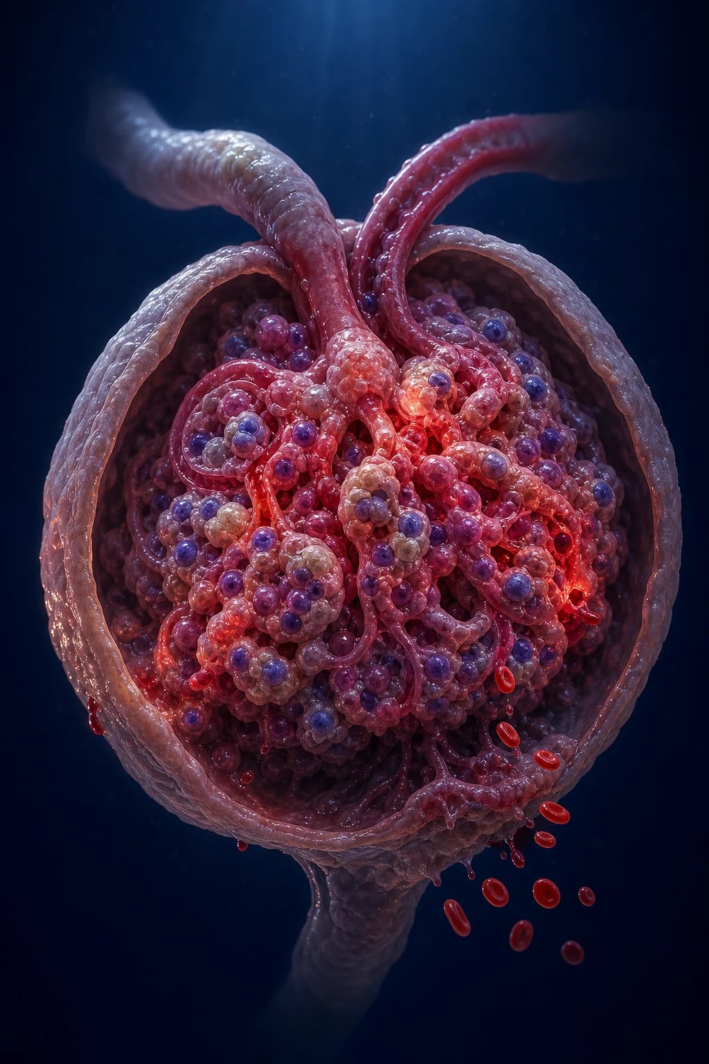
Nephritic syndrome is the clinical expression of active glomerular inflammation. In contrast to nephrotic syndrome, where the dominant problem is a leaky filtration barrier losing protein, the nephritic glomerulus is inflamed and proliferating: cellular proliferation (mesangial and endocapillary), inflammatory-cell infiltration (neutrophils, macrophages, T cells), and disruption of the capillary wall let blood (red cells and red-cell casts) leak into the urine alongside subnephrotic amounts of protein. The defining pentad is: [1]
- Haematuria — dysmorphic red cells and red-cell casts (the cardinal microscopy finding).
- Subnephrotic proteinuria — typically under 3.5 g/day (nephrotic-range proteinuria may occur in overlap, especially lupus class IV/V).
- Hypertension — from sodium/water retention and renin-angiotensin activation.
- Oedema — periorbital and dependent, from sodium retention and reduced GFR.
- Acute kidney injury (AKI) — a rapid rise in creatinine with oliguria from inflamed, failing glomeruli.[1][13]
The single most important triage tool at the bedside is serum complement (C3 and C4). A low C3 points to immune-complex and infectious causes (post-streptococcal GN, membranoproliferative GN and C3 glomerulopathy, lupus nephritis, endocarditis- and shunt-associated GN, cryoglobulinaemia), whereas a normal complement points to IgA nephropathy, IgA vasculitis (Henoch-Schonlein purpura), anti-GBM disease, and ANCA-associated vasculitis. The pivotal clinical decision is to distinguish a self-limiting post-streptococcal GN from a renal emergency such as rapidly progressive (crescentic) GN — the latter destroys glomeruli within days to weeks and demands urgent immunosuppression.[2][14]
Nephritic vs nephrotic vs RPGN vs asymptomatic haematuria
| Syndrome | Hallmark | Typical proteinuria | Complement |
|---|---|---|---|
| Nephritic syndrome | Haematuria (RBC casts) + hypertension + oedema + AKI | Under 3.5 g/day | Low or normal (cause-dependent) |
| Nephrotic syndrome | Heavy proteinuria + oedema + hypoalbuminaemia + hyperlipidaemia | Over 3.5 g/day | Usually normal |
| Rapidly progressive GN (RPGN) | Loss over 50% of GFR within under 3 months; crescents on biopsy | Variable | Low or normal |
| Asymptomatic haematuria | Isolated microscopic haematuria, no proteinuria/AKI/hypertension | Under 0.5 g/day | Normal |
| Chronic GN | Slowly progressive CKD with proteinuria, hypertension, shrunken kidneys | Variable | Variable |
Classification
Nephritic syndrome is classified along two axes — complement level (the bedside discriminator) and histological pattern on immunofluorescence (the biopsy discriminator that defines RPGN). [1]

LOW complement (C3)
Immune-complex & infectious
- **Post-streptococcal GN (PSGN)** — 1 to 3 weeks after GAS pharyngitis or 2 to 4 weeks after pyoderma; low C3, raised ASO; self-limiting in children
- **Membranoproliferative GN (MPGN) & C3 glomerulopathy** — persistent low C3, tram-track BM; hepatitis B/C, lupus, monoclonal gammopathy or alternative-pathway dysregulation
- **Lupus nephritis** (class IV diffuse proliferative) — low C3 AND C4, ANA and anti-dsDNA, full-house IF
- **Endocarditis-associated GN** and **shunt nephritis** — chronic bacteraemia; treat the infection
- **Cryoglobulinaemia** — low C4 (classical pathway), palpable purpura, hepatitis C
NORMAL complement
Pauci-immune & IgA
- **IgA nephropathy (Berger disease)** — commonest primary GN worldwide; synpharyngitic haematuria; mesangial IgA deposits
- **IgA vasculitis (Henoch-Schonlein purpura)** — palpable purpura, arthritis, abdominal pain, IgA nephritis; children
- **Anti-GBM disease (Goodpasture)** — linear IgG against alpha3(IV)NC1; pulmonary haemorrhage; plasma exchange
- **ANCA-associated vasculitis** — pauci-immune crescentic GN; anti-PR3 (c-ANCA, GPA) or anti-MPO (p-ANCA, MPA); pulmonary-renal
- **Double-positive ANCA + anti-GBM** — manage as anti-GBM (add plasma exchange)
RPGN by IF pattern
Crescents on biopsy
- **Type I** — linear anti-GBM (Goodpasture); anti-alpha3(IV)NC1 antibody; pulmonary haemorrhage
- **Type II** — granular immune-complex (lupus, post-infectious, IgA)
- **Type III** — pauci-immune (ANCA-associated: GPA, MPA, EGPA)
- All three carry the histological hallmark of **crescents** — cellular/fibrocellular proliferations of parietal epithelial cells and macrophages compressing the tuft
- All need urgent steroids + cyclophosphamide or rituximab; plasma exchange for anti-GBM and severe ANCA
By time-course
Clinical tempo
- **Acute GN** — onset over days to weeks (e.g. PSGN, IgA flare)
- **Rapidly progressive GN** — over 50% fall in GFR within under 3 months; crescents
- **Chronic GN** — slowly progressive CKD; small kidneys, interstitial fibrosis
- **Recurrent macroscopic haematuria** — IgA nephropathy, Alport carrier
- **Asymptomatic haematuria** — incidental finding; work up for glomerular vs urological
Special subtypes
- **C3 glomerulopathy** — alternative pathway dysregulation, C3 nephritic factor; recurrent after transplant
- **Cryoglobulinaemic vasculitis** — type II mixed (HCV); tetrad of purpura, weakness, arthralgia, neuropathy
- **Monoclonal Ig-related GN (POEMS, MGUS)** — proliferative GN with monoclonal deposits
- **HIV-associated immune-complex kidney disease (HIVICK)**
- **Drug-induced ANCA vasculitis** — hydralazine, propylthiouracil, levamisole-adulterated cocaine
Rapidly progressive GN — the three immunofluorescence types
RPGN is not a single disease but a syndrome of rapid glomerular destruction unified by the histological finding of crescents in over 50% of glomeruli. It is classified by immunofluorescence: [1]
- Type I — anti-GBM (linear IgG): anti-GBM disease/Goodpasture; antibody against the non-collagenous domain of the alpha-3 chain of type IV collagen (alpha3(IV)NC1); typically a pulmonary-renal syndrome with diffuse alveolar haemorrhage.[3][14]
- Type II — immune-complex (granular): lupus nephritis class IV, post-infectious GN, IgA nephropathy, cryoglobulinaemia; the underlying disease dictates treatment.[8]
- Type III — pauci-immune (little/no deposition): ANCA-associated vasculitis — granulomatosis with polyangiitis (GPA, PR3/c-ANCA), microscopic polyangiitis (MPA, MPO/p-ANCA), eosinophilic granulomatosis with polyangiitis (EGPA/Churg-Strauss).[15]
Epidemiology & Risk Factors
Incidence and demographics by cause
- IgA nephropathy (Berger disease) is the commonest primary glomerulonephritis worldwide, with biopsy-detected incidence highest in Asian populations (China, Japan, Korea) and a peak in young adult men. Many cases are never biopsied, so the true prevalence is higher than registry data suggest.
- Post-streptococcal GN remains the commonest post-infectious GN globally and the commonest acute GN in children (ages 2 to 12). It is endemic in resource-limited tropical regions (impetigo/pyoderma-related) but still occurs sporadically worldwide.
- ANCA-associated vasculitis has an annual incidence of around 10 to 20 per million, peaking in older adults (60 to 70 years). MPA is slightly more common than GPA in Asian populations; the reverse is seen in European populations.
- Anti-GBM disease is rare (0.5 to 1 per million per year) but has a bimodal age distribution: young men (20 to 30) typically presenting with severe pulmonary haemorrhage and rapidly progressive GN, and older women (60+) more often with renal-limited disease.[14]
- Lupus nephritis affects young women of childbearing age (female:male around 9:1); class IV diffuse proliferative is the most nephritic class.
- IgA vasculitis (Henoch-Schonlein purpura) is the commonest systemic vasculitis of childhood, peaking at ages 4 to 6, with a slight male predominance.[10][11]
Nephritogenic streptococcal strains
Post-streptococcal GN is caused by group A beta-haemolytic streptococcus (Streptococcus pyogenes) of certain M types that carry nephritogenic antigens: notably SpeB (streptococcal pyrogenic exotoxin B, a cysteine protease) and NAPlr (nephritis-associated plasmin receptor, glyceraldehyde-3-phosphate dehydrogenase). These antigens deposit in glomeruli, activate complement, and form the characteristic subepithelial immune humps.[1][2]
- Pharyngitis-associated M types: 1, 2, 4, 12.
- Skin/pyoderma-associated M types: 2, 49, 55, 57, 60.
- Seasonal pattern: pharyngitis-related PSGN peaks in winter/early spring; pyoderma-related PSGN in summer/early autumn (tropical climates). [1]
Risk factors
- Recent group A streptococcal infection (pharyngitis, pyoderma, cellulitis).
- Chronic infection: infective endocarditis, deep-seated abscess, ventriculoatrial shunt, osteomyelitis, chronic hepatitis B (membranous, MPGN), hepatitis C (cryoglobulinaemia, MPGN, membranous), HIV (HIVICK, collapsing FSGS).[8][16]
- Systemic autoimmune disease: SLE (lupus nephritis), rheumatoid arthritis, Sjogren syndrome.
- Drugs: hydralazine, propylthiouracil, levamisole-adulterated cocaine, allopurinol, penicillamine, sulfasalazine (all can trigger drug-induced ANCA vasculitis).
- Malignancy: monoclonal gammopathies (MGUS, myeloma, lymphoma) producing proliferative GN with monoclonal deposits.
- Family history: Alport syndrome carriers (haematuria), familial IgA nephropathy, complement regulatory gene mutations (factor H deficiency → C3 glomerulopathy).
Pathophysiology
The glomerular filtration barrier and how it is breached
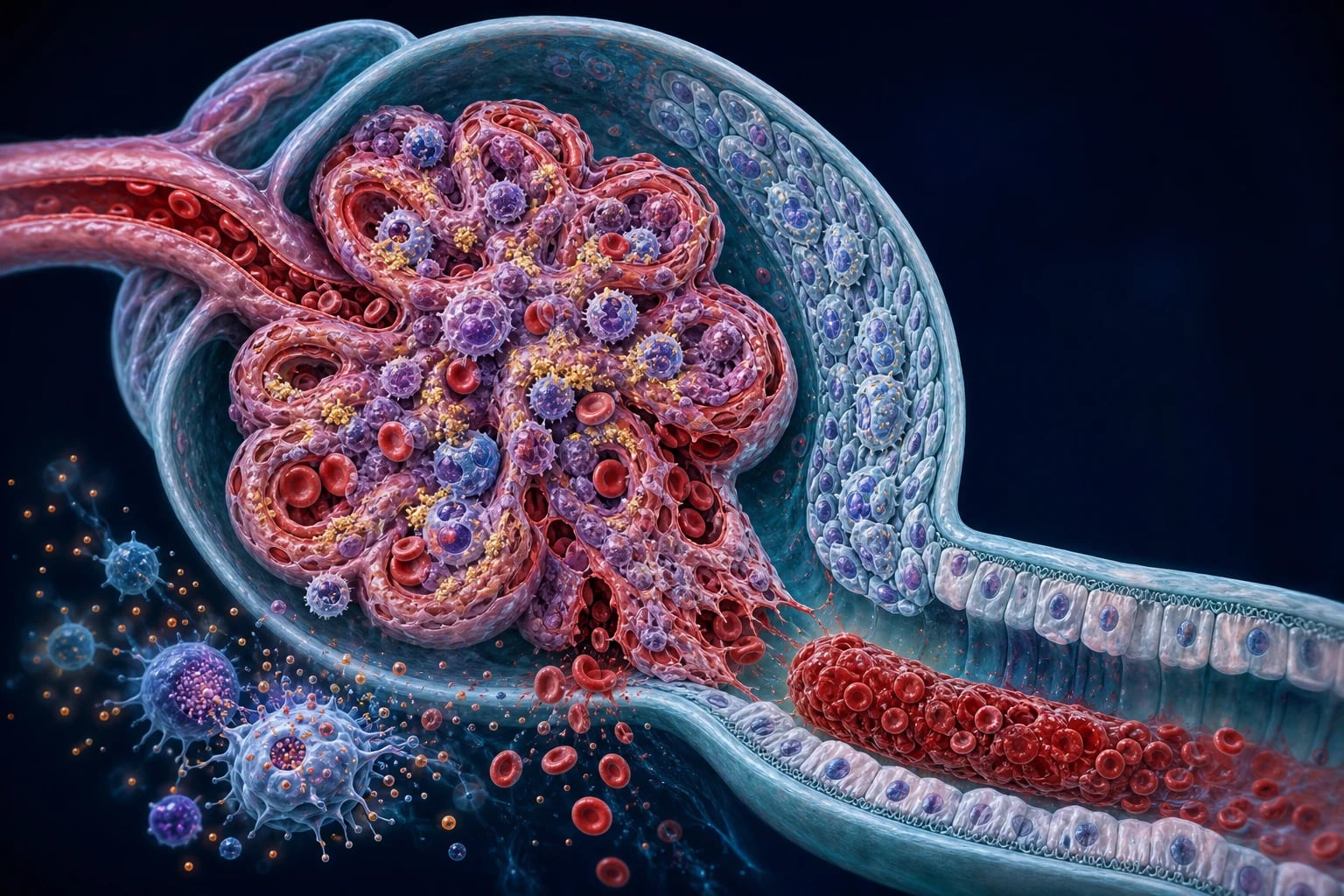
The normal filtration barrier has three layers: a fenestrated endothelium (charge and size selectivity via glycocalyx), the glomerular basement membrane (GBM) rich in type IV collagen (alpha-3, -4, -5 chains — the target of anti-GBM disease), and podocyte foot processes joined by the slit diaphragm (nephrin, podocin). In nephritic syndromes the injury is to the endothelium, mesangium and GBM rather than to the podocyte (as in nephrotic syndrome): immune-complex deposition, complement activation, leucocyte infiltration and cellular proliferation physically rupture the capillary wall, allowing red cells to pass through and form casts in the tubule. [1]
Post-streptococcal GN — the archetype of immune-complex proliferative GN
The classical sequence in PSGN is: [1]
- Nephritogenic streptococcal infection (pharyngitis or pyoderma) with circulating SpeB and NAPlr antigens.
- Antigenaemia leads to formation of immune complexes that deposit in glomeruli, especially in situ in the subepithelial space, with cross-linking by antibodies forming large subepithelial humps.
- Complement activation — predominantly via the alternative and lectin pathways, with consumption of C3 (C3 is low; early classical components C1q and C4 are typically normal or only mildly reduced).
- Inflammation and proliferation — neutrophils and macrophages infiltrate the tuft, mesangial and endocapillary cells proliferate, producing a diffuse endocapillary proliferative pattern.
- Capillary-wall disruption lets red cells and red-cell casts enter the urine and subnephrotic protein leak; the inflamed glomerulus has reduced GFR (oliguria, AKI) and avidly reabsorbs sodium (oedema, hypertension).[1][2]
Complement C3 typically normalises within 6 to 8 weeks as the immune complexes are cleared; persistence beyond this signals an alternative diagnosis (MPGN, C3 glomerulopathy, lupus). [1]
IgA nephropathy — the multi-hit hypothesis
The currently accepted four-hit pathogenesis explains why IgA nephropathy runs in families and in certain ethnic groups: [1]
- Hit 1 — production of galactose-deficient IgA1 (Gd-IgA1), an aberrantly glycosylated IgA1 hinge-region O-glycan (a heritable trait).
- Hit 2 — formation of autoantibodies (IgG or IgA) that recognise the exposed GalNAc residues on Gd-IgA1.
- Hit 3 — formation of large circulating immune complexes (Gd-IgA1 plus autoantibody) that deposit in the mesangium.
- Hit 4 — mesangial cell activation with release of cytokines (IL-6, TNF-alpha), chemokines, and growth factors, driving mesangial proliferation, extracellular matrix expansion, podocyte injury and oxidative stress — the histological lesion. [1]
The same mechanism underlies IgA vasculitis (Henoch-Schonlein purpura), in which IgA immune complexes deposit systemically in skin, gut, joint and kidney, producing the characteristic tetrad. [1]
Anti-GBM disease — the linear antibody
Autoantibodies (IgG, rarely IgA) target the non-collagenous domain of the alpha-3 chain of type IV collagen (alpha3(IV)NC1), an antigen sequestered in the mature GBM and alveolar basement membrane. Binding produces linear IgG deposition along the GBM on immunofluorescence, activates complement (C3) and triggers Fc-receptor-mediated neutrophil and macrophage injury that ruptures the capillary wall, causing crescent formation and (in the lung) alveolar haemorrhage. The disease is the prototype of a tissue-fixed antibody disease — hence the central role of plasma exchange to remove circulating antibody.[3][14]
ANCA-associated vasculitis — the pauci-immune, NET-driven lesion
ANCA are IgG autoantibodies against neutrophil granule proteins: anti-MPO (perinuclear p-ANCA) and anti-PR3 (cytoplasmic c-ANCA). The pathogenesis involves: [1]
- Priming of neutrophils by cytokines (IL-1, TNF) during infection or inflammation — moving MPO and PR3 to the cell surface.
- Binding of ANCA to surface antigens, with cross-linking and neutrophil activation via Fc-gamma receptors.
- Degranulation and NETosis — release of reactive oxygen species, proteolytic enzymes, and neutrophil extracellular traps (NETs) that damage the endothelium.
- Necrotising, crescentic GN with little or no immune deposition (pauci-immune on IF), and (depending on subtype) granulomatous inflammation in the upper and lower respiratory tract (GPA) or asthma and eosinophilia (EGPA).[15]
Drugs such as hydralazine, propylthiouracil and levamisole-adulterated cocaine can induce MPO-ANCA vasculitis, often with anti-histone and anti-MPO co-positivity. [1]
Lupus nephritis — full-house immune-complex deposition
In SLE, defective clearance of apoptotic debris leads to autoantibodies against nuclear antigens (dsDNA, nucleosomes, ribonucleoproteins). Resulting immune complexes deposit in the mesangium, subendothelium and subepithelium of the glomerulus, activating complement via the classical pathway (hence low C3 and C4) and producing full-house immunofluorescence (IgG, IgA, IgM, C3, C1q, C4). The ISN/RPS 2003 classification recognises six classes, of which class IV (diffuse proliferative) is the most nephritic and severe and class III (focal proliferative) is also nephritic; class V (membranous) is predominantly nephrotic but can overlap. [1]
Crescent formation — the histological hallmark of RPGN
Whatever the cause, severe capillary-wall injury ruptures the GBM, allowing fibrin, macrophages and plasma proteins to enter Bowman's space. This stimulates parietal epithelial cells (and podocytes that detach and adopt a proliferative phenotype) to divide, producing crescent-shaped cellular and fibrocellular masses that compress and ultimately obliterate the tuft. Crescents in over 50% of glomeruli defines RPGN; once fibrous (chronic), they are irreversible, which is why treatment must be rapid. [1]
Mechanism of hypertension, oedema and AKI
In nephritic syndromes the dominant mechanism of oedema is sodium retention from a reduced GFR with intact tubular reabsorption (the overfill mechanism), in contrast to the underfill mechanism (hypoalbuminaemia) of nephrotic syndrome. Hypertension results from volume expansion plus activation of the intrarenal renin-angiotensin-aldosterone system by ischaemic juxtaglomerular cells. AKI is glomerular in origin — the inflamed, proliferative tuft simply under-filters, producing oliguria and a rising creatinine. [1]
Clinical Presentation
Classical nephritic presentation
- Haematuria — most often macroscopic ('cola', 'tea', 'smoky' or frankly bloody urine) at presentation in IgA nephropathy and PSGN, but often only microscopic in lupus and ANCA vasculitis.
- Periorbital oedema, worse in the morning, and dependent (sacral, ankle) oedema.
- Hypertension — often mild to moderate but may be severe enough to cause hypertensive encephalopathy (headache, visual disturbance, seizures) especially in children with PSGN.
- Oliguria and a rise in serum creatinine (AKI).
- Lethargy, anorexia, malaise, sometimes flank discomfort. [1]
Temporal relationship to infection — a key discriminator
| Cause | Latent period from infection | Complement |
|---|---|---|
| IgA nephropathy | 0 to 2 days (synpharyngitic, with the URTI) | Normal |
| IgA vasculitis (HSP) | 1 to 2 weeks after a URTI | Normal |
| PSGN (pharyngitis) | 1 to 3 weeks | Low C3 |
| PSGN (pyoderma) | 2 to 4 weeks | Low C3 |
| Endocarditis/shunt GN | Weeks to months of bacteraemia | Low C3/C4 |
| Cryoglobulinaemia | Years of chronic HCV | Low C4 |
Cause-specific systemic clues
- PSGN — recent sore throat or skin infection;cola-coloured urine; periorbital oedema; hypertension.
- IgA nephropathy — painless gross haematuria within a day or two of an upper respiratory infection (synpharyngitic); flank discomfort.
- IgA vasculitis (HSP) — palpable purpura on extensor surfaces and buttocks, abdominal pain, arthritis, scrotal swelling (children).
- Lupus nephritis — malar rash, photosensitivity, oral ulcers, alopecia, arthritis, serositis, Raynaud, cytopenias.[6][7]
- Granulomatosis with polyangiitis (GPA) — sinusitis, nasal crusting, epistaxis, saddle-nose deformity, pulmonary nodules/cavities, conductive/sensorineural deafness, subglottic stenosis.
- Microscopic polyangiitis (MPA) — pulmonary haemorrhage (without granulomatous ENT disease), neuropathy, palpable purpura.
- EGPA (Churg-Strauss) — adult-onset asthma, eosinophilia, sinusitis, mononeuritis multiplex.
- Anti-GBM (Goodpasture) — rapidly progressive haemoptysis with renal failure (pulmonary-renal syndrome).
- Cryoglobulinaemia — palpable purpura, weakness, arthralgia, peripheral neuropathy, Raynaud; chronic hepatitis C.[16]
- Endocarditis-associated GN — fever, new or changing murmur, splinter haemorrhages, Osler nodes, Janeway lesions, emboli.
Atypical presentations
- Elderly — may present with pulmonary oedema, confusion from hypertensive encephalopathy, or unexplained AKI rather than visible haematuria; ANCA vasculitis is the dominant cause of crescentic GN in this group.
- Diabetics — may have a mixed nephritic-nephrotic picture that obscures an overlapping GN; the absence of diabetic retinopathy and rapid onset should prompt biopsy.
- Pregnant — pre-eclampsia can mimic nephritic syndrome (hypertension, oedema, AKI, proteinuria) but has normal complements and no RBC casts; lupus nephritis may flare, especially postpartum.
- Immunocompromised — atypical infections (fungal, mycobacterial, opportunistic viral) producing immune-complex GN.
- Children with classic PSGN — periorbital oedema, tea-coloured urine, hypertension; usually a benign self-limiting illness, but hypertensive emergency can occur. [1]
Pulmonary-renal syndromes — recognise early
The combination of haemoptysis (or rapidly progressive dyspnoea from alveolar haemorrhage) with rapidly progressive GN is a medical emergency. The two dominant causes are anti-GBM disease (Goodpasture) and ANCA-associated vasculitis (GPA, MPA); lupus and cryoglobulinaemia are rarer causes. Immediate management is plasma exchange plus high-dose steroids plus cyclophosphamide (or rituximab) — delay costs the kidney and the lung.[3][14][15]
Differential Diagnosis
The differential is organised first by complement and second by tempo and pattern. [1]
Low-complement causes
- Post-streptococcal GN — recent GAS infection; C3 low, normalises in 6 to 8 weeks; ASO and anti-DNase B raised; subepithelial humps on EM.
- Membranoproliferative GN (MPGN) — persistent low C3 (low C4 if immune-complex); tram-track double-contour GBM; associated with hepatitis B/C, lupus, monoclonal gammopathy.[8]
- C3 glomerulopathy — alternative pathway dysregulation; low C3 with normal C4; C3 nephritic factor and anti-factor H may be positive; recurrent after transplant.[9]
- Lupus nephritis — low C3 AND C4; ANA, anti-dsDNA; full-house IF; extra-renal lupus features.[6][7]
- Endocarditis-associated GN and shunt nephritis — chronic bacteraemia; positive blood cultures; treat the infection.
- Cryoglobulinaemia — low C4 (out of proportion to C3); palpable purpura; hepatitis C.[16]
Normal-complement causes
- IgA nephropathy — commonest primary GN; synpharyngitic haematuria; mesangial IgA-dominant deposits.
- IgA vasculitis (HSP) — palpable purpura, arthritis, abdominal pain, IgA nephritis.[10][11]
- Anti-GBM disease — anti-GBM antibody positive; pulmonary haemorrhage; linear IgG.[3][14]
- ANCA-associated vasculitis — anti-MPO/PR3 positive; ENT, lung, neuropathy.[15]
Distinguishing nephritic from urological and other causes of haematuria/AKI
- Urological causes of haematuria (stones, tumour, UTI, BPH) — no RBC casts, isomorphic (non-dysmorphic) RBCs, no proteinuria or complement changes; pain or clots in urine suggest a urological source.
- Acute interstitial nephritis — drug exposure (beta-lactams, NSAIDs, PPIs), eosinophiluria (historical), white-cell casts, fever, rash; no RBC casts or low complement.
- Acute tubular necrosis — ischaemic or nephrotoxic context; muddy-brown granular casts; no RBC casts.
- Pre-renal AKI — responsive to fluid; bland urine; no casts.
- Hypertensive emergency with renal failure — may mimic GN; look for RBC casts and complement to distinguish. [1]
C3 glomerulopathy vs immune-complex MPGN
Both look alike on light microscopy (membranoproliferative pattern, low C3). C3 glomerulopathy shows dominant C3 staining on IF (C3 intensity at least two orders of magnitude greater than any immunoglobulin) and is driven by alternative pathway dysregulation (C3 nephritic factor, anti-factor H, factor H/I deficiency). Immune-complex MPGN shows full immunoglobulin and complement staining and is driven by a chronic antigen load (hepatitis B/C, lupus) or monoclonal gammopathy. The distinction matters: C3G may respond to complement blockade (eculizumab, pegcetacoplan), while immune-complex MPGN requires treatment of the underlying disease.[8][9]
Clinical & Bedside Assessment
Focused examination
- Volume status and BP — accurate blood pressure (often raised), JVP, weight, and serial fluid balance; periorbital, sacral and pedal oedema; basal crackles and pulmonary oedema.
- Abdomen — hepatosplenomegaly, ascites (especially lupus with serositis), renal masses, palpable bladder.
- Skin — malar rash, palpable purpura (lower limbs in HSP, cryoglobulinaemia, ANCA), livedo reticularis, necrotising lesions (GPA, antiphospholipid), oral and nasal ulcers.
- ENT — sinus tenderness, nasal crusting, septal perforation, saddle nose (GPA); red tongue with strawberry (in PSGN from preceding pharyngitis).
- Chest — crackles (pulmonary oedema, pulmonary haemorrhage), pleural effusion, consolidations (GPA cavities).
- Cardiovascular — new or changing murmur (endocarditis), pericardial rub (lupus, uraemia).
- Neurological — focal deficits, mononeuritis multiplex (vasculitis), seizures or reduced GCS (hypertensive encephalopathy, lupus cerebritis).
- Fundoscopy — hypertensive retinopathy, cytoid bodies (lupus), choroiditis, Roth spots (endocarditis).
- Joints — synovitis (lupus, HSP, rheumatoid-associated vasculitis). [1]
Targeted history
- Recent infection — sore throat, skin sores, fever; onset-to-symptom interval.
- Drug history — hydralazine, propylthiouracil, allopurinol, penicillamine, sulfasalazine, minocycline, levamisole-cocaine, anti-TNF agents.
- Chronic infection — hepatitis B/C, HIV, endocarditis risk (IVDU, prosthetic valve, dialysis access).
- Systemic symptoms — rash, arthralgia, mouth ulcers, alopecia, photosensitivity, sinus disease, haemoptysis, abdominal pain, neuropathy.
- Travel, exposure, family history — TB exposure; family history of renal disease, deafness (Alport), autoimmune disease.
- Pregnancy — current or recent, pre-eclampsia, postpartum flare of lupus. [1]
Investigations
Urine — the cardinal specimen
| Test | Finding in nephritic syndrome |
|---|---|
| Dipstick | Blood 1+ to 4+ with protein (subnephrotic, under 3.5 g/day); leucocytes may be present |
| Phase-contrast microscopy | Dysmorphic red cells (especially acanthocytes) and red-cell casts — pathognomonic for glomerular bleeding |
| Protein quantification | Spot urine protein-to-creatinine ratio under 300 to 350 mg/mmol or 24-hour protein under 3.5 g (nephrotic-range suggests overlap) |
| Other | Absence of muddy-brown granular casts (ATN) and white-cell/eosinophil casts (AIN); no crystals or bacteria |
Red-cell casts are the single most discriminating microscopy finding: their presence confirms a glomerular source of bleeding and mandates a glomerular work-up. Their absence does not exclude a GN, especially in ANCA vasculitis where the lesion may be focal. [1]
Baseline bloods
- Urea, creatinine, eGFR — quantify AKI; trend daily.
- FBC — anaemia of chronic inflammation; PRCA in GPA (anti-PR3); eosinophilia in EGPA; thrombocytopenia in lupus.
- ESR/CRP — markedly raised in vasculitis, endocarditis.
- Albumin — low in nephrotic overlap.
- Electrolytes — hyperkalaemia, hyperphosphataemia, metabolic acidosis in AKI. [1]
The serological workup — splits the differential
Nephritic serology — the key panels
- Complement — C3 and C4 (and total haemolytic complement CH50 if C3/C4 normal but suspicion high). In PSGN, C3 is low and returns to normal by 6 to 8 weeks; persistent low C3 beyond 8 weeks is atypical and mandates biopsy to exclude MPGN/C3G, lupus, endocarditis.[1]
- Anti-streptococcal serology — ASO titre rises after pharyngitis (but not reliably after pyoderma) and anti-DNase B antibody rises after both; a rising titre over 2 to 3 weeks is more useful than a single value.
- Autoimmune — ANA, anti-dsDNA (high specificity for lupus), anti-Sm, complement; anti-C1q (lupus nephritis activity); lupus anticoagulant and anticardiolipin (antiphospholipid).
- ANCA with MPO and PR3 specificities — anti-PR3 (cytoplasmic c-ANCA) → GPA; anti-MPO (perinuclear p-ANCA) → MPA. Drug-induced ANCA is often high-titre p-ANCA with anti-MPO and anti-histone.
- Anti-GBM antibody — enzyme immunoassay against the alpha3(IV)NC1 antigen; positive in Goodpasture.[3]
- Infection screen — hepatitis B surface antigen, hepatitis C antibody with PCR if positive, HIV, cryoglobulins (with RF and C4), blood cultures and echocardiography if endocarditis suspected.
- Monoclonal gammopathy work-up — serum protein electrophoresis, serum free light chains, urine for Bence-Jones; consider bone marrow if abnormal (monoclonal Ig-related proliferative GN).
Renal biopsy — when and what it shows
Indications: [1]
- All adults with nephritic syndrome (unless classic self-limiting PSGN with rapid resolution).
- Any rapidly progressive GN or pulmonary-renal syndrome.
- Persistent low complement (over 8 weeks) or atypical features (heavy proteinuria, persistent renal dysfunction, no clear post-infectious trigger).
- Children with atypical PSGN, nephrotic-range proteinuria, or steroid-resistant disease. [1]
Histological patterns (light microscopy, immunofluorescence, electron microscopy): [1]
| Cause | Light microscopy | Immunofluorescence | Electron microscopy |
|---|---|---|---|
| PSGN | Diffuse endocapillary proliferative GN; neutrophils | Coarse granular IgG/C3; starry-sky | Subepithelial humps |
| IgA nephropathy | Mesangial (and endocapillary) proliferation | Mesangial IgA-dominant (with C3) | Mesangial electron-dense deposits |
| Anti-GBM | Crescents, necrotising lesions | Linear IgG along GBM | GBM disruption, no deposits |
| ANCA vasculitis | Necrotising crescentic GN, fibrinoid necrosis | Pauci-immune (little/no Ig) | No immune deposits |
| Lupus class IV | Diffuse endocapillary proliferation, wire-loop, crescents | Full-house (IgG, IgA, IgM, C3, C1q, C4) | Subendothelial, mesangial, subepithelial deposits |
| MPGN | Double-contour (tram-track) GBM, mesangial insertion | IgG/C3 granular (immune-complex) or C3-dominant (C3G) | Subendothelial and mesangial deposits |
| Cryoglobulinaemia | MPGN-like pattern, intracapillary thrombi | IgM, IgG, C3 | Subendothelial organised deposits |
| HSP/IgA vasculitis | Mesangial proliferation, crescents in severe | IgA-dominant mesangial | Mesangial deposits (identical to IgA nephropathy) |
Imaging
- Renal ultrasound — kidney size (normal to enlarged in acute GN; small and echogenic in chronic disease), exclude obstruction; small kidneys contraindicate aggressive immunosuppression.
- Chest X-ray / CT chest — pulmonary oedema, diffuse alveolar haemorrhage, cavitating nodules (GPA), pleural effusion.
- CT sinuses in suspected GPA.
- Echocardiography and blood cultures if endocarditis suspected. [1]
Special tests
- C3 nephritic factor and anti-factor H for suspected C3 glomerulopathy.
- ANCA subtyping (MPO, PR3); anti-GBM titre quantitation to monitor plasma exchange.
- Renal biopsy MEST-C score for IgA nephropathy prognostication (Mesangial hypercellularity, Endocapillary hypercellularity, Segmental sclerosis, Tubular atrophy/interstitial fibrosis, Crescents).
- ISN/RPS 2003 lupus nephritis class (I to VI) and activity/chronicity indices. [1]
Management — Resuscitation & General

Immediate ABC and life-threats
- Airway/Breathing — oxygen if hypoxic; secure the airway if massive pulmonary haemorrhage (anti-GBM/ANCA) with anaesthetic support.
- Circulation — IV access; treat hyperkalaemia (calcium gluconate 10 mL of 10% IV over 5 to 10 min for membrane stabilisation; insulin 10 units with 25 to 50 g of 50% dextrose IV; salbutamol 10 to 20 mg nebulised; sodium bicarbonate if acidotic; calcium resonium or RRT for removal).
- Pulmonary oedema — sit upright, high-flow oxygen, IV furosemide 40 to 80 mg, nitrates if hypertensive, consider non-invasive ventilation and RRT if refractory. [1]
Hypertensive emergency
Reduce BP gradually — no more than 25% in the first hour, then to 160/100 mmHg over 2 to 6 hours to avoid cerebral and coronary hypoperfusion. Agents: [1]
- IV labetalol 20 to 80 mg bolus every 10 min, or infusion 0.5 to 2 mg/min.
- IV nicardipine infusion 5 to 15 mg/h (titrate).
- IV clevidipine infusion 1 to 16 mg/h.
- Oral nifedipine long-acting or amlodipine for urgent but non-emergency hypertension.
- AVOID sublingual nifedipine (erratic absorption, precipitous hypotension, stroke risk).
- In eclampsia, IV magnesium sulphate 4 g loading then 1 to 2 g/h. [1]
Fluid overload and oedema
- Salt restriction under 2 g/day; fluid restriction to previous day's urine output plus 500 mL insensible losses.
- Loop diuretic — oral furosemide 40 mg daily, titrate to weight; IV furosemide 40 to 80 mg if severe or gut oedema.
- Add a thiazide (e.g. metolazone 2.5 to 5 mg) or amiloride for resistant oedema.
- Dialysis for refractory pulmonary oedema, oligoanuric AKI, or hyperkalaemia (AEIOU criteria below). [1]
Indications for urgent renal replacement therapy — AEIOU
When to dialyse in AKI — AEIOU
AEIOU
Refractory metabolic acidosis (pH under 7.1) unresponsive to bicarbonate
Refractory hyperkalaemia (K+ over 6.5 mmol/L) unresponsive to medical therapy
Toxins or drug overdose amenable to dialysis (lithium, salicylate, metformin)
Refractory volume overload (pulmonary oedema unresponsive to diuretics)
Symptomatic uraemia — pericarditis, encephalopathy, uraemic bleeding
Supportive care for all causes
- Treat the precipitating infection — antibiotics for PSGN do not change the GN course once established (the immune injury is already underway), but they treat the source, prevent rheumatic fever and limit spread. Use oral penicillin V (or benzathine penicillin IM) for GAS pharyngitis; erythromycin or clindamycin if penicillin-allergic.
- Bed rest during the acute phase; daily monitoring of weight, BP, urine output, electrolytes, creatinine.
- Nutrition — adequate calories, controlled protein (0.8 g/kg/day) and potassium/phosphate restriction in AKI.
- VTE prophylaxis if immobilised or nephrotic-range proteinuria.
- Infection prophylaxis during immunosuppression — Pneumocystis jirovecii prophylaxis with trimethoprim-sulfamethoxazole 480 mg daily (or 960 mg three times weekly); antifungal and antiviral as needed; screen for latent TB, hepatitis B/C, HIV before immunosuppression; vaccinate (pneumococcal, influenza, hepatitis B) where possible. [1]
Management — Definitive & Stepwise
Definitive treatment is cause-specific. The principle: PSGN is supportive and self-limiting; IgA nephropathy uses ACEi/ARB-led supportive care with immunosuppression for high-risk disease; RPGN (anti-GBM, ANCA, severe immune-complex) requires urgent induction immunosuppression. [1]
The supportive care bundle for ALL glomerular disease (KDOQI/KDIGO)
- RAAS blockade — ACE inhibitor (e.g. ramipril 2.5 to 10 mg daily) or ARB (e.g. losartan 50 to 100 mg daily) to reduce proteinuria and intraglomerular pressure; titrate to BP and potassium.
- BP target — under 130/80 mmHg (under 125/75 mmHg if proteinuric), per KDIGO.
- SGLT2 inhibitor — dapagliflozin 10 mg daily (or empagliflozin) for proteinuric CKD; reduces proteinuria and slows progression.
- Statin for hyperlipidaemia and cardiovascular risk.
- Smoking cessation, weight management, salt restriction.
- Vaccination (influenza annually, pneumococcal, hepatitis B). [1]
IgA nephropathy — supportive-first, then risk-stratified immunosuppression
- Optimised supportive care for at least 90 days — ACEi/ARB titrated, BP target under 130/80 mmHg, SGLT2 inhibitor, salt restriction, smoking cessation, weight, statin.
- If persistent proteinuria over 0.75 to 1 g/day despite optimised supportive care: a 6-month course of glucocorticoids (KDIGO 2021/2024, informed by TESTING and STARPROM) — typically prednisolone 0.6 to 0.8 mg/kg/day tapering over 6 months, with gastroprotection, bone protection and PJP prophylaxis; weigh the risk of infection, hyperglycaemia and bone toxicity.
- Targeted-release budesonide (Nefecon) — locally acting steroid in the ileal Peyer patches, reducing Gd-IgA1 production; for persistent proteinuria despite maximal supportive care.
- Sparsentan (dual endothelin-A and angiotensin-II type-1 antagonist; PROTECT trial) and complement inhibitors (avacopan C5aR inhibitor; iptacopan factor B inhibitor) for selected patients.
- Fish oil (omega-3 fatty acids) has modest, debated efficacy and is widely used because of low toxicity. [1]
ANCA-associated vasculitis — induction
- Glucocorticoid pulse — methylprednisolone 500 to 1000 mg IV daily for 3 days (avoid in severe infection; consider reduced dose in elderly).
- Then oral prednisolone 1 mg/kg/day (max 60 to 80 mg), tapering over months to a maintenance dose under 5 mg/day.
- PLUS rituximab 375 mg/m² IV weekly for 4 weeks (RAVE trial showed non-inferiority to cyclophosphamide and superiority for relapsing disease), OR cyclophosphamide 15 mg/kg IV every 2 weeks for 3 doses then every 3 weeks (6-month course, dose-reduce for age and renal function).[4][15]
- Avacopan (C5a receptor inhibitor) 30 mg twice daily can allow earlier steroid taper in selected patients (ADVOCATE trial).
- Plasma exchange (MEPEX regimen) for severe renal failure (creatinine over 5.7 mg/dL or dialysis-dependence) or diffuse alveolar haemorrhage — 7 to 14 exchanges of 60 mL/kg over 2 to 4 weeks.[5]
Anti-GBM disease — combination of plasma exchange, steroids and cyclophosphamide
- Plasma exchange — daily or alternate-day, 4 L exchanges with 5% human albumin and fresh frozen plasma if bleeding; continue until anti-GBM antibody undetectable (typically 2 to 3 weeks).
- Methylprednisolone pulse 500 to 1000 mg IV daily for 3 days, then oral prednisolone 1 mg/kg/day tapering over months.
- Cyclophosphamide 2 mg/kg/day orally (dose-reduce for age and renal function) for 2 to 3 months.
- Avoid late transplantation until anti-GBM antibody has been undetectable for at least 6 months.[3][14]
Lupus nephritis class III/IV — induction and maintenance
- Induction (3 to 6 months): mycophenolate mofetil (MMF) 2 to 3 g/day (target 3 g/day) OR low-dose IV cyclophosphamide (Euro-Lupus regimen 500 mg every 2 weeks for 6 doses), PLUS glucocorticoids (methylprednisolone pulse then prednisolone 0.5 to 0.8 mg/kg/day tapering).[6]
- Add voclosporin 23.7 mg twice daily (AURORA 1 and 2 trials showed higher renal response with voclosporin triple therapy) or tacrolimus as triple therapy.[7]
- Hydroxychloroquine 200 to 400 mg daily for all lupus patients (improves renal and overall outcomes).
- Maintenance (after induction): MMF 1 to 2 g/day or azathioprine 1 to 2 mg/kg/day plus low-dose prednisolone (target under 5 mg/day); consider rituximab for refractory disease.
- Class V (membranous) lupus nephritis with nephrotic-range proteinuria treated as membranous nephropathy (calcineurin inhibitor or MMF plus steroids).
Crescentic immune-complex GN
Treat the underlying disease (lupus, post-infectious with crescents, severe IgA) with glucocorticoid pulse plus cyclophosphamide or mycophenolate, as for the parent disease. [1]
C3 glomerulopathy and MPGN
- Supportive care (ACEi/ARB, BP, SGLT2 inhibitor).
- Mycophenolate for proteinuria and progressive decline.
- Complement blockade — eculizumab (anti-C5) or pegcetacoplan (anti-C3) in selected patients with progressive disease and confirmed alternative pathway dysregulation.[9]
- Treat the underlying cause of immune-complex MPGN — antivirals for hepatitis B/C, treat lupus, treat monoclonal gammopathy (clone-directed therapy).
Cryoglobulinaemic vasculitis
- Hepatitis C-related — direct-acting antivirals (DAAs) achieve sustained virological response in most; severe vasculitis adds rituximab ± plasma exchange.[16]
- Essential and non-HCV — rituximab plus steroids; plasma exchange for crisis (hyperviscosity, severe renal failure, neuropathy).
Maintenance, prophylaxis and relapse
- ANCA vasculitis maintenance — rituximab 1 g at 0 and 2 weeks, then every 4 to 6 months for 18 to 24 months; or azathioprine 2 mg/kg/day. Relapse (rising BVAS) triggers re-induction.
- Lupus nephritis maintenance — MMF or azathioprine plus low-dose steroid; hydroxychloroquine indefinitely; renal flare (rising proteinuria, falling complement, active sediment) triggers re-induction.
- PJP prophylaxis — trimethoprim-sulfamethoxazole 480 mg daily (or 960 mg three times weekly) for the duration of high-dose steroids, cyclophosphamide or rituximab.
- Mesna and forced hydration with high-dose cyclophosphamide to prevent haemorrhagic cystitis; PCV and cervical screening (cyclophosphamide carcinogenicity); fertility preservation (sperm/embryo banking) where possible. [1]
Specific Subtypes & Scenarios
Post-streptococcal GN (PSGN)
- Presentation: 1 to 3 weeks after GAS pharyngitis or 2 to 4 weeks after pyoderma in a child (or adult); periorbital oedema, cola-coloured urine, hypertension, AKI.
- Investigation: low C3 returning to normal by 6 to 8 weeks; raised ASO and anti-DNase B; urine with dysmorphic RBCs and RBC casts; biopsy (rarely needed) shows diffuse endocapillary proliferative GN with subepithelial humps.
- Management: supportive — fluid and sodium balance, BP control, loop diuretic for oedema; treat the streptococcal infection with penicillin; dialysis rarely needed.
- Prognosis: excellent in children (over 95% complete recovery; microscopic haematuria may persist for up to a year); adults have a higher rate of residual renal impairment, especially elderly diabetics. Atypical features (heavy proteinuria, persistent low C3 beyond 8 weeks, rapidly progressive course) warrant biopsy.[1][2]
IgA nephropathy (Berger disease)
- Presentation: synpharyngitic gross haematuria in a young adult; or incidental microscopic haematuria with mild proteinuria; occasionally RPGN with crescents.
- Investigation: normal complement; mesangial IgA-dominant deposits on biopsy; MEST-C score for prognosis.
- Management: ACEi/ARB + SGLT2 inhibitor for proteinuria; 6-month steroids or targeted-release budesonide for persistent proteinuria over 0.75 to 1 g/day despite 90 days of supportive care; sparsentan or complement inhibitors in selected cases.
- Prognosis: variable — 20 to 40% progress to ESKD over 20 to 30 years; predictors are sustained proteinuria over 1 g/day, hypertension, reduced eGFR at diagnosis, MEST-C high-score lesions. [1]
IgA vasculitis (Henoch-Schonlein purpura)
- Presentation: palpable purpura on extensor surfaces and buttocks, arthritis, abdominal pain (intussusception risk), and renal disease with IgA nephritis; children under 10.
- Investigation: normal complement; serum IgA may be raised; biopsy of skin (leucocytoclastic vasculitis with IgA deposition) or kidney (IgA-dominant mesangial deposits).
- Management: supportive in most; steroids for severe GI disease, severe arthritis, or nephritis with proteinuria over 1 g/day or crescents; severe nephritis treated as IgA nephropathy (ACEi/ARB, steroids, sometimes immunosuppression).
- Prognosis: usually self-limiting in children; adults have higher risk of CKD; recurrence after transplant is recognised.[10][11]
Rapidly progressive GN (RPGN) — the renal emergency
- Definition: loss of over 50% of GFR within under 3 months, with crescents on biopsy.
- Types: Type I anti-GBM, Type II immune-complex (lupus, post-infectious, IgA), Type III pauci-immune (ANCA).
- Management: urgent — methylprednisolone pulse plus cyclophosphamide or rituximab; plasma exchange for anti-GBM and severe ANCA; supportive care and RRT if needed; biopsy should not delay immunosuppression if the picture is classic. [1]
Lupus nephritis
- Class IV diffuse proliferative is the most nephritic and most severe; class III focal proliferative is also nephritic.
- Class V membranous is predominantly nephrotic but can overlap with III or IV.
- Investigation: low C3 AND C4, ANA, anti-dsDNA, anti-Sm; biopsy with ISN/RPS 2003 classification and activity/chronicity indices.
- Management: induction with MMF or cyclophosphamide plus steroids and voclosporin/tacrolimus; maintenance with MMF or azathioprine plus low-dose steroid; hydroxychloroquine throughout.[6][7]
ANCA-associated vasculitis — the three subtypes
| Subtype | ANCA | Hallmark |
|---|---|---|
| GPA (Wegener) | PR3 (c-ANCA) | Granulomatous ENT + lung disease, saddle nose, sinusitis, nodules/cavities |
| MPA | MPO (p-ANCA) | Renal-predominant, pulmonary haemorrhage without granulomatous ENT disease, neuropathy |
| EGPA (Churg-Strauss) | MPO in ~40% | Adult-onset asthma, eosinophilia, sinusitis, mononeuritis multiplex |
Induction with rituximab or cyclophosphamide plus steroids; plasma exchange for severe renal failure or pulmonary haemorrhage; PJP prophylaxis throughout.[4][15]
Membranoproliferative GN and C3 glomerulopathy
- Immune-complex MPGN — hepatitis B/C, lupus, monoclonal gammopathy; full immunoglobulin and complement staining; treat the underlying cause.
- C3 glomerulopathy — alternative pathway dysregulation (C3 nephritic factor, factor H/I deficiency); C3-dominant staining with little immunoglobulin; low C3 with normal C4; risk of recurrence after transplant; complement blockade (eculizumab, pegcetacoplan) in selected cases.[8][9]
Cryoglobulinaemic vasculitis
- Type II mixed (IgM kappa with RF activity against polyclonal IgG) is classically associated with chronic hepatitis C.
- Tetrad: palpable purpura, weakness, arthralgia, neuropathy; low C4 out of proportion to C3; renal biopsy shows MPGN-like pattern with intracapillary thrombi.
- Management: DAAs for HCV; rituximab and plasma exchange for severe disease.[16]
Endocarditis- and shunt-associated GN
- Chronic bacteraemia drives immune-complex deposition; low C3 and C4.
- Treat the infection — antibiotics and (for endocarditis) valve surgery; for shunt nephritis, removal of the infected shunt. [1]
Complications & Pitfalls
Acute complications
- Severe hypertension — hypertensive encephalopathy, seizures, posterior reversible encephalopathy syndrome (PRES), intracerebral haemorrhage.
- Pulmonary oedema and AKI requiring dialysis.
- Pulmonary haemorrhage in anti-GBM and ANCA disease — can be catastrophic.
- Thrombosis — renal vein thrombosis in nephrotic overlap; venous thromboembolism with severe nephrotic-range proteinuria and immobilisation.
- Infection — from immunosuppression (PJP, herpes zoster, CMV reactivation, opportunistic fungal and mycobacterial infection); encapsulated organisms if nephrotic.
- Contrast nephropathy from imaging — weigh against the need; use iso-osmolar contrast with pre-hydration. [1]
Chronic complications
- Progression to CKD/ESKD — particularly anti-GBM with oligoanuria at presentation, severe ANCA, crescentic lupus, IgA with persistent proteinuria.
- Persistent hypertension, cardiovascular disease from CKD.
- Recurrent disease — IgA nephropathy (30 to 50% recurrence in transplant grafts but graft loss in only 5 to 10%), lupus (rare recurrence), ANCA (relapse rate 30 to 50% over 5 years), C3 glomerulopathy (high recurrence in transplant).
- Drug toxicity — cyclophosphamide (haemorrhagic cystitis, infertility, myelodysplasia, bladder cancer), steroids (diabetes, osteoporosis, avascular necrosis, cataract, infection), rituximab (hypogammaglobulinaemia, progressive multifocal leukoencephalopathy — rare), calcineurin inhibitors (nephrotoxicity, hypertension, diabetes), MMF (teratogenic, cytopenia). [1]
Classic pitfalls
- Assuming all nephritic presentations are PSGN and missing RPGN — any rapidly progressive AKI with haematuria mandates urgent work-up.
- Not checking complement in a young person with haematuria.
- Failing to biopsy a rapidly progressive AKI with haematuria and RBC casts.
- Delaying plasma exchange in anti-GBM disease with pulmonary haemorrhage.
- Giving high-dose immunosuppression without excluding infection — especially active TB, hepatitis B (reactivation), HIV, and untreated endocarditis.
- Forgetting PJP prophylaxis on cyclophosphamide/rituximab/high-dose steroids.
- Missing double-positive (ANCA + anti-GBM) disease — these should be managed as anti-GBM (add plasma exchange).
- Forgetting mesna with high-dose cyclophosphamide to prevent haemorrhagic cystitis.
- Treating a chronic lesion (fibrous crescents, small kidneys) aggressively — irreversible fibrosis will not respond and exposes the patient to immunosuppressive harm. [1]
Prognosis & Disposition
By cause
| Cause | Prognosis |
|---|---|
| PSGN (children) | Excellent; over 95% complete recovery; C3 normalises in 6 to 8 weeks; microscopic haematuria may persist for a year |
| PSGN (adults) | Worse than children; residual renal impairment in 10 to 20%, higher in elderly diabetics |
| IgA nephropathy | 20 to 40% progress to ESKD over 20 to 30 years; predictors: proteinuria over 1 g/day, hypertension, reduced eGFR, MEST-C score, persistent microscopic haematuria |
| Anti-GBM disease | Guarded; high ESKD and mortality if oligoanuric or dialysis-dependent at presentation; better if treated before dialysis |
| ANCA vasculitis | 5-year survival 70 to 80% with modern therapy; ESKD in 20 to 40% depending on severity; relapse 30 to 50% over 5 years |
| Lupus nephritis class IV | 10-year renal survival 80 to 90% with modern therapy; worse in non-adherent, non-white, antiphospholipid-positive patients |
| MPGN/C3G | Variable; 50% progress to ESKD over 10 years; high recurrence after transplant (C3G) |
| Cryoglobulinaemia | Depends on HCV control; antivirals have transformed the outlook |
| IgA vasculitis (HSP) | Usually self-limiting in children; adults have higher CKD risk |
Predictors of poor renal outcome
- Sustained heavy proteinuria (over 1 g/day) — the single most powerful predictor across all causes.
- Hypertension at presentation.
- Reduced eGFR at presentation.
- Crescents on biopsy (especially over 50% of glomeruli), and interstitial fibrosis and tubular atrophy.
- Older age, dialysis-dependence at presentation, oligoanuria.
- For lupus: high activity index, antiphospholipid antibodies, non-adherence. [1]
Indications for nephrology referral and admission
- Emergency admission: any suspected RPGN or pulmonary-renal syndrome (haemoptysis, rapidly progressive GN); severe hypertension or hypertensive encephalopathy; pulmonary oedema; hyperkalaemia.
- Urgent referral: persistent heavy proteinuria, rising creatinine, low complement, systemic features.
- Biopsy-driven management: all adults (unless classic self-limiting PSGN), any atypical or rapidly progressive course. [1]
Indications for renal replacement therapy and transplant
- ESKD requiring dialysis or transplant.
- Transplant suitable for most causes once disease is quiescent. Disease-specific considerations:
- Anti-GBM — wait 6 to 12 months after anti-GBM antibody is undetectable.
- ANCA — quiescent for 6 to 12 months on maintenance therapy.
- IgA nephropathy — recurrence in 30 to 50% of grafts but graft loss lower; recurrence usually late and slowly progressive.
- MPGN/C3 glomerulopathy — high recurrence (up to 50 to 80% for C3G).
- Lupus nephritis — recurrence rare (under 5%). [1]
Special Populations
Children
- PSGN is commonest and usually self-limiting — supportive care only; biopsy reserved for atypical features (low complement persisting beyond 8 weeks, nephrotic-range proteinuria, rapidly progressive course, no clear post-infectious trigger).
- IgA vasculitis (HSP) nephritis is treated as IgA nephropathy in severe cases (proteinuria over 1 g/day or crescents): ACEi/ARB, steroids, sometimes immunosuppression.
- Weight-based dosing of all drugs (mg/kg, max adult doses). [1]
Elderly
- ANCA vasculitis is the dominant cause of crescentic GN.
- Higher mortality from immunosuppression; reduce cyclophosphamide dose for age and renal function (e.g. Euro-Lupus low-dose regimen).
- Balance immunosuppression against infection and malignancy risk; consider rituximab-based regimens to avoid cumulative cyclophosphamide toxicity.
- Vasculitis in older patients may present non-specifically (weight loss, fatigue, raised inflammatory markers) — a high index of suspicion is needed. [1]
Pregnancy
- Lupus nephritis may flare, especially in the postpartum period.
- Pre-eclampsia mimics nephritic/nephrotic syndrome (hypertension, oedema, AKI, proteinuria) but has normal complements and no RBC casts, and uric acid is often high; serum FMS-like tyrosine kinase 1 (sFlt-1) is high and placental growth factor (PlGF) is low in pre-eclampsia.
- Switch ACEi/ARB (teratogenic) to labetalol, nifedipine, methyldopa for BP.
- Avoid mycophenolate and cyclophosphamide (teratogenic) — use azathioprine, tacrolimus, hydroxychloroquine and steroids.
- Antiphospholipid syndrome co-exists in some lupus patients — needs aspirin ± low-molecular-weight heparin. [1]
Diabetic patients
- May have superimposed glomerular disease (diabetic nephropathy plus another GN); biopsy if atypical (rapid onset, RBC casts, low complement, no diabetic retinopathy).
- SGLT2 inhibitor is the cornerstone of diabetic kidney disease and reduces proteinuria and progressive CKD.
- Avoid iodinated contrast in AKI where possible. [1]
Immunocompromised (HIV, transplant, post-chemotherapy)
- HIV-associated immune-complex kidney disease (HIVICK) and collapsing FSGS; treat the underlying HIV with antiretrovirals.
- Hepatitis B and C with MPGN, membranous or cryoglobulinaemic GN; antiviral therapy transforms the outlook.
- Transplant patients: drug interactions (calcineurin inhibitors with azoles, macrolides); consider transplant recurrence per disease. [1]
Anticoagulated patients
- Weigh bleeding risk against the need for renal biopsy (consider transjugular biopsy if INR not correctable).
- Warfarin interacts with steroids and azathioprine; DOACs require renal dose adjustment. [1]
Evidence, Guidelines & Regional Differences
Landmark trials
| Trial | Population | Finding |
|---|---|---|
| RAVE (Stone, NEJM 2010)[4] | ANCA vasculitis induction | Rituximab non-inferior to cyclophosphamide; superior in relapsing disease; cyclophosphamide-free induction |
| MEPEX (Jayne, JASN 2007)[5] | Severe ANCA with creatinine over 5.7 mg/dL | Plasma exchange better than methylprednisolone for renal recovery at 3 months |
| ALMS (Appel, JASN 2009)[6] | Lupus nephritis induction | MMF non-inferior to cyclophosphamide; higher response in non-white and Hispanic patients |
| AURORA 1 (Rovin, Lancet 2021)[7] | Lupus nephritis | Voclosporin plus standard-of-care increased renal response at 52 weeks; AURORA 2 confirmed durability at 2 years |
| TESTING (informed KDIGO 2024) | High-risk IgA nephropathy | Steroids reduced proteinuria and progression but with increased serious infection (dose modification matters) |
| PROTECT | IgA nephropathy | Sparsentan reduced proteinuria more than losartan |
| ADVOCATE | ANCA vasculitis | Avacopan (C5aR inhibitor) allowed glucocorticoid sparing with non-inferior remission |
Guidelines
- KDIGO 2021 Glomerular Diseases Guideline and the 2024/2025 KDIGO IgA Nephropathy Update — the international standard for evaluation and treatment, with the supportive-care-first philosophy for IgA and the immunosuppressive induction regimens for RPGN and lupus.[12][13]
- EULAR 2023 recommendations for the management of systemic lupus erythematosus and lupus nephritis (MMF or low-dose cyclophosphamide induction, MMF or azathioprine maintenance, hydroxychloroquine throughout, voclosporin or tacrolimus as triple therapy).
- ACR/VF 2021 Guideline for the management of ANCA-associated vasculitis (rituximab or cyclophosphamide induction, plasma exchange in severe disease).
- IPNA 2020 childhood glomerular disease recommendations.
Regional differences
- Globally: empiric supportive management for classic paediatric PSGN avoids biopsy unless atypical.
- India and resource-limited settings: endemic PSGN from skin infection (tropical climates); limited renal biopsy access; rising incidence of IgA nephropathy in young adults; cost and access constraints for rituximab, complement inhibitors (eculizumab, pegcetacoplan), sparsentan and voclosporin shape treatment choices; cyclophosphamide and steroids remain first-line for RPGN.
- High-income settings: broader use of rituximab, voclosporin, complement inhibitors, targeted-release budesonide, sparsentan.
- Hepatitis B/C-endemic regions: higher incidence of MPGN, membranous, and cryoglobulinaemic GN; direct-acting antivirals transform HCV-cryoglobulinaemia management. [1]
Exam Pearls
- Nephritic pentad: haematuria (dysmorphic RBCs and RBC casts), subnephrotic proteinuria, hypertension, oedema, AKI — from glomerular inflammation/proliferation.
- Complement split — PSLICE: Post-strep GN, SLE/lupus, subacute L (use Lupus), Infectious/shunt/endocarditis, Cryoglobulinaemia — all low-complement causes. Normal complement: IgA nephropathy, IgA vasculitis/HSP, anti-GBM, ANCA vasculitis.
- RBC casts = glomerular bleeding — the single most discriminating microscopy finding.
- PSGN: 1 to 3 weeks after pharyngitis, low C3, raised ASO, subepithelial humps, supportive and self-limiting in children. IgA: synpharyngitic, normal complement, mesangial IgA deposits.
- Crescents on biopsy = RPGN — a renal emergency. Type I anti-GBM (linear IgG, pulmonary haemorrhage, plasma exchange). Type II immune-complex. Type III pauci-immune (ANCA).
- Pulmonary-renal syndrome: anti-GBM (Goodpasture) and ANCA (GPA, MPA) — plasma exchange plus steroids plus cyclophosphamide or rituximab.
- Lupus nephritis class IV diffuse proliferative = most nephritic; low C3 AND C4, ANA/anti-dsDNA positive, full-house IF.
- ANCA: anti-PR3 = c-ANCA, GPA (sinuses, nose, lung cavities); anti-MPO = p-ANCA, MPA (renal-predominant, pulmonary haemorrhage, neuropathy). Treat with rituximab or cyclophosphamide plus steroids; plasma exchange if dialysis-dependent or pulmonary haemorrhage.
- Cryoglobulinaemia: low C4, palpable purpura, arthralgia, weakness, neuropathy, hepatitis C; treat with antivirals and rituximab.
- Crescentic anti-GBM: anti-alpha3(IV)NC1 antibody; do NOT transplant until antibody negative for 6 months.
- PJP prophylaxis (trimethoprim-sulfamethoxazole) on cyclophosphamide, rituximab or high-dose steroids; mesna with high-dose cyclophosphamide to prevent haemorrhagic cystitis.
- Plasma exchange is essential in anti-GBM disease (removes circulating antibody) and used in severe ANCA (dialysis-dependent or pulmonary haemorrhage).
- Complement normalises in 6 to 8 weeks in PSGN — persistence mandates biopsy.
- Sustained proteinuria over 1 g/day is the single most powerful predictor of progression in IgA nephropathy (and most GN). [1]
Exam application bank (NEET-PG / INICET)
One-line answer
Nephritic syndrome is the glomerular inflammatory counterpart of nephrotic syndrome: haematuria (dysmorphic red cells and red-cell casts), subnephrotic proteinuria (typically under 3.5 g/day), hypertension, oedema and an acute rise in creatinine (AKI), caused by glomerular inflammation and cellular proliferation. Causes are split by serum complement: low C3/C4 (post-streptococcal GN, membranoproliferative GN, lupus nephritis, endocarditis-associated GN, cryoglobulinaemia, shunt nephritis) versus normal complement (IgA nephropathy — the commonest primary GN worldwide, IgA vasculitis/Henoch-Schonlein purpura, anti-GBM disease/Goodpasture, ANCA-associated vasculitis). The pivotal clinical decision is distinguishing a self-limiting post-streptococcal GN (supportive care, excellent prognosis in children) from a renal emergency such as rapidly progressive (crescentic) GN, which destroys glom [1]
Worked stems (answer without another resource)
Stem 1 — Classic presentation. Map symptoms to mechanism; name the first investigation and first treatment step with dose/route if drug therapy is standard. [1]
Stem 2 — Unstable / complicated. List red flags that force immediate resuscitation, theatre, ICU, antidote, or reperfusion — and what you do in the first 15 minutes. [1]
Stem 3 — Atypical group. Elderly, pregnancy, child, or immunocompromised: how presentation and thresholds change. [1]
Stem 4 — Differential trap. Name the three closest mimics and one discriminator for each. [1]
Stem 5 — Disposition. Who goes home with safety-netting, who is admitted, who needs HDU/ICU/theatre, and what follow-up is mandatory. [1]
Rapid viva checklist
- Definition + classification
- Pathophysiology chain
- Bedside signs / criteria
- Score with exact components (if any)
- Emergency bundle
- Definitive therapy with doses
- Complications of disease and of treatment
- Special populations
- Guideline/trial name if classic
- Three exam traps
Coverage self-check
If you cannot answer any stem above from this page alone, re-read the matching section — the page is intended to be self-sufficient for final-prof and NEET-PG/INICET questions on Nephritic Syndrome & Acute Glomerulonephritis.
References
- [1]Balasubramanian R, Marks SD. Post-infectious glomerulonephritis Paediatr Int Child Health, 2017.PMID 28891413
- [2]Rodriguez-Iturbe B. Autoimmunity in Acute Poststreptococcal GN: A Neglected Aspect of the Disease J Am Soc Nephrol, 2021.PMID 33531351
- [3]Ponticelli C, Cresseri D, Tarantino A, Moroni G. Anti-glomerular basement membrane vasculitis Autoimmun Rev, 2023.PMID 36252931
- [4]Stone JH, Merkel PA, Spiera R, et al. Rituximab versus cyclophosphamide for ANCA-associated vasculitis N Engl J Med, 2010.PMID 20647199
- [5]Jayne DR, Gaskin G, Rasmussen N, et al. Randomized trial of plasma exchange or high-dosage methylprednisolone as adjunctive therapy for severe renal vasculitis J Am Soc Nephrol, 2007.PMID 17582159
- [6]Appel GB, Contreras G, Dooley MA, et al. Mycophenolate mofetil versus cyclophosphamide for induction treatment of lupus nephritis J Am Soc Nephrol, 2009.PMID 19369404
- [7]Rovin BH, Teng YKO, Ginzler EM, et al. Efficacy and safety of voclosporin versus placebo for lupus nephritis (AURORA 1): a double-blind, randomised, multicentre, placebo-controlled, phase 3 trial Lancet, 2021.PMID 33971155
- [8]Sethi S, Fervenza FC. Membranoproliferative glomerulonephritis: pathogenetic heterogeneity and proposal for a new classification Semin Nephrol, 2011.PMID 21839367
- [9]Bomback AS, Smith RJ, Barile GR, et al. Eculizumab in the treatment of membranoproliferative glomerulonephritis Nephron Clin Pract, 2014.PMID 25402185
- [10]Pillebout E, Jamin A, Aydin S, et al. IgA vasculitis Semin Immunopathol, 2021.PMID 34170395
- [11]McCarthy HJ, Tizard EJ. Clinical practice: Diagnosis and management of Henoch-Schönlein purpura Eur J Pediatr, 2010.PMID 20012647
- [12]Floege J, Barbour SJ, Cattran DC, et al. Management and treatment of glomerular diseases (part 1): conclusions from a Kidney Disease: Improving Global Outcomes (KDIGO) Controversies Conference Kidney Int, 2019.PMID 30665568
- [13]Rovin BH, Caster DJ, Cattran DC, et al. Management and treatment of glomerular diseases (part 2): conclusions from a Kidney Disease: Improving Global Outcomes (KDIGO) Controversies Conference Kidney Int, 2019.PMID 30665569
- [14]Reggiani F, Esposito P, Togliatto L, et al. Goodpasture syndrome and anti-glomerular basement membrane disease Clin Exp Rheumatol, 2023.PMID 36995324
- [15]Kronbichler A, Subramanian AK, Smith RM, Jayne DRW. Diagnosis and management of ANCA-associated vasculitis Lancet, 2024.PMID 38368016
- [16]Terrier B, Marie I, Lacraz A, et al. Non HCV-related infectious cryoglobulinemia vasculitis: Results from the French nationwide CryoVas survey and systematic review of the literature J Autoimmun, 2015.PMID 26320984