Nephrology · General Medicine
Nephrolithiasis (Kidney & Ureteric Stones)
Also known as Nephrolithiasis · Renal calculi · Kidney stones · Ureteric colic · Urolithiasis · Renal colic
Nephrolithiasis (renal and ureteric stones) is the formation of crystalline calculi within the urinary tract, driven by urinary supersaturation of stone-forming salts, and presenting classically as acute, severe colicky flank pain radiating from loin to groin with nausea, restlessness and haematuria. Calcium oxalate is the commonest stone type (~75%, radio-opaque); the other three pillars are uric acid (radio-lucent, acidic urine, gout, dissolvable with alkalinisation), struvite (urease-producing Proteus infection, alkaline urine, staghorn calculi) and cystine (autosomal-recessive cystinuria, hexagonal crystals). Risk factors cluster around low fluid intake, high sodium/animal-protein/fructose intake, obesity, gout, primary hyperparathyroidism, inflammatory bowel disease and renal tubular acidosis. Non-contrast CT KUB is the diagnostic standard (95 to 98 percent sensitive). Most stones under 5 mm pass spontaneously with NSAIDs, fluids and an alpha-blocker; larger or obstructing stones need ESWL, ureteroscopy with laser, or PCNL, chosen by size and site. Prevention rests on fluids over 2.5 to 3 L/day, low salt, low animal protein, normal dietary calcium, and targeted metabolic therapy (thiazide, potassium citrate, allopurinol, tiopronin). The single most dangerous scenario is an obstructing stone with infection, which destroys a kidney within hours and demands urgent decompression before definitive stone treatment.
On this page & tools
Your progress
Saved locally on this device.
Practise this topic
Exam tags
Red flags
Overview & Definition
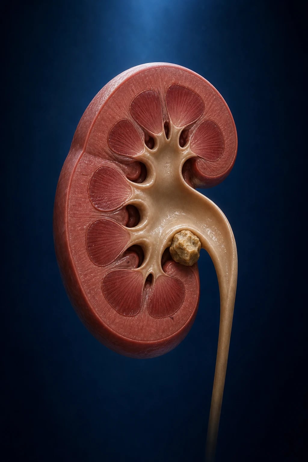
Nephrolithiasis (literally "stone in the kidney") and the broader term urolithiasis ("stone in the urinary tract") describe the formation of solid crystalline calculi anywhere along the urinary tract — in the renal parenchyma or calices (nephrolithiasis), the ureter (ureterolithiasis) or the bladder (cystolithiasis). Stones are not foreign bodies; they are biominerals precipitated from urine that has become supersaturated with respect to one of a handful of stone-forming salts.[1]
Clinically the disease matters for four reasons. First, an acute episode produces pain that patients rank among the most severe human beings experience. Second, an obstructing stone can destroy a kidney — acutely when combined with infection (pyonephrosis, urosepsis), and chronically through hydronephrosis and atrophy. Third, stones are highly recurrent: about half of untreated first-time stone formers will form another stone within ten years, so the clinician's job is not only to relieve the acute attack but to prevent the next one through targeted metabolic therapy. Fourth, stones are an independent risk factor for chronic kidney disease, hypertension and, in severe phenotypes (cystinuria, primary hyperoxaluria, struvite staghorn), end-stage renal failure.[1][3]
The clinical task in any patient with suspected renal colic is therefore a layered one: (1) control pain, (2) decide whether the stone is complicated (infection, AKI, solitary or transplanted kidney, bilateral obstruction, pregnancy) — because complicated stones are emergencies, (3) image and locate the stone, (4) choose a modality to clear it (conservative passage, ESWL, ureteroscopy, PCNL), and (5) initiate prevention by metabolic work-up and long-term measures.[2][4]
Classification
Stones are classified by mineral composition, which in turn predicts the radiographic appearance, the underlying metabolic defect, and the preventive therapy. The four classical types account for over 95 percent of all stones:[1]
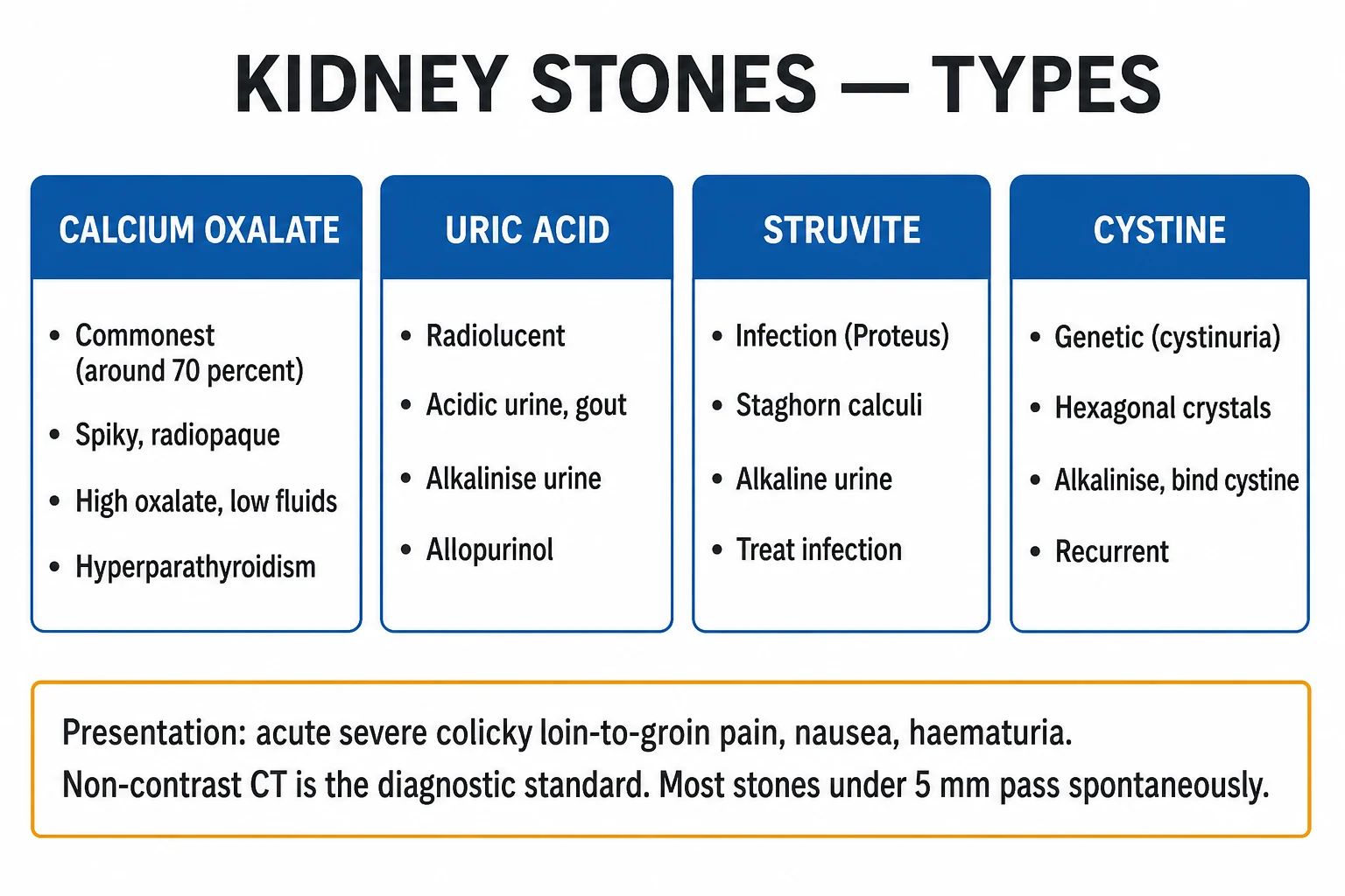
- Calcium stones (80 to 85 percent): predominantly calcium oxalate (most common), with a minority calcium phosphate (brushite). Radio-opaque. Driven by hypercalciuria, hyperoxaluria, hypocitraturia and a high urine pH in the calcium-phosphate subgroup.
- Uric acid stones (5 to 10 percent): radio-lucent on plain radiograph (low attenuation ~150 to 300 Hounsfield units on CT). Form in persistently acidic urine (pH under 5.5); associated with gout, myeloproliferative disease, rapid cell turnover, chronic diarrhoea and ileostomy, and metabolic syndrome. Uniquely dissolvable by alkalinising the urine — a key exam point.
- Struvite stones (10 to 15 percent): magnesium ammonium phosphate, produced when urease-splitting organisms (Proteus, Klebsiella, Providencia, Pseudomonas, Staphylococcus saprophyticus) hydrolyse urea to ammonia, raising urine pH above 7.2. Grow into staghorn calculi that fill the renal pelvis and calices. Common in women and recurrent-UTI patients.
- Cystine stones (~1 percent): hexagonal crystals, semi-opaque ("ground-glass") on plain film. Caused by autosomal-recessive cystinuria — a defect in the renal/intestinal dibasic-amino-acid transporter (genes SLC3A1, SLC7A9). Present from childhood; the cyanide-nitroprusside test is positive.
- Miscellaneous: drug stones (indinavir, triamterene, sulfadiazine, topiramate, excessive vitamin C → oxalate, excessive vitamin D and calcium → hypercalciuria) and rare matrix (proteinaceous) stones. [1]
Epidemiology & Risk Factors
The lifetime risk of a kidney stone in a developed country is 10 to 15 percent, and it is rising — driven by westernised diet, the obesity and diabetes epidemic, and warming climates. The peak age is 20 to 50 years and there is a male predominance of about 2:1 for calcium stones, though the gap is narrowing as female obesity and dietary patterns converge. Recurrence is the rule, not the exception: approximately 15 percent at 1 year, 30 to 40 percent at 5 years, and about 50 percent at 10 years in untreated patients — which is why a first stone always merits a preventive plan.[1]
Geographically, the global "stone belt" runs through hot, arid regions; in India the high-incidence belt spans the north and west (Gujarat, Rajasthan, Punjab, Delhi), attributable to heat, dehydration, dietary oxalate/animal protein, and in places fluoride/contaminated water. Stones are also more common in sedentary occupations, drivers, and those with limited daytime fluid access. [1]
Risk factors and why they matter
The dietary and metabolic risk factors to elicit at the bedside are: low fluid intake, high sodium (increases urinary calcium), high animal protein (acid load and uric-acid load), high fructose/sucrose, high dietary oxalate (spinach, rhubarb, nuts, chocolate, tea), and low or high dietary calcium — paradoxically, a low-calcium diet increases stone risk because intestinal calcium normally binds oxalate; without it, free oxalate is absorbed and excreted. Systemic conditions that predispose are obesity and metabolic syndrome, gout and hyperuricaemia, primary hyperparathyroidism, type 2 diabetes, renal tubular acidosis (especially distal/type 1 → calcium phosphate stones), inflammatory bowel disease and intestinal bypass (enteric hyperoxaluria, low citrate), chronic diarrhoea and ileostomy (acidic, concentrated urine → uric-acid stones), recurrent UTI with urease-producing organisms, medullary sponge kidney, hypercortisolism, and immobility (bone resorption → hypercalciuria). A positive family history and cystinuria complete the screen.[1][2]
Pathophysiology
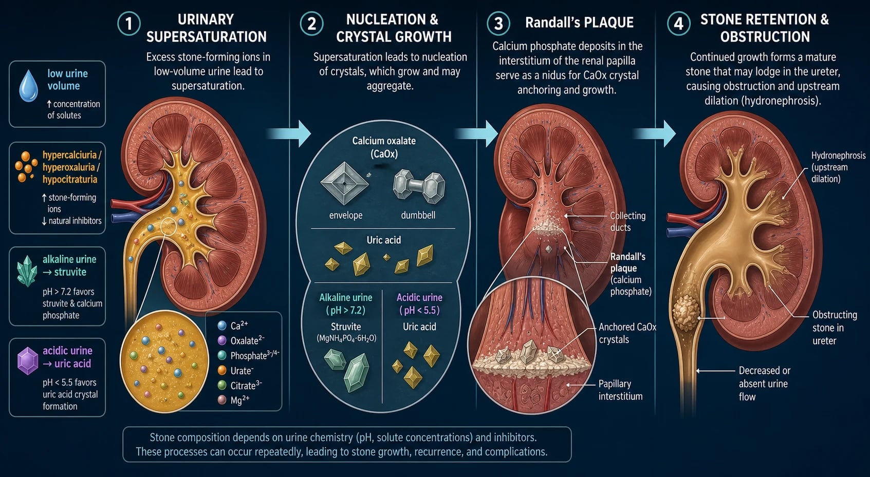
The unifying mechanism is urinary supersaturation. Urine holds dissolved ions in metastable solution; when the concentration of a stone-forming salt exceeds its formation product, ions leave solution, nucleate a solid crystal, and the crystal grows and aggregates. Healthy urine resists this because it contains inhibitors of crystallisation — citrate (which complexes calcium and reduces free ion activity), magnesium (complexes oxalate), pyrophosphate, Tamm-Horsfall protein (uromodulin) and nephrocalcin. Stones form when supersaturation overwhelms these inhibitors, or when inhibitors themselves are deficient (notably hypocitraturia).[1]
The stone phenotypes map onto distinct physicochemical failures: [1]
- Calcium oxalate — the dominant phenotype. Driven by hypercalciuria (the commonest abnormality, classified as absorptive, resorptive from hyperparathyroidism, or renal-leak), hyperoxaluria (dietary, enteric from fat malabsorption where unabsorbed fatty acids bind luminal calcium leaving oxalate free for absorption, or primary hyperoxaluria — an autosomal-recessive hepatic enzyme defect), and hypocitraturia. In the idiopathic majority, Randall's plaques — deposits of calcium phosphate (apatite) in the renal papillary interstitium — originate in the thin limbs of the loop of Henle, migrate to the papillary surface, erode through the urothelium, and serve as the nidus to which calcium-oxalate crystals adhere. The presence of Randall's plaques at ureteroscopy is the pathognomonic finding of idiopathic calcium-stone disease.[1]
- Calcium phosphate (brushite) — favours a persistently alkaline urine (high urine pH), as in distal (type 1) renal tubular acidosis and primary hyperparathyroidism; rTA also causes hypocitraturia, compounding risk.
- Uric acid — uric acid is the protonated, undissociated form of urate at a pH below its pKa of 5.5; below this pH it is poorly soluble and precipitates. The dominant driver is therefore persistently acidic urine (pH under 5.5), classically in gout, chronic diarrhoea/ileostomy (loss of alkali), myeloproliferative disorders and tumour-lysis, and increasingly metabolic syndrome (insulin resistance reduces renal ammoniagenesis, dropping urine pH). Because solubility rises steeply with pH, uric-acid stones are the one type that can be dissolved medically by alkalinising the urine to 6.5 to 7.0.
- Struvite (magnesium ammonium phosphate) — strictly an infection phenomenon. Urease-producing bacteria hydrolyse urinary urea (NH₂CONH₂) into two molecules of ammonia and one of carbon dioxide; the ammonia raises urinary pH above 7.2, and at this alkaline pH magnesium, ammonium and phosphate co-precipitate as struvite. The stone grows rapidly and layers itself around bacteria, which persist within it — hence the staghorn morphology and the need for complete surgical removal plus culture-directed antibiotics; medical dissolution is impossible. The classical organism is Proteus mirabilis; E. coli does not produce urease (a frequent MCQ trap).
- Cystine — an inherited defect (autosomal recessive) in the rBAT/b⁰⁺⁺ amino-acid transporter (genes SLC3A1 type A and SLC7A9 type B) in the proximal tubule and small intestine. The result is massive urinary loss of the dibasic amino acids cystine, ornithine, arginine and lysine (mnemonic COAL); cystine is the least soluble, precipitating at physiological urine pH. Disease presents in childhood and is relentlessly recurrent.
The pain of renal colic is visceral: ureteric spasm, distension of the renal capsule and pelvis, and increased peristaltic pressure stimulate C-fibre and A-delta visceral afferents that follow the sympathetic chains of T11 to L2. This segmental innervation explains the loin-to-groin radiation (T11–L2 dermatomes supply flank, groin and upper thigh) and the genital/scrotal or labial referred pain as the stone approaches the vesico-ureteric junction; distal-ureteric stones also produce urogenital and bladder symptoms (frequency, urgency, strangury) by irritating the trigone.[1]
Clinical Presentation
The classical presentation is acute ureteric colic: [1]
- Pain — sudden in onset, severe, fluctuating in colicky waves, felt in the flank/loin and radiating to the groin, testicle or labia. As a stone descends the pain migrates from loin to lower abdomen and finally to the groin/urethra. The patient is writhing, restless, unable to find a comfortable position — the single most useful bedside discriminator from peritoneal pain, where the patient lies motionless.
- Gastrointestinal — nausea and vomiting from visceral afferent stimulation (gastric plexus overlap); ileus is common.
- Urinary — macroscopic or microscopic haematuria (about 90 percent), and — when the stone sits at the vesico-ureteric junction — frequency, urgency, dysuria and strangury (the desire but inability to void).
- Systemic — typically afebrile. Fever, rigors or haemodynamic instability signal infection and convert the episode into an emergency. [1]
Atypical presentations are important and frequently examined: [1]
- Elderly — may complain only of diffuse abdominal pain, confusion, or a fall; an obstructed infected stone may present as delirium and sepsis without a clear pain history. Always image the older patient with unexplained sepsis of urinary origin.
- Diabetic / immunocompromised — blunted symptoms; risk of emphysematous pyelonephritis (gas-forming organisms in necrotic renal parenchyma), papillary necrosis, and a stone acting as a nidus for fungating infection. Minimal pain with severe systemic illness is the danger signal.
- Pregnant — renal colic is the commonest non-obstetric cause of abdominal pain requiring admission in pregnancy; it can mimic labour, trigger uterine contractions and preterm labour, and there is a right-sided predominance (dextrorotation and ureteric compression by the gravid uterus). Imaging and analgesia are constrained (see Special Populations).
- Children — nonspecific abdominal or flank pain, vomiting, isolated microscopic haematuria, or recurrent UTI; a thorough metabolic work-up is mandatory because an underlying cystinuria, primary hyperoxaluria or distal RTA is far more likely than in adults.
- Silent obstruction — a staghorn or pelvic stone may produce no colic and present late with CKD, hypertension or recurrent UTI. [1]
Differential Diagnosis
The differential of acute flank/loin pain is broad and missing a surgical or vascular emergency is the principal danger. Each candidate diagnosis has a distinguishing feature that breaks the pattern of renal colic:[1]
Ureteric colic (stone)
- Loin-to-groin, colicky waves, **writhing restless** patient
- Microscopic/macroscopic **haematuria** (~90%)
- Afebrile unless infected; CVA tenderness mild
- CT KUB shows the stone and hydronephrosis
Acute appendicitis
- Migratory **periumbilical → RLQ** pain, **lies still**, peritonism
- Anorexia, low-grade fever, raised inflammatory markers
- No haematuria; CT shows inflamed appendix
Acute pyelonephritis
- **Fever, rigors**, unwell, persistent (not colicky) loin pain
- Tender **CVA**, positive urine culture, leucocytes/nitrites on dipstick
- Usually no haematuria; no stone on imaging
Biliary colic / cholecystitis
- **RUQ** pain after fatty food, **Murphy's sign**
- No haematuria; ultrasound shows gallstones/thickened wall
Ectopic pregnancy / ovarian torsion / PID
- **β-hCG** mandatory in any woman of childbearing age
- Lower abdominal/pelvic pain, vaginal bleeding/discharge
- Pelvic ultrasound is diagnostic; torsion = surgical emergency
Testicular torsion
- Acute scrotum; **high-riding, horizontal lie**, **absent cremasteric reflex**
- Pain may radiate to groin/abdomen — always examine the **scrotum**
- 6-hour window — surgical emergency
Leaking/ruptured AAA
- Older male, **back/flank pain**, hypotension, **pulsatile abdominal mass**
- Can masquerade as renal colic — CT if any suspicion
Mesenteric ischaemia
- **Pain out of proportion**, metabolic acidosis, raised **lactate**
- Elderly with vascular disease/atrial fibrillation
Herpes zoster
- **Dermatomal** burning pain, normal urinalysis, vesicular rash appears within days
The non-negotiable rule: in any woman of childbearing age perform a β-hCG before CT, and in any patient examine the scrotum to exclude torsion — a stone work-up that misses an ectopic or a torsion is a catastrophic, classic error. [1]
Clinical & Bedside Assessment
History should establish: the onset, site, character and radiation of pain; prior stone episodes and family history; fluid and dietary habits (fluid volume, salt, animal protein, oxalate-rich foods, vitamin C/D/calcium supplements); bowel disease or surgery; gout, hyperparathyroidism or RTA; the drug list (topiramate, indinavir, sulfadiazine, triamterene, carbonic anhydrase inhibitors, loop diuretics, vitamin D, calcium); and systemic symptoms of infection. [1]
Examination: [1]
- Vital signs — fever, tachycardia and hypotension herald sepsis and the obstructed-infected system; do not be reassured by a normal blood pressure early.
- Abdomen — typically soft and non-rigid; there may be mild costovertebral-angle (CVA) tenderness. Guarding or rigidity redirects the diagnosis to a peritoneal/vascular cause.
- Genital examination — to exclude testicular torsion (acute scrotum, absent cremasteric reflex) and epididymo-orchitis.
- Rectal / pelvic examination — assess the prostate (BPH, prostatism) and exclude pelvic pathology.
- Hydration and sepsis screen — capillary refill, mucous membranes; calculate qSOFA if sepsis suspected. [1]
The distinguishing bedside sign is the writhing, restless patient (visceral pain) versus the motionless patient (peritoneal/somatic pain). CVA punch tenderness localises pathology to the upper urinary tract. [1]
Investigations
The diagnostic standard is non-contrast CT of the kidneys, ureters and bladder (CT KUB) — low-dose where possible. It is 95 to 98 percent sensitive and 96 to 98 percent specific, detects all stone types (including radio-lucent uric acid, which is seen as a low-attenuation focus rather than missed), defines size, location and degree of obstruction (hydronephrosis), and excludes competing diagnoses (appendicitis, AAA, diverticulitis). It is the first-line investigation in adults presenting with uncomplicated flank pain.[3]
Imaging — which test and when
Urinalysis and microscopy — dipstick shows haematuria in about 90 percent; the urine pH is diagnostic of type (pH under 5.5 → uric acid; pH over 7.2 with leucocytes/nitrites → struvite). Crystals on microscopy: envelope/dumbbell (calcium oxalate), coffin-lid (struvite), hexagonal (cystine — the pathognomonic crystal), and rhomboid/needle (uric acid). Sterile pyuria with a sterile culture raises the possibility of renal tuberculosis or partially treated infection. [1]
Bloods — urea, electrolytes and creatinine (renal function — essential before contrast and before NSAIDs), full blood count (leucocytosis = infection), CRP, corrected calcium and phosphate (hypercalcaemia → measure PTH), urate, bicarbonate and chloride (hyperchloraemic metabolic acidosis with alkaline urine → distal RTA), and glucose. [1]
Stone analysis — every passed or retrieved stone should be sent for infrared spectroscopy or X-ray diffraction, the definitive composition test that anchors preventive therapy. [1]
Metabolic work-up — indicated in recurrent stone formers, a strong family history, children, bilateral or staghorn stones, a solitary kidney, gout, or any systemic predisposition. Perform two 24-hour urine collections (volume, calcium, oxalate, citrate, urate, sodium, phosphate, and a cystine screen) plus a cystine screen (cyanide-nitroprusside test) if cystinuria is suspected, and serum calcium/PTH. The goal is a biochemical phenotype (e.g. hypercalciuric, hypocitraturic, hyperuricosuric) that dictates targeted drug therapy.[2][3]
The intravenous urogram (IVU) is now largely historical, replaced by CT. A plain KUB film still has a role in follow-up of known radio-opaque stones and in assessing stone fragmentation after lithotripsy. [1]
Management — Resuscitation

Resuscitation begins with ABCs and an immediate judgement: is this a complicated stone? The complicated stone is an emergency and is defined by any of: fever/sepsis, acute kidney injury, a solitary or transplanted obstructed kidney, bilateral obstruction, intractable pain or vomiting, or pregnancy with obstruction. These patients need admission, antibiotics and decompression, not analgesia and waiting.[3][4]
Pain control — NSAIDs are first-line and are non-inferior to opioids while causing less vomiting and fewer rescue doses:[2]
- Diclofenac 75 mg IM or IV, or ketorolac 30 mg IV — reduce renal pelvic pressure and ureteric spasm via prostaglandin inhibition.
- Morphine 5 to 10 mg IV (or fentanyl) — reserve for when NSAIDs are contraindicated (chronic kidney disease, active peptic ulcer, third-trimester pregnancy, aspirin-sensitive asthma, hypovolaemia) or inadequate. [1]
Anti-emetic — ondansetron 4 mg IV or metoclopramide 10 mg IV for nausea/vomiting. [1]
Hydration — oral if tolerated; IV isotonic crystalloid if vomiting or dehydrated. Note: aggressive IV hydration does not accelerate stone passage; the goal is euvolaemia, not forced diuresis. [1]
Septic/obstructed system — the urological emergency: [1]
- Blood and urine cultures, IV broad-spectrum antibiotics (e.g. co-amoxiclav 1.2 g IV plus gentamicin, or piperacillin-tazobactam 4.5 g IV per local policy), and resuscitation.
- Urgent urology referral — the obstruction must be decompressed before definitive stone treatment. Options are a retrograde JJ stent (placed cystoscopically under anaesthetic) or a percutaneous nephrostomy (radiologically guided). Do not perform ESWL or ureteroscopy in the face of untreated infection.
- Definitive stone removal is undertaken once the patient is afebrile and cultures are clear. [1]
AKI from bilateral obstruction or a solitary obstructed kidney likewise needs urgent decompression — obstruction relief is the treatment. Strain all urine through a filter to capture any passed stone for analysis. [1]
Management — Definitive & Stepwise
Definitive management is decided by stone size, site, composition and patient factors, and is structured in three tiers — conservative/medical expulsive therapy, lithotripsy/endoscopy, and percutaneous surgery.[4]
1. Conservative and medical expulsive therapy (MET)
The probability of spontaneous passage is size-dependent: stones under 5 mm pass in about 80 percent of cases, 5 to 7 mm in about 60 percent, 7 to 10 mm in 30 to 50 percent, and stones over 10 mm rarely pass (under 10 percent). For stones under 10 mm with controlled pain and no complication, offer MET: [1]
- Adequate analgesia (NSAIDs ± opioids) and hydration.
- Alpha-blocker — tamsulosin 0.4 mg nocte for up to 4 weeks. Alpha-blockers relax the smooth muscle of the distal ureter (α1-adrenergic receptors predominate at the vesico-ureteric junction). Evidence (SUSPEND, MUSIC) shows the benefit is modest and concentrated in larger (5 to 10 mm) distal-ureteric stones; MET is no longer routinely recommended for stones under 5 mm.
- Strain the urine, review in 2 to 4 weeks with imaging, and escalate to intervention if the stone fails to pass, pain is uncontrolled, or renal function deteriorates. [1]
2. Extracorporeal shock-wave lithotripsy (ESWL)
External shock waves are focused onto the stone to fragment it; the fragments then pass spontaneously. Indications: renal stones and proximal/mid-ureteric stones under 20 mm with favourable anatomy. Contraindications: pregnancy, uncorrected bleeding diathesis/anticoagulation, distal obstruction, cystine and calcium-oxalate-monohydrate (very hard) stones, calcified abdominal aortic aneurysm, and a long skin-to-stone distance in obesity. Complications include steinstrasse (a column of fragments obstructing the ureter), haematuria, sepsis and perinephric haematoma. [1]
3. Ureteroscopy (URS) with Holmium:YAG laser lithotripsy
A rigid or flexible scope is passed transurethrally to the stone, which is fragmented in situ with a Holmium laser and the fragments basketed. Indications: ureteric stones of any size (especially distal), kidney stones under 20 mm (flexible URS / retrograde intrarenal surgery, RIRS). Stone-free rates exceed 90 percent for distal-ureteric stones. A JJ stent may be left for 1 to 2 weeks, particularly after a difficult procedure. Complications: ureteric injury/stricture, sepsis, and stent-related symptoms. [1]
4. Percutaneous nephrolithotomy (PCNL)
A tract is created through the flank into the kidney and the stone removed directly (often after ultrasonic or pneumatic fragmentation). Indications: the gold standard for kidney stones over 20 mm and for staghorn calculi, and where ESWL/URS have failed. Mini-PCNL is an option for 10 to 20 mm stones. Stone-free rates exceed 90 percent even for large stones. Complications: bleeding (renal angiography/embolisation rarely), sepsis, colonic or pleural injury. Laparoscopic or open stone removal is now rare, reserved for complex anatomical situations. [1]
Prevention — long-term metabolic therapy
Every first-time stone former should receive general measures; recurrent or metabolically abnormal formers add targeted drug therapy.[2]
General measures (all stone types):
- Fluids: over 2.5 to 3 L/day, distributed through the day and night, to keep urine output over 2 L/day (over 3 L/day in cystinuria); this is the single most effective intervention.
- Low sodium (under 100 mmol/day) — sodium and calcium share tubular reabsorption; less sodium = less urinary calcium.
- Moderate animal protein (0.8 to 1 g/kg/day) — reduces acid and urate load.
- Normal dietary calcium (1000 to 1200 mg/day) — not a low-calcium diet; intestinal calcium binds dietary oxalate and reduces oxalate absorption. (Borghi, NEJM.)
- Restrict oxalate (spinach, rhubarb, nuts, chocolate, tea, beetroot) in calcium-oxalate formers. [1]
Targeted therapy by phenotype: [1]
| Phenotype | Drug, dose & rationale |
|---|---|
| Calcium + hypercalciuria | Thiazide (hydrochlorothiazide 25 to 50 mg OD, chlorthalidone 25 mg, or indapamide) — induces mild volume depletion → increased proximal sodium/calcium reabsorption → reduces urinary calcium. Monitor potassium and add potassium citrate to correct hypokalaemia-induced hypocitraturia. |
| Calcium + hypocitraturia | Potassium citrate (e.g. 10 mEq three times daily, titrated) — citrate complexes calcium and directly inhibits crystallisation; also corrects metabolic acidosis of RTA. |
| Uric acid | Alkalinise urine to pH 6.5 to 7.0 (potassium citrate or sodium bicarbonate) — dissolves existing and prevents new stones; allopurinol 100 to 300 mg OD if hyperuricaemia/gout/urate overproduction. |
| Struvite | Complete surgical removal (PCNL) + culture-directed antibiotics ± acetohydroxamic acid (a urease inhibitor, limited by side-effects). Eradicate urease-producing infection; otherwise recurrence is rapid. |
| Cystine | Aggressive hydration (urine over 3 L/day) + alkalinise urine to pH above 7.0 + cysteine-binding thiols: tiopronin (alpha-mercaptopropionylglycine) 800 to 1200 mg/day (first-line) or D-penicillamine 1 to 2 g/day (second-line, more side-effects). The thiol breaks the cystine disulphide bond forming a more soluble mixed disulphide. |
| Primary hyperparathyroidism | Parathyroidectomy — curative for the underlying cause. |
Specific Subtypes & Scenarios
- Calcium oxalate — radio-opaque, spiky, commonest; work up hypercalciuria, hyperoxaluria, hypocitraturia, hyperparathyroidism; thiazide + citrate backbone.
- Calcium phosphate (brushite) — favours alkaline urine; look for distal RTA (metabolic acidosis, alkaline urine, hypocitraturia, nephrocalcinosis) and primary hyperparathyroidism; treat the cause plus citrate.
- Uric acid — the dissolvable stone; alkalinisation to 6.5 to 7.0 can dissolve stones without surgery; check gout, myeloproliferative disease, chronic diarrhoea, metabolic syndrome.
- Struvite / staghorn — infection stone; complete surgical clearance (PCNL, sometimes staged) is the goal because residual fragments harbour bacteria and regrow; long-term antibiotics; almost never ESWL as sole therapy.
- Cystine — childhood onset, hexagonal crystals, positive cyanide-nitroprusside; aggressive fluids + alkalinisation + thiol chelators; lifelong; specialist metabolic clinic.
- Drug stones — review the drug list: indinavir (radio-lucent HIV protease inhibitor), triamterene, sulfadiazine, topiramate/acetazolamide (chronic metabolic acidosis → calcium phosphate), vitamin C excess → oxalate, vitamin D/calcium excess → hypercalciuria.
- Staghorn calculus — fills the renal pelvis and calices; usually struvite; PCNL is the gold standard; untreated it causes progressive renal failure, xanthogranulomatous pyelonephritis and squamous-cell carcinoma of the renal pelvis in the long term.
- Nephrocalcinosis — diffuse renal calcification (medullary more than cortical); causes include medullary sponge kidney, distal RTA, hyperparathyroidism, sarcoidosis, milk-alkali syndrome; distinct from discrete stones. [1]
Complications & Pitfalls
Complications of the disease: [1]
- Obstructive uropathy and AKI — bilateral obstruction or a solitary obstructed kidney can cause acute, reversible kidney injury; chronic obstruction causes atrophy.
- Pyonephrosis, pyelonephritis and urosepsis — the obstructed-infected system is the lethal combination; a kidney can be destroyed within hours.
- Hydronephrosis and renal atrophy — from chronic, often silent, obstruction.
- Xanthogranulomatous pyelonephritis — chronic obstruction plus infection in which the renal parenchyma is replaced by lipid-laden ("xanthoma") macrophages; mimics a renal tumour and usually needs nephrectomy.
- CKD and ESRF — 1 to 3 percent of stone formers progress to ESRF; higher in cystinuria, primary hyperoxaluria and recurrent staghorn disease.
- Hypertension, reduced quality of life, and (in some cohorts) increased cardiovascular and fracture risk. [1]
Complications of the procedures:
- ESWL — steinstrasse, haematuria, perinephric haematoma, sepsis, failure in hard stones.
- Ureteroscopy — ureteric injury, ureteric stricture (late), sepsis, JJ-stent symptoms (frequency, dysuria, flank pain on voiding).
- PCNL — bleeding (occasionally needing renal angiography/embolisation), sepsis, injury to adjacent colon, pleura, liver or spleen. [1]
Pitfalls: [1]
- Treating pain and missing sepsis — a "stone" patient who is febrile or hypotensive is not a routine colic; decompress first.
- Ordering CT without a β-hCG in a woman of childbearing age — ectopic pregnancy mimics colic.
- Not examining the scrotum — testicular torsion referred to the groin/abdomen is a surgical emergency.
- Forgetting the AAA — an older vascular patient with "renal colic" may be rupturing.
- Recommending a low-calcium diet — it paradoxically increases oxalate absorption and stone risk; prescribe normal calcium.
- Omitting the metabolic work-up in a recurrent or young former — prevention is the whole point of follow-up.
- Using NSAIDs in advanced CKD, volume depletion or third-trimester pregnancy — renal perfusion depends on prostaglandins. [1]
Prognosis & Disposition
Spontaneous passage is size-dependent: under 5 mm ~80 percent, 5 to 7 mm ~60 percent, 7 to 10 mm 30 to 50 percent, over 10 mm unlikely. Stone-free rates by modality: ESWL 70 to 85 percent for kidney stones under 20 mm; ureteroscopy over 90 percent for distal-ureteric stones; PCNL over 90 percent for kidney stones over 20 mm and staghorn.[4]
Recurrence — about 15 percent at 1 year, 30 to 40 percent at 5 years, and ~50 percent at 10 years without prevention; general measures plus thiazide or citrate halve this rate. Long-term, stone formers have an increased risk of CKD, hypertension and cardiovascular disease, and the rare aggressive phenotypes (cystinuria, primary hyperoxaluria, recurrent staghorn) can reach ESRF. [1]
Disposition:
- Discharge the patient with an uncomplicated stone under 10 mm, controlled pain, able to tolerate oral intake, with a supply of NSAIDs, an alpha-blocker if 5 to 10 mm, a urine strainer, written advice, and outpatient imaging in 2 to 4 weeks plus a metabolic work-up and urology follow-up if not passed.
- Admit any complicated stone (sepsis, AKI, solitary/transplanted obstructed kidney, bilateral obstruction, intractable pain/vomiting, pregnancy with obstruction, large stone needing admission for PCNL). Discharge instructions must flag the return-immediately warning signs: fever, rigors, uncontrollable pain, vomiting, anuria, or failure to pass the stone within the expected window. [1]
Special Populations
- Pregnancy — renal colic is the commonest non-obstetric cause of abdominal pain requiring admission. Ultrasound is first-line (avoid ionising radiation); a limited low-dose CT is reserved for cases where ultrasound is non-diagnostic and the risks of missing a dangerous stone outweigh the radiation dose. Analgesia: paracetamol first; avoid NSAIDs after 32 weeks (premature closure of the ductus arteriosus and oligohydramnios) and in the third trimester generally; opioids sparingly. ESWL is contraindicated. An obstructed/infected system in pregnancy is decompressed with a temporary JJ stent or percutaneous nephrostomy, with definitive therapy deferred to the postpartum period.[3]
- Children — every child with a stone needs a comprehensive metabolic work-up (cystinuria, primary hyperoxaluria, distal RTA, hypercalciuria). Ultrasound is first-line imaging; CT is minimised. MET (alpha-blockers) is used cautiously. Recurrence over a lifetime is the concern, so preventive habits are installed early.
- Solitary or transplanted kidney — any obstruction is an emergency: urgent decompression to preserve the only functioning renal unit. Transplant patients may develop stones at the uretero-ureteric anastomosis, and immunosuppression blunts the signs of infection.
- Diabetic / immunocompromised — high risk of emphysematous pyelonephritis and papillary necrosis; a high index of suspicion and early imaging are essential.
- Anticoagulated / bleeding diathesis — NSAIDs are hazardous; assess and reverse coagulopathy before ESWL or PCNL, but never withhold decompression of an obstructed infected system.
- Cystinuria — a lifelong metabolic disease; pregnancy is managed on tiopronin (stopped pre-conception) plus hydration and alkalinisation; patients need a specialist metabolic-stone clinic.
- The elderly — atypical pain, comorbidity, polypharmacy (NSAID toxicity), and a higher likelihood that flank pain is not a stone (AAA, mesenteric ischaemia, malignancy, zoster).
Evidence, Guidelines & Regional Differences
Landmark evidence and guidelines: [1]
- EAU (European Association of Urology) Guidelines on Urolithiasis (Türk et al., 2016, updated 2024) — the current international reference for diagnosis, conservative management and interventional treatment.[3][4]
- AUA (American Urological Association) Medical Management of Kidney Stones guideline (Pearle et al., 2014) — fluid, diet and pharmacological prevention.[2]
- Khan SR et al., "Kidney stones" (Nature Reviews Disease Primers, 2016) — the definitive modern pathophysiology review.[1]
- Alpha-blocker MET trials (SUSPEND, Pickard 2015; MUSIC; STONE) — α-blockers (tamsulosin) show no significant overall benefit for small ureteric stones, with a modest benefit for larger (5 to 10 mm) distal-ureteric stones; MET is no longer routine for stones under 5 mm.
- NSAIDs vs opioids (Holdgate, Cochrane) — NSAIDs are non-inferior for pain control and reduce the need for rescue analgesia.
- Dietary calcium (Borghi et al., NEJM 2002) — a normal-calcium, low-salt, low-protein diet halves recurrence compared with a low-calcium diet, debunking the "avoid dairy" myth.
Regional deltas: [1]
- India / South Asia — emphasis on dietary oxalate and animal protein, vitamin B6 deficiency (a cofactor in oxalate metabolism), heat/dehydration, and the high-incidence "stone belt" across the north and west. Bladder stones (endemic cystolithiasis from dietary oxalate and low fluid intake in children) remain a problem in some regions.
- UK (NICE) — low-dose non-contrast CT KUB is first-line in adults without sepsis; ultrasound first in pregnancy and children.
- US (AUA) — ultrasound often offered first in the emergency setting with CT if the diagnosis is unclear (ALARA principle).
- Australia (RANZCR) — emphasises the "as low as reasonably achievable" radiation principle and a low-dose CT protocol.
- Struvite management — universal agreement that complete surgical clearance plus antibiotics is required; ESWL monotherapy is not acceptable for staghorn. [1]
Exam Pearls
Four stone types — COMUS
COMUS
Commonest (~75%); radio-opaque, spiky envelope crystals; hypercalciuria, hyperPTH, IBD; thiazide + citrate
Calcium, struvite and cystine are radio-opaque; uric acid is radio-LUcent
Struvite — urease-producing Proteus → alkaline urine pH >7.2 → staghorn; complete surgical clearance
Radio-lucent, acidic urine (pH under 5.5), gout; DISSOLVES with alkalinisation; allopurinol
Autosomal-recessive cystinuria (SLC3A1/SLC7A9); hexagonal crystals; cyanide-nitroprusside; tiopronin/penicillamine
The high-yield examination facts: [1]
- CT KUB (non-contrast) is the gold standard — 95 to 98 percent sensitive; first-line in adults; ultrasound is first-line in pregnancy and children.
- Calcium oxalate is the commonest stone (~75%, radio-opaque, spiky). Uric acid is radio-lucent + dissolvable + gout. Struvite is staghorn + Proteus + alkaline urine. Cystine is hexagonal + genetic + cyanide-nitroprusside.
- The restless, writhing patient = renal colic; the motionless patient = peritoneal pain — the classic bedside discriminator.
- Passage by size: under 5 mm ~80%, 5 to 10 mm ~50%, over 10 mm — intervene. Tamsulosin helps only larger distal stones.
- Surgery by size/site: kidney under 20 mm → ESWL or RIRS; ureter under 20 mm → ureteroscopy with laser (± ESWL proximal); kidney over 20 mm or staghorn → PCNL.
- Prevention = fluids over 2.5 to 3 L/day + low salt + low animal protein + NORMAL calcium (not low). Add thiazide (hypercalciuria), citrate (hypocitraturia/urate), allopurinol (urate), tiopronin (cystine).
- Obstructed + infected system = emergency → IV antibiotics + urgent decompression (JJ stent or nephrostomy) BEFORE definitive stone treatment.
- Hypercalcaemia + recurrent calcium stones → measure PTH → primary hyperparathyroidism → parathyroidectomy.
- Randall's plaques (calcium phosphate at the renal papilla) anchor idiopathic calcium-oxalate stones.
- E. coli does NOT produce urease (only Proteus, Klebsiella, Providencia, Pseudomonas, Staph saprophyticus do) — a frequent MCQ trap.
- Distal RTA → calcium phosphate stones (alkaline urine, hypocitraturia, hyperchloraemic metabolic acidosis, nephrocalcinosis).
- The cyanide-nitroprusside test is positive in cystinuria; the hexagonal crystal is pathognomonic. [1]
Exam application bank (NEET-PG / INICET)
One-line answer
Nephrolithiasis (renal and ureteric stones) is the formation of crystalline calculi within the urinary tract, driven by urinary supersaturation of stone-forming salts, and presenting classically as acute, severe colicky flank pain radiating from loin to groin with nausea, restlessness and haematuria. Calcium oxalate is the commonest stone type (~75%, radio-opaque); the other three pillars are uric acid (radio-lucent, acidic urine, gout, dissolvable with alkalinisation), struvite (urease-producing Proteus infection, alkaline urine, staghorn calculi) and cystine (autosomal-recessive cystinuria, hexagonal crystals). Risk factors cluster around low fluid intake, high sodium/animal-protein/fructose intake, obesity, gout, primary hyperparathyroidism, inflammatory bowel disease and renal tubular acidosis. Non-contrast CT KUB is the diagnostic standard (95 to 98 percent sensitive). Most stones u
Worked stems (answer without another resource)
Stem 1 — Classic presentation. Map symptoms to mechanism; name the first investigation and first treatment step with dose/route if drug therapy is standard. [1]
Stem 2 — Unstable / complicated. List red flags that force immediate resuscitation, theatre, ICU, antidote, or reperfusion — and what you do in the first 15 minutes. [1]
Stem 3 — Atypical group. Elderly, pregnancy, child, or immunocompromised: how presentation and thresholds change. [1]
Stem 4 — Differential trap. Name the three closest mimics and one discriminator for each. [1]
Stem 5 — Disposition. Who goes home with safety-netting, who is admitted, who needs HDU/ICU/theatre, and what follow-up is mandatory. [1]
Rapid viva checklist
- Definition + classification
- Pathophysiology chain
- Bedside signs / criteria
- Score with exact components (if any)
- Emergency bundle
- Definitive therapy with doses
- Complications of disease and of treatment
- Special populations
- Guideline/trial name if classic
- Three exam traps
Coverage self-check
If you cannot answer any stem above from this page alone, re-read the matching section — the page is intended to be self-sufficient for final-prof and NEET-PG/INICET questions on Nephrolithiasis (Kidney & Ureteric Stones).
References
- [1]Khan SR, Pearle MS, Robertson WG, Gambaro G, Canales BK, Moe OW, et al. Kidney stones Nat Rev Dis Primers, 2016.PMID 27188687
- [2]Pearle MS, Goldfarb DS, Assimos DG, Curhan G, Denu-Ciocca CJ, Matlaga BR, et al. Medical management of kidney stones: AUA guideline J Urol, 2014.PMID 24857648
- [3]Türk C, Petřík A, Sarica K, Seitz C, Skolarikos A, Straub M, Knoll T. EAU Guidelines on Diagnosis and Conservative Management of Urolithiasis Eur Urol, 2016.PMID 26318710
- [4]Türk C, Petřík A, Sarica K, Seitz C, Skolarikos A, Straub M, Knoll T. EAU Guidelines on Interventional Treatment for Urolithiasis Eur Urol, 2016.PMID 26344917