Nephrology · General Medicine
Nephrotic Syndrome
Also known as Nephrotic syndrome · Nephrosis
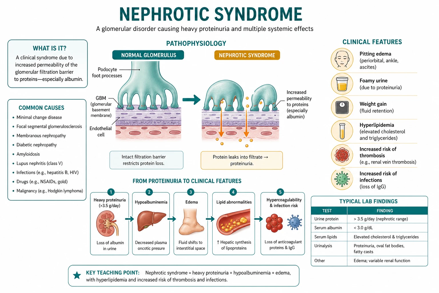
Nephrotic syndrome is the clinical expression of severe glomerular filtration-barrier (podocyte) injury, defined by the tetrad of heavy proteinuria (over 3.5 g/day, or over 50 mg/kg/day in children, or urine protein-to-creatinine ratio over 300 mg/mmol), hypoalbuminaemia (under 30 g/L, often under 25 g/L), oedema and hyperlipidaemia with lipiduria. It is distinguished from the nephritic syndrome by proteinuria-dominant features (few cells in the sediment) versus haematuria, hypertension, renal failure and low complement. In children (peak age 2-6 years, male predominance) the commonest cause is minimal change disease; in adults (equal sex ratio) membranous nephropathy and FSGS lead, with secondary causes — diabetic nephropathy, lupus nephritis, amyloidosis, malignancy, pre-eclampsia, and drugs (NSAIDs, pamidronate, lithium) — important. Malignancy associations are high-yield: membranous = solid-organ carcinoma (lung, colon, stomach); MCD = Hodgkin lymphoma. Complications arise from urinary losses — infection (encapsulated organisms from IgG and complement factor B/D loss), thromboembolism (renal vein, DVT, PE from antithrombin-III loss), hyperlipidaemia, AKI and malnutrition. Diagnosis combines protein quantification, urine microscopy, complement, autoimmune/viral/anti-PLA2R serology and renal biopsy (all adults). Management is to treat the cause (e.g. prednisolone 60 mg/m²/day for MCD), reduce proteinuria (ACE inhibitor/ARB; SGLT2 inhibitor for diabetic kidney disease), control fluid and lipids, and prevent complications (anticoagulation, vaccination).
On this page & tools
Your progress
Saved locally on this device.
Practise this topic
Exam tags
Red flags
Overview & Definition
Nephrotic syndrome is the clinical expression of severe glomerular filtration-barrier (podocyte) injury, allowing the leak of plasma proteins into the urine in quantities the kidney was never designed to lose. The defining tetrad is heavy proteinuria over 3.5 g/day (over 50 mg/kg/day in children, or a spot urine protein-to-creatinine ratio over 300 mg/mmol), hypoalbuminaemia under 30 g/L (often under 25 g/L), oedema, and hyperlipidaemia with lipiduria. The protein losses drive the entire syndrome — oedema and hypoalbuminaemia are direct consequences of albumin leak, and the dangerous complications of infection and thrombosis flow from loss of specific circulating proteins (immunoglobulins, complement factors, antithrombin III). The clinician's task is to confirm the syndrome, identify the cause (primary glomerular disease vs secondary systemic disease), and treat both the disease and its complications.[1][2]
The distinction between nephrotic and nephritic syndromes is the single most testable branch-point in glomerular medicine. Nephrotic is proteinuria-dominant — the tetrad above, with few or no cells in the urine sediment (no RBC casts). Nephritic is inflammation-dominant — haematuria with dysmorphic red cells, red-cell casts, hypertension, renal impairment/oliguria, and frequently a low serum complement. A mixed picture exists (for example lupus class IV can be both), but the pure nephrotic pattern points to minimal change disease, membranous nephropathy, FSGS, diabetic nephropathy and amyloidosis; the pure nephritic pattern to post-streptococcal GN, IgA nephropathy and rapidly progressive GN. Urinalysis and the serum complement are the two discriminators at the bedside.[1]
Classification — paediatric steroid-response categories
In paediatric practice — where minimal change disease dominates — disease is classified by the response to the initial steroid course, and these categories drive every subsequent management decision.[2][9]
Relapse is defined as urine protein 3+ or greater (or protein-to-creatinine ratio over 200 mg/mmol) for 3 consecutive days, having previously been in remission. FRNS and SDNS prompt steroid-sparing agents (calcineurin inhibitor or rituximab) to spare the child cumulative steroid toxicity (growth failure, Cushingoid habitus, osteopenia, cataracts, hypertension). SRNS carries the worst prognosis and the highest risk of progression to ESKD.[2]
Causes at a glance — by age
The aetiology is strikingly age-dependent, and stating the commonest cause for each age bracket is a guaranteed exam point.[1][2]

Children (1-10 yr)
- Minimal change disease (commonest, steroid-responsive)
- FSGS (steroid-resistant, rising)
- Congenital: Finnish type (NPHS1/nephrin), diffuse mesangial sclerosis
- Secondary: hepatitis B, syphilis, malaria, SLE (rare pre-puberty)
Adolescents
- FSGS and MCD both common
- Lupus nephritis (class V) emerges in females
- IgA nephropathy (occasionally nephrotic)
Adults (16-60 yr)
- Membranous nephropathy #1 (30-40%, PLA2R-positive)
- FSGS ~20-25%
- Minimal change disease ~15% (often younger adults)
- Secondary: diabetic, lupus, HBV/HCV/HIV, amyloidosis, drugs
Elderly (over 60 yr)
- Membranous nephropathy (screen for malignancy)
- Amyloidosis (AA from chronic inflammation)
- Diabetic nephropathy
- Paraproteinaemic disease (myeloma/AL)
Pathophysiology

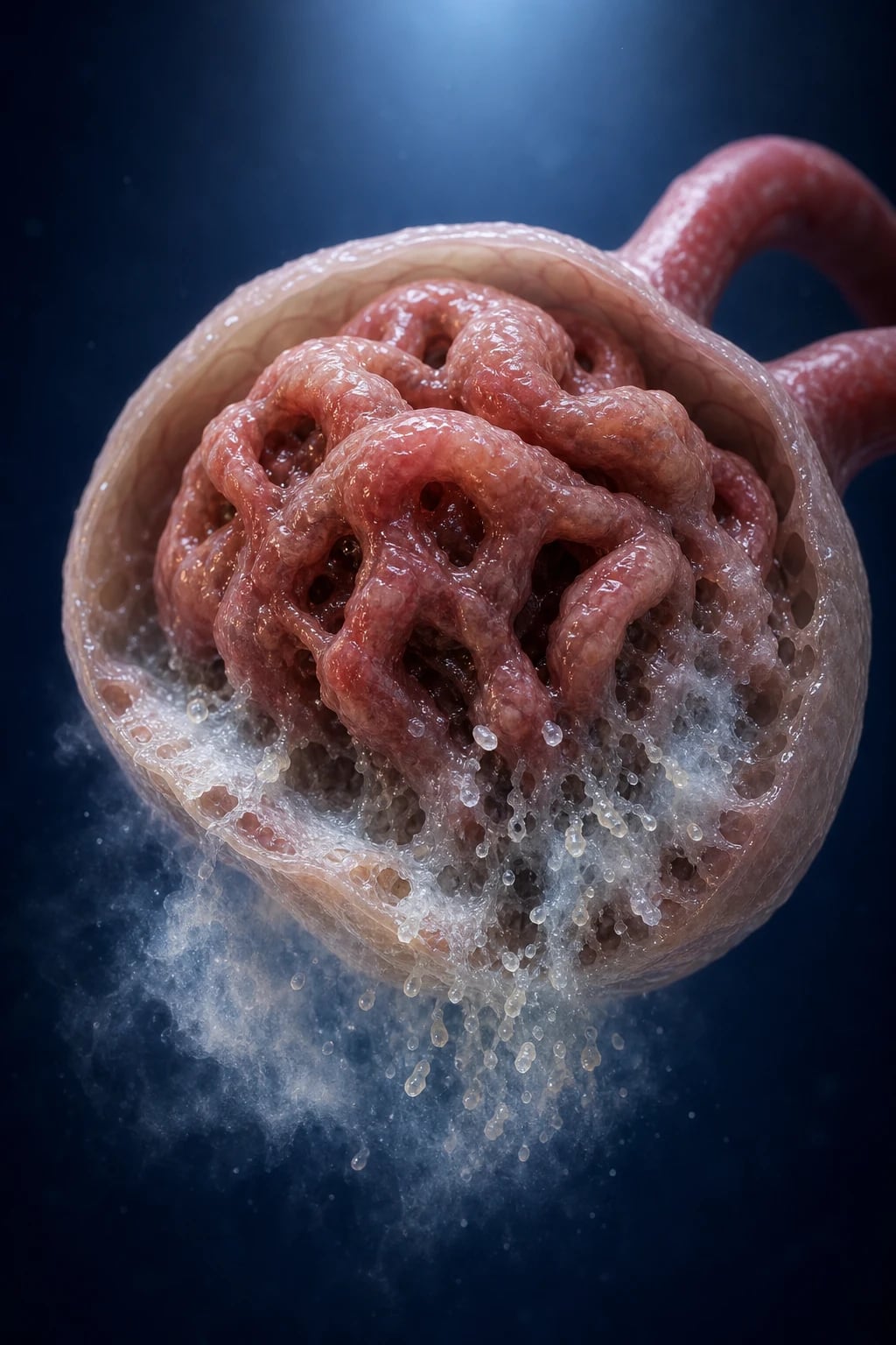
The glomerular filtration barrier has three layers: the fenestrated endothelium, the glomerular basement membrane (GBM), and the podocyte foot processes joined by the slit diaphragm. The barrier is both size- and charge-selective — it carries a net negative charge (from heparan sulphate proteoglycans) that electrostatically repels albumin (also negatively charged), so that under normal conditions only trace amounts of albumin cross into the urine (under 30 mg/day). In nephrotic syndrome, injury to the podocyte (the terminally differentiated, non-dividing visceral epithelial cell) effaces the foot processes and disrupts the slit diaphragm proteins (nephrin, podocin, CD2AP, synaptopodin). The barrier then loses both charge and size selectivity, and albumin and smaller plasma proteins pour into the urine — a massive, relatively non-selective proteinuria that overwhelms proximal tubular reabsorption.[1][9]
The downstream consequences follow directly from which proteins are lost and how the body compensates:[1]
Oedema — the underfill vs overfill hypotheses. The underfill hypothesis holds that hypoalbuminaemia lowers plasma oncotic pressure, causing fluid to shift from the intravascular to the interstitial compartment; the resulting plasma-volume contraction activates the renin-angiotensin-aldosterone system (RAAS), which drives renal sodium and water retention that perpetuates oedema. The overfill (primary renal sodium retention) hypothesis holds that the proteinuric kidney has a primary defect in sodium excretion — enhanced activity of the epithelial sodium channel (ENaC) in the collecting duct, possibly driven by filtered serine proteases (plasminogen activator) that activate ENaC — so patients are often volume-overloaded with a raised JVP rather than contracted. This is why aggressive diuresis risks precipitating AKI: the kidney is sodium-avid for reasons beyond simple volume depletion. In practice both mechanisms coexist, with the overfill/primary-sodium-retention model dominating once oedema is established.[1]
Compensatory hepatic synthesis. The liver, sensing the fall in oncotic pressure and plasma protein mass, ramps up protein synthesis — but it cannot selectively make albumin, so it overproduces lipoproteins (increased cholesterol, increased triglycerides, leading to hyperlipidaemia and lipiduria) and clotting factors, especially fibrinogen and factor VIII, while albumin synthesis still cannot keep up with the urinary loss. This dual response explains two cardinal features at once: the hyperlipidaemia of the tetrad, and the hypercoagulable state (more fibrinogen combined with urinary antithrombin-III loss).[1]
The two signature complications follow directly from the identity of the urinary protein losses:[1]
- Infection: loss of IgG and alternative-pathway complement factors B and D impairs opsonisation of encapsulated organisms (Streptococcus pneumoniae, Haemophilus influenzae, Escherichia coli) → spontaneous bacterial peritonitis, cellulitis, pneumococcal sepsis. The risk is highest when albumin is under 20 g/L and is amplified by immunosuppressive therapy.
- Thrombosis: loss of antithrombin III (the key natural inhibitor of thrombin and factor Xa), increased fibrinogen and factor VIII, platelet activation, and hemoconcentration/hyperviscosity from over-diuresis → renal vein thrombosis, DVT and PE. The thrombotic risk is highest in membranous nephropathy (up to 40% lifetime risk) and correlates with the degree of hypoalbuminaemia.[1][11]
Causes — Primary Glomerular & Secondary
The diagnosis of nephrotic syndrome is a syndrome label, not a disease label. The work that matters is identifying the underlying cause, because management and prognosis depend entirely on it. Causes divide into primary (idiopathic) glomerular diseases and secondary systemic diseases.[1][5]
Causes in CHILDREN
In children the cause is overwhelmingly a primary podocytopathy, and the peak age distribution is itself diagnostic.[2][9]
Minimal change disease
- Peak age 2-6 years; male:female 2:1
- Steroid-responsive in 90-95%
- Normal light microscopy; foot-process effacement on EM
- No immune deposits; idiopathic (T-cell cytokine / suPAR hypothesis)
FSGS
- Steroid-resistant (the commonest cause of SRNS)
- Genetic: NPHS2 (podocin), NPHS1, WT1, COL4A3/4/5
- Secondary: obesity, reduced renal mass, sickle cell
- Rising incidence; higher risk of ESKD
Congenital nephrotic syndrome
- Finnish type: NPHS1 (nephrin) — most common
- Diffuse mesangial sclerosis: NPHS2, WT1, PLCE1
- Infective: syphilis, CMV, toxoplasmosis, malaria
- Drug: NSAIDs; requires genetic testing + renal transplant
Secondary (children)
- SLE class V (lupus) — emerging post-puberty
- Hepatitis B-associated membranous
- Malaria (quartan) — endemic regions
Congenital nephrotic syndrome (CNS) deserves special attention because it is a distinct, life-threatening entity presenting at birth or within the first three months of life. The Finnish type (autosomal recessive, NPHS1 mutation encoding nephrin) is the classic and commonest form, endemic in Finland; it presents with massive placental oedema (placenta over 25% of birth weight), premature delivery, severe oedema, and proteinuria detectable from the first days of life. Other genetic causes include diffuse mesangial sclerosis (NPHS2/podocin, WT1, PLCE1) and Pierson syndrome (LAMB2). Non-genetic causes that must be excluded in any neonate with nephrotic-range proteinuria are congenital infections (syphilis, CMV, toxoplasmosis, rubella, malaria) and maternal diabetes. Management is exceptionally challenging — ACE inhibitor/ARB ± indomethacin to reduce proteinuria, aggressive nutritional support and daily albumin infusions to maintain oncotic pressure until bilateral nephrectomy followed by peritoneal dialysis and renal transplantation (the only definitive cure, though recurrence in the graft from anti-nephrin antibodies is a recognised risk).[8]
Causes in ADULTS
In adults the sex ratio is roughly equal, and the leading primary causes are membranous nephropathy and FSGS, with diabetic nephropathy the single commonest secondary cause overall.[1]
- Membranous nephropathy — the commonest cause in Caucasian adults (30-40%); defined by subepithelial immune deposits and anti-PLA2R antibody (IgG4) positivity in 70-80% of primary cases.
- FSGS — 20-25% and rising worldwide; frequently steroid-resistant; collapsing variant strongly associated with HIV.
- Minimal change disease — 10-15%, often in younger adults and in association with NSAIDs or Hodgkin lymphoma.
- Diabetic nephropathy — the commonest secondary cause; typical picture (long-standing diabetes + retinopathy + gradual onset) needs no biopsy.
- Amyloidosis — AA type from chronic inflammation (rheumatoid, IBD, chronic infection, familial Mediterranean fever); AL type from plasma-cell dyscrasia; Congo-red positive.
- Lupus nephritis (class V membranous) — low complement, ANA/anti-dsDNA positive.
- HIV-associated nephropathy (HIVAN) — collapsing FSGS, rapid progression, predominantly in patients of African descent.
- Hepatitis B (membranous), hepatitis C (membranoproliferative/cryoglobulinaemic).
- Paraproteinaemic disease — multiple myeloma (AL amyloid or light-chain deposition disease).
- Drugs — NSAIDs (minimal change disease or acute interstitial nephritis), pamidronate (collapsing nephropathy/FSGS), lithium (FSGS), gold, penicillamine, captopril (historical, membranous), heroin.
- Malignancy — see below.[1][5]
NEPHROTIC
Malignancy associations (memorise)
A new nephrotic syndrome in an older adult is a pointer to occult malignancy until excluded. The two classic, high-yield associations are:[1]
- Membranous nephropathy ↔ solid-organ carcinoma — lung (commonest), colon, stomach, breast, prostate, kidney. Screen an elderly patient with new membranous with age-appropriate malignancy workup (chest imaging, colonoscopy, mammography, prostate-specific antigen).
- Minimal change disease ↔ Hodgkin lymphoma (and less often non-Hodgkin lymphoma). The Reed-Sternberg cell produces a cytokine (likely IL-13) that injures podocytes; the nephrotic syndrome may precede the lymphoma diagnosis and remits with treatment of the tumour.[1]
Clinical Presentation
Presenting features reflect fluid accumulation and protein loss. The most common first symptom in a child is periorbital oedema, especially on waking (parents notice puffy eyes and are often told it is an allergy), progressing to dependent oedema (ankles, sacrum), ascites, pleural effusions, scrotal or labial swelling and generalised anasarca. Adults more often present with dependent oedema and ascites. Patients describe frothy urine (sometimes frankly fatty), fatigue, anorexia and weight gain from fluid retention. Urine microscopy shows oval fat bodies (tubular cells laden with lipid) with a "Maltese-cross" birefringence under polarised light — the microscopic signature of lipiduria.[1]
Cause-specific clues to seek in the history and examination narrow the differential rapidly:[1]
- Diabetes and/or hypertension with long-standing disease → diabetic nephropathy.
- Chronic rash, arthritis, alopecia, oral ulcers, photosensitivity → lupus nephritis.
- Chronic inflammatory disease (rheumatoid, IBD, chronic osteomyelitis) with macroglossia and periorbital purpura → amyloidosis.
- Weight loss, smoking history, haemoptysis, change in bowel habit → malignancy-associated membranous.
- Fever, night sweats, painless lymphadenopathy → Hodgkin lymphoma (MCD).
- HIV risk factors, hepatitis B/C risk factors — active IV drug use, transfusion, high-risk sexual exposure.
- Pregnancy with new hypertension after 20 weeks → pre-eclampsia.
- Drug history — NSAIDs, pamidronate, lithium, gold, captopril.
- Family history — congenital or genetic SRNS (consanguinity, early-onset disease in siblings).[1]
Complication presentations may be the first sign that brings the patient to hospital:[1]
- Sudden flank or loin pain, haematuria and a rising creatinine → renal vein thrombosis (image and anticoagulate).
- Fever, abdominal pain/peritonism or cellulitis → encapsulated-organism infection, spontaneous bacterial peritonitis.
- Dyspnoea, pleuritic chest pain, tachycardia → pulmonary embolism (or pulmonary oedema).
- Xanthomata (tuberous, tendinous, or eruptive) → severe hyperlipidaemia.[1]
Atypical presentation — adults versus children. Adults far more often have secondary disease and require biopsy; an elderly patient with new nephrotic syndrome must be screened for malignancy. In children, primary (spontaneous) pneumococcal peritonitis is a classic emergency in nephrotic ascites and may be the first presentation of occult nephrotic syndrome.[2]
Differential Diagnosis
The diagnosis is nephrotic syndrome (a clinical syndrome), not a disease; the differential is the underlying cause, distinguished by biopsy and serology. Two branch-points dominate the differential.[1][5]
Branch-point 1: Nephrotic vs nephritic
As above — proteinuria-dominant with bland sediment is nephrotic; haematuria/RBC casts/hypertension/renal failure/low complement is nephritic. Urinalysis and complement decide at the bedside.[1][5]
Branch-point 2: The glomerular causes, distinguished by histology and serology
Minimal change disease (MCD)
- Light microscopy: NORMAL glomeruli
- Immunofluorescence: no immune deposits
- Electron microscopy: diffuse foot-process effacement
- Classic in 2-6 yr child; steroid-responsive 90-95%
- Can complicate Hodgkin lymphoma or NSAIDs
Membranous nephropathy
- LM: thickened GBM, spike-and-dome (silver stain)
- IF: granular subepithelial IgG4 deposits
- EM: subepithelial immune deposits
- Serum anti-PLA2R positive (~70-80%)
- Highest thrombosis risk; carcinoma association in elderly
FSGS
- LM: focal segmental glomerular sclerosis ± hyalinosis
- IF: variable IgM/C3 in scarred segments
- EM: foot-process effacement
- Often steroid-resistant; collapsing variant = HIVAN
- Genetic (NPHS2) and secondary (obesity, reduced renal mass) forms
Diabetic nephropathy
- LM: nodular glomerulosclerosis (Kimmelstiel-Wilson)
- No biopsy if typical (retinopathy + long-standing DM)
- Gradual onset; ACEi/ARB + SGLT2i cornerstone
Lupus nephritis class V
- ANA + anti-dsDNA positive; LOW C3/C4
- Subepithelial immune deposits (full-house IF)
- Treat per lupus immunosuppression protocol
Amyloidosis
- Congo-red: apple-green birefringence
- AA type: chronic inflammation; AL: plasma-cell dyscrasia
- Macroglossia, periorbital purpura (AL)
- Serum free light chains, serum/urine electrophoresis
Oedema of nephrotic syndrome versus cardiac, hepatic, or malnutrition causes is separated by the urinalysis — heavy proteinuria with few cells is unique to the nephrotic kidney (cardiac and hepatic oedema have bland urine; malnutrition has a low albumin but no proteinuria). Membranous carries the highest thrombosis risk and is the one most associated with carcinoma; MCD with Hodgkin; collapsing FSGS with HIV.[1]
Clinical & Bedside Assessment
A focused examination quantifies the oedema, assesses fluid status, hunts for a secondary cause, and screens for complications — all in a few minutes.[1]
- Oedema and fluid status — grade oedema (periorbital, dependent/sacral, ascites, pleural effusion, genital/scrotal); check JVP, blood pressure and weight; auscultate for basal crackles (volume overload) versus signs of volume depletion (over-diuresis). Document the daily weight — the single most reliable index of fluid balance.
- Screen for a secondary cause — fundoscopy (diabetic or hypertensive retinopathy); skin (lupus rash, purpura, xanthomata, striae); joints (synovitis); macroglossia (amyloidosis — a specific, easily-missed clue); abdominal masses and organomegaly; lymphadenopathy (Hodgkin/lymphoma, malignancy).
- Screen for complications at the bedside — calf tenderness, unilateral leg swelling and a positive Homan sign (DVT — though Homan sign is insensitive); raised JVP and basal crackles (volume overload, pulmonary oedema); fever, abdominal tenderness/peritonism (spontaneous bacterial peritonitis); cellulitis; and signs of pulmonary embolism (tachypnoea, pleuritic pain, tachycardia, hypoxia).
- Targeted history — drugs (NSAIDs, lithium, pamidronate, gold), infection (HIV, hepatitis B/C), malignancy, and family history (genetic SRNS, congenital nephrotic syndrome, consanguinity).[1]
Investigations
Investigations confirm the syndrome and classify the cause. The workup has three layers: confirm and quantify proteinuria, characterise the urine sediment, and identify the underlying disease with serology and biopsy.[1][4]
Step 1: Confirm and quantify the syndrome
- Protein quantification — a 24-hour urine protein over 3.5 g/day (over 50 mg/kg/day in children) confirms nephrotic-range proteinuria; in routine practice, a spot urine protein-to-creatinine ratio over 300 mg/mmol (over 2 g/g) correlates well and avoids the collection errors of 24-hour urine.
- Serum chemistry — albumin under 30 g/L, renal function (urea, creatinine, eGFR), lipid profile (raised total cholesterol and triglycerides), and FBC (haemoconcentration may mask anaemia).
- Urine microscopy — oval fat bodies / Maltese-cross under polarised light (lipiduria); hyaline casts; few or no red cells and no red-cell casts (distinguishes from nephritic). Bence-Jones protein if myeloma suspected.[1]
Step 2: Serological workup to classify the cause
The serological battery is targeted at the common secondary causes:[1][4]
- Glucose and HbA1c — diabetes.
- ANA and anti-dsDNA — lupus.
- Complement C3 and C4 — low in lupus, membranoproliferative GN, post-infectious GN, cryoglobulinaemia (normal in MCD, membranous, FSGS).
- HBsAg, anti-HCV, HIV serology — infective secondary causes.
- Serum electrophoresis, serum free light chains and urine for Bence-Jones protein — myeloma and AL amyloid.
- Serum anti-PLA2R antibody — the hallmark of primary membranous nephropathy (~70-80% sensitivity); also used to monitor disease activity and relapse after immunosuppression or transplant.[4]
- Pregnancy test in women of childbearing age (pre-eclampsia).
Step 3: Renal biopsy — the decisive investigation
Renal biopsy is indicated in all adults with nephrotic syndrome, because the histological pattern drives treatment (steroids for MCD, rituximab/Ponticelli for membranous, steroids for primary FSGS). In children, biopsy is reserved for atypical presentations — age under 1 or over 10 years, hypertension, haematuria, low complement, renal impairment, or steroid-resistant disease — because the empiric steroid trial is both diagnostic and therapeutic in the classic paediatric case. The biopsy is read on light microscopy (LM), immunofluorescence (IF), and electron microscopy (EM):[1][5]
- MCD — normal LM, no immune deposits on IF, diffuse foot-process effacement on EM.
- Membranous — thickened GBM with spike-and-dome on silver stain (LM); granular subepithelial IgG4 (IF); subepithelial immune deposits (EM); anti-PLA2R positive.
- FSGS — focal segmental sclerosis with hyalinosis (LM); variable IgM/C3 (IF); foot-process effacement (EM); the collapsing variant (HIVAN) shows collapsing glomerulopathy with podocyte hypertrophy/hyperplasia.
- Amyloid — Congo-red apple-green birefringence under polarised light, with amyloid P component on IF.
- Diabetic — nodular glomerulosclerosis (Kimmelstiel-Wilson nodules) if biopsied, though biopsy is unnecessary if the clinical picture is typical.[1][5]
Baseline workup before immunosuppression — glucose, TB screen (Mantoux/IGRA), hepatitis B/C and HIV, bone-density (DEXA) before long steroids, and vaccination status (pneumococcal, influenza, hepatitis B should be up to date before immunosuppression).[5]
Imaging — renal ultrasound (kidney size, exclude obstruction or venous thrombosis; increased echogenicity suggests medical renal disease); renal Doppler ultrasound or CT venography for suspected renal vein thrombosis; age-appropriate malignancy screen (chest imaging, colonoscopy, mammography, PSA) in an elderly patient with new membranous nephropathy.[1]
Management — General Measures
General measures apply to every nephrotic patient regardless of cause, and their disciplined application prevents most of the preventable morbidity (infection, thrombosis, over-diuresis, accelerated vascular disease).[1]
- Salt and fluid — salt restriction (under 2 g, i.e. under 85 mmol sodium per day) is the single most effective non-drug oedema measure; fluid restriction is added only if hyponatraemic. Control blood pressure (target under 130/80 mmHg).
- Diuretic therapy for oedema — oral furosemide 40 mg daily, titrating the dose to the daily weight; add a potassium-sparing diuretic — amiloride 5-10 mg or spironolactone 25-50 mg — for resistant oedema (these also antagonise the aldosterone/ENaC-driven sodium retention of the overfill state). For refractory oedema with severe hypoalbuminaemia (under 20 g/L), gut oedema limits oral absorption — give intravenous furosemide 40-80 mg combined with intravenous 20-25% albumin (e.g. 25 g) to restore oncotic pressure and deliver the diuretic to the tubule; weigh daily and avoid over-diuresis, which precipitates AKI.
- Treat suspected infection promptly — a nephrotic patient with fever is septic from an encapsulated organism until proven otherwise; give empirical broad-spectrum antibiotics immediately (e.g. IV ceftriaxone 1-2 g for suspected pneumococcal peritonitis or sepsis, with the addition of cloxacillin or vancomycin for cellulitis, adjusting to local resistance). Do not delay for cultures in the septic patient — obtain cultures first, then treat.
- Hospitalise for severe oedema, AKI, suspected thrombosis or infection, or for biopsy/immunosuppression.[1]
Management — Definitive & Stepwise
Disease-specific therapy follows histology. Alongside the cause-specific treatment, two principles hold for every nephrotic patient: reduce proteinuria (ACEi/ARB ± SGLT2i) and prevent complications (anticoagulation, vaccination).[1][3]
Minimal change disease (MCD)
Prednisolone
Dose
60 mg/m²/day (maximum 60-80 mg/day) — equivalent to 1 mg/kg/day
For frequently-relapsing nephrotic syndrome (FRNS) (two or more relapses within 6 months or four or more within 12 months) or steroid-dependent nephrotic syndrome (SDNS) (two consecutive relapses during taper or within 14 days of stopping), add a steroid-sparing agent: a calcineurin inhibitor (ciclosporin 4-5 mg/kg/day or tacrolimus 0.1 mg/kg/day, both monitored by trough levels) or rituximab (375 mg/m² per dose, typically two doses). Steroid-resistant nephrotic syndrome (SRNS) — failure to remit after 4-6 weeks of daily steroids — mandates biopsy (exclude FSGS) and genetic testing; treat with calcineurin inhibitors, but the long-term ESKD risk is higher.[2][9]
Membranous nephropathy
Membranous nephropathy is risk-stratified at diagnosis. Low-risk patients (albumin over 30 g/L, normal renal function, proteinuria under 4 g/day) are managed conservatively with antiproteinuric therapy and close observation, because about one-third undergo spontaneous remission within five years. High-risk patients (declining renal function, persistent proteinuria over 8 g/day for over 6 months, or severe symptomatic nephrotic syndrome) need immunosuppression. Two regimens are standard:[3][5]
- Rituximab (first-line per KDIGO 2021 and the MENTOR trial) — 1 g IV on day 1 and day 15, repeated at 6 months. MENTOR (Fervenza, NEJM 2019) showed rituximab was non-inferior to ciclosporin at 12 months and superior at 24 months (lower relapse) in primary membranous nephropathy.[3]
- Ponticelli regimen (corticosteroid + alkylating agent) — alternating monthly cycles: methylprednisolone 1 g IV daily for 3 days at the start of months 1, 3, 5 followed by oral prednisolone 0.5 mg/kg/day for the rest of the month, alternating with oral cyclophosphamide 2-2.5 mg/kg/day in months 2, 4, 6 — a 6-month course. The 1998 Ponticelli trial established that cyclophosphamide was as effective as and less toxic than chlorambucil in idiopathic membranous nephropathy.[6]
- Calcineurin inhibitors (ciclosporin or tacrolimus) — an alternative when rituximab is unavailable or contraindicated; effective but relapse-prone on cessation.
Focal segmental glomerulosclerosis (FSGS)
Primary FSGS (an immune-mediated podocytopathy) is treated with high-dose steroids — prednisolone 1 mg/kg/day (maximum 80 mg) for up to 16 weeks before declaring steroid resistance, then a calcineurin inhibitor. Secondary FSGS (obesity, reduced renal mass, reflux nephropathy, sickle cell, heroin, HIV) is not immunosuppression-responsive and is managed by treating the cause plus ACEi/ARB. The DUPLEX trial (Rheault, NEJM 2023) showed sparsentan (a dual endothelin-A and angiotensin-II receptor antagonist) achieved a greater proteinuria reduction than irbesartan at 36 weeks in primary FSGS, establishing it as a new option.[10] Prognosis is guarded — up to 50% of primary FSGS progresses to ESKD within 5-10 years, and FSGS recurs in the transplant in up to 30% (often within days, as collapsing recurrence from a circulating permeability factor).[1]
Congenital nephrotic syndrome
There is no curative medical therapy for genetic CNS. Management is supportive and staged: ACE inhibitor/ARB ± indomethacin to reduce proteinuria; aggressive nutritional support (high-calorie, high-protein feeds, often via nasogastric tube); daily intravenous albumin infusions (4-5 g/kg 20% albumin) to maintain oncotic pressure; thyroxine and vitamin D supplementation (loss of thyroxine-binding globulin and cholecalciferol-binding protein); anticoagulation for severe hypoalbuminaemia; and bilateral nephrectomy followed by peritoneal dialysis and renal transplantation once the child reaches adequate size (the only definitive cure, though anti-nephrin antibody-mediated recurrence in the graft is a recognised risk in Finnish-type disease).[8]
Antiproteinuric cornerstone — for ALL causes
ACE inhibitor (ramipril) / ARB (losartan)
Dose
Ramipril 1.25-10 mg/day OR losartan 50-100 mg/day
DAPA-CKD (Heerspink, NEJM 2020) demonstrated that dapagliflozin 10 mg/day reduced the composite of sustained eGFR decline, ESKD, or renal/cardiovascular death by 39% versus placebo across a broad population with CKD, including patients with glomerular disease — making SGLT2 inhibition a new cornerstone of antiproteinuric management.[7]
Lipids and anticoagulation
- Lipids — statin (e.g. atorvastatin 20-40 mg at night) for persistent hyperlipidaemia; consider in all adults with nephrotic syndrome, given the accelerated atherosclerotic risk.
- Anticoagulation — nephrotic patients are hypercoagulable (antithrombin-III loss, high fibrinogen, platelet activation, hemoconcentration). Prophylactic anticoagulation is reasonable when serum albumin is under 25-30 g/L, especially in membranous nephropathy (the highest-risk histology). After any thrombotic event, fully anticoagulate (LMWH/warfarin or a DOAC) for as long as nephrotic-range proteinuria persists. The decision balances bleeding risk against the high prevalence of silent/asymptomatic thrombosis.[11]
Vaccination
Pneumococcal (conjugate PCV13 then polysaccharide PPSV23), annual influenza, and hepatitis B vaccination — ideally before the start of immunosuppression. Avoid live vaccines (BCG, MMR, varicella, oral polio) while on immunosuppression.[1]
Complications & Pitfalls
Most complications arise directly from urinary protein loss — the identity of the lost protein determines the complication:[1]
- Thromboembolism — renal vein thrombosis, DVT, and PE from antithrombin-III loss and high fibrinogen; highest in membranous nephropathy (lifetime risk up to 40%). Anticoagulate when albumin is under 25-30 g/L or after any thrombosis. Presentation is sudden flank pain, haematuria, and a deteriorating GFR; image with Doppler or CT venography.
- Infection — encapsulated bacteria (pneumococcus, H. influenzae, E. coli); spontaneous bacterial peritonitis (classically pneumococcal, in nephrotic ascites), cellulitis, and pneumococcal sepsis. The mechanism is urinary IgG and alternative-complement (factor B/D) loss. Vaccinate; treat empirically and early.
- Hyperlipidaemia — accelerated atherosclerosis and cardiovascular risk; also acute pancreatitis from severe hypertriglyceridaemia. Treat with a statin.
- AKI — from volume depletion, diuretic overuse, bilateral renal vein thrombosis, or progression of the underlying disease. Monitor creatinine and weight; avoid over-diuresis.
- Malnutrition and metabolic — protein malnutrition (negative nitrogen balance); hypocalcaemia and vitamin D deficiency (loss of cholecalciferol-binding protein); iron-deficiency anaemia (transferrin loss); hypothyroidism (loss of thyroxine-binding globulin, especially in congenital nephrotic syndrome).
- Acute tubular injury — the heavy protein load damages proximal tubular cells, contributing to the AKI risk and to interstitial fibrosis over time.[1]
Classic pitfalls to avoid: not anticoagulating a severely hypoalbuminaemic patient; missing renal vein thrombosis; not vaccinating; treating oedema with over-diuresis causing AKI; forgetting the ACEi/ARB; failing to biopsy an adult; and not screening an elderly patient with membranous nephropathy for malignancy.[1]
Prognosis & Disposition
Prognosis depends almost entirely on the underlying cause.[1][2]
Minimal change disease
- 95% steroid-responsive; normal life expectancy
- Relapses common but diminish with age
- Long-term renal prognosis excellent
- Small minority become steroid-dependent or resistant
Membranous nephropathy
- ~1/3 spontaneous remission
- ~1/3 persistent proteinuria, stable function
- ~1/3 progress to CKD/ESKD
- Risk-stratified immunosuppression and anticoagulation improve outcome
FSGS
- Up to 50% ESKD within 5-10 years (primary)
- Steroid-resistant forms fare worst
- Recurs in transplant in up to 30%
- Genetic forms do not recur in transplant
Diabetic/amyloid/HIVAN
- Tight glycaemic/BP control slows diabetic nephropathy
- Amyloid: treat underlying inflammation
- HIVAN: ART + ACEi markedly improved outcomes
- All can progress to ESKD
Predictors of a poor renal outcome (the four that recur across every glomerular disease): persistent heavy proteinuria, hypertension, reduced renal function at presentation, and interstitial fibrosis and tubular atrophy (IFTA) on biopsy.[1]
Disposition — refer to nephrology for biopsy and immunosuppression as indicated; plan for renal replacement therapy (dialysis/transplant) in progressive ESKD, noting that FSGS may recur in the transplant (warranting early aggressive treatment with plasmapheresis and rituximab) whereas genetic FSGS does not recur.[1]
Special Populations
- Children — MCD commonest; empiric prednisolone 60 mg/m²/day without biopsy for a classic presentation (age 1-10 years, normotensive, no haematuria, normal complement and renal function); lower threshold to biopsy if atypical or steroid-resistant. Weight-based dosing throughout. Pneumococcal peritonitis is a classic childhood emergency and may be the first presentation of occult nephrotic syndrome.[2]
- Elderly — membranous nephropathy and secondary causes (malignancy, amyloidosis) dominate; actively screen for solid-organ malignancy; higher thrombosis risk and higher drug-toxicity considerations.
- Pregnancy — distinguish nephrotic syndrome from pre-eclampsia (new hypertension after 20 weeks, hyperuricaemia, low complement in some, resolves post-partum); switch ACEi/ARB to labetalol or nifedipine (ACEi/ARB are fetotoxic, especially in the 2nd and 3rd trimester); monitor fetal growth and maternal renal function; manage oedema and thrombosis risk.
- Diabetic patients — diabetic nephropathy is the commonest secondary cause; do not routinely biopsy if typical (retinopathy, long-standing diabetes, gradual onset); ACEi/ARB and SGLT2 inhibitor are the cornerstone.
- Immunocompromised / HIV — HIV-associated nephropathy (HIVAN) presents as a collapsing FSGS with rapid progression, predominantly in patients of African descent; screen (HIV test) and start antiretroviral therapy plus ACEi, which has transformed the prognosis.
Evidence, Guidelines & Regional Differences
MENTOR — Rituximab vs Ciclosporin in Membranous Nephropathy
PMID 31269364
Key finding
Rituximab non-inferior to ciclosporine at 12 months and superior at 24 months (lower relapse) in primary membranous nephropathy — the basis for rituximab as first-line immunosuppression in high-risk disease.
DAPA-CKD — Dapagliflozin in Chronic Kidney Disease
PMID 32970396
Key finding
Dapagliflozin 10 mg/day reduced the composite of sustained eGFR decline, ESKD, or renal/cardiovascular death by 39% vs placebo across a broad CKD population including glomerular disease.
DUPLEX — Sparsentan vs Irbesartan in FSGS
PMID 37921461
Key finding
Sparsentan achieved greater proteinuria reduction than irbesartan at 36 weeks in primary FSGS — a new non-immunosuppressive option.
- KDIGO 2021 Clinical Practice Guideline for the Management of Glomerular Diseases is the international standard for evaluation and treatment of nephrotic syndrome — defining biopsy indications, the role of anti-PLA2R antibody (avoiding biopsy in some primary membranous), and risk-stratified immunosuppression (rituximab-based for membranous).[5]
- Anti-PLA2R antibody (Beck LH Jr, NEJM 2009) established the M-type phospholipase A2 receptor as the target antigen in approximately 70-80% of primary membranous nephropathy; it is now both a diagnostic marker and an activity/relapse-monitoring marker.[4]
- Ponticelli regimen (Ponticelli C, JASN 1998) — the randomised comparison of methylprednisolone plus chlorambucil versus methylprednisolone plus cyclophosphamide that established the modern steroid-plus-cyclophosphamide regimen for idiopathic membranous nephropathy.[6]
- IPDN / KDIGO paediatric practice standardises the management of childhood SSNS and SRNS — first-line prednisolone 60 mg/m²/day, definitions of frequent relapse/steroid-dependence/steroid-resistance, and the early use of calcineurin inhibitors/rituximab for FRNS/SDNS, with biopsy and genetics for SRNS.[2]
Regional / resource-limited (Indian) practice. Children are typically treated empirically with prednisolone without biopsy (biopsy access and cost limit early histology); late presentation and late biopsy of adults is common. Pneumococcal peritonitis and infection are major causes of mortality. Quartan malaria (Plasmodium malariae)-associated nephrotic syndrome remains endemic in parts of sub-Saharan Africa and is typically steroid-resistant. Emphasise prednisolone availability, vaccination and empirical antibiotics as the practical priorities. The African ancestry–APOL1 association with FSGS, HIVAN and progressive non-diabetic kidney disease is a major genetic determinant of disease severity and transplant recurrence risk in those populations.[1][2]
Exam Pearls & High-Yield Minutiae
Red Flags
Exam application bank (NEET-PG / INICET)
One-line answer
Nephrotic syndrome is the clinical expression of severe glomerular filtration-barrier (podocyte) injury, defined by the tetrad of heavy proteinuria (over 3.5 g/day, or over 50 mg/kg/day in children, or urine protein-to-creatinine ratio over 300 mg/mmol), hypoalbuminaemia (under 30 g/L, often under 25 g/L), oedema and hyperlipidaemia with lipiduria. It is distinguished from the nephritic syndrome by proteinuria-dominant features (few cells in the sediment) versus haematuria, hypertension, renal failure and low complement. In children (peak age 2-6 years, male predominance) the commonest cause is minimal change disease; in adults (equal sex ratio) membranous nephropathy and FSGS lead, with secondary causes — diabetic nephropathy, lupus nephritis, amyloidosis, malignancy, pre-eclampsia, and drugs (NSAIDs, pamidronate, lithium) — important. Malignancy associations are high-yield: membranous [1]
Worked stems (answer without another resource)
Stem 1 — Classic presentation. Map symptoms to mechanism; name the first investigation and first treatment step with dose/route if drug therapy is standard. [1]
Stem 2 — Unstable / complicated. List red flags that force immediate resuscitation, theatre, ICU, antidote, or reperfusion — and what you do in the first 15 minutes. [1]
Stem 3 — Atypical group. Elderly, pregnancy, child, or immunocompromised: how presentation and thresholds change. [1]
Stem 4 — Differential trap. Name the three closest mimics and one discriminator for each. [1]
Stem 5 — Disposition. Who goes home with safety-netting, who is admitted, who needs HDU/ICU/theatre, and what follow-up is mandatory. [1]
Rapid viva checklist
- Definition + classification
- Pathophysiology chain
- Bedside signs / criteria
- Score with exact components (if any)
- Emergency bundle
- Definitive therapy with doses
- Complications of disease and of treatment
- Special populations
- Guideline/trial name if classic
- Three exam traps
Coverage self-check
If you cannot answer any stem above from this page alone, re-read the matching section — the page is intended to be self-sufficient for final-prof and NEET-PG/INICET questions on Nephrotic Syndrome.
References
- [1]Kodner C. Diagnosis and Management of Nephrotic Syndrome in Adults Am Fam Physician, 2016.PMID 26977832
- [2]Downie ML, Gallibois C, Parekh RS, et al. Nephrotic syndrome in infants and children: pathophysiology and management Paediatr Int Child Health, 2017.PMID 28914167
- [3]Fervenza FC, Appel GB, Barbour SJ, et al. Rituximab or Cyclosporine in the Treatment of Membranous Nephropathy N Engl J Med, 2019.PMID 31269364
- [4]Beck LH Jr, Bonegio RG, Lambeau G, et al. M-type phospholipase A2 receptor as target antigen in idiopathic membranous nephropathy N Engl J Med, 2009.PMID 19571279
- [5]Kidney Disease: Improving Global Outcomes (KDIGO) Glomerular Diseases Work Group. KDIGO 2021 Clinical Practice Guideline for the Management of Glomerular Diseases Kidney Int, 2021.PMID 34556256
- [6]Ponticelli C, Altieri P, Scolari F, et al. A randomized study comparing methylprednisolone plus chlorambucil versus methylprednisolone plus cyclophosphamide in idiopathic membranous nephropathy J Am Soc Nephrol, 1998.PMID 9513907
- [7]Heerspink HJL, Stefánsson BV, Correa-Rotter R, et al. Dapagliflozin in Patients with Chronic Kidney Disease N Engl J Med, 2020.PMID 32970396
- [8]Jalanko H. Congenital nephrotic syndrome Pediatr Nephrol, 2009.PMID 17968594
- [9]Vivarelli M, Massella L, Ruggiero B, Emma F. Minimal Change Disease Clin J Am Soc Nephrol, 2017.PMID 27940460
- [10]Rheault MN, Alpers CE, Barratt J, et al. Sparsentan versus Irbesartan in Focal Segmental Glomerulosclerosis N Engl J Med, 2023.PMID 37921461
- [11]Zou H, Li Y, Hao C, et al. Management of anticoagulation and antiplatelet therapy in patients with primary membranous nephropathy BMC Nephrol, 2019.PMID 31791286