Nephrology · General Medicine
Polycystic Kidney Disease
Also known as Polycystic kidney disease · ADPKD · Adult polycystic kidney disease · Autosomal dominant polycystic kidney disease
Autosomal dominant polycystic kidney disease (ADPKD) is the commonest inherited kidney disease (prevalence 1 in 400 to 1 in 1000), caused in 95 percent of families by mutations in PKD1 (85 percent, chromosome 16, severe) or PKD2 (15 percent, chromosome 4, milder). It produces bilateral, progressively enlarging renal cysts arising from any nephron segment, leading to hypertension, grossly enlarged kidneys, and progression to end-stage kidney disease by the fifth or sixth decade (about 50 percent by age 60). Important extrarenal features include liver cysts (commonest, ~80 percent by age 60), intracranial berry aneurysms (~10 percent, with subarachnoid haemorrhage risk), mitral valve prolapse (~25 percent), pancreatic cysts and diverticular disease. Diagnosis is by ultrasound using age-adjusted cyst count (Pei criteria), cross-sectional imaging for height-adjusted total kidney volume (Mayo classification), or genetic testing. Management centres on strict blood-pressure control (ACE inhibitor or ARB, target under 110 over 80), tolvaptan (a vasopressin V2 antagonist) in rapidly progressive disease, treatment of cyst complications (infection, bleeding, pain), and dialysis or transplantation for ESKD — with selective aneurysm screening when there is a family history of haemorrhage.
On this page & tools
Your progress
Saved locally on this device.
Practise this topic
Exam tags
Red flags

Overview & Definition
Autosomal dominant polycystic kidney disease (ADPKD) is a systemic, heritable disorder of cystogenesis driven by defects in the polycystin-1 and polycystin-2 proteins, producing relentlessly enlarging bilateral renal cysts and a characteristic constellation of extrarenal manifestations (liver cysts, intracranial aneurysm, cardiac valve defects, pancreatic cysts, diverticular disease).[1]
It is the commonest monogenic kidney disease (prevalence 1 in 400 to 1 in 1000 live births worldwide) and accounts for 5–10 percent of all end-stage kidney disease (ESKD).[1] The disease is fully penetrant (essentially 100 percent by age 70 with modern imaging) but highly variable in severity, even within the same family. Because the kidneys enlarge for decades before renal failure, family screening, blood-pressure control and tolvaptan in the rapidly progressive form can meaningfully slow progression.[1][2]
Classification
ADPKD is classified genetically (by mutated gene), radiologically (Mayo imaging classification of cyst burden) and by age at onset.[1][8]
Genetic classification: [1]
ADPKD vs ARPKD — the two must-not-confuse inherited polycystic diseases
ADPKD
- Inheritance **autosomal dominant**; gene **PKD1 (85%)** chr 16 or **PKD2 (15%)** chr 4
- Protein **polycystin-1 / polycystin-2** (PC1/PC2 complex)
- Onset **adult** (cysts detectable from adolescence); ESKD by **50s–60s**
- Kidneys **massively enlarged** with cysts of all sizes
- Liver: **bile-duct hamartomas → cysts** (commonest extrarenal site)
- Prevalence **1:400–1:1000**
ARPKD
- Inheritance **autosomal recessive**; gene **PKHD1** (chr 6)
- Protein **fibrocystin / polyductin**
- Onset **neonatal / infancy**; many die in the neonatal period
- Kidneys **huge, echogenic, fusiform dilatation of collecting ducts**
- Liver: **congenital hepatic fibrosis** with portal hypertension
- Prevalence **1:10,000–1:40,000**
Molecular (modern) classification — now refined beyond PKD1/PKD2:[1]
- Typical ADPKD —
PKD1orPKD2mutation with the classic bilateral cystic phenotype (Mayo class 1A–1E). - ADPKD-like disease with non-PKD genes — monoallelic pathogenic variants in
ALG8,ALG9,GANAB,DNAJB11,IFT140(in ~2 percent of clinically diagnosed ADPKD); generally milder, often atypical imaging.[1] - Atypical polycystic kidney disease — bilateral cysts not meeting ADPKD imaging criteria (Mayo class 2); consider mimics and non-PKD genes.
Phenotypic / Mayo imaging classification uses height-adjusted total kidney volume (htTKV) to stratify the typical (class 1, expanding cysts) into 1A (slowest) through 1E (most rapidly enlarging) and identifies atypical patterns (class 2). Mayo class 1C–1E defines rapidly progressive disease and is a key trigger for tolvaptan.[8]
Very early onset (VEO) ADPKD — diagnosis in utero or within the first 18 months of life; bilateral hugely enlarged echogenic kidneys; often severe but variable course.[2]
Epidemiology & Risk Factors
- Prevalence: 1 in 400 to 1 in 1000 live births across all populations; the commonest life-threatening monogenic disorder.[1]
- Burden: accounts for 5–10 percent of all ESKD worldwide, making it the 4th or 5th leading cause of ESKD.[10]
- Penetrance: essentially 100 percent by age 70 with modern imaging, but expressivity is highly variable.
- Inheritance: autosomal dominant; de novo mutations account for 5–10 percent of cases (no family history).[1]
Genetic risk factors for severe disease[1]:
PKD1(vsPKD2) — earlier ESKD.- Truncating
PKD1mutation (vs non-truncating / in-frame) — most severe. - Biallelic / hypomorphic
PKD1and the contiguousTSC2-PKD1deletion syndrome produce severe early-onset disease. - Modifier genes (
HNF1B,HNF4A,DZIP1L) and digenic inheritance (PKD1+HNF1B) modify expressivity. [1]
Modifiable and phenotypic risk factors that accelerate progression:[1][10]
- Male sex, hypertension before age 35, gross haematuria episodes, onset of ESKD before age 55 in affected parent.
- Rapid kidney growth (height-adjusted total kidney volume ≥5 percent per year or Mayo 1D–1E).
- Proteinuria / albuminuria (each g/g increase predicts faster decline).
- Obesity, smoking, high dietary sodium (>6 g/day), low water intake, frequent NSAID use, uncontrolled hypertension, hyperuricaemia. [1]
Pathophysiology
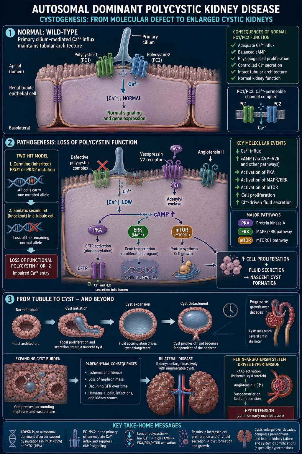
ADPKD is a ciliopathy — a disorder of the primary (non-motile) cilium of the tubular epithelial cell.[1]
1. The polycystin complex. Polycystin-1 (PC1, PKD1, chromosome 16p13.3) is a large mechanosensory transmembrane receptor; polycystin-2 (PC2, PKD2, chromosome 4q22.1) is a non-selective calcium-permeable cation channel of the TRP family (TRPP2). The two proteins form a 1:1 heterodimeric complex in the primary cilium, plasma membrane and ER. By sensing tubular flow (bending of the cilium), the PC1/PC2 complex normally permits a calcium influx that maintains intracellular calcium and suppresses cAMP.[1]
2. The two-hit model (Knudson). Although inheritance is autosomal dominant, individual cysts are focal because a germline mutation in one allele is necessary but not sufficient for cyst formation: each cyst requires a somatic 'second hit' (mutation, loss of heterozygosity, or epigenetic silencing) in the other, wild-type allele of PKD1 or PKD2. This explains the focal, asymmetric, expanding cyst population.[1] The rate of second hits (raised by oxidative stress, DNA damage) partly determines severity.
3. The downstream signalling cascade. Loss of functional polycystin produces a coherent biochemical phenotype:[1]
- ↓ intracellular Ca²⁺ (loss of PC2 channel activity).
- ↑ cAMP — disinhibition of adenylyl cyclase 5/6; low Ca²⁺ favours AC6 and inhibits PDE1, so cAMP accumulates.
- ↑ vasopressin V2-receptor signalling — vasopressin stimulates AC6 → more cAMP. Vasopressin levels rise with low water intake; this is the pharmacological target of tolvaptan.[4]
- cAMP → PKA → B-Raf / MAPK (ERK) drives epithelial proliferation (cyst-lining cells multiply).
- cAMP → CFTR-mediated Cl⁻ (and fluid) secretion into the cyst lumen — explains why cysts are fluid-filled and why CFTR inhibitors slow growth in vitro.
- mTOR activation (via disinhibition of the TSC1/2–Rheb axis and ER stress) drives protein synthesis and proliferation; mTOR inhibitors (sirolimus, everolimus) reduce cyst growth in models but have disappointed clinically.
- RAAS and inflammation — cyst compression of neighbouring vasculature causes ischaemia, local renin release, endothelial dysfunction (↓ nitric oxide) and interstitial inflammation and fibrosis.[1]
- Wnt, JAK-STAT, Hippo-YAP, Hedgehog and metabolic reprogramming (Warburg-like glycolysis) further amplify cyst-cell proliferation.
4. Cyst detachment and growth. Early cysts remain connected to the tubule but, as they dilate beyond ~2 mm, the epithelial segment pinches off — the cyst is now isolated from the nephron and continues to enlarge by polarised fluid secretion (Cl⁻/Na⁺/K⁻ driven by the basolateral Na⁺/K⁺-ATPase and the apical CFTR/PKA axis) plus ongoing proliferation of the lining epithelium. The cyst fluid accumulates secreted growth factors (EGF, VEGF), cytokines and antimicrobial peptides.[1]
5. Genotype–phenotype correlation.[1]
PKD1truncating — median age at ESKD ~55 years (most severe).PKD1non-truncating (in-frame) — median ESKD ~65 years.PKD2— median ESKD ~74 years (milder, fewer and later cysts, lower hypertension burden).- Contiguous
TSC2-PKD1deletion — severe, very-early-onset disease combined with tuberous sclerosis features. [1]
Clinical Presentation
ADPKD typically declares itself in the 3rd to 5th decade, but cysts are detectable from childhood and the disease can present at any age.[1][10]
Renal manifestations
Frequency of renal manifestations in ADPKD by the time of diagnosis
- Hypertension — the earliest and commonest manifestation, often preceding any fall in GFR by years to decades; it occurs in up to 60 percent at diagnosis and over 80 percent with established disease, and is itself a driver of cyst growth and cardiovascular mortality. The mechanism is multifactorial and begins before GFR falls: expanding cysts compress intrarenal arterioles causing focal ischaemia and local renin release (intrarenal RAAS activation); sympathetic overactivity (driven by polycystin-deficient baroreceptors); endothelial dysfunction with reduced nitric-oxide bioavailability; and impaired renal-pressure natriuresis. Because hypertension accelerates cyst enlargement, LVH and aneurysm risk, early detection and aggressive treatment with an ACEi or ARB is the cornerstone of disease modification.[1][7]
- Flank or abdominal pain — from cyst enlargement, stretch of the renal capsule, haemorrhage, infection, stone, or rarely a coexisting tumour.
- Palpable bilateral irregular renal mass — large kidneys are palpable in advanced disease.
- Haematuria — microscopic (common) or macroscopic (rupture of a cyst into the collecting system, ~40 percent lifetime risk of an episode).
- Nephrolithiasis — uric acid stones (low urinary pH from hypocitraturia) and calcium oxalate stones, in ~20 percent.
- Urinary tract infection — cystitis, pyelonephritis, infected cyst (fever, flank pain, positive cultures).
- Proteinuria / albuminuria — usually under 1 g/day; heavier proteinuria suggests another glomerular lesion.
- Progressive CKD to ESKD — ~50 percent by age 60; cyst compression of neighbouring nephrons and vasculature drives ischaemia, interstitial inflammation and fibrosis, so GFR remains well preserved for decades while total kidney volume climbs, then declines progressively once a critical parenchymal threshold is crossed (the "burnt-out" phase).[1]
Because flank pain is the single commonest reason an ADPKD patient seeks acute care, the four causes must be separated at the bedside — they have different investigations and different treatments.[10]
Flank pain in ADPKD — the four causes every candidate must distinguish
Cyst haemorrhage or rupture
- **Sudden**, often **unilateral severe** flank or loin pain ± **macroscopic haematuria**
- **Low-grade fever** (inflammatory), **no positive cultures**; usually **self-limited over 2 to 7 days**
- **CT**: **hyperdense (40–80 HU) cyst**, fluid level, **no enhancement**
- **Treat:** bed rest, hydration, analgesia (**paracetamol ± opioid; AVOID NSAIDs**); stop anticoagulants; tranexamic acid if refractory
Infected cyst
- **Fever, rigors, flank tenderness**, positive **blood or urine cultures**, raised WBC/CRP
- Pain **worsened by sitting upright or bending** (cyst stretches capsule); may have **no dysuria** (cyst disconnected from urine)
- **CT/MRI**: cyst with **thickened wall, gas, debris, surrounding stranding**; **FDG-PET** localises occult infection
- **Treat:** **cyst-penetrating antibiotic** (**ciprofloxacin, clindamycin, chloramphenicol**) for **2 to 6 weeks**; **drain** if greater than 3 to 5 cm or refractory
Nephrolithiasis
- **Acute colicky** pain **radiating to groin**, restless, **microscopic or macroscopic haematuria**
- **Uric acid** (low urinary pH from hypocitraturia) and **calcium oxalate** stones predominate
- **Non-contrast CT** is diagnostic; **ultrasound** may miss small radio-lucent stones
- **Treat:** hydration, analgesia, **potassium citrate**, thiazide for hypercalciuria; **ESWL or ureteroscopy** for obstructing stones
Coexisting renal cell carcinoma
- **Atypical pain**, **weight loss**, **persistent haematuria**, or a **complex cyst** (Bosniak III or IV)
- ADPKD carries a **modestly increased RCC risk** — never assume a bleed is benign
- **Contrast CT**: **enhancing mural nodule, thick irregular septation**, neovascularity
- **Treat:** **urological referral**, **partial or radical nephrectomy**; do NOT sclerose a possibly malignant cyst
Haematuria deserves its own work-up. Microscopic haematuria is common and reflects cyst-wall micro-bleeding; it requires urinalysis, blood pressure review and avoidance of NSAIDs and anticoagulants where possible. Macroscopic haematuria — classically after minor trauma, vigorous exercise, or in the anticoagulated patient — is usually a cyst rupture into the collecting system and is managed with bed rest, oral or IV hydration to prevent clot colic, and analgesia; bleeding settles within days in most cases. Persistent or painless macroscopic haematuria, or any haematuria with constitutional symptoms, must be imaged to exclude a coexisting renal cell carcinoma in a complex cyst.[1][10]
Extrarenal manifestations
[1]Extrarenal manifestations of ADPKD — cumulative prevalence
Polycystic liver disease (PLD) is the commonest extrarenal manifestation — present in roughly half of patients by age 40 and up to 80 percent by age 60, more numerous and larger in women and with multiparity and oestrogen exposure. Liver cysts arise by dilatation of biliary microhamartomas (von Meyenburg complexes) and, because they compress rather than destroy hepatocytes, synthetic liver function is almost always preserved — PLD kills by mass effect (pain, early satiety, cachexia, inferior vena cava or portal compression) or by complications (infection, haemorrhage, torsion).[1][10]
Intracranial berry aneurysms occur in about 8 to 10 percent of ADPKD patients (versus 1 to 2 percent in the general population), cluster in the anterior circulation (internal carotid and middle cerebral artery bifurcations) and are the most feared extrarenal complication because rupture causes subarachnoid haemorrhage with a case fatality of 30 to 50 percent. Rupture risk rises with aneurysm size (greatest above 7 mm), posterior-circulation location, uncontrolled hypertension, smoking, prior rupture, and a family history of aneurysm or SAH. The rupture is often heralded by a thunderclap ("worst-ever") headache, sometimes with photophobia, neck stiffness or loss of consciousness.[9][10]
Cardiac valve abnormalities are a direct consequence of polycystin deficiency in valvular connective tissue: mitral valve prolapse in roughly 25 percent, with aortic regurgitation, tricuspid regurgitation and aortic root dilatation (which rarely progresses to dissection). Most are haemodynamically insignificant, but a systolic click-murmur on the bedside exam of a young hypertensive patient is a clue to ADPKD. Other associations include seminal-vesicle and epididymal cysts (male infertility is uncommon), colonic diverticulosis with higher perforation risk in ESKD, inguinal and umbilical hernias, and bronchiectasis.[1]
Atypical presentations
- Elderly — incidental bilateral renal cysts on imaging; some never reach ESKD (especially
PKD2or non-PKDgenes). - Pregnancy — usually preserved fertility; new hypertension or pre-eclampsia is more common; large kidneys may cause pain and rarely obstruct labour.
- Diabetic — combined diabetes + ADPKD accelerates CKD; exclude diabetic nephropathy as a superimposed cause of heavier proteinuria.
- Immunocompromised / post-transplant — risk of infected cysts and BK/CMV interposed with native kidney cyst complications.
- Anticoagulated — markedly higher risk of cyst haemorrhage and macroscopic haematuria; reassess risk–benefit.
- Very early onset — bilateral hugely echogenic kidneys in utero/neonate, often oligohydramnios, pulmonary hypoplasia.[2]
Differential Diagnosis
A complete differential, with distinguishing features, is essential — not all bilateral renal cysts are ADPKD.[1]
The key differentials (each with the feature that separates it from ADPKD): [1]
- Multiple simple renal cysts (Bosniak I) — few, small, smooth-walled, normal-sized kidneys, no family history; very common in the elderly.
- Acquired cystic kidney disease (ACKD) — develops in dialysis-dependent ESKD; small shrunken kidneys (not enlarged); associated with secondary hyperparathyroidism and a higher renal-cell-carcinoma risk.
- Tuberous sclerosis complex (TSC1/TSC2) — angiofibromas, ash-leaf macules, seizures, subependymal nodules, renal angiomyolipomas; the contiguous TSC2-PKD1 deletion syndrome causes severe early-onset polycystic kidneys.
- Von Hippel-Lindau disease — renal cell carcinoma, pancreatic cysts, haemangioblastoma, retinal angioma, phaeochromocytoma.
- Medullary sponge kidney — papillary collecting-duct ectasia with nephrocalcinosis, normal kidney size, no ESKD.
- Autosomal recessive PKD (ARPKD) — neonatal huge echogenic kidneys, congenital hepatic fibrosis, PKHD1.
- Renal cell carcinoma in a cyst (Bosniak III/IV) — enhancing mural nodule or septation; requires resection.
- Bilateral hydronephrosis — dilated pelvicalyceal system with thin parenchyma and an obstructive cause; no parenchymal cysts. [1]
| Condition | Key distinguishing features |
|---|---|
| Multiple simple renal cysts (Bosniak I) | Few, small, round/smooth, thin-walled, anechoic, normal-sized kidneys; no family history; very common in elderly. |
| Acquired cystic kidney disease (ACKD) | Develops in dialysis-dependent ESKD; small, shrunken kidneys (unlike the huge kidneys of ADPKD); associated with secondary hyperparathyroidism and a higher renal cell carcinoma risk. |
Tuberous sclerosis complex (TSC1/TSC2) | Angiofibromas (face), ash-leaf macules, shagreen patch, seizures, subependymal nodules/giant-cell astrocytoma; renal angiomyolipomas (fat-density on CT). The contiguous TSC2-PKD1 deletion syndrome combines severe early polycystic kidneys with TSC features. |
| von Hippel-Lindau disease | Renal cell carcinoma (often bilateral), pancreatic cysts, cerebellar/spinal haemangioblastoma, retinal angioma, phaeochromocytoma; VHL gene. |
| Medullary sponge kidney | Papillary collecting duct ectasia, nephrocalcinosis and recurrent stones; normal kidney size; does not cause ESKD. |
| Autosomal recessive PKD (ARPKD) | Neonatal onset, huge echogenic fusiform kidneys, congenital hepatic fibrosis (portal hypertension), PKHD1. |
| Renal cell carcinoma in a cyst (Bosniak III/IV) | Enhancing mural nodule/septation, irregular thick wall; needs resection; not ADPKD per se but ADPKD does modestly increase RCC risk. |
| Bilateral hydronephrosis | Dilated pelvicalyceal system, thin parenchyma, obstructive cause (BPH, stones, retroperitoneal fibroma); no parenchymal cysts. |
Clinical & Bedside Assessment
- History. Three-generation pedigree; age of ESKD and SAH in affected relatives; onset and duration of hypertension; episodes of flank pain, macroscopic haematuria, UTI, stone passage; urinary frequency/dysuria; family history of aneurysm or SAH, intracerebral haemorrhage, sudden death; drug history (NSAIDs, anticoagulants, ACEi/ARB); smoking, alcohol, salt and water intake.
- Examination. Blood pressure (both arms), bilateral ballotable irregular renal masses, palpable liver (polycystic liver disease), abdominal hernias, mitral click/murmur (MVP), stigmata of CKD (anaemia, pallor, uraemic flap, peripheral oedema), cutaneous features of TSC (ash-leaf macules, angiofibromas) to exclude the contiguous gene syndrome.
- Neurological assessment if any thunderclap headache — GCS, meningeal signs, focal deficit (suspected SAH is an emergency). [1]
Investigations
1. Imaging — diagnostic (with family history)
Pei unified age-adjusted ULTRASOUND criteria for an at-risk first-degree relative of an ADPKD patient:[3]
[1]Age-adjusted CT / MRI criteria (higher sensitivity):[3]
- Age 15–40 yr: ≥ 3 cysts (bilateral or unilateral).
- Age >40 yr: ≥ 5 cysts in each kidney. [1]
2. Mayo imaging classification (prognosis & treatment triage)
The Mayo classification stratifies ADPKD by height-adjusted total kidney volume (htTKV) on MRI/CT:[8]
- Class 1 (typical) — bilateral expanding cysts, sub-classified 1A → 1E by the annual growth rate of htTKV (a surrogate for cyst burden that predicts the age at ESKD):[8]
- 1A — slowest (htTKV growth under ~1.5 percent per year); ESKD unlikely before age 70.
- 1B — ~1.5 to 3 percent per year; intermediate.
- 1C — ~3 to 4.5 percent per year.
- 1D — ~4.5 to 6 percent per year.
- 1E — over 6 percent per year; the most rapidly progressive.
- Classes 1C through 1E define rapidly progressive disease — the key trigger for tolvaptan.
- Class 2 (atypical) — bilateral cysts not meeting the typical pattern (e.g. asymmetric, segmental, lopsided, or unilateral); consider mimics and non-
PKDgenes; tolvaptan is generally not indicated.
MRI is the preferred modality for htTKV measurement; ultrasound is adequate for screening. [1]
3. Genetic testing
- Atypical or mild phenotype, very early onset, or no family history.
- Evaluation of a potential living related kidney donor (genetics + imaging together; an at-risk relative under 30 may need both).
- Reproductive counselling (preimplantation genetic diagnosis).
- Confirming ADPKD in a young adult with hypertension and equivocal imaging. [1]
Next-generation sequencing of PKD1/PKD2 (with copy-number analysis for the PKD1 5′-duplicated region) plus a panel of non-PKD genes (ALG8, ALG9, GANAB, DNAJB11, IFT140, HNF1B, TSC2). A pathogenic variant is identified in ~75–90 percent of typical ADPKD. [1]
4. Baseline laboratory panel
- U&E, creatinine, eGFR (CKD-EPI 2021).
- Urinalysis — haematuria, proteinuria.
- Urine ACR / UPCR (albuminuria predicts progression).
- FBC, iron studies (anaemia — note ADPKD has better-preserved haemoglobin than other ESKD causes because of preserved erythropoietin).
- LFTs (baseline before tolvaptan).
- Lipid profile, urate, fasting glucose/HbA1c, calcium-phosphate, PTH, vitamin D, 25-OH-D.
- Urine culture if symptoms. [1]
5. Selective intracranial aneurysm screening
MRA (time-of-flight, no contrast) or CTA — selective, not routine. Indications:[9][10]
- Family history of intracranial aneurysm or SAH.
- Prior aneurysm rupture or known aneurysm.
- Uncontrolled hypertension, smoker, age >50, female, or high-risk occupation (e.g. pilot).
- Before major elective surgery, transplant work-up, or at patient request after counselling. [1]
Routine screening of all ADPKD patients is not recommended (low yield, false-positive risk). [1]
Management — Resuscitation (time-critical scenarios)

- Suspected SAH (thunderclap headache): airway-breathing-circulation; intravenous labetalol or nicardipine for BP control; emergency non-contrast CT (sensitivity ~100 percent in the first 24 h); lumbar puncture for xanthochromia if CT negative but suspicion persists; urgent neurosurgical / interventional neuroradiology referral for endovascular coiling (preferred for posterior circulation and most aneurysms under 10 mm) or surgical clipping.[10]
- Cyst haemorrhage (gross haematuria / flank pain): bed rest; oral/IV fluids to prevent clot obstruction; analgesia — paracetamol 1 g QDS, or tramadol 50–100 mg QDS/PRN, or short-course opioid; AVOID NSAIDs (worsen GFR, increase bleeding); stop anticoagulants; consider tranexamic acid 1 g IV/PO TDS or aminocaproic acid for refractory bleeding; selective renal arterial embolisation for uncontrolled or recurrent life-threatening haemorrhage; transfuse if haemodynamically significant.
- Cyst infection with sepsis: blood and urine cultures; cyst-penetrating antibiotic (lipophilic, active in cyst fluid) — ciprofloxacin 500 mg PO BD (or 400 mg IV BD), clindamycin 600 mg IV TDS, or chloramphenicol; continue for 2 weeks minimum (up to 4–6 weeks for deep infections); drain an infected cyst ≥3–5 cm under imaging. Avoid aminoglycosides and β-lactams alone (poor cyst penetration).
- ESKD complications: treat hyperkalaemia, acidosis, volume overload, and uraemia per standard AKI/CKD bundle; dialyse if refractory.
Management — Definitive & Stepwise
Step 1 — Strict blood-pressure control
Target BP under 130/80 mmHg (KDIGO 2025). A lower target of 110/80 mmHg may be considered in young adults with eGFR over 30, based on the HALT-PKD trial A, where rigorous control (95–110/60–75) with an ACEi or ARB slowed kidney growth without accelerating the GFR decline.[7][9][10]
- First-line: ACE inhibitor OR ARB (not both) — ramipril, lisinopril, enalapril; or telmisartan, losartan, valsartan. Rationale: RAAS is central to ADPKD hypertension and progression; ACEi/ARB also reduce proteinuria.
- Monitoring: U&E within 1–2 weeks of initiation and after dose changes (risk of hyperkalaemia, rise in creatinine); review 6–monthly.
- Second-line: long-acting dihydropyridine calcium-channel blocker (amlodipine 5–10 mg OD); β-blocker; loop diuretic only if volume overloaded. Avoid combined ACEi + ARB (no additive benefit, more harm).
- Lifestyle: salt restriction < 6 g NaCl/day (proven beneficial in HALT-PKD),[7] weight loss, regular aerobic exercise, stop smoking, limit alcohol, moderate protein intake (0.8 g/kg/day — high-protein diets accelerate CKD).
Step 2 — Disease-modifying therapy (tolvaptan)
Tolvaptan — a selective vasopressin V2-receptor antagonist that suppresses cAMP in the collecting duct, slowing cyst growth and eGFR decline.[4][5]
When and how to use tolvaptan — the 'PROGRESS' checklist
NICE, EMA and KDIGO 2025 endorse tolvaptan for rapidly progressive ADPKD with preserved-to-moderately-impaired renal function.[6][9]
Step 3 — Treat complications
- Pain — paracetamol first; short courses of opioid sparingly; avoid NSAIDs (and COX-2 inhibitors). For chronic pain: nerve block, cyst aspiration-sclerotherapy (with ethanol or minocycline) for dominant symptomatic cysts, laparoscopic cyst fenestration or renal denervation; nephrectomy only for refractory pain, recurrent infection/haemorrhage, or to make space for a transplant.
- Nephrolithiasis (~20 percent; uric-acid stones from low urinary pH/hypocitraturia and calcium oxalate stones) — high fluid intake (urine output greater than 3 L/day), oral potassium citrate 10–20 mEq PO TDS (alkalinises urine, corrects hypocitraturia), thiazide diuretic (e.g. hydrochlorothiazide 25 mg OD or chlorthalidone) for hypercalciuria, dietary salt and purine restriction; extracorporeal shock-wave lithotripsy (ESWL) or flexible ureteroscopy with laser lithotripsy for obstructing or refractory stones (percutaneous approaches are technically harder in very large kidneys).[1]
- Cyst infection — see resuscitation (ciprofloxacin / clindamycin / chloramphenicol; drain large infected cysts).
- Cyst haemorrhage — see resuscitation.
- Proteinuria — optimise ACEi/ARB, BP and sodium; address SGLT2-inhibitor emerging evidence cautiously.[9]
Step 4 — ESKD care and screening
- Dialysis — haemodialysis or peritoneal dialysis (PD technically harder with very large kidneys; PD associated with higher mechanical failure rates).
- Transplantation — preemptive living-donor kidney transplant is preferred; screen all potential living related donors for ADPKD by age-appropriate imaging AND genetics (an at-risk relative under 30 needs both). Native kidneys may be removed before transplant if very large or recurrently infected/bleeding, to accommodate the graft.
- Aneurysm surveillance — known aneurysm: serial MRA every 2–5 years depending on size; intervene if >7 mm, growing, symptomatic, or in a high-risk location — endovascular coiling (preferred) or surgical clipping. Strict BP control, smoking cessation.[9]
Step 5 — Polycystic liver disease
Most patients are asymptomatic. Symptomatic options: cyst aspiration-sclerotherapy (single dominant cyst), laparoscopic fenestration, hepatic resection, selective hepatic artery embolisation (multiple cysts with preserved parenchyma), somatostatin analogue (lanreotide 90–120 mg SC monthly or octreotide LAR 20–40 mg IM monthly) to reduce liver volume, liver transplant for massive disease with cachexia/venous obstruction.[10]
Step 6 — Family screening and prevention
ADPKD is an autosomal dominant, fully penetrant disease, so each child of an affected parent has a 50 percent risk. Screening is a nephrology and genetics responsibility, not an incidental finding.[2][9]
Screening pathway for at-risk relatives
**Genetic counselling first** — take a three-generation pedigree; determine whether the family mutation is known ( cascade testing of the identified `PKD1`/`PKD2` variant is cheapest and most accurate).
**Asymptomatic child or young adult, known family mutation** — offer **pre-symptomatic genetic testing** after counselling, ideally from age 16 to 18 (allow the young person autonomy).
**No known family mutation** — **age-adjusted ultrasound (Pei criteria 3-2-4)** from age 15; a normal scan at age 20 has a high negative predictive value, but a single normal scan does not exclude ADPKD.
**Selective intracranial aneurysm screening (MRA)** only if a **family history of aneurysm/SAH**, prior rupture, uncontrolled hypertension, or before major elective surgery or transplant work-up. Routine screening of all patients is **not** recommended.
**Living related kidney-donor candidate** — **exclude ADPKD** with **age-appropriate imaging AND genetic testing**; an at-risk relative under 30 needs both, since ultrasound can miss early cysts.
**Preventive counselling** — strict BP control, low-salt diet, high water intake, **avoid contact/collision sports**, avoid NSAIDs, cautious use of oestrogen-containing contraception, and **stop smoking** (vascular and aneurysm risk).
Primary prevention is not possible (the mutation is present from conception), but disease-modifying prevention — early BP control, salt restriction, hydration, and tolvaptan in rapid progressors — delays ESKD by years. Caffeine was historically discouraged on the basis that it raises cAMP; current evidence is insufficient to recommend routine caffeine avoidance, though moderation is reasonable.[10]
Specific Subtypes & Scenarios
PKD1-truncating ADPKD — most severe; consider early tolvaptan, vigilant BP control, family screening from adolescence.PKD2ADPKD — milder, later ESKD (~74 yr); many never reach dialysis; standard management.- Atypical / non-
PKDADPKD (ALG8,ALG9,GANAB,DNAJB11,IFT140) — milder, often isolated cysts; manage by phenotype; do not over-treat with tolvaptan unless clearly progressive. - Contiguous
TSC2-PKD1deletion — severe early polycystic kidneys with tuberous sclerosis features; combined nephrology + genetics + dermatology/neurology care. - Very early onset (VEO) ADPKD — paediatric nephrology; control BP (target below 75th percentile), preserve renal function, manage dialysis/transplant pathway early; tolvaptan is not approved under 18 in most regions.[2]
- Polycystic liver-dominant disease — hepatology for symptomatic liver disease even when renal function preserved.
Complications & Pitfalls
Renal
- ESKD (~50 percent by age 60), refractory hypertension, recurrent cyst infection and haemorrhage, nephrolithiasis, chronic flank pain, proteinuria, renal cell carcinoma (modestly increased risk — distinguish complex cyst with Bosniak classification).
Cardiovascular
- Hypertensive heart disease and left ventricular hypertrophy (LVH), mitral valve prolapse (~25 percent), aortic regurgitation, aortic root dilatation, intracranial aneurysm and SAH (most feared).
Hepatobiliary
- Polycystic liver disease (commonest extrarenal), liver cyst infection/haemorrhage, biliary obstruction, rarely portal hypertension.
Gastrointestinal
- Diverticular disease (sigmoid, with higher perforation risk in ESKD), pancreatic cysts, hernias (inguinal, umbilical, incisional).
Reproductive
- Seminal vesicle / epididymal cysts, infertility (rare), pregnancy complications (hypertension, pre-eclampsia).
Pitfalls
- Assuming all renal cysts in an ADPKD family are benign — always consider RCC in a complex cyst (Bosniak III/IV).
- Attributing macroscopic haematuria to a "cyst bleed" when the cause is RCC or a stone — image and follow-up.
- Using an aminoglycoside or β-lactam alone for an infected cyst — poor cyst penetration; use ciprofloxacin / clindamycin / chloramphenicol.
- Starting ACEi/ARB without monitoring potassium and creatinine.
- Forgetting SAH in any ADPKD patient with thunderclap headache.
- Prescribing NSAIDs for chronic ADPKD pain (accelerates CKD).
- Using combined ACEi + ARB (no benefit, more harm).
- Failing to screen living related kidney donors with both imaging and genetics.
- Initiating tolvaptan without baseline LFTs and a contraception plan (teratogenic; hepatotoxic).
Prognosis & Disposition
ADPKD is progressive and irreversible, but its trajectory is now modifiable:[1][10]
- Overall: ~50 percent reach ESKD by age 60;
PKD1reaches ESKD ~20 years earlier thanPKD2. - Predictors of rapid progression:
PKD1truncating mutation, male sex, hypertension before 35, gross haematuria episodes, Mayo class 1D–1E, proteinuria, rapid kidney growth (htTKV ≥5 percent/yr). - Tolvaptan delays ESKD by 2–3 years in the TEMPO and REPRISE trial follow-up, and 5–10 years when started early in high-risk disease and continued long-term — a major reason it is now a core disease-modifying therapy.[4][5][6]
- Transplantation offers excellent outcomes (5-year graft survival >85 percent).
- Family screening enables early intervention.
Disposition: all patients under nephrology; transplant-eligible ESKD to the transplant pathway; asymptomatic carriers to family screening clinic. Counsel genetics, contraception, donor evaluation and aneurysm-screening triggers. [1]
Special Populations
- Children and at-risk young adults. Offer genetic testing and/or age-appropriate ultrasound screening from age 15; manage BP aggressively (target below 75th/90th percentile); avoid contact sports; long-term nephrology follow-up.[2]
- Pregnancy. Counselling pre-conception: stop ACEi/ARB (teratogenic); switch to labetalol/methyldopa/nifedipine. Risk of pre-eclampsia is modestly increased; monitor BP and urine closely; large kidneys rarely obstruct labour. Oestrogen-containing contraceptives may worsen hypertension (prefer progesterone-only or IUD).[9]
- Elderly. Many never reach ESKD; conservative management; beware polypharmacy, NSAID overuse, falls; do not over-treat asymptomatic cysts.
- Living kidney donor evaluation. Exclude ADPKD with age-appropriate imaging AND genetics before donation; an at-risk relative under 30 needs both; MRI if uncertain.[9]
- Anticoagulated patient (warfarin/DOAC). Higher risk of cyst haemorrhage and macroscopic haematuria; reassess risk-benefit; treat bleeding promptly; do not start DOACs in advanced ADPKD without weighing bleeding risk.
- Diabetic patient. Combined diabetes + ADPKD accelerates CKD; screen for superimposed diabetic nephropathy if proteinuria is heavy; SGLT2 inhibitor evidence emerging.[9]
Evidence, Guidelines & Regional Differences
The four landmark trials/guidelines that decide an ADPKD viva answer
TEMPO 3:4 (2012)
- Torres VE et al, **NEJM 2012** (PMID 23121377)
- Tolvaptan vs placebo in **early** ADPKD (eGFR ≥60)
- **Slowed TKV growth** and **eGFR decline** over 3 years
- Aquaresis, thirst, hepatotoxicity
REPRISE (2017)
- Torres VE et al, **NEJM 2017** (PMID 29105594)
- Tolvaptan in **later-stage** ADPKD (eGFR 25–65)
- Slowed eGFR decline; similar safety profile
- Stepwise withdrawal-to-treatment design
HALT-PKD (2014–17)
- Schrier RW et al; Torres VE et al 2017 (PMID 27993381)
- **Rigorous BP (95–110/60–75) with ACEi/ARB** slowed kidney growth
- **Salt restriction under 6 g/day** beneficial
- Combining ACEi + ARB not beneficial
KDIGO 2025 ADPKD
- Global guideline (PMID 41864657, KDOQI US commentary)
- Endorses **tolvaptan for rapid progressors**
- **Selective** aneurysm screening
- SGLT2 inhibitor data emerging; contraception, AED counselling
- NICE (UK) — funds tolvaptan only under specific criteria (rapidly progressive, eGFR thresholds, monitored LFTs); MRA preferred for aneurysm screening.
- EMA — tolvaptan licensed for rapidly progressive ADPKD with evidence of declining renal function; LFT monitoring mandatory (monthly for 18 months).
- FDA (USA) — labelled for adults at risk of rapidly progressive decline; REMS for hepatotoxicity.
- India (ICMR) — variable genetic-testing access; MRA over CTA in young (iodinated contrast risk); cost-limited tolvaptan use; emphasis on BP control, low salt, high water intake as the affordable backbone.
- Australasia (CARI) / Canada (CSN) — broadly concordant with KDIGO 2025; tolvaptan in designated centres only.
- ERA-EDTA (Europe) — broadly concordant with KDIGO 2025; recommends tolvaptan for Mayo class 1C–1E with eGFR 25–89, with mandatory monthly LFT monitoring for 18 months then 3-monthly, and a centralised prescribing model; supports pre-symptomatic genetic testing in at-risk adults with adequate counselling; somatostatin analogues considered second-line for polycystic liver disease.[6]
Exam Pearls
ADPKD in ten exam-ready lines
Exam application bank (NEET-PG / INICET)
One-line answer
Autosomal dominant polycystic kidney disease (ADPKD) is the commonest inherited kidney disease (prevalence 1 in 400 to 1 in 1000), caused in 95 percent of families by mutations in PKD1 (85 percent, chromosome 16, severe) or PKD2 (15 percent, chromosome 4, milder). It produces bilateral, progressively enlarging renal cysts arising from any nephron segment, leading to hypertension, grossly enlarged kidneys, and progression to end-stage kidney disease by the fifth or sixth decade (about 50 percent by age 60). Important extrarenal features include liver cysts (commonest, ~80 percent by age 60), intracranial berry aneurysms (~10 percent, with subarachnoid haemorrhage risk), mitral valve prolapse (~25 percent), pancreatic cysts and diverticular disease. Diagnosis is by ultrasound using age-adjusted cyst count (Pei criteria), cross-sectional imaging for height-adjusted total kidney volume (Mayo
Worked stems (answer without another resource)
Stem 1 — Classic presentation. Map symptoms to mechanism; name the first investigation and first treatment step with dose/route if drug therapy is standard. [1]
Stem 2 — Unstable / complicated. List red flags that force immediate resuscitation, theatre, ICU, antidote, or reperfusion — and what you do in the first 15 minutes. [1]
Stem 3 — Atypical group. Elderly, pregnancy, child, or immunocompromised: how presentation and thresholds change. [1]
Stem 4 — Differential trap. Name the three closest mimics and one discriminator for each. [1]
Stem 5 — Disposition. Who goes home with safety-netting, who is admitted, who needs HDU/ICU/theatre, and what follow-up is mandatory. [1]
Rapid viva checklist
- Definition + classification
- Pathophysiology chain
- Bedside signs / criteria
- Score with exact components (if any)
- Emergency bundle
- Definitive therapy with doses
- Complications of disease and of treatment
- Special populations
- Guideline/trial name if classic
- Three exam traps
Coverage self-check
If you cannot answer any stem above from this page alone, re-read the matching section — the page is intended to be self-sufficient for final-prof and NEET-PG/INICET questions on Polycystic Kidney Disease.
References
- [1]Bergmann C, Guay-Woodford LM, Harris PC, Horie S, Peters DJM, Torres VE. Polycystic kidney disease Nat Rev Dis Primers, 2018.PMID 30523303
- [2]Gimpel C, Bergmann C, Bockenhauer D, et al. International consensus statement on the diagnosis and management of autosomal dominant polycystic kidney disease in children and young people Nat Rev Nephrol, 2019.PMID 31118499
- [3]Pei Y, Obaji J, Dupuis A, et al. Unified criteria for ultrasonographic diagnosis of ADPKD J Am Soc Nephrol, 2009.PMID 18945943
- [4]Torres VE, Chapman AB, Devuyst O, et al. (TEMPO 3:4 Trial Investigators). Tolvaptan in patients with autosomal dominant polycystic kidney disease N Engl J Med, 2012.PMID 23121377
- [5]Torres VE, Chapman AB, Devuyst O, et al. (REPRISE Trial Investigators). Tolvaptan in Later-Stage Autosomal Dominant Polycystic Kidney Disease N Engl J Med, 2017.PMID 29105594
- [6]Müller RU, Messchendorp AL, Böger R, et al. An update on the use of tolvaptan for autosomal dominant polycystic kidney disease: consensus statement on behalf of the ERA Working Group on Inherited Kidney Disorders, the European Rare Kidney Disease Reference Network and Polycystic Kidney Disease International Nephrol Dial Transplant, 2022.PMID 35134221
- [7]Torres VE, Abebe KZ, Chapman AB, et al. (HALT-PKD Trial Investigators). Dietary salt restriction is beneficial to the management of autosomal dominant polycystic kidney disease Kidney Int, 2017.PMID 27993381
- [8]Bae KT, Shi T, Kim B, et al. Expanded Imaging Classification of Autosomal Dominant Polycystic Kidney Disease J Am Soc Nephrol, 2020.PMID 32487558
- [9]Dahl NK, August P, Bae KT, et al. KDOQI US Commentary on the KDIGO 2025 Clinical Practice Guideline for the Evaluation, Management, and Treatment of Autosomal Dominant Polycystic Kidney Disease (ADPKD) Am J Kidney Dis, 2026.PMID 41864657
- [10]Gordon CE, Garimella PS, Bhatt N, et al. Autosomal Dominant Polycystic Kidney Disease: Core Curriculum 2025 Am J Kidney Dis, 2025.PMID 40844441