Nephrology · General Surgery / General Medicine
Renal Cell Carcinoma
Also known as Renal cell carcinoma · RCC · Hypernephroma · Grawitz tumour · Renal adenocarcinoma · Kidney cancer
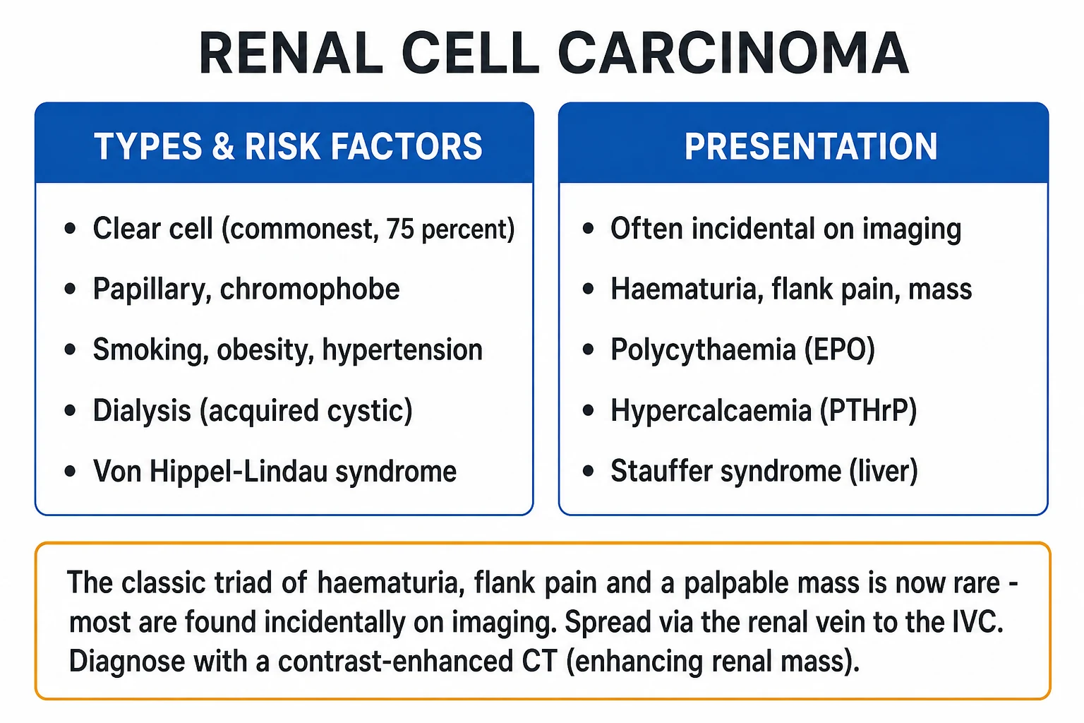
Renal cell carcinoma (RCC) is an adenocarcinoma arising from the proximal convoluted tubular epithelium and the commonest primary malignancy of the kidney (about 80 to 90 percent of all renal malignancies). Clear-cell RCC is the dominant subtype (about 75 percent) and is driven by bi-allelic inactivation of the VHL tumour-suppressor gene on chromosome 3p25, with consequent accumulation of hypoxia-inducible factor (HIF) and up-regulation of VEGF, PDGF, EPO, GLUT-1 and carbonic anhydrase IX. Established risk factors are smoking, obesity, hypertension, end-stage kidney disease with acquired cystic change, and the hereditary syndromes (Von Hippel-Lindau). Most tumours are now detected incidentally on imaging; the classic triad of haematuria, flank pain and a palpable mass is now rare. RCC is notorious for paraneoplastic syndromes (polycythaemia from ectopic erythropoietin, hypercalcaemia from PTHrP, Stauffer syndrome of non-metastatic hepatic dysfunction) and for venous spread via the renal vein into the inferior vena cava. Diagnosis is by multiphase contrast-enhanced CT showing an enhancing renal mass (more than 15 to 20 Hounsfield units). Localised disease is cured by partial or radical nephrectomy; advanced disease, which is chemo-resistant (multi-drug-resistance / P-glycoprotein pumps), is now treated with immunotherapy combinations (nivolumab plus ipilimumab, pembrolizumab plus axitinib or lenvatinib) and tyrosine-kinase inhibitors (sunitinib, cabozantinib) — a revolution that has transformed metastatic RCC from a uniformly fatal disease into a survivable, often chronic one.
On this page & tools
Your progress
Saved locally on this device.
Exam tags
Red flags
Overview & Definition
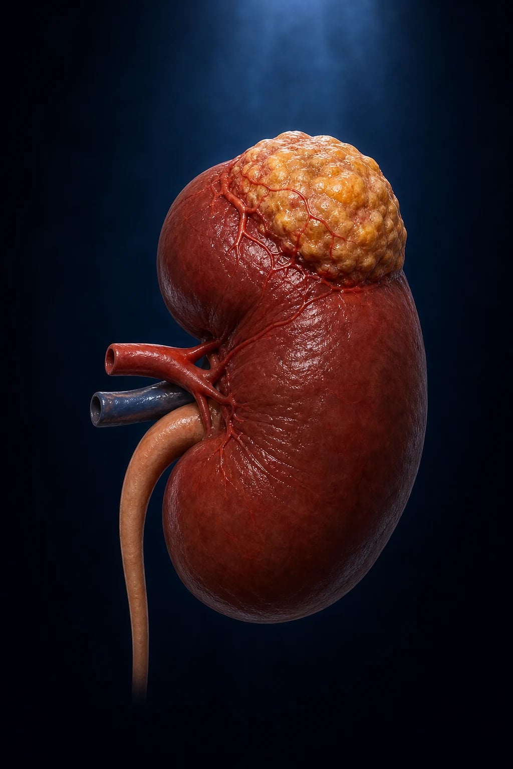
Renal cell carcinoma (RCC) is a heterogeneous group of adenocarcinomas arising from the epithelium of the renal tubules, of which the clear-cell subtype is dominant (about 75 percent) and the most aggressive. RCC accounts for about 80 to 90 percent of all primary renal malignancies in adults and roughly 2 to 3 percent of all adult cancers worldwide, making it the commonest primary kidney cancer.[1][8]
The clinical flavour of RCC is stealthy: most tumours are now discovered incidentally on imaging performed for an unrelated reason, and the classic triad of haematuria, flank pain and a palpable flank mass is now seen in fewer than 10 percent of cases and usually indicates advanced disease.[1] RCC is also the archetypal "internist's tumour" — it is famous for its paraneoplastic syndromes (polycythaemia, hypercalcaemia, Stauffer syndrome) and for propensity to invade the renal vein and spread along the inferior vena cava as a tumour thrombus. The disease is notably chemo-resistant (high expression of P-glycoprotein / multi-drug-resistance pumps and a low proliferative index), which made it a grim diagnosis in the cytokine era but, since 2005, has become the proving ground for targeted anti-angiogenics and, since 2015, for immune checkpoint inhibitor combinations — together one of the great success stories of modern oncology.[2][9]
The two clinical skills that decide an RCC answer are (i) recognising a solid enhancing renal mass as RCC until proven otherwise and staging it; and (ii) choosing between curative surgery for localised disease and modern immunotherapy/targeted therapy for advanced disease, stratified by IMDC prognostic score.[3][6]
Classification
RCC is classified on three axes — histological subtype, hereditary vs sporadic, and the Bosniak system for cystic lesions (which determines whether a renal cyst is benign or malignant on imaging). [1]
Histological subtypes (the 2016/2022 WHO classification — high-yield)[1][9]
| Subtype | Frequency | Cell / genetic driver | Behaviour |
|---|---|---|---|
| Clear-cell RCC | ~75% | VHL loss (3p25), lipid/glycogen-rich clear cytoplasm, highly vascular | Most aggressive; chemo-resistant; responsive to TKIs and ICI |
| Papillary type 1 | ~5% | MET gain (chromosome 7 trisomy) | Indolent; hereditary papillary RCC |
| Papillary type 2 | ~10% | NRF2-ARE activation; FH loss (HLRCC) | More aggressive |
| Chromophobe | ~5% | Multiple chromosome losses (1, 2, 6, 10, 13, 17, 21); FLCN (Birt-Hogg-Dube) | Often indolent |
| Collecting-duct | under 1% | Aggressive; medullary subtype in sickle-cell trait | Very poor prognosis |
| Translocation (MiT-family, TFE3/TFEB) | rare | Younger patients; translocation t(6;11), Xp11 | Distinct biology |
| Medullary | rare | Sickle-cell trait; young Black patients | Extremely aggressive |
Hereditary vs sporadic RCC[2][9]
| Syndrome | Gene (locus) | Subtype | Hallmarks |
|---|---|---|---|
| Von Hippel-Lindau | VHL (3p25) | Clear-cell (bilateral, multifocal) | CNS + retinal haemangioblastomas, phaeochromocytoma, pancreatic neuroendocrine tumours |
| Hereditary papillary RCC | MET (7q31) | Papillary type 1 | Late-onset, indolent |
| Birt-Hogg-Dube | FLCN (17p11) | Chromophobe / oncocytic hybrid | Lung cysts, spontaneous pneumothorax, skin fibrofolliculomas |
| Hereditary leiomyomatosis-RCC (HLRCC) | FH (fumarate hydratase, 1q43) | Papillary type 2 | Uterine and cutaneous leiomyomas; aggressive RCC |
| Tuberous sclerosis | TSC1 / TSC2 | Angiomyolipoma + (occasionally) clear-cell | Hamartomas, epilepsy, adenoma sebaceum |
Sporadic RCC
- Single tumour, unilateral, older patient (6th to 7th decade)
- Bi-allelic VHL inactivation by mutation plus 3p loss in ~75% of clear-cell cases (a two-hit mechanism)
- Smoking, obesity, hypertension and dialysis are the modifiable drivers
- Outcome determined by stage and IMDC prognostic score
Hereditary RCC
- Younger patients, bilateral and multifocal tumours
- Each syndrome maps to a single gene and a characteristic subtype (VHL, MET, FH, FLCN, TSC)
- Syndromic stigmata give the diagnosis (haemangioblastomas, leiomyomas, lung cysts)
- Surveillance is lifelong; nephron-sparing surgery preferred; genetic counselling
Bosniak cyst classification
- I — benign simple cyst: water density, hairline wall, no enhancement (malignant risk ~0%)
- II — few hairline septa, fine calcification, no enhancement (~0%)
- IIF — more septa, thickened wall, no enhancement; requires follow-up imaging (~5 to 10%)
- III — thick septa/wall with measurable enhancement (~50% malignant — biopsy or excise)
- IV — clearly malignant enhancing nodular mass (~90%) — surgical
Epidemiology & Risk Factors
RCC accounts for roughly 431,000 new cases and 155,000 deaths annually worldwide (GLOBOCAN 2020); incidence is two to three times higher in men than in women, peaks in the 6th to 7th decade, and is substantially higher in developed countries — partly driven by incidental detection on cross-sectional imaging.[8]
Renal cell carcinoma — epidemiology at a glance
Risk factors — recalled with the mnemonic "SOAP plus dialysis and genes"[1][8]
- S — Smoking. A dose-dependent relationship; smokers have a roughly 50 percent higher risk than never-smokers; risk falls after cessation. The strongest single environmental risk factor.
- O — Obesity. Each 5 kg/m² rise in BMI increases RCC risk by about 25 to 30 percent; the link is mediated by oestrogens, insulin-like growth factor-1, adipokines (leptin), lipid peroxidation, and chronic inflammation. RCC is one of the cancers most strongly linked to obesity.
- A — Adult / older age. Peak incidence in the 6th to 7th decade.
- P — long-standing hypertension (H) and analgesic phenacetin abuse (historical). Hypertension is an independent risk factor — partly confounded by antihypertensive drugs, but the association persists.
- Dialysis / ESKD — acquired cystic kidney disease. After 3 to 5 years on dialysis, up to 90 percent of patients develop acquired cystic change, with a 3- to 7-fold increased incidence of RCC; tumours are often multiple and in shrunken native kidneys.
- Genes / hereditary syndromes — see above; VHL is the prototype.
- Occupational / chemical exposures — cadmium, trichloroethylene, asbestos, petroleum products, and chronic lead exposure are weakly associated.
- Race / geography — incidence is higher in North American and European populations; in India and many low- and middle-income countries, RCC presents at a more advanced stage (less incidental detection).[8]
Pathophysiology
The molecular biology of clear-cell RCC is one of the best-understood of any solid tumour, and it directly explains every modern therapy.[2][9]
The VHL-HIF axis in clear-cell RCC
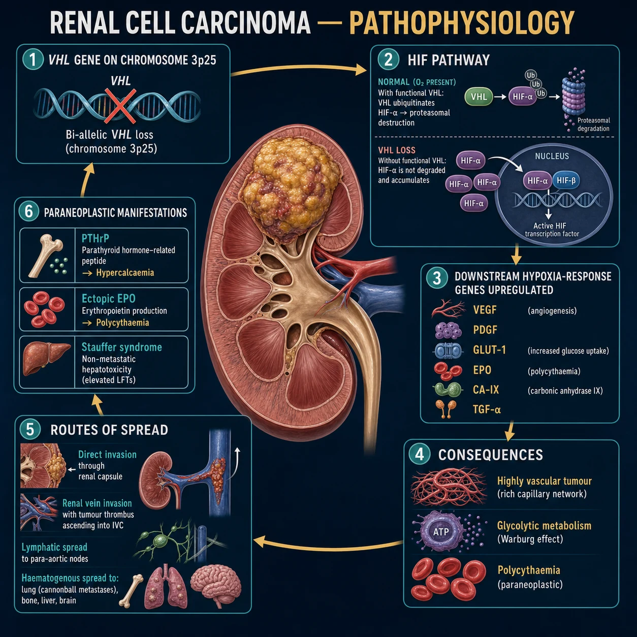
In normal physiology the VHL protein (encoded on chromosome 3p25) is part of an E3 ubiquitin-ligase complex that, under normoxic conditions, recognises and ubiquitinates the alpha subunit of hypoxia-inducible factor (HIF-1alpha and HIF-2alpha), tagging it for proteasomal degradation. HIF-alpha hydroxylation by prolyl hydroxylase (an oxygen-, iron- and 2-oxoglutarate-dependent enzyme) is the molecular signal of "oxygen present" that allows VHL to bind and destroy HIF. [1]
In clear-cell RCC, bi-allelic inactivation of VHL (one copy lost by chromosome 3p deletion, the second by point mutation or promoter hypermethylation — a classic Knudson two-hit mechanism) removes this brake. HIF-alpha accumulates even in the presence of oxygen, translocates to the nucleus, dimerises with HIF-beta, and drives transcription of a cassette of hypoxia-response genes:[2][9]
- VEGF (vascular endothelial growth factor) and PDGF — drive the exuberant angiogenesis that makes RCC a golden-yellow, hypervascular tumour.
- GLUT-1 and glycolytic enzymes — produce the Warburg effect (aerobic glycolysis) and the clear, lipid- and glycogen-laden cytoplasm that gives the cell its name.
- Erythropoietin (EPO) — explains the paraneoplastic polycythaemia unique to RCC (the tumour arises in the very cell that, physiologically, regulates EPO).
- Carbonic anhydrase IX (CA-IX / G250) — a sensitive immunohistochemical marker of clear-cell RCC and a therapeutic target.
- TGF-alpha, EGFR ligands — autocrine growth stimulation. [1]
Other molecular subtypes
- Papillary type 1 — activating mutations / gain of MET (often chromosome 7 trisomy), driving the HGF-MET-RAF-MAPK pathway.[9]
- Papillary type 2 (HLRCC) — loss of fumarate hydratase (FH); accumulation of fumarate inhibits prolyl hydroxylases (pseudohypoxia) and causes 2-succinocysteine accumulation; aggressive.
- Chromophobe — combined loss of multiple chromosomes (1, 2, 6, 10, 13, 17, 21); FLCN loss in Birt-Hogg-Dube.[9]
Why RCC is chemo-resistant
Clear-cell RCC over-expresses P-glycoprotein (MDR-1 / ABCB1) multi-drug-resistance pumps, has a low proliferative index, and expresses high levels of detoxifying enzymes (glutathione-S-transferases) — all of which render conventional cytotoxic chemotherapy largely ineffective, motivating the development of targeted and immune-based therapies.[1]
Routes of spread
- Direct — growth through the renal capsule into perinephric fat and beyond Gerota fascia (T3 / T4).
- Venous (the classic) — invasion of the renal vein, then ascent as a tumour thrombus into the IVC and occasionally the right atrium; may produce lower-limb oedema, Budd-Chiari physiology, or pulmonary tumour embolism.
- Lymphatic — to para-aortic, paracaval, and retroperitoneal nodes.
- Haematogenous — to lung (cannonball metastases), bone (osteolytic, expansile, pathological fracture), liver, brain, and skin (rapidly growing vascular nodules). [1]
Paraneoplastic syndromes — reproduced verbatim[1][2]
| Syndrome | Mediator | Feature |
|---|---|---|
| Polycythaemia | Ectopic erythropoietin (EPO) from tumour cells | Raised haematocrit; RCC is the classic cause of paraneoplastic polycythaemia |
| Hypercalcaemia | PTHrP (parathyroid hormone-related peptide) | Symptomatic hypercalcaemia; a negative prognostic marker (IMDC criterion) |
| Stauffer syndrome | Unknown (cytokine, ?IL-6) | Non-metastatic, reversible hepatic dysfunction — raised ALP, hypoalbuminaemia, prolonged PT, abnormal bilirubin; resolves after nephrectomy if the tumour is removed |
| Cushing syndrome | Ectopic ACTH | Rare |
| Anaemia / amyloidosis | IL-6, AA amyloid | Anaemia disproportionate to stage is paradoxically common in advanced disease |
| Neuromyopathy / polymyalgia | Autoimmune / paraneoplastic | Proximal weakness |
Clinical Presentation
The modern presentation is incidental: an asymmetric solid renal mass found on ultrasound or CT performed for another reason (abdominal pain, investigation of hypertension, incidental finding on trauma CT, antenatal ultrasound). Up to 50 to 60 percent of RCCs are now diagnosed incidentally, and these tumours are typically smaller and at a lower stage.[1]
The classic triad (now rare)[1]
- Haematuria — gross or microscopic, typically painless, from tumour erosion into the collecting system.
- Flank pain — from capsular stretch, haemorrhage into the tumour, or clot colic.
- Palpable flank or abdominal mass — a firm, non-tender, ballotable mass. [1]
When all three coexist, advanced disease is likely. The classic "too-late" presentations remain important exam favourites:[1]
- Cannonball lung metastases with haemoptysis on a chest X-ray.
- Pathological fracture from a lytic bone metastasis (especially rib, spine, pelvis, long bones).
- New-onset left varicocele in a man, especially one that does not empty on lying down — from obstruction of the left testicular vein by a left renal tumour thrombus. (A right-sided varicocele, which is rare anatomically, suggests IVC obstruction.) [1]
Systemic / B symptoms
Weight loss, fatigue, fever (an under-recognised paraneoplastic feature — RCC is in the differential of fever of unknown origin), night sweats, and anaemia disproportionate to disease stage (from chronic inflammation / IL-6). [1]
Atypical presentations[1]
- Elderly — anaemia, weight loss, vague abdominal discomfort; the haematuria may be dismissed; the mass may be attributed to "constipation".
- Dialysis / ESKD patient — new mass in a shrunken native kidney, falling EPO requirement (the tumour may paradoxically raise EPO), haematuria in an anuric patient.
- Pregnancy — incidental finding on obstetric ultrasound; management tailored to gestation and risk.
- Hereditary syndrome patient (VHL) — known diagnosis; multifocal, bilateral tumours from the second decade onwards; surveillance imaging drives detection.
- Young patient with sickle-cell trait — consider renal medullary carcinoma (devastatingly aggressive). [1]
Differential Diagnosis
Not every renal mass is RCC. The complete differential of a renal mass, with distinguishing features:[1]
| Differential | Key distinguishing features |
|---|---|
| Simple renal cyst (Bosniak I) | Anechoic, water density, hairline wall, no enhancement on CT; extremely common; asymptomatic |
| Complex cyst (Bosniak IIF/III/IV) | Septa, calcification, wall thickening, enhancement → stratify by Bosniak |
| Angiomyolipoma | Fat density (under -20 HU) on CT; associated with tuberous sclerosis; may bleed if large (>4 cm) |
| Fat-poor angiomyolipoma | Diagnostic pitfall — looks like RCC; HMB-45 positive on biopsy |
| Oncocytoma | Benign; classically a central scar, but radiologically indistinguishable from RCC — diagnosis often on histology after resection |
| Transitional cell (urothelial) carcinoma of renal pelvis | Filling defect on pyelogram / CTU, haematuria dominant, may be associated with bladder cancer; needs nephroureterectomy with bladder cuff |
| Wilms tumour (nephroblastoma) | Child under 5; large, rapidly growing abdominal mass |
| Renal abscess / pyonephrosis | Fever, rigors, positive urine culture, diabetes, obstructing stone; gas within collection |
| Xanthogranulomatous pyelonephritis | Chronic, staghorn calculus, non-functioning kidney, replacement by lipid-laden macrophages |
| Renal tuberculosis | Cavitating, calcified "putty kidney", sterile pyuria, constitutional symptoms |
| Renal infarct | Sudden flank pain, atrial fibrillation, wedge-shaped non-enhancing lesion |
| Renal lymphoma | Usually multiple, bilateral, hypovascular masses or diffuse infiltration; widespread disease |
| Metastasis to kidney | From lung, breast, melanoma; usually in advanced systemic disease |
| Adrenal tumour | Separated from the kidney on multiplanar imaging; endocrine features |
Renal mass biopsy is indicated for (i) metastatic disease before commencing systemic therapy, (ii) enrolling in active surveillance, (iii) suspicion of lymphoma, infection, or metastasis, or (iv) syndromic/atypical tumours. It is avoided when the imaging is classically malignant and the patient will proceed straight to surgery (historical concern about tract seeding, now small but real).[1]
Clinical & Bedside Assessment
Abdominal examination of a renal mass
- Inspection — abdominal distension, visible mass in a thin patient, scars of previous surgery.
- Palpation — bimanual (ballottement) technique. With the patient relaxed and supine, place one hand anteriorly in the lumbar region and the other posteriorly in the renal angle; on deep inspiration the kidney descends and can be "ballotted" between the two hands. A normal right kidney is occasionally palpable in a thin patient; the left kidney is virtually never palpable unless enlarged.
- Percussion — a renal mass is dull to percussion; resonance suggests an overlying gas-filled bowel.
- Auscultation — listen for a renal artery bruit (renovascular disease co-existing). [1]
Examination for paraneoplastic features and metastases
- Hands and face — plethora / plethora (polycythaemia), clubbing (paraneoplastic), leuconychia (hypoalbuminaemia / Stauffer).
- Cervical nodes — Virchow's node (left supraclavicular, Troisier's sign).
- Chest — varicocele (especially left), signs of pleural effusion or cannonball lung metastases.
- Abdomen — hepatomegaly / hepatotoxicity (Stauffer), ascites, caput medusae (IVC obstruction).
- Lower limbs — oedema from IVC thrombus; deep-vein thrombosis (RCC is a hypercoagulable state).
- Spine / bones — tenderness at sites of metastasis, neurological deficit (cord compression).
- Skin — vascular cutaneous metastases; fibrofolliculomas (Birt-Hogg-Dube). [1]
Bedside assessment of severity
Vital signs, performance status (Karnofsky / ECOG), evidence of haemorrhagic shock (massive haematuria), hypercalcaemic crisis (dehydration, confusion, constipation), and cord compression (back pain, weakness, sensory level, urinary retention — a surgical and oncological emergency). [1]
Investigations
First-line work-up of a confirmed or suspected renal mass[1]
- Multiphase contrast-enhanced CT of the abdomen and pelvis — the gold standard. Phases: non-contrast (baseline density, ~20 to 40 HU), corticomedullary (early arterial), nephrogenic (parenchymal), and excretory (collecting system). An enhancement of more than 15 to 20 HU between non-contrast and contrast phases defines a solid mass suspicious for RCC. CT also defines size, renal-vein / IVC thrombus, nodal disease, and adrenal/liver involvement.
- CT chest (or chest X-ray) — to detect lung metastases (cannonball lesions).
- Full blood count, U&E, LFT, calcium, coagulation — anaemia, polycythaemia, Stauffer syndrome pattern (raised ALP, abnormal PT, hypoalbuminaemia), hypercalcaemia, renal function.
- Urinalysis — haematuria; cytology if upper-tract TCC suspected.
- MRI — when CT contrast is contraindicated (allergy, AKI/CKD, pregnancy), for detailed mapping of IVC tumour thrombus, and for characterising small indeterminate masses.
- Bone scan — only if bone pain, raised alkaline phosphatase, or pathological fracture; not routine (RCC bone metastases are often osteolytic and can be fludeoxyglucose-avid).
- Renal mass biopsy — see Differential Diagnosis above.
- Erythropoietin level, PTHrP — selected cases to confirm a paraneoplastic syndrome. [1]
Bosniak cyst classification (reproduced verbatim)[1]
| Category | Imaging features | Malignancy risk | Management |
|---|---|---|---|
| I | Simple benign cyst: water density, hairline-thin wall, no septa / calcification / solid component / enhancement | ~0% | No follow-up |
| II | Minimally complex: a few hairline septa, fine calcification, uniformly high-attenuation (under 3 cm) non-enhancing | ~0% | No follow-up |
| IIF | "Follow-up": multiple hairline septa, minimally thickened wall/septa, no measurable enhancement, hyperdense (over 3 cm) | ~5 to 10% | Serial imaging (6, 12, 24 months) |
| III | Indeterminate / malignant: thick irregular wall/septa, measurable enhancement | ~50% | Biopsy or surgical excision (partial nephrectomy) |
| IV | Clearly malignant: enhancing soft-tissue component adjacent to wall/septa | ~90% | Surgical resection |
AJCC 8th edition TNM staging (summary)[1]
- T1a — tumour 4 cm or less, limited to kidney.
- T1b — tumour over 4 cm up to 7 cm, limited to kidney.
- T2a — tumour over 7 cm up to 10 cm, limited to kidney.
- T2b — tumour over 10 cm, limited to kidney.
- T3a — into major renal vein / perinephric fat / renal sinus, not beyond Gerota fascia.
- T3b — into the vena cava below the diaphragm.
- T3c — into the vena cava above the diaphragm or its wall.
- T4 — beyond Gerota fascia (including ipsilateral adrenal).
- N0 / N1 — none / metastasis in regional nodes.
- M0 / M1 — none / distant metastasis. [1]
IMDC (Heng) prognostic score for metastatic RCC — reproduced verbatim[3][6]
One point each for: [1]
- Time from diagnosis to initiation of systemic therapy under 1 year.
- Karnofsky performance status under 80.
- Haemoglobin below the lower limit of normal.
- Corrected calcium above the upper limit of normal.
- Neutrophil count above the upper limit of normal.
- Platelet count above the upper limit of normal. [1]
Risk groups: favourable = 0; intermediate = 1 to 2; poor = 3 to 6. The IMDC score (a refinement of the original Memorial Sloan-Kettering / Motzer score) is the central prognostic tool and drives first-line choice in metastatic RCC. [1]
Management — Resuscitation
Although most RCCs are detected electively, several time-critical scenarios demand emergency management:[1]
Massive gross haematuria from a renal tumour
- ABCDE; two large-bore cannulae; cross-match blood; intravenous fluid resuscitation; transfuse for haemodynamic instability or symptomatic anaemia.
- Three-way urinary catheter with bladder irrigation to prevent clot retention; consider cystoscopic clot evacuation.
- Selective renal artery embolisation (interventional radiology) — first-line to arrest bleeding in unfit patients or as a bridge to surgery.
- Palliative or emergency nephrectomy if embolisation fails or the tumour is resectable. [1]
Hypercalcaemic crisis (paraneoplastic, PTHrP-mediated)
- Aggressive isotonic saline (3 to 6 L over 24 hours) — restores intravascular volume and promotes calciuria.
- IV bisphosphonate — zoledronic acid 4 mg IV over 15 minutes (or pamidronate 60 to 90 mg); onset over 2 to 4 days.
- Denosumab 120 mg subcutaneously (consider in refractory disease or renal impairment).
- Calcitonin 4 to 8 IU/kg subcutaneously every 12 hours for rapid (but transient) effect while bisphosphonate works.
- Treat the tumour — definitive systemic therapy or nephrectomy where possible. [1]
IVC tumour thrombus with embolic risk
- Consider therapeutic anticoagulation (low-molecular-weight heparin preferred) weighed against bleeding risk.
- Define the cephalad extent of thrombus with MRI / contrast CT — levels (Novick / Mayo):
- Level 0 — renal vein only.
- Level 1 — IVC under 2 cm above renal vein.
- Level 2 — IVC over 2 cm below the hepatic veins.
- Level 3 — at / above hepatic veins (intrahepatic IVC).
- Level 4 — suprahepatic IVC into the right atrium (requires cardiopulmonary bypass, often a combined cardiothoracic-urological procedure).[1]
- Pre-operative renal artery embolisation may be used to reduce vascularity in large tumours.
Metastatic spinal cord compression (RCC bone metastasis)
- Dexamethasone 16 mg orally or IV immediately, then 16 mg daily (in divided doses) — reduces oedema.
- Urgent whole-spine MRI (within 24 hours).
- Neurosurgical / orthopaedic opinion for decompression and stabilisation if there is spinal instability, a single compressive site, or neurological deficit (esp. if non-ambulatory at presentation).
- Urgent palliative radiotherapy (RCC is radio-resistant but radiotherapy relieves compression and pain).
- Treat the systemic disease. [1]
Management — Definitive & Stepwise
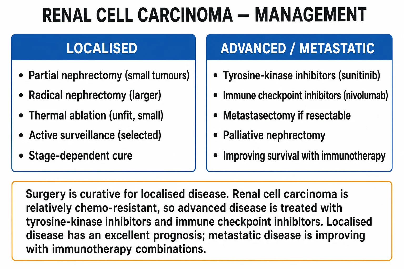
Localised RCC (T1 to T3, M0) — surgery is curative[1]
The choice between partial and radical nephrectomy depends on tumour size, location, the contralateral kidney, and patient comorbidity. [1]
- Partial nephrectomy (nephron-sparing surgery) — preferred for T1 tumours (especially under 4 cm), solitary kidney, bilateral tumours, chronic kidney disease, and hereditary syndromes. Removes the tumour with a margin of normal parenchyma while preserving renal function. Equivalent oncological outcome to radical nephrectomy for T1 tumours; lower risk of long-term CKD.
- Radical nephrectomy — for larger (T2) tumours, central tumours not amenable to partial nephrectomy, or when partial is technically not feasible. Removes the kidney, perinephric fat and Gerota fascia, with ipsilateral adrenalectomy when the tumour involves the upper pole or directly invades the adrenal; hilar lymph node dissection for staging.
- Minimally invasive (laparoscopic / robotic) approaches — less pain, shorter stay, equivalent oncology, especially for T1 to T2 tumours.
- Thermal ablation — radiofrequency ablation (RFA) and cryoablation for small (under 3 to 4 cm) tumours in elderly / unfit patients or those with multiple comorbidities; lower morbidity but higher local recurrence than surgery.
- Active surveillance — for small renal masses (under 2 to 4 cm) in patients with limited life expectancy; serial imaging with intervention if growth. Validated by the DISSRM registry.[1]
Advanced / metastatic RCC — chemo-resistant; immunotherapy and TKI combinations[1][3]
RCC is the poster-child for modern immuno-oncology. Cytotoxic chemotherapy has no meaningful role. Cytokine therapy (high-dose IL-2, interferon-alpha) was historical first-line; IL-2 can produce durable complete remissions in a small minority but is highly toxic and now largely superseded.[5]
Modern first-line combinations — agent, target, regimen, evidence
| Regimen | Target | Typical regimen | Landmark trial |
|---|---|---|---|
| Nivolumab + ipilimumab | PD-1 + CTLA-4 | Nivolumab 3 mg/kg plus ipilimumab 1 mg/kg every 3 weeks x 4, then nivolumab 6 mg/kg (or 240 mg flat) every 2 weeks (or 480 mg every 4 weeks) | CheckMate 214 — superior overall survival vs sunitinib in intermediate/poor-risk ccRCC; 8-year survival over 25 percent[3] |
| Pembrolizumab + axitinib | PD-1 + VEGFR TKI | Pembrolizumab 200 mg every 3 weeks (or 400 mg every 6 weeks); axitinib 5 mg twice daily | KEYNOTE-426 — superior OS and PFS vs sunitinib[6] |
| Pembrolizumab + lenvatinib | PD-1 + VEGFR1-3 + FGFR + PDGFR TKI | Pembrolizumab 200 mg every 3 weeks; lenvatinib 20 mg once daily | CLEAR — improved OS, PFS and response vs sunitinib; the new standard for many patients[7] |
| Nivolumab + cabozantinib | PD-1 + VEGFR + MET + AXL TKI | Nivolumab 240 mg every 2 weeks; cabozantinib 40 mg daily | CheckMate 9ER |
| Sunitinib (single-agent TKI) | VEGFR2, PDGFR, KIT, FLT3, RET | 50 mg once daily, 4 weeks on / 2 weeks off, or 2 weeks on / 1 week off | The original targeted therapy (2006) that displaced cytokines[5] |
| Pazopanib | VEGFR1-3, PDGFR, KIT | 800 mg once daily | COMPARZ — non-inferior to sunitinib |
| Cabozantinib | VEGFR2, MET, AXL | 60 mg once daily | first-line or after ICI/TKI |
| Lenvatinib + everolimus | TKI + mTOR | Lenvatinib 18 mg plus everolimus 5 mg daily | After anti-angiogenic failure |
| Everolimus / temsirolimus | mTOR | Everolimus 10 mg daily; temsirolimus 25 mg IV weekly | Later-line; temsirolimus first-line in poor-risk |
Cytoreductive nephrectomy and metastasectomy
- Cytoreductive nephrectomy (removing the primary tumour in the presence of metastatic disease) was a cornerstone of the cytokine era. In the modern immunotherapy era its role is more selective — the CARMENA trial showed that sunitinib alone was non-inferior to nephrectomy followed by sunitinib in patients with intermediate/poor-risk metastatic ccRCC, so nephrectomy is now reserved for fit patients with limited metastatic burden, a good performance status, and a primary tumour that is symptomatic or has a heavy tumour thrombus.[1]
- Metastasectomy (surgical excision of oligometastatic disease, e.g. a single lung metastasis or solitary bone lesion) can produce prolonged disease-free survival in selected patients.
Management algorithm by IMDC risk group[3][6][7]
- Favourable-risk (IMDC 0) — TKI monotherapy (e.g. sunitinib, pazopanib) or an ICI + TKI combination (pembrolizumab + axitinib); ICI combination toxicity must be weighed against the indolent biology.
- Intermediate / poor-risk (IMDC 1 to 6) — ICI-based doublet first-line: nivolumab + ipilimumab, pembrolizumab + axitinib, or pembrolizumab + lenvatinib are all current standards.
- Progression after ICI/TKI — switch TKI class (e.g. to cabozantinib), lenvatinib + everolimus, or tivozanib.
- Non-clear-cell histologies — extrapolated regimens; clinical trial enrolment encouraged. [1]
Specific Subtypes & Scenarios
Clear-cell RCC (75 percent)[1][2]
The dominant subtype; VHL-driven, highly vascular, golden-yellow, with clear, lipid-laden cytoplasm and a delicate "chicken-wire" vascular network. Chemo-resistant; responsive to anti-VEGF TKIs and immune checkpoint inhibitors. CA-IX positive, PAX8 / RCC-marker / CD10 / vimentin positive, CK7 negative. [1]
Papillary RCC — type 1 and type 2[9]
- Type 1 — papillae lined by a single layer of small cells with scant pale cytoplasm; MET-driven; indolent; basis of hereditary papillary RCC; CD10, AMACR positive, CK7 positive.
- Type 2 — pseudostratified, eosinophilic cells, FH loss (HLRCC); aggressive; often advanced at diagnosis. [1]
Chromophobe RCC[9]
Pale, eosinophilic, plant-cell-like cells with perinuclear halos; combined chromosome losses; FLCN (Birt-Hogg-Dube); usually indolent; CK7 positive, CD117 positive. Differential from oncocytoma can be challenging. [1]
Collecting-duct and renal medullary carcinoma
Both rare, both devastatingly aggressive. Collecting-duct carcinoma arises in the Bellini duct of the medulla, presents at an advanced stage, and is chemo-resistant. Renal medullary carcinoma occurs almost exclusively in patients with sickle-cell trait or disease (typically young Black patients), is rapidly fatal, and presents with haematuria, flank pain and weight loss. [1]
Translocation (MiT-family) RCC
Defined by translocations involving TFE3 (Xp11) or TFEB (t(6;11)); affects children and young adults; PSA / melanocytic markers may be positive; distinct biology and chemoresistance. [1]
Cystic RCC and the Bosniak IV cyst
A multilocular cystic renal neoplasm of low malignant potential (formerly "multilocular cystic RCC") — cysts lined by clear cells; excellent prognosis after resection. Bosniak IV cysts are managed as malignant. [1]
Von Hippel-Lindau disease[2][9]
Autosomal-dominant, VHL gene on 3p25. Patients develop bilateral, multifocal clear-cell RCC (and CNS and retinal haemangioblastomas, phaeochromocytoma, pancreatic neuroendocrine tumours and cysts, endolymphatic sac tumours, epididymal/broad-ligament cystadenomas). Management: lifelong surveillance imaging from adolescence, nephron-sparing surgery for tumours over 3 cm, genetic counselling and testing of relatives. A "canary in the coal mine" for the VHL-HIF biology that also drives sporadic ccRCC. [1]
RCC in ESKD / dialysis (acquired cystic disease)[1]
Patients on long-term dialysis develop acquired cystic kidney disease, with a 3- to 7-fold increased risk of RCC — often multifocal and in shrunken native kidneys. Many centres perform annual or biennial screening ultrasound of native kidneys in dialysis patients; native nephrectomy may be considered at transplantation. [1]
Complications & Pitfalls
Local
- Renal vein and IVC tumour thrombus — risk of pulmonary embolism, Budd-Chiari physiology, lower-limb oedema, caput medusae; dictates surgical complexity.
- Local recurrence after nephrectomy (especially with positive surgical margins or T3 disease).
- Tumour haemorrhage — spontaneous bleed into the tumour causing flank pain, Wunderlich syndrome (massive non-traumatic renal haemorrhage).
- Obstruction of the collecting system with clot or tumour → hydronephrosis. [1]
Distant
- Cannonball lung metastases with haemorrhage / haemoptysis.
- Pathological fracture (osteolytic, expansile).
- Spinal cord compression — emergency.
- Brain metastases — haemorrhagic, neurological emergency.
- Hepatic failure from massive liver involvement. [1]
Paraneoplastic
- Hypercalcaemia (PTHrP), polycythaemia (EPO), Stauffer syndrome, AA amyloidosis (nephrotic syndrome), non-metastatic neuromyopathy. [1]
Treatment-related
- Post-nephrectomy CKD progression — a major reason partial nephrectomy is preferred for small tumours; protect residual function with ACE-inhibitor / SGLT2-inhibitor, BP control.
- Immunotherapy-related adverse events (irAEs) — immune pneumonitis, colitis, hepatitis, endocrinopathy (thyroiditis, hypophysitis, type-1 diabetes, adrenal insufficiency), dermatitis, myocarditis; managed with corticosteroids and holding the ICI (severe).
- TKI toxicities — hypertension (driven by VEGF blockade), hand-foot skin reaction, hypothyroidism, proteinuria, cardiotoxicity (heart failure), diarrhoea, fatigue, hair depigmentation.
- mTOR-inhibitor toxicities — pneumonitis, hyperglycaemia, mucositis, hyperlipidaemia. [1]
Classic diagnostic pitfalls
- Assuming a complex cyst is benign — under-stage a Bosniak III/IV cyst.
- Missing an IVC tumour thrombus on pre-operative imaging (catastrophic at surgery).
- Over-biopsying a typical angiomyolipoma (diagnosed radiologically by fat).
- Failing to screen / image the contralateral kidney in VHL and other syndromes.
- Missing synchronous metastases (always stage with a chest CT before surgery).
- Confusing renal medullary carcinoma (sickle-cell trait, young) with a more indolent tumour. [1]
Prognosis & Disposition
Five-year survival by AJCC stage[1]
- Stage I (T1 N0 M0) — approximately 80 to 95 percent (excellent after surgery).
- Stage II (T2 N0 M0) — approximately 70 to 80 percent.
- Stage III (T3 / N1, M0) — approximately 50 to 70 percent.
- Stage IV (any T, any N, M1) — approximately 10 to 15 percent historically, rising toward 25 to 30 percent and beyond with modern ICI combinations in selected groups.[3]
Predictors of poor outcome
- High ISUP / Fuhrman nuclear grade and sarcomatoid differentiation.
- Tumour necrosis, lymphovascular invasion.
- Nodal or distant metastases, high IMDC score, anaemia, hypercalcaemia, low KPS, short time-to-treatment.[3]
- Venous tumour thrombus (worsens outcome; level-dependent).
- Non-clear-cell histologies (collecting-duct, medullary, HLRCC) generally worse.
Surveillance after curative nephrectomy[1]
Driven by risk of recurrence (stage, grade, histology, margins). Typical EAU / NCCN-style schedule: [1]
- History, examination, bloods (eGFR, FBC, LFT, calcium) at each visit.
- Chest imaging (CXR or CT) and abdominal imaging (US or CT) at intervals scaled to risk — low risk: yearly for 3 to 5 years; high risk: CT abdomen and chest every 6 months for 3 years, then annually to 5 years.
- Renal function surveillance is essential (risk of CKD progression in the solitary kidney). [1]
Special Populations
- Pregnancy — MRI and ultrasound are the imaging modalities of choice (avoid radiation and iodinated contrast where possible). For small, low-risk tumours discovered in pregnancy, active surveillance with definitive surgery post-partum is reasonable; large or symptomatic tumours may require surgery during pregnancy (second trimester preferred).
- Chronic kidney disease / dialysis — lower threshold to screen native kidneys; nephron-sparing strongly favoured; consider native-kidney nephrectomy at transplantation for known tumours; CKD patients face competing risks.
- Hereditary syndromes (VHL, BHD, HLRCC, hereditary papillary) — bilateral, multifocal disease from a young age; lifelong surveillance; nephron-sparing surgery to defer dialysis; genetic counselling of the proband and first-degree relatives.
- Elderly / frail — active surveillance for small renal masses; ablation preferred when intervention indicated; weigh competing mortality.
- Solitary kidney — partial nephrectomy strongly preferred; ablative options; dialysis risk must be discussed.
- IVC / atrial tumour thrombus — multidisciplinary planning with cardiothoracic and vascular surgery; cardiopulmonary bypass for atrial (level 4) thrombus.
- Anticoagulated patient — balance bleeding (renal mass, surgery) vs thrombosis (IVC thrombus, malignancy); bridge peri-operatively with LMWH; avoid neuraxial procedures while anticoagulated.
- Sickle-cell trait — young patients with haematuria: consider renal medullary carcinoma. [1]
Evidence, Guidelines & Regional Differences
Landmark trials (know these by name)[3][4][5][6][7]
- Motzer 2007 — sunitinib vs interferon-alpha (NEJM, PMID 17215529) — first targeted therapy to improve progression-free and overall survival in metastatic RCC; the beginning of the TKI era.[5]
- Motzer / Escudier 2015 — CheckMate 025 (NEJM, PMID 26406148) — nivolumab (PD-1) vs everolimus after TKI failure; first immune checkpoint inhibitor to improve overall survival in RCC.[4]
- Motzer 2018 — CheckMate 214 (NEJM, PMID 29562145) — nivolumab + ipilimumab vs sunitinib first-line; superior OS in intermediate/poor-risk ccRCC; established dual ICI as first-line.[3]
- Rini 2019 — KEYNOTE-426 (NEJM, PMID 30779529) — pembrolizumab + axitinib vs sunitinib; superior OS, PFS, response.[6]
- Motzer 2021 — CLEAR (NEJM, PMID 33616314) — lenvatinib + pembrolizumab (or lenvatinib + everolimus) vs sunitinib; the strongest first-line OS/PFS benefit to date.[7]
- CARMENA — questioned the routine role of cytoreductive nephrectomy in the immunotherapy era.
- DISSRM registry — validated active surveillance for small renal masses.[1]
Regional guideline differences[1]
- EAU (European Association of Urology) 2024 and NCCN (US) 2024 broadly converge on ICI-based combinations first-line for intermediate/poor-risk metastatic ccRCC, with TKI monotherapy for favourable-risk disease or unfit patients.
- AUA 2017 active surveillance guideline codifies the role of surveillance for small renal masses (cT1).
- India / LMIC patterns (NCI / ICMR registries) — RCC presents at a more advanced stage (less incidental detection); access to ICI and TKI is variable, with cost a real constraint; sunitinib and pazopanib generics have improved affordability; interferon/IL-2 still used in resource-limited settings.[8]
Controversies
- Renal mass biopsy — historical avoidance (seeding) vs modern use in metastatic / surveillance settings.
- Cytoreductive nephrectomy in the immunotherapy era — selective rather than routine.
- Adrenalectomy at radical nephrectomy — increasingly omitted when the adrenal is radiologically normal (sparing does not compromise oncology).
- Surgical vs ablation vs surveillance for small renal masses — patient-centred. [1]
Exam Pearls
Risk factors — recall 'SOAP + DAG'
SOAP
Dose-dependent; ~50 percent higher risk
Each 5 kg/m² adds ~25 to 30 percent risk
Peak in the 6th to 7th decade
Independent risk factor
3- to 7-fold increased RCC risk after years on dialysis
Hereditary syndromes by gene and subtype
Paraneoplastic triad — recall 'E-P-Liver'
EPL
RCC is the classic paraneoplastic cause of polycythaemia
Symptomatic; an IMDC prognostic factor
Non-metastatic, reversible cholestasis; resolves after nephrectomy
Hereditary RCC syndromes by gene
G-V-M-B-H-T
Clear-cell; bilateral, multifocal; CNS/retinal haemangioblastomas
Hereditary papillary RCC type 1
Chromophobe / oncocytic; lung cysts, skin fibrofolliculomas
Papillary type 2; uterine/cutaneous leiomyomas; aggressive
Angiomyolipoma and occasional RCC
- Any solid renal mass that enhances over 15 to 20 HU on multiphase CT is RCC until proven otherwise.[1]
- RCC = adenocarcinoma of the proximal convoluted tubule (not the collecting system — that is TCC/urothelial).
- The classic triad (haematuria, flank pain, palpable mass) is rare (under 10 percent) and indicates advanced disease.
- New left varicocele that does not empty on lying down in a man = left renal tumour in the renal vein — image.
- RCC is the unique cancer that causes polycythaemia — via ectopic erythropoietin from the tumour cell itself.
- Cannonball metastases (well-circumscribed round lung lesions): differential = RCC, choriocarcinoma, sarcoma, occasionally thyroid.
- Clear-cell RCC = chromosome 3p deletion / VHL loss; papillary type 1 = chromosome 7 trisomy / MET; HLRCC = FH; Birt-Hogg-Dube = FLCN.
- Stauffer syndrome resolves after nephrectomy — a clinical pearl confirming its paraneoplastic nature.
- RCC is chemo-resistant (MDR-1 / P-glycoprotein); surgery cures local disease; ICI/TKI for advanced.
- First-line ICI combos for intermediate/poor-risk: nivolumab + ipilimumab (CheckMate 214), pembrolizumab + axitinib (KEYNOTE-426), pembrolizumab + lenvatinib (CLEAR).
- Bosniak III and IV cysts are managed as malignant; IIF requires follow-up imaging.
- Sunitinib (VEGFR TKI) is the textbook prototype targeted therapy for RCC (4 weeks on / 2 weeks off).[5]
- Cytoreductive nephrectomy is selective in the immunotherapy era (CARMENA).
Exam application bank (NEET-PG / INICET)
One-line answer
Renal cell carcinoma (RCC) is an adenocarcinoma arising from the proximal convoluted tubular epithelium and the commonest primary malignancy of the kidney (about 80 to 90 percent of all renal malignancies). Clear-cell RCC is the dominant subtype (about 75 percent) and is driven by bi-allelic inactivation of the VHL tumour-suppressor gene on chromosome 3p25, with consequent accumulation of hypoxia-inducible factor (HIF) and up-regulation of VEGF, PDGF, EPO, GLUT-1 and carbonic anhydrase IX. Established risk factors are smoking, obesity, hypertension, end-stage kidney disease with acquired cystic change, and the hereditary syndromes (Von Hippel-Lindau). Most tumours are now detected incidentally on imaging; the classic triad of haematuria, flank pain and a palpable mass is now rare. RCC is notorious for paraneoplastic syndromes (polycythaemia from ectopic erythropoietin, hypercalcaemia fro
Worked stems (answer without another resource)
Stem 1 — Classic presentation. Map symptoms to mechanism; name the first investigation and first treatment step with dose/route if drug therapy is standard. [1]
Stem 2 — Unstable / complicated. List red flags that force immediate resuscitation, theatre, ICU, antidote, or reperfusion — and what you do in the first 15 minutes. [1]
Stem 3 — Atypical group. Elderly, pregnancy, child, or immunocompromised: how presentation and thresholds change. [1]
Stem 4 — Differential trap. Name the three closest mimics and one discriminator for each. [1]
Stem 5 — Disposition. Who goes home with safety-netting, who is admitted, who needs HDU/ICU/theatre, and what follow-up is mandatory. [1]
Rapid viva checklist
- Definition + classification
- Pathophysiology chain
- Bedside signs / criteria
- Score with exact components (if any)
- Emergency bundle
- Definitive therapy with doses
- Complications of disease and of treatment
- Special populations
- Guideline/trial name if classic
- Three exam traps
Coverage self-check
If you cannot answer any stem above from this page alone, re-read the matching section — the page is intended to be self-sufficient for final-prof and NEET-PG/INICET questions on Renal Cell Carcinoma.
References
- [1]Hsieh JJ, Purdue MP, Signoretti S, et al. Renal cell carcinoma Nat Rev Dis Primers, 2017.PMID 28276433
- [2]Jonasch E, Walker CL, Rathmell WK. Clear cell renal cell carcinoma ontogeny and mechanisms of lethality Nat Rev Nephrol, 2021.PMID 33144689
- [3]Motzer RJ, Tannir NM, McDermott DF, et al. (CheckMate 214). Nivolumab plus Ipilimumab versus Sunitinib in Advanced Renal-Cell Carcinoma N Engl J Med, 2018.PMID 29562145
- [4]Motzer RJ, Escudier B, McDermott DF, et al. (CheckMate 025). Nivolumab versus Everolimus in Advanced Renal-Cell Carcinoma N Engl J Med, 2015.PMID 26406148
- [5]Motzer RJ, Hutson TE, Tomczak P, et al. Sunitinib versus interferon alfa in metastatic renal-cell carcinoma N Engl J Med, 2007.PMID 17215529
- [6]Rini BI, Plimack ER, Stus V, et al. (KEYNOTE-426). Pembrolizumab plus Axitinib versus Sunitinib for Advanced Renal-Cell Carcinoma N Engl J Med, 2019.PMID 30779529
- [7]Motzer R, Alekseev B, Rha SY, et al. (CLEAR). Lenvatinib plus Pembrolizumab or Everolimus for Advanced Renal Cell Carcinoma N Engl J Med, 2021.PMID 33616314
- [8]Sung H, Ferlay J, Siegel RL, et al. Global Cancer Statistics 2020: GLOBOCAN Estimates of Incidence and Mortality Worldwide for 36 Cancers in 185 Countries CA Cancer J Clin, 2021.PMID 33538338
- [9]Linehan WM, Ricketts CJ. The Metabolic Basis of Kidney Cancer Cancer Discov, 2019.PMID 31088840