Nephrology · Nephrology
Renal Replacement Therapy
Also known as Renal replacement therapy · RRT · Dialysis · Haemodialysis · Peritoneal dialysis · Kidney transplantation
Renal replacement therapy (RRT) is the substitution of the kidney's excretory, fluid-balance and electrolyte/acid-base functions when they fail — applied either as planned maintenance therapy in end-stage kidney disease (ESKD) or as emergency support in severe acute kidney injury (AKI). There are three modalities — haemodialysis (HD) (extracorporeal clearance across a semipermeable membrane by diffusion; AV fistula is the best access, 3 times weekly, target Kt/V over 1.2; risks access infection, intradialytic hypotension, amyloidosis), peritoneal dialysis (PD) (the peritoneal membrane as the filter, glucose osmotic gradient, home-based, gentler; main risk peritonitis, usually Staph epidermidis), and kidney transplantation (the best survival and quality of life, living-donor and pre-emptive preferred, requiring lifelong immunosuppression — calcineurin inhibitor + mycophenolate + steroid; twin threats rejection and opportunistic infection CMV/BK/PJP). A fourth, legitimate option is conservative (non-dialytic) care for the frail elderly. Start dialysis for symptoms (eGFR around 5-10) or for AEIOU emergencies (Acidosis, Electrolytes, Ingestion, Overload, Uraemia) — never for a number alone.
On this page & tools
Your progress
Saved locally on this device.
Exam tags
Red flags
Overview & Definition
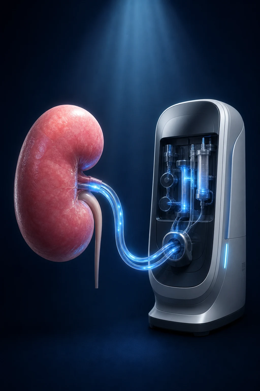
Renal replacement therapy (RRT) refers to any treatment that substitutes for the excretory, fluid-balance, electrolyte and acid-base functions of the failing kidney — and, in the case of transplantation, also restores some of the kidney's endocrine roles (erythropoietin, active vitamin D). The term is applied in two distinct settings, and the distinction drives every decision:[1]
- Maintenance RRT in end-stage kidney disease (ESKD), also called kidney failure with replacement therapy (KFRT) — CKD Stage 5 where kidney function has fallen irreversibly and the patient needs lifelong support. This is a planned pathway: the goal is to prepare early (vascular access, vaccination, transplant assessment), choose the modality with the patient, and preserve residual function, nutrition and quality of life.
- Acute RRT in severe acute kidney injury (AKI) — a temporary, supportive measure while the underlying insult (sepsis, nephrotoxin, obstruction) is treated and the kidney recovers. The goal is physiological support, not chronic replacement, and timing is driven by complications (AEIOU), not a creatinine number.[2][4]
The clinical skill in RRT is not the mechanics of the machine; it is (1) deciding when to start, (2) choosing the right modality for the individual, (3) planning access and transplantation early, and (4) recognising the modality-specific emergencies (access infection, peritonitis, rejection) that determine survival and quality of life. A fifth, often-overlooked option — conservative (non-dialytic) care — is a legitimate, evidence-supported choice for the frail elderly or those with heavy comorbidity, focusing on symptom control and dignity.[6]
Classification
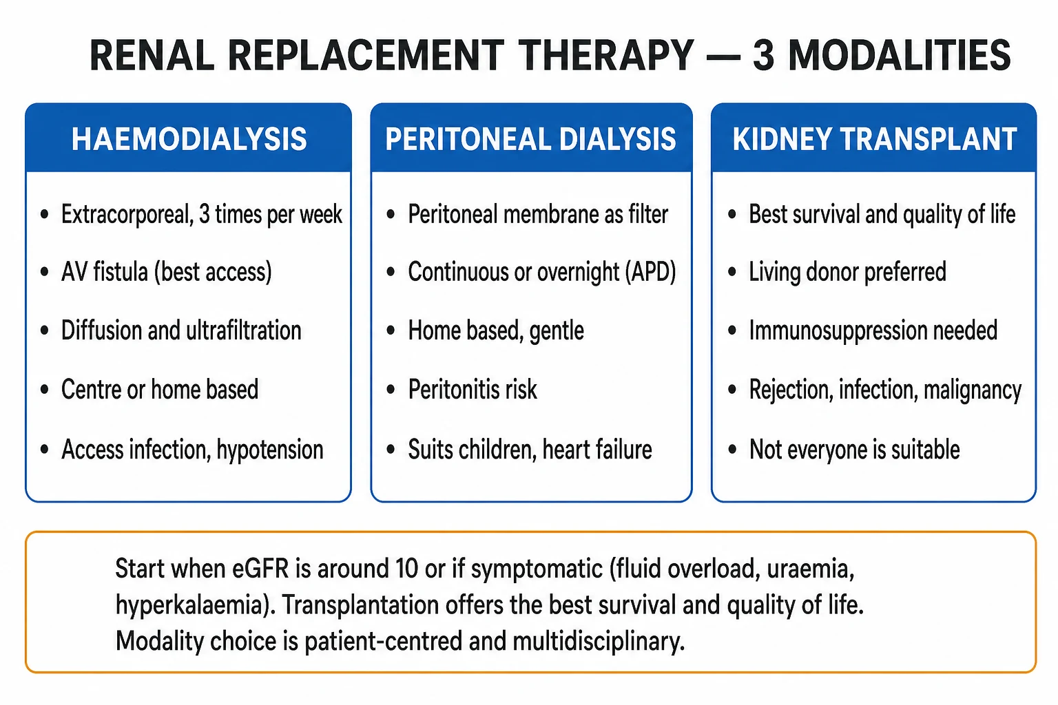
RRT is classified along three axes — modality, setting/frequency, and access: [1]
By modality
| Modality | Principle | Setting | Key access | Best for |
|---|---|---|---|---|
| Haemodialysis (HD) | Extracorporeal diffusion across a synthetic membrane; ultrafiltration by hydrostatic pressure | Centre or home, 3x/week | AV fistula (best); AV graft; tunnelled catheter | Most adults; rapid clearance; toxin removal |
| Peritoneal dialysis (PD) | Peritoneal membrane as semipermeable filter; glucose/icodextrin osmotic gradient | Home (CAPD or APD) | Tenckhoff catheter | Children, heart failure, residual function, home independence |
| Kidney transplant | Donor kidney implanted in iliac fossa; restores filtration and endocrine function | Surgery; lifelong outpatient | Vascular + ureteric anastomoses | All suitable patients — best survival and QoL |
| Conservative care | No dialysis; symptom-focused management | Community/hospice | None | Frail elderly, high comorbidity, patient choice |
By frequency/setting (HD)
- Conventional in-centre HD — 3 times/week, 3-4 hours per session (the global default).
- Short daily HD — 5-6 sessions/week, 2-3 hours; better phosphate and BP control (FHN trials).
- Nocturnal HD — 3-6 nights/week, 6-8 h overnight; the most physiological clearance.
- Home HD — patient or partner trained; greater independence and QoL. [1]
By PD modality
- Continuous ambulatory PD (CAPD) — manual exchanges, 3-5 per day, dwell volumes 1.5-2.5 L; the original PD method.
- Automated PD (APD / continuous cyclic PD, CCPD) — a cycler machine runs overnight (8-12 h) with a daytime dwell; suits children, working adults and high transporters. [1]
By transplant donor source
- Living donor — related (genetically) or unrelated (emotionally/spousally); best graft and patient survival; can be planned pre-emptively.
- Deceased donor after brain death (DBD) — heart-beating; standard deceased source.
- Deceased donor after circulatory death (DCD) — non-heart-beating; expanding the donor pool.
- Expanded-criteria donor (ECD) — older donors or with comorbidity; used selectively to expand the pool. [1]
Haemodialysis
- Extracorporeal, **diffusion** clearance across synthetic membrane
- **AV fistula** best access; matures **6-12 weeks**
- **3 sessions/week, 3-4 h each**; Kt/V over 1.2 target
- Rapid solute/fluid removal; suits most adults
- Risks: access infection/thrombosis, intradialytic hypotension, **beta-2 microglobulin amyloidosis**, hepatitis B/C
Peritoneal dialysis
- **Peritoneal membrane** as filter; **three-pore model**
- **Glucose osmotic gradient** pulls water and solutes
- **Home-based**: CAPD (manual, 4/day) or APD (overnight cycler)
- Gentle, continuous; preserves residual renal function longer
- Main risk **peritonitis** (cloudy effluent); *Staph epidermidis* commonest
Kidney transplant
- **Best survival and quality of life**
- **Living-donor, pre-emptive** preferred (before dialysis)
- Donor kidney in **right iliac fossa**; vascular + ureteric anastomoses
- **Lifelong immunosuppression**: tacrolimus + mycophenolate + steroid
- Twin threats: **rejection** and **opportunistic infection** (CMV, BK, PJP); malignancy (skin, PTLD)
Epidemiology & Risk Factors
The global burden of ESKD/KFRT is large and rising, driven by the diabetes and hypertension epidemic and by ageing populations.[1]
- Incidence of treated ESKD is approximately 130-150 per million population per year in developed countries (higher in the US, lower in parts of Europe); in India, registry data under-represent the true burden but incidence is rising rapidly.
- Prevalence of RRT exceeds 1,000-2,000 per million in many high-income countries; the dialysis population is growing at 5-7% per year.
- Leading causes of ESKD are diabetic kidney disease (rising, now the commonest in many regions), hypertensive nephrosclerosis, chronic glomerulonephritis (especially IgA nephropathy), and adult polycystic kidney disease. [1]
Modality split worldwide: haemodialysis is dominant (~80-89%), peritoneal dialysis (~9-11%), with transplant varying widely by country. PD is over-represented in children, in resource-limited settings, and in countries with active home-dialysis programmes (Mexico, Hong Kong).[7]
Risk factors that predict earlier need for dialysis: poor glycaemic and BP control, rapidly progressive GN, nephrotoxic exposures, recurrent AKI, and low nephron endowment (prematurity, low birth weight). [1]
Transplant outcomes set the standard for survival benefit: [1]
- Living-donor kidney transplant — 1-year graft survival over 95%, 10-year patient survival over 60%.
- Deceased-donor transplant — 1-year graft survival around 89-95%, graft half-life around 10-15 years.
- Annual mortality on dialysis is approximately 10-20%, dominated by cardiovascular disease (the leading cause of death in ESKD, accounting for ~40-50% of deaths), followed by infection and withdrawal of treatment.[8]
RRT — the numbers that decide an answer
Pathophysiology
RRT replaces the kidney's work by moving solute and water across a semipermeable membrane. Understanding how each modality does this determines the choice, the dose and the complications. [1]
Haemodialysis — diffusion across a synthetic membrane
Blood leaves the patient via the vascular access, passes through a hollow-fibre dialyser (thousands of capillary-like membranes), and returns cleansed. Dialysate flows on the outside of the fibres in the opposite direction (counter-current) to blood — this maintains the maximal concentration gradient along the entire membrane length, the principle that makes HD efficient.[7]
- Diffusion is the dominant clearance mechanism — solutes move down their concentration gradient from blood (high urea, creatinine, potassium) to dialysate (low). Small molecules (urea MW 60, creatinine 113, potassium 39) clear well; middle molecules (beta-2 microglobulin MW 11,800) clear poorly on low-flux membranes and need high-flux membranes.
- Ultrafiltration removes fluid — a trans-membrane hydrostatic pressure gradient (controlled by the machine) pushes water out of blood into dialysate. The volume removed per session equals the interdialytic weight gain plus any prescribed negative balance.
- Clearance is governed by Fick's law — flux is proportional to membrane area, permeability, and concentration gradient. The dialysis dose is summarised as Kt/V (see Investigations).
- Dialysate composition is adjusted to correct the patient's biochemistry — bicarbonate (or acetate) buffer, sodium ~140, potassium usually 2, calcium 1.25-1.5 mmol/L. [1]
Peritoneal dialysis — the peritoneum as a living membrane
In PD the peritoneum (visceral + parietal, surface area ~1-2 m² in adults) acts as the semipermeable membrane, with the peritoneal capillaries on the "blood side". Dialysate is instilled through a Tenckhoff catheter; solutes and water move between capillary blood and dialysate across the three-pore model:[1]
- Small pores (~4-5 nm) — the bulk pathway; small solutes (urea, creatinine, sodium) diffuse down their gradient; the rate depends on peritoneal solute transport rate, measured by the peritoneal equilibration test (PET).
- Ultrasmall pores (aquaporin-1) — water-only channels; responsible for free-water clearance during the early (hypotonic) phase of a dwell.
- Large pores (>15 nm) — permit macromolecules (albumin) to leak; explains the protein loss in PD (~5-15 g/day). [1]
The osmotic gradient is created by the dialysate glucose concentration (1.36%, 2.27%, 3.86% dextrose) or by icodextrin (a polyglucose, iso-osmotic but oncotic — used for the long overnight dwell to sustain ultrafiltration without glucose absorption). Over a dwell, the glucose gradient dissipates as glucose is absorbed, so ultrafiltration is maximal early and falls with dwell time — which is why high transporters (rapid glucose absorption) suit short-dwell APD, while low transporters suit long-dwell CAPD. [1]
Kidney transplantation — vascular and immunological principles
A donor kidney is placed extraperitoneally in the right or left iliac fossa. The renal artery is anastomosed end-to-side to the external iliac artery, the renal vein to the external iliac vein, and the ureter is implanted into the bladder (ureteroneocystostomy). The native kidneys are usually left in situ (except in polycystic disease with massive enlargement, or uncontrolled hypertension/infection). [1]
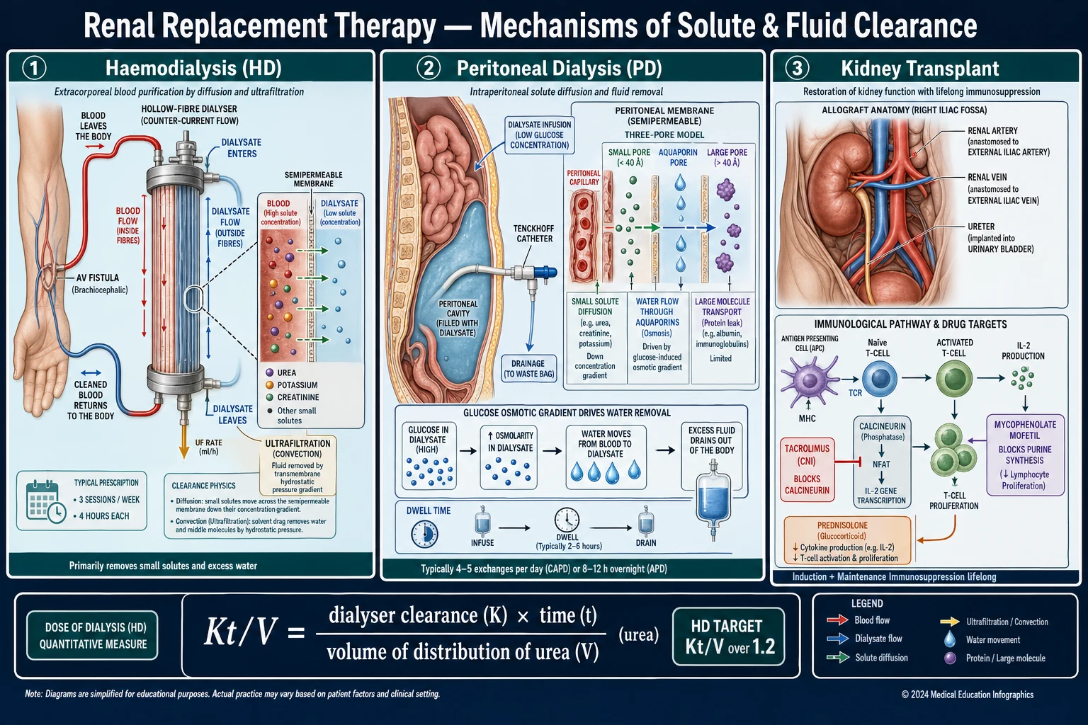
The immunology of rejection centres on the T-cell response to donor antigen, and the immunosuppressive drugs target the three signals of T-cell activation: [1]
- Signal 1 (antigen recognition) — the T-cell receptor (TCR) binds donor peptide presented on MHC molecules. Calcineurin inhibitors (tacrolimus, ciclosporin) act downstream of signal 1, blocking the calcineurin–NFAT pathway that transcribes IL-2.
- Signal 2 (co-stimulation) — CD28 on the T-cell binds B7 (CD80/86) on the antigen-presenting cell. Belatacept (a CTLA-4 Ig fusion protein) blocks this signal.
- Signal 3 (proliferation) — IL-2 and other cytokines drive T-cell division via the mTOR pathway. Sirolimus/everolimus (mTOR inhibitors) and mycophenolate (blocks inosine monophosphate dehydrogenase, IMPDH — de-novo purine synthesis, to which lymphocytes are uniquely dependent) act here. [1]
Antibody-mediated rejection is driven by preformed or de-novo donor-specific antibodies (DSA) against donor HLA, which activate complement (C4d deposition in peritubular capillaries) and cause endothelial injury — the basis of plasmapheresis, IVIG, rituximab and bortezomib in treatment. [1]
Why an AV fistula needs time
An AV fistula joins an artery to a vein (classically the radial artery to cephalic vein at the wrist — the Cimino-Brescia fistula). The sudden high-pressure arterial flow into the vein triggers flow-mediated dilatation and wall thickening (arterialisation) over 6-12 weeks, producing a vein large enough to be cannulated repeatedly with large-bore needles at high flow rates (300-400 mL/min). Early cannulation risks blow-out, aneurysm and infiltration — which is why fistulae are created 6-12 months before anticipated dialysis. This single fact — that a working fistula cannot be improvised at the moment of need — is the strongest argument for early nephrology referral.[7]
Clinical Presentation
The clinical presentation spans the symptoms of advanced kidney failure (which prompt dialysis planning), the emergencies that demand dialysis now (AEIOU), and the modality-specific complications that recur through a patient's dialysis career. [1]
Symptoms prompting RRT (advanced CKD/ESKD)
- Uraemic symptoms — anorexia, nausea, vomiting, weight loss, metallic taste, fatigue, pruritus, restless legs, cold intolerance, sexual dysfunction, impaired concentration and memory, insomnia.
- Fluid overload — peripheral oedema, breathlessness (pulmonary oedema), hypertension.
- Biochemical derangement — symptomatic anaemia, hyperkalaemia, metabolic acidosis (Kussmaul breathing), mineral bone disease (bone pain, fractures).
- Bleeding tendency — uraemic platelet dysfunction (bruising, GI bleeding). [1]
Emergencies requiring dialysis — the AEIOU presentation
A patient with severe AKI or decompensated ESKD may present acutely with one of the AEIOU emergencies: [1]
- A — Acidosis — severe metabolic acidosis (pH under 7.1-7.15) refractory to bicarbonate.
- E — Electrolytes — refractory hyperkalaemia (K over 6.5 mmol/L or with ECG changes: peaked T waves, widened QRS, sine wave) not responding to medical therapy.
- I — Ingestion — a dialysable toxin: lithium, salicylate, methanol, ethylene glycol, metformin (in lactic acidosis).
- O — Overload — refractory pulmonary oedema unresponsive to high-dose diuretics.
- U — Uraemia — uraemic pericarditis (pericardial rub, effusion, tamponade), uraemic encephalopathy (asterixis, seizures, coma), or severe uraemic bleeding. [1]
Modality-specific presentations
- HD access complications — exit-site/tunnel infection (redness, pus, pain), bacteraemia (fever, rigors during or after dialysis — suspect Staphylococcus aureus septicaemia, endocarditis, metastatic infection), thrombosis/stenosis (swollen arm, prolonged bleeding from needles, recirculation, high venous pressure alarms), steal syndrome (ischaemic hand — cold, painful, decreased pulses).
- HD intradialytic symptoms — hypotension (commonest — nausea, cramps, dizziness during ultrafiltration), muscle cramps, dialysis disequilibrium (headache, nausea, seizures, coma in the first dialysis of a heavily uraemic patient — cerebral oedema from rapid solute shift), anaphylactoid reactions (rare — to the membrane, sterilant ethylene oxide, or heparin).
- PD presentations — peritonitis (cloudy effluent, abdominal pain, fever — the cardinal triad), exit-site/tunnel infection (erythema, purulent discharge), catheter malfunction (poor inflow/outflow), hernia (inguinal, incisional), hydrothorax (right-sided, glucose-rich pleural effusion from a diaphragmatic defect), encapsulating peritoneal sclerosis (long-term PD — bloody effluent, bowel obstruction, malnutrition).
- Transplant presentations — acute rejection (rising creatinine, graft tenderness, fever, oliguria), opportunistic infection (fever with a normal graft — see timeline below), new skin lesions or rapidly growing lymphadenopathy (squamous cell skin cancer, post-transplant lymphoproliferative disorder, PTLD), CNI toxicity (tremor, gingival hypertrophy, rising creatinine with high trough levels). [1]
Opportunistic infection timeline after transplant
0-1 month
- Donor-derived infection
- Wound/urinary/catheter infection (nosocomial)
- Herpes simplex reactivation (rare with prophylaxis)
1-6 months
- **CMV** — the classic opportunistic infection (fever, leukopenia, pneumonitis, colitis, hepatitis)
- **BK polyomavirus** nephropathy (rising creatinine, Decoy cells in urine)
- **Pneumocystis jirovecii (PJP)** — fever, hypoxia, diffuse infiltrates (prevented by co-trimoxazole)
After 6 months
- Community-acquired infections
- **Mycobacterium tuberculosis** reactivation (high-risk in endemic areas)
- **Late opportununist** — cryptococcus, listeria, nocardia
- **Malignancy** — non-melanoma skin cancer (100-fold risk), PTLD, cervical, Kaposi sarcoma
Atypical presentations
- Elderly/frail — functional decline, anorexia, falls, delirium; uraemia may be mistaken for dementia or depression. A lower threshold to investigate and to consider conservative care is appropriate.
- Children — failure to thrive, growth retardation, delayed puberty, poor school performance; PD is preferred and transplant is the goal.
- Pregnancy on dialysis — reduced fertility; intensified HD (daily, 20+ h/week) can restore fertility and improve fetal survival to over 70%.
- Diabetic — cardiovascular dominant mortality; may present with severe peripheral vascular disease, autonomic neuropathy causing intradialytic hypotension, and foot ulcers. [1]
Differential Diagnosis
The RRT decisions an examiner tests are mostly modality-against-modality comparisons, plus distinguishing complications. [1]
Distinguishing the modalities
| Factor | Haemodialysis | Peritoneal dialysis | Transplant |
|---|---|---|---|
| Clearance principle | Diffusion (counter-current) | Diffusion + ultrafiltration (peritoneum) | Native kidney |
| Cardiovascular stability | Often unstable (rapid UF) | Stable (continuous) | Normal |
| Residual renal function | Lost faster | Preserved longer | N/A |
| Infection risk | Access bacteraemia (S. aureus) | Peritonitis (Staph epidermidis) | Opportunistic (CMV, BK, PJP) |
| Suitable for children | Less suitable | Preferred | Definitive |
| Home-based | Possible (home HD) | Yes (CAPD/APD) | Outpatient |
| Survival | Annual mortality ~10-20% | Comparable to HD (2-3 yr) | Best (halves mortality) |
| Contraindications | Severe hypotension, no access | Abdominal surgery/adhesions, hernia | Active infection, malignancy, non-adherence |
Distinguishing vascular access types
| Access | Primary patency | Infection risk | Preferred? |
|---|---|---|---|
| AV fistula | Best (years) | Lowest | Yes — fistula first |
| AV graft (PTFE) | Intermediate (months-years) | Intermediate | If fistula fails or veins poor |
| Tunnelled catheter | Months | Highest (bacteraemia, stenosis) | Temporary/bridge only |
Distinguishing rejection types
Hyperacute
- **Minutes-hours** after reperfusion
- Preformed **anti-donor antibody** (ABO/HLA mismatch, positive crossmatch)
- Complement activation, thrombosis, graft necrosis
- **Graft loss** — requires immediate nephrectomy
- Prevented by **crossmatch**
Acute cellular (ACR)
- **Days-weeks** to months
- **T-cell mediated** (tubulitis, intimal arteritis)
- Rising creatinine, graft tenderness, fever
- **Responds to IV methylprednisolone**; ATG if resistant
- Banff graded by tubulitis/arteritis
Antibody-mediated (ABMR)
- **Weeks-months**
- **Donor-specific antibody** + complement (**C4d** in peritubular capillaries)
- Rising creatinine, graft dysfunction
- Plasmapheresis + IVIG + rituximab ± bortezomib
- Often in sensitised patients
Chronic (CAMR / CAN)
- **Months-years**
- Interstitial fibrosis + tubular atrophy, **transplant glomerulopathy**
- Insidious rise in creatinine, proteinuria
- **Irreversible** — slow graft loss
- Driven by chronic ABMR, CNI toxicity, recurrence
Rising creatinine after transplant — the differential
A rising creatinine in a transplant recipient is not always rejection. Biopsy is the definitive test. Distinguish: [1]
- Acute cellular rejection — biopsy shows tubulitis, intimal arteritis; responds to steroids.
- Antibody-mediated rejection — C4d positive, DSA present; plasmapheresis/IVIG.
- Calcineurin inhibitor (CNI) nephrotoxicity — high tacrolimus/ciclosporin trough; biopsy shows isometric vacuolisation of tubules, arteriolar hyalinosis; reduces on dose-lowering.
- BK polyomavirus nephropathy — rising BK PCR, decoy cells in urine; biopsy shows intranuclear viral inclusions in tubular cells; treat by reducing immunosuppression.
- Ureteric obstruction — hydronephrosis on ultrasound; ureteric stenosis/clot.
- Recurrence of native disease — FSGS (early, nephrotic-range proteinuria), IgA nephropathy, MPGN, atypical HUS.
- Volume depletion, ATN (delayed graft function), drug nephrotoxicity (NSAIDs, contrast, ACE inhibitors). [1]
PD peritonitis vs other causes of cloudy effluent
- Bacterial peritonitis — cloudy effluent, WBC over 100/mL with over 50% neutrophils, abdominal pain; culture positive (~70-90%).
- Culture-negative peritonitis — sterile-appearing but cloudy; often resolves empirically.
- Fungal peritonitis — recurrent, often after bacterial peritonitis; may require catheter removal.
- Eosinophilic peritonitis — sterile, eosinophils in effluent; often air or allergic reaction to catheter.
- Encapsulating peritoneal sclerosis (EPS) — chronic, bloody effluent, bowel obstruction, intra-abdominal calcification on CT; a late, feared complication of long-term PD.
- Catheter malposition / constipation — poor drainage, clear effluent, normal cell count. [1]
Clinical & Bedside Assessment
The pre-dialysis (ESKD planning) assessment
- Volume status — JVP, peripheral and sacral oedema, lung bases, blood pressure (interdialytic hypertension), weight trend.
- Nutritional status — BMI, subjective global assessment (SGA), mid-arm circumference; malnutrition predicts poor dialysis outcomes.
- Cardiovascular exam — murmurs (valvular disease common and undertreated in ESKD), signs of heart failure, peripheral pulses and ABI (vascular disease).
- Access examination — inspect existing access; Allen test before radio-cephalic fistula creation (confirm ulnar supply to the hand to avoid steal).
- Neurological — for uraemic neuropathy and restless legs. [1]
Examining the AV fistula
A patent, mature fistula has three cardinal signs — examine every dialysis patient for them: [1]
- Thrill — palpable, present throughout the cardiac cycle (not just systolic); confirms flow.
- Bruit — auscultate over the fistula; a continuous machinery bruit (systolic and diastolic).
- Pulse augmentation — compress the fistula proximally; the pulse should augment. [1]
Red flags on fistula examination: [1]
- Absent thrill or bruit — thrombosis; urgent vascular review (the fistula can often be salvaged by thrombolysis/thrombectomy if caught early).
- Erythema, swelling, pus, tenderness — infection; cultures, IV antibiotics (cover S. aureus, including MRSA).
- Cold, painful, pale hand with weak pulses — steal syndrome; urgent vascular review.
- Aneurysm/pseudoaneurysm — localised dilatation at needling sites; risk of rupture.
- Swollen arm, prominent chest-wall veins — central venous stenosis (especially after previous subclavian catheters). [1]
Examining the PD patient
- Exit-site — inspect for erythema, discharge; score with the Twardowski criteria (0 = excellent, 4 = tunnel infection).
- Abdomen — hernias (inguinal, incisional, umbilical), leaks around the catheter, signs of peritonitis (guarding, rebound).
- Volume status — PD patients can be volume-overloaded or -depleted; assess clinically. [1]
Examining the transplant recipient
- Graft — palpate in the iliac fossa (tenderness suggests rejection or infection); auscultate for a bruit (anastomotic stenosis).
- Blood pressure — hypertension is common (CNI, steroids, graft).
- Infection screen — fever workup (the immunosuppressed patient may have subtle signs); examine skin, oropharynx, perianal area.
- Malignancy screen — full skin examination (non-melanoma skin cancer is 100-fold more common), lymphadenopathy/splenomegaly (PTLD).
- Graft function trends — the trajectory of creatinine matters more than any single value; a 25% rise warrants investigation. [1]
Investigations
Indications to start dialysis — the AEIOU and the symptom threshold
AEIOU — emergency dialysis indications (reproduced verbatim)
| Letter | Indication | Threshold / example |
|---|---|---|
| A | Acidosis (refractory metabolic) | pH under 7.1-7.15 despite bicarbonate |
| E | Electrolytes (refractory hyperkalaemia) | K over 6.5 mmol/L or any K with ECG changes, not responding to medical therapy |
| I | Ingestion (dialysable toxin) | Lithium, salicylate, methanol, ethylene glycol, metformin (in severe lactic acidosis) |
| O | Overload (refractory) | Pulmonary oedema unresponsive to high-dose diuretics |
| U | Uraemia | Pericarditis (rub, effusion), encephalopathy (asterixis, seizures, coma) |
Symptom threshold in ESKD
Start maintenance dialysis when symptoms or complications of uraemia appear, typically at eGFR 5-10 mL/min. The IDEAL trial (Cooper 2010) showed no survival or QoL benefit of early initiation at eGFR 10-14 over late initiation at eGFR 5-7 — start for the patient, not for a number. An asymptomatic patient can be monitored, with access created and transplant assessment underway, until symptoms or an AEIOU-type complication intervenes.[6]
Dialysis adequacy — what to measure
Haemodialysis dose — Kt/V and URR
- Kt/V — the standard dose metric. K = dialyser urea clearance, t = treatment time, V = volume of urea distribution (~total body water). The target is single-pool Kt/V at least 1.2 per session (target 1.4), measured by pre- and post-dialysis urea. Minimum session length 3-4 hours, 3 times/week.
- URR (urea reduction ratio) — simpler; URR = (pre-urea − post-urea)/pre-urea × 100; target at least 65%.[7]
Peritoneal dialysis dose
- Total Kt/V at least 1.7 per week (renal + peritoneal clearance combined); creatinine clearance targets 50-60 L/week/1.73 m².
- PD preserves residual renal function longer than HD — a major advantage (residual function predicts survival).
- Peritoneal equilibration test (PET) — classifies transport status using a 4-hour dwell with 2.27% dextrose: high, high-average, low-average, low transporters. High transporters absorb glucose rapidly, lose the osmotic gradient, and suit short-dwell APD; low transporters have slow solute equilibration and suit long-dwell CAPD.[1]
Transplant work-up — what to check before listing
- ABO blood group compatibility (or ABO-incompatible desensitisation).
- HLA typing — HLA-A, -B, -C, -DR, -DQ; DR and DQ mismatches most impact graft survival.
- Panel-reactive antibody (PRA) — measures anti-HLA antibodies from prior exposure (pregnancy, transfusion, transplant); a high PRA means a highly sensitised patient.
- Crossmatch — complement-dependent cytotoxicity (CDC) and flow cytometric crossmatch; a positive T-cell crossmatch is an absolute contraindication to transplantation (hyperacute rejection).
- Virology — HBV (HBsAg, anti-HBs, anti-HBc), HCV antibody, HIV, CMV IgG (donor/recipient status — D+/R− is the high-risk mismatch), EBV.
- Infection screen — TB (IGRA/Mantoux), BK virus, urinary tract, dental, cardiac valves.
- Cancer screening — age-appropriate; active malignancy is a contraindication.
- Cardiac assessment — echocardiography, coronary assessment (ESKD patients have high cardiovascular risk).
- Psychosocial — adherence capacity, social support. [1]
The Banff classification of acute rejection
The Banff schema grades renal allograft biopsy lesions, the basis for diagnosing rejection: [1]
- Acute cellular rejection (ACR) — graded by tubulitis (t) and intimal arteritis (v): borderline changes, IA (mild tubulitis), IB (moderate), IIA (intimal arteritis), IIB (severe arteritis, fibrinoid necrosis), III (transmural arteritis).
- Antibody-mediated rejection (ABMR) — requires (1) histological injury (microvascular inflammation — glomerulitis g, peritubular capillaritis ptc), (2) C4d positivity in peritubular capillaries, and (3) serological evidence of donor-specific antibody (DSA).
- Chronic active rejection / chronic allograft nephropathy — interstitial fibrosis and tubular atrophy (ci/ct), transplant glomerulopathy (cg), transplant arteriopathy.[10]
PD peritonitis diagnosis
- Cloudy effluent with white cell count over 100/mL and over 50% neutrophils (after a dwell of at least 2 h).
- Culture of effluent (send a large volume, concentrate by centrifugation); Gram stain is usually negative.
- Commonest organisms: coagulase-negative staphylococci (Staph epidermidis), then S. aureus, then Gram-negatives; fungal in recurrent cases.[9]
Routine monitoring on dialysis
Every patient on dialysis needs regular monitoring: monthly U&E, creatinine, bicarbonate, calcium, phosphate, albumin, FBC (anaemia), ferritin/TSAT; 3-monthly PTH (target 2-9x upper limit of normal in CKD-MBD), Kt/V/URR (HD); annual echocardiography, vascular access surveillance, HBV/HCV serology, and vaccination review. [1]
Management — Resuscitation
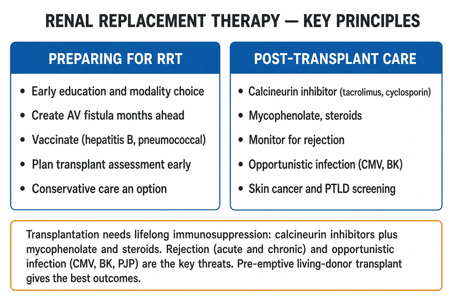
When a dialysis patient (or a patient with severe AKI) presents with a life-threatening complication, ABCDE first, then arrange urgent dialysis for any AEIOU indication. [1]
Hyperkalaemia emergency (the commonest pre-dialysis crisis)
- Assess and stabilise — oxygen, IV access, 12-lead ECG (peaked T waves, flattened/absent P waves, prolonged PR, widened QRS, sine wave — the latter pre-arrest).
- Membrane stabilisation — calcium gluconate 10% (10 mL = 10 mmol) IV over 5-10 min if ECG changes; protects the myocardium (does not lower K).
- Shift K into cells — insulin 10 units + 50% dextrose 50 mL IV over 15-30 min (or 10 units in 500 mL 10% dextrose over 60 min in the elderly to avoid hypoglycaemia); salbutamol 10-20 mg nebulised (additive effect); sodium bicarbonate 1.26% if acidotic.
- Remove K — potassium binders (sodium polystyrene sulfonate 15-30 g orally/rectally; newer patiromer/zirconium cyclosilicate); diuretics if residual function; dialysis if refractory or severe (K over 7, ongoing rise, or ECG changes persisting).[3]
Pulmonary oedema (refractory)
- Sit upright, high-flow oxygen, CPAP or NIV (reduces work of breathing and preload).
- IV furosemide (high dose, e.g. 250 mg or more) only if there is residual renal function — in anuric dialysis patients diuretics are useless.
- IV GTN (venodilator) if hypertensive.
- Urgent haemodialysis with ultrafiltration — the definitive treatment in established ESKD; remove 1-2 L during the session. [1]
Uraemic pericarditis / tamponade
- Urgent intensive dialysis (daily until resolved); analgesia (NSAIDs/colchine if no contraindication).
- Echocardiogram to assess effusion size and tamponade physiology; pericardiocentesis if haemodynamically compromised. [1]
Emergency access
If dialysis is needed immediately and no mature fistula exists, place a central venous dialysis catheter (non-tunnelled for acute need): [1]
- Internal jugular is the preferred site — lowest infection and stenosis rate.
- Femoral is acceptable in emergencies or coagulopathy, but higher infection risk.
- Avoid subclavian if at all possible — it causes central venous stenosis that destroys future fistula veins in that arm. [1]
Clotted AV fistula
- Urgent vascular access review — thrombolysis or surgical/radiological thrombectomy can salvage a thrombosed fistula if caught within 24-48 h.
- If dialysis is needed before salvage, place a temporary catheter.
- Do not cannulate an infected fistula. [1]
PD peritonitis (empirical)
- Send effluent for cell count and culture immediately.
- Start empirical intraperitoneal antibiotics within 6 hours per ISPD 2016 — gram-positive cover (vancomycin or first-generation cephalosporin) plus gram-negative cover (ceftazidime or an aminoglycoside), both given intraperitoneally.[9]
- Add heparin 500-1000 units/L IP to reduce fibrin and catheter blockage.
- Most cases respond within 48-72 h; catheter removal is indicated for refractory, fungal, or non-tunnelled relapsing peritonitis.
Management — Definitive & Stepwise
The RRT planning ladder (ESKD)
A planned, structured approach to ESKD dramatically improves outcomes — late referral (within 3 months of starting dialysis) is associated with higher mortality, more catheter use, and lower transplant rates. The ladder: [1]
Open the ESKD planning ladder
- Early nephrology referral — at CKD Stage 4 (eGFR under 30 mL/min), ideally at least 12 months before anticipated dialysis. Allows time for access creation, vaccination, transplant assessment.
- Modality education — multidisciplinary discussion of HD, PD, transplant and conservative care; patient-centred choice informed by lifestyle, home support, comorbidity.
- Vascular access creation — AV fistula created 6-12 months before anticipated need; maturation takes 6-12 weeks. AV graft if veins poor; tunnelled catheter only as a bridge.
- Vaccination before immunosuppression — hepatitis B series (double dose, check anti-HBs titre over 10 IU/L), pneumococcal, influenza (annual), COVID-19, plus HBV/HAV in high-risk.
- Transplant assessment and listing — tissue typing, immunological, cardiac, infection and cancer screening; pre-emptive listing (before dialysis) when eGFR under 20 mL/min if a living donor is identified.
- Conservative care discussion — for the frail elderly or high comorbidity; a legitimate, evidence-based pathway.
- Dialysis initiation — when symptomatic (eGFR around 5-10) or for AEIOU; not for a creatinine number alone.[6]
Haemodialysis prescription
- Frequency — conventional 3 sessions/week; short daily (5-6x) or nocturnal in selected patients.
- Duration — 3-4 hours per session; longer for large patients or high interdialytic weight gain.
- Blood flow — 300-400 mL/min through a mature fistula.
- Dialysate flow — 500-800 mL/min, counter-current.
- Ultrafiltration goal — set to the interdialytic weight gain + prescribed negative balance; limit UF rate to under 10-13 mL/kg/h to avoid intradialytic hypotension and myocardial stunning.
- Dose target — single-pool Kt/V at least 1.2 (target 1.4); URR at least 65%.
- Dialysate composition — bicarbonate buffer; sodium ~140; potassium usually 2; calcium 1.25-1.5 mmol/L.
- Anticoagulation — unfractionated heparin or low-molecular-weight heparin to prevent clotting in the extracorporeal circuit; heparin-free dialysis (with saline flushes) in active bleeding. [1]
Peritoneal dialysis prescription
- CAPD — 3-5 manual exchanges/day, dwell volumes 1.5-2.5 L, dialysate glucose 1.36%, 2.27%, 3.86% as needed for ultrafiltration.
- APD — cycler runs 8-12 h overnight with 3-5 cycles, plus a daytime "last-fill" dwell (often icodextrin for the long daytime dwell).
- Dose — total Kt/V at least 1.7/week; adjust to residual function.
- Adapt to PET — high transporters → APD (short dwells); low transporters → CAPD (long dwells) or increased dwell volume. [1]
Kidney transplant — immunosuppression
Post-transplant immunosuppression — the triple therapy
TIM
calcineurin inhibitor (CNI) — blocks calcineurin/NFAT, suppresses IL-2 transcription; target trough 5-10 ng/mL; side-effects nephrotoxicity, diabetes, tremor, alopecia
antiproliferative — blocks inosine monophosphate dehydrogenase (IMPDH), de-novo purine synthesis; dose 1-2 g/day; side-effects leukopenia, GI upset, teratogenic (switch to azathioprine pre-pregnancy)
steroid — broad anti-inflammatory (blocks NF-kB, cytokines); induction IV methylpred, maintenance prednisolone weaned to ~5 mg; side-effects diabetes, osteoporosis, Cushingoid
Induction therapy — given peri-operatively to most recipients:
- Basiliximab (interleukin-2 receptor antagonist) — for low-immunological-risk recipients.
- Anti-thymocyte globulin (ATG) — for high-risk recipients (sensitised, delayed graft function, re-transplant). [1]
Maintenance — triple therapy (CNI + antiproliferative + steroid), with steroid withdrawal/minimisation in low-risk recipients to reduce diabetes and osteoporosis. Alternatives to tacrolimus — ciclosporin (more hirsutism, gingival hypertrophy, hypertension; less diabetes), sirolimus/everolimus (mTOR inhibitors — used to spare CNI nephrotoxicity; side-effects hyperlipidaemia, impaired wound healing, proteinuria, mouth ulcers). [1]
Prophylaxis post-transplant:
- Co-trimoxazole (trimethoprim-sulfamethoxazole 480 mg daily, or 960 mg 3x/week) for 6-12 months — prevents PJP, also toxoplasmosis, listeria, nocardia.
- Valganciclovir for CMV D+/R− mismatch — typically 900 mg daily for 3-6 months.
- Nystatin or fluconazole — oral candidiasis prophylaxis for 1 month.
- Antiviral for HBV — entecavir or tenofovir for HBsAg-positive recipients, to prevent reactivation under immunosuppression. [1]
Treating acute rejection
- Acute cellular rejection — IV methylprednisolone 500 mg-1 g daily for 3 days; if steroid-resistant, anti-thymocyte globulin (ATG).
- Antibody-mediated rejection — plasmapheresis (3-6 sessions), IV immunoglobulin (e.g. 2 g/kg, often divided), rituximab (anti-CD20), and in refractory cases bortezomib (proteasome inhibitor targeting plasma cells).
- Optimise baseline immunosuppression — ensure tacrolimus in range, adherence confirmed. [1]
Managing complications of long-term dialysis
- Anaemia — IV iron (target TSAT over 20%, ferritin over 200 µg/L) then ESA (epoetin/darbepoetin) to Hb 100-115 g/L (avoid over 130 — cardiovascular risk).
- Mineral bone disease (CKD-MBD) — phosphate binders (calcium acetate/carbonate, sevelamer, lanthanum) with meals; activated vitamin D (calcitriol/paricalcitol) and cinacalcet (calcimimetic) for secondary hyperparathyroidism (target PTH 2-9x upper limit); parathyroidectomy for refractory disease.
- Acidosis — oral sodium bicarbonate to keep bicarbonate over 22.
- Hypertension — ACE inhibitor/ARB (cautious), calcium channel blocker, beta-blocker; dry-weight reduction is the most powerful intervention.
- Cardiovascular risk — statin (simvastatin/atorvastatin; note simvastatin dose-limiting with amlodipine), aspirin if indicated; smoking cessation; exercise. [1]
Evidence on RRT timing in AKI
Two landmark trials transformed practice: [1]
- AKIKI (Gaudry 2016, NEJM) — in critically ill adults with KDIGO Stage 3 AKI, no mortality benefit of early (immediate) vs delayed (KDIGO criteria + AEIOU, or after 72 h of Stage 3) RRT initiation. ~50% of the delayed-Strategy group never needed dialysis.[5]
- STARRT-AKI (2020, NEJM) — in critically ill patients with severe AKI, an accelerated (early) strategy did not reduce 90-day mortality and increased adverse events (persistent RRT dependence, infections).[4]
Conclusion: in AKI, start RRT for AEIOU, not for a creatinine number. A wait-and-watch strategy (with close monitoring) is appropriate in stable KDIGO Stage 3 AKI. [1]
Specific Subtypes & Scenarios
Continuous vs intermittent RRT in the ICU
In haemodynamically unstable critically ill patients, continuous renal replacement therapy (CRRT) or sustained low-efficiency dialysis (SLED) may be preferred over intermittent HD: [1]
- CRRT — runs 24 h/day; modalities include CVVH (continuous veno-venous haemofiltration — convective clearance), CVVHD (diffusive), CVVHDF (combined). Gentle fluid removal suits the shocked patient; disadvantages — anticoagulation (regional citrate often), immobility, cost, ongoing clearance losses of drugs and nutrients.
- SLED — hybrid, 6-8 h sessions, daily or alternate; gentler than IHD, less resource-intensive than CRRT; increasingly used in ICUs.
- No clear mortality difference between CRRT and intermittent HD in AKI; choice driven by haemodynamic stability, anticoagulation, and centre expertise. [1]
RRT in children
- PD is the preferred initial modality (APD overnight preserves school attendance and family routine); infant-sized PD catheters.
- Transplant is the goal — best for growth and neurodevelopment; growth hormone for short stature; living-donor transplant preferred.
- Weight-based drug and dialysate dosing; meticulous attention to nutrition and growth. [1]
Pre-emptive transplant
- Transplantation before dialysis is started — the best survival and QoL outcomes — achievable from a living donor (planned).
- Deceased-donor pre-emptive listing is possible once eGFR under 20 mL/min.
- Avoids dialysis access complications, preserves residual function, reduces sensitisation from transfusions. [1]
Highly sensitised transplant candidates
A high PRA (e.g. from multiple pregnancies, prior transfusions or transplant) makes finding a compatible donor hard. Options: [1]
- Desensitisation — plasmapheresis + IVIG ± rituximab to lower anti-HLA antibody.
- Paired kidney exchange (kidney swap) — incompatible donor-recipient pairs are matched with another incompatible pair, transplanting in a cycle.
- Acceptable mismatch programmes (Eurotransplant) for highly sensitised patients. [1]
Simultaneous kidney-pancreas transplant (SPK)
- In type 1 diabetes with ESKD, SPK cures both diseases, improves survival and quality of life, and prevents diabetic complications.
- Pancreas after kidney (PAK), or islet transplantation in selected non-uraemic patients with severe hypoglycaemia unawareness. [1]
Dialysis in pregnancy
- Intensified HD (daily, or 5-6 sessions/week, targeting 20+ h/week) improves fetal survival to over 70%; PD is rarely used in pregnancy (reduced capacity, mechanical issues).
- Conception after transplant — usually deferred 1-2 years with stable graft function and low immunosuppression; use tacrolimus + azathioprine + prednisolone (mycophenolate is teratogenic and must be switched pre-conception); close obstetric-nephrology co-management. [1]
Conservative (non-dialytic) care
- A legitimate, evidence-supported option for the frail elderly or those with heavy comorbidity in whom the burden of dialysis outweighs benefit.
- Focuses on symptom control — anaemia (ESA), fluid (diuretics), itch, restless legs, depression, advance care planning, and a community/palliative pathway.
- In the over-80s with high comorbidity, mean survival on conservative care is comparable to dialysis (often under 2 years either way), but with fewer hospital days and better QoL. [1]
Complications & Pitfalls
Haemodialysis complications
- Access complications — infection (exit-site, tunnel, bacteraemia — S. aureus septicaemia with metastatic infection: endocarditis, osteomyelitis, septic emboli), stenosis/thrombosis, steal syndrome (hand ischaemia), aneurysm, central venous stenosis.
- Intradialytic hypotension — commonest symptom; from rapid ultrafiltration exceeding vascular refill; myocardial stunning with recurrent episodes.
- Muscle cramps — from rapid fluid shifts and osmotic changes.
- Dialysis disequilibrium syndrome — cerebral oedema from rapid solute shifts in the first dialysis of a heavily uraemic patient (urea over 35-40 mmol/L); headache, nausea, seizures, coma; prevented by a short, gentle first session (2 h, low blood flow), reduced dialysate flow, and avoiding over-rapid UF.
- Allergic/anaphylactoid reactions — to the dialyser membrane (AN69), sterilant (ethylene oxide), or heparin.
- Dialysis-related amyloidosis — beta-2 microglobulin deposition (poorly cleared by low-flux membranes) over 5-10+ years; carpal tunnel syndrome, arthropathy, bone cysts; reduced by high-flux membranes.
- Hepatitis B and C — historical scourge of dialysis units; now controlled by isolation, vaccination (HBV), and universal precautions; HCV curable with DAAs.
- Accelerated cardiovascular disease — the leading cause of death; from hypertension, dyslipidaemia, vascular calcification, inflammation.
- Anaemia, mineral bone disease, malnutrition, depression, reduced fertility — shared with all dialysis. [1]
Peritoneal dialysis complications
- Peritonitis — the cardinal complication; cloudy effluent + abdominal pain +/- fever; coagulase-negative staphylococci commonest, then S. aureus, Gram-negatives; fungal in recurrent disease (often needs catheter removal).[9]
- Exit-site/tunnel infection — Twardowski grading; S. aureus and Pseudomonas; precedes peritonitis.
- Catheter malfunction — poor inflow/outflow (constipation, omental wrap, migration); resolves with laxatives, repositioning, or omentopexy.
- Hernias (inguinal, incisional, umbilical) — from raised intra-abdominal pressure.
- Hydrothorax — glucose-rich right-sided pleural effusion from a diaphragmatic defect.
- Hyperglycaemia and weight gain — from glucose absorption (~100-300 g/day on standard PD); icodextrin reduces this.
- Encapsulating peritoneal sclerosis (EPS) — a feared late complication of long-term PD (after 5+ years); bowel obstruction, bloody effluent, weight loss, intra-abdominal calcification on CT; high mortality; treatment with tamoxifen, nutritional support, surgery in expert centres.
- Loss of residual renal function and membrane failure — over years; technique survival falls, often requiring transfer to HD.
Transplant complications
Rejection
- Hyperacute (minutes) — preformed Ab, graft loss
- Acute cellular (days-weeks) — T-cell; steroids
- Antibody-mediated (weeks-months) — C4d; plasmapheresis/IVIG/rituximab
- Chronic (months-years) — fibrosis/atrophy, transplant glomerulopathy; irreversible
Opportunistic infection
- **CMV** (1-6 mo) — fever, leukopenia, pneumonitis, colitis
- **BK nephropathy** — rising creatinine, decoy cells; reduce immunosuppression
- **PJP** — fever, hypoxia, diffuse infiltrates; prevented by co-trimoxazole
- **TB** reactivation in endemic areas; mycobacteria, cryptococcus late
Malignancy
- **Non-melanoma skin cancer** (100x risk) — sun protection, surveillance
- **PTLD** (EBV-driven B-cell lymphoma) — especially EBV D+/R−
- Cervical (HPV), Kaposi (HHV-8), renal cell
- Annual dermatology review, sun protection
Drug & other
- **CNI nephrotoxicity** — arteriolar hyalinosis, chronic fibrosis
- Post-transplant diabetes (tacrolimus, steroids)
- Cardiovascular disease — still the leading cause of death
- Recurrence of native disease (FSGS, IgA, MPGN, atypical HUS)
Classic pitfalls
- Late referral — fistula not ready; dialysis via catheter (higher infection, mortality); transplant assessment not done.
- Missing transplant as an option — every suitable patient should be assessed and listed; age alone is not a contraindication.
- Using subclavian catheters — destroys future fistula veins by central stenosis; internal jugular always preferred.
- Missing PD peritonitis — any cloudy effluent is peritonitis until proven otherwise; treat empirically within 6 h.
- Attributing a rising creatinine to rejection without biopsy — could be CNI toxicity, BK nephropathy, obstruction, or recurrence; each has a different treatment.
- Under-immunising — HBV, pneumococcal, influenza, COVID-19 vaccines before transplant; live vaccines are contraindicated after.
- Missing CMV or BK — fever with leukopenia, rising creatinine, or unexplained graft dysfunction warrants PCR.
- Starting dialysis for a number — IDEAL/AKIKI/STARRT-AKI: start for symptoms (ESKD) or AEIOU (AKI), not for eGFR or creatinine alone.
- Overlooking conservative care as a valid, compassionate option in the frail elderly.
- Forgetting dialysis disequilibrium in the first session of a heavily uraemic patient. [1]
Prognosis & Disposition
Survival comparisons
- Living-donor transplant — the gold standard; 10-year patient survival over 60% (vs ~30-40% on dialysis). Pre-emptive transplant is best.
- Deceased-donor transplant — 1-year graft survival over 89-95%; graft half-life ~10-15 years; chronic allograft nephropathy and death-with-a-functioning-graft (cardiovascular) are the main late losses.
- Wolfe 1999 (NEJM) — transplantation halves long-term mortality compared with remaining on dialysis on the waiting list.[8]
- Dialysis survival — annual mortality ~10-20%, dominated by cardiovascular disease (40-50% of deaths), then infection and withdrawal.
- PD vs HD survival — broadly comparable for the first 2-3 years; PD may have an early survival advantage in low-comorbidity patients (residual function, gentler UF), but technique survival falls over time (membrane failure, transfer to HD).
- Conservative care — in the over-80s with high comorbidity, mean survival is often comparable to dialysis (under 2 years either way), but with better QoL and fewer hospital days.
Prognostic factors for dialysis survival
- Age, comorbidity (especially diabetes and cardiovascular disease), albumin (nutrition), residual renal function, dialysis adequacy (Kt/V), access type (fistula better than catheter — catheter use carries 2-3x the mortality of a fistula), and interdialytic weight gain. [1]
Disposition logic
- CKD Stage 4 (eGFR under 30) — nephrology referral, RRT education, access planning, transplant assessment.
- CKD Stage 5 with symptoms — initiate dialysis (HD or PD) or proceed to transplant if pre-emptive; conservative care pathway if chosen.
- Severe AKI with AEIOU — ICU and RRT (CRRT if unstable, IHD if stable); review timing per AKIKI/STARRT-AKI.
- Frail elderly/high comorbidity — conservative care discussion; shared decision-making. [1]
Special Populations
- Children/infants — PD preferred (APD overnight); transplant the goal; growth hormone for short stature; weight-based drug and dialysate dosing; meticulous nutrition.
- Pregnancy — intensified HD (daily, 20+ h/week); conceive 1-2 years post-transplant with stable function; switch mycophenolate to azathioprine pre-conception; tacrolimus + azathioprine + prednisolone is the safe regimen.
- Elderly/frail — weigh dialysis burden against benefit; conservative care is valid; shared decision-making with the patient and family.
- Diabetic — cardiovascular dominant mortality; PD glucose absorption worsens glycaemia (icodextrin preferred for the long dwell); simvastatin dose-limited with amlodipine; simultaneous kidney-pancreas transplant option in type 1; high amputation risk.
- Hepatitis B/C or HIV — isolation HD machine for HBV-positive patients; HCV now curable with DAAs before transplant; HIV is no longer a contraindication to transplant; outcomes improving.
- Highly sensitised (PRA high) — desensitisation, paired exchange, acceptable mismatch programmes. [1]
Evidence, Guidelines & Regional Differences
International standards
- KDIGO 2012 AKI Guideline — the international standard for AKI definition, staging, and RRT timing (initiation, modality, dose in CRRT).[3]
- KDIGO 2012 CKD Guideline — classification and ESKD planning, including referral thresholds.
- KDOQI (USA) — vascular access (fistula-first), dialysis adequacy, and anaemia/mineral bone disease guidelines; the Fistula First Catheter Last initiative.
- ERA-EDTA (Europe) and ISPD (peritoneal dialysis) — the 2016 ISPD peritonitis recommendations are the global standard for PD peritonitis prevention and treatment.[9]
- NICE (UK) — guidelines on dialysis and transplant assessment; conservative care formally endorsed.
Landmark trials
- Wolfe 1999 (NEJM) — established that transplant halves mortality vs remaining on dialysis on the waiting list.[8]
- IDEAL (Cooper 2010, NEJM) — in ESKD, no survival or QoL benefit of early (eGFR 10-14) vs late (eGFR 5-7) dialysis initiation.[6]
- AKIKI (Gaudry 2016, NEJM) — in critically ill Stage 3 AKI, no mortality benefit of early vs delayed RRT; ~50% of the delayed group never needed dialysis.[5]
- STARRT-AKI (2020, NEJM) — an accelerated (early) RRT strategy did not reduce 90-day mortality and increased adverse events (persistent RRT, infection) in severe AKI.[4]
- HEMO trial (2002) — high-flux vs low-flux membranes: no overall survival benefit (subgroup benefit in long-dialysis-vintage patients).
- FHN trials (daily/nocturnal HD) — more frequent HD improved LV mass, BP, phosphate but no clear survival benefit.
Regional differences
- India (NOTTO/ICMR) — percutaneous fistula creation increasing; deceased-donor organ scarcity (cultural and infrastructural); commercial/paid donation ethically prohibited (Transplantation of Human Organs Act 1994) but a black market persists; government HD programmes expanding under PM-JAY; PD under-utilised but growing; transplant tourism an ethical concern.
- Spain — opt-out (presumed consent) legislation; highest deceased-donor transplant rate in the world (national transplant coordination network).
- UK — PD declining (under 20% of incident); AV fistula first; conservative care validated and offered; opt-in system.
- USA — high catheter use (an ongoing quality problem despite Fistula First); deceased-donor dominant; long transplant waiting list; PD growing with home-dialysis initiatives.
- Mexico, Hong Kong — PD-first policies (PD as the default incident modality, driven by cost and home-care models). [1]
Exam Pearls
When to start emergency dialysis — AEIOU
AEIOU
refractory metabolic acidosis, pH under 7.1-7.15
refractory hyperkalaemia, K over 6.5 mmol/L or ECG changes
dialysable toxins — lithium, salicylate, methanol, ethylene glycol, metformin
refractory pulmonary oedema unresponsive to diuretics
pericarditis (rub, effusion), encephalopathy (seizures, coma)
Exam application bank (NEET-PG / INICET)
One-line answer
Renal replacement therapy (RRT) is the substitution of the kidney's excretory, fluid-balance and electrolyte/acid-base functions when they fail — applied either as planned maintenance therapy in end-stage kidney disease (ESKD) or as emergency support in severe acute kidney injury (AKI). There are three modalities — haemodialysis (HD) (extracorporeal clearance across a semipermeable membrane by diffusion; AV fistula is the best access, 3 times weekly, target Kt/V over 1.2; risks access infection, intradialytic hypotension, amyloidosis), peritoneal dialysis (PD) (the peritoneal membrane as the filter, glucose osmotic gradient, home-based, gentler; main risk peritonitis, usually Staph epidermidis), and kidney transplantation (the best survival and quality of life, living-donor and pre-emptive preferred, requiring lifelong immunosuppression — calcineurin inhibitor + mycophenolate + steroid
Worked stems (answer without another resource)
Stem 1 — Classic presentation. Map symptoms to mechanism; name the first investigation and first treatment step with dose/route if drug therapy is standard. [1]
Stem 2 — Unstable / complicated. List red flags that force immediate resuscitation, theatre, ICU, antidote, or reperfusion — and what you do in the first 15 minutes. [1]
Stem 3 — Atypical group. Elderly, pregnancy, child, or immunocompromised: how presentation and thresholds change. [1]
Stem 4 — Differential trap. Name the three closest mimics and one discriminator for each. [1]
Stem 5 — Disposition. Who goes home with safety-netting, who is admitted, who needs HDU/ICU/theatre, and what follow-up is mandatory. [1]
Rapid viva checklist
- Definition + classification
- Pathophysiology chain
- Bedside signs / criteria
- Score with exact components (if any)
- Emergency bundle
- Definitive therapy with doses
- Complications of disease and of treatment
- Special populations
- Guideline/trial name if classic
- Three exam traps
Coverage self-check
If you cannot answer any stem above from this page alone, re-read the matching section — the page is intended to be self-sufficient for final-prof and NEET-PG/INICET questions on Renal Replacement Therapy.
[1]References
- [1]Villa G, Ricci Z, Ronco C. Renal Replacement Therapy Crit Care Clin, 2015.PMID 26410148
- [2]Neri M, Villa G, Garzotto F, et al. Nomenclature for renal replacement therapy in acute kidney injury: basic principles Crit Care, 2016.PMID 27719682
- [3]Kellum JA, Lameire N, for the KDIGO AKI Guideline Work Group. Diagnosis, evaluation, and management of acute kidney injury: a KDIGO summary (Part 1) Crit Care, 2013.PMID 23394211
- [4]STARRT-AKI Investigators. Timing of Initiation of Renal-Replacement Therapy in Acute Kidney Injury N Engl J Med, 2020.PMID 32668114
- [5]Gaudry S, Hajage D, Schortgen F, et al. Initiation Strategies for Renal-Replacement Therapy in the Intensive Care Unit N Engl J Med, 2016.PMID 27181456
- [6]Cooper BA, Branley P, Bulfone L, et al. A randomized, controlled trial of early versus late initiation of dialysis N Engl J Med, 2010.PMID 20581422
- [7]Himmelfarb J, Ikizler TA. Hemodialysis N Engl J Med, 2010.PMID 21047227
- [8]Wolfe RA, Ashby VB, Milford EL, et al. Comparison of mortality in all patients on dialysis, patients on dialysis awaiting transplantation, and recipients of a first cadaveric transplant N Engl J Med, 1999.PMID 10580071
- [9]Li PK, Szeto CC, Piraino B, et al. ISPD Peritonitis Recommendations: 2016 Update on Prevention and Treatment Perit Dial Int, 2016.PMID 27282851
- [10]Nankivell BJ, Borrows RJ, Fung CL, et al. The natural history of chronic allograft nephropathy N Engl J Med, 2003.PMID 14668458