Neurology · Neurology
Brain Tumours
Also known as Intracranial neoplasm · Glioma · Glioblastoma · Meningioma · Brain metastasis
Brain tumours are intracranial neoplasms — primary (arising from glia, meninges, cranial nerves, pituitary or germ cells) or secondary (metastatic, roughly five to ten times commoner than primary). The WHO 2021 classification defines them by integrated histology and molecular markers (IDH mutation, 1p/19q codeletion, MGMT methylation, BRAF, H3 K27). Presentation: raised ICP (morning headache, vomiting, papilloedema), seizures, progressive focal deficit. MRI with gadolinium is the imaging gold standard. Management: maximal safe resection, radiotherapy, chemotherapy (Stupp protocol for glioblastoma — radiotherapy 60 Gy plus concomitant and adjuvant temozolomide), dexamethasone for vasogenic oedema, anticonvulsants for seizures. Glioblastoma median survival is about 15 months with Stupp protocol.
On this page & tools
Your progress
Saved locally on this device.
Exam tags
Red flags
Overview & Definition
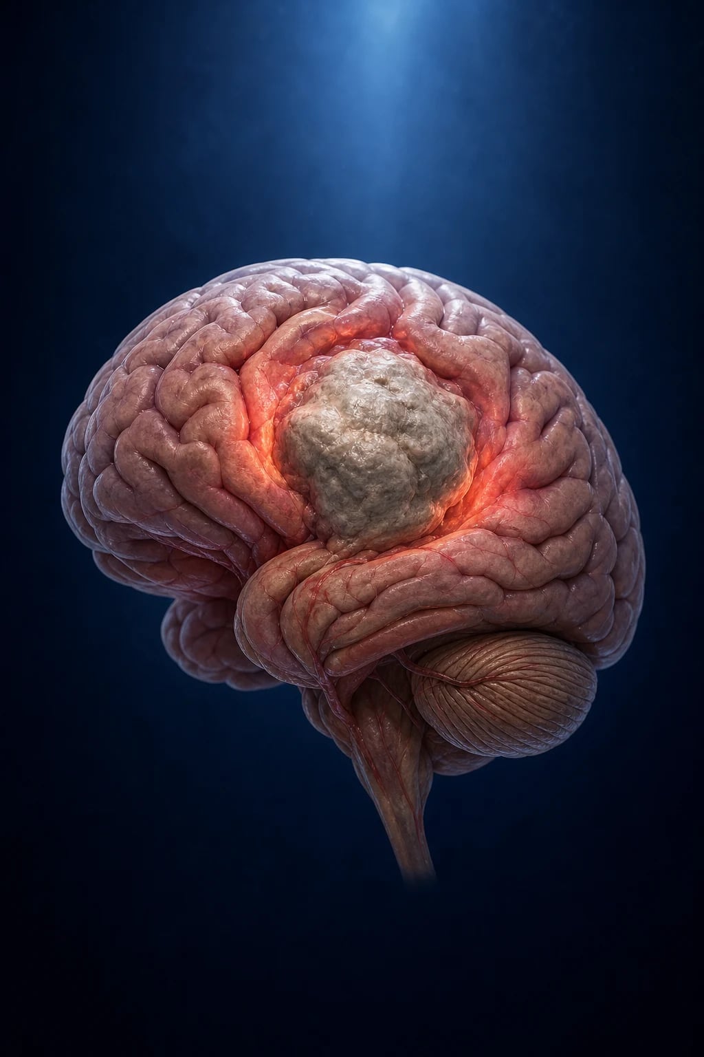
A brain tumour is any neoplasm arising within the cranial cavity — from the brain parenchyma itself, its coverings (meninges), the cranial nerves, the pituitary or pineal glands, or as a secondary deposit from a systemic cancer. The single most important first split, made at the bedside, is between primary tumours (originating in the CNS) and secondary (metastatic) tumours, because the implications for investigation, prognosis and treatment are entirely different. Metastases outnumber primary tumours by roughly five to ten times in adults, yet the public and many clinical questions fixate on primary glioma — the examiner will test both directions.[7]
The second conceptual anchor is the WHO 2021 classification, which defines each tumour by integrated histology plus molecular marker rather than by microscopy alone.[1] Two gliomas that look identical down a microscope can have different diagnoses, different treatments and different prognoses once their IDH status and chromosomal codeletion pattern are known. This molecular turn is the most important development in neuro-oncology in two decades and is examined explicitly.
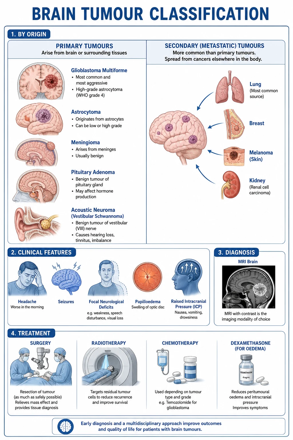
Whatever the histology, all brain tumours ultimately cause symptoms through a small set of shared mechanisms — mass effect and raised intracranial pressure, direct infiltration or destruction of functioning brain, peritumoural vasogenic oedema, seizure generation from cortical irritation, and distortion or obstruction of cerebrospinal fluid pathways producing hydrocephalus. Understanding these mechanisms unifies the clinical presentation, the imaging, and the medical management (steroids for oedema, anticonvulsants for seizures, shunts for hydrocephalus).[1]
Classification
Primary versus secondary, and the WHO 2021 framework
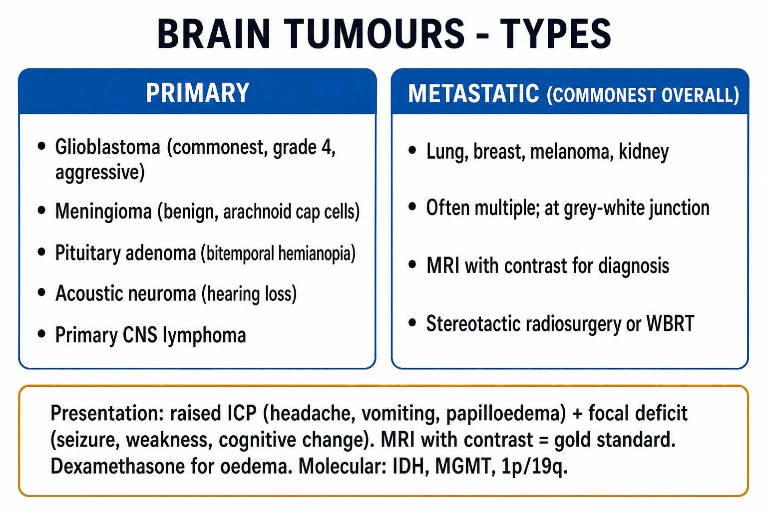
The clinically useful first division is the primary/metastatic split. Among primary tumours, the WHO 2021 classification groups tumours by the cell of origin and, increasingly, by defining molecular alteration.[1]
Gliomas
- Arise from glial cells — astrocytes, oligodendrocytes, ependymocytes
- Astrocytoma, IDH-mutant (WHO grade 2 to 4)
- Oligodendroglioma, IDH-mutant AND 1p/19q codeleted (grade 2 to 3)
- Glioblastoma, IDH-wildtype (grade 4) — commonest primary malignant brain tumour in adults
- Defined by integrated histology plus molecular markers, not microscopy alone
Meningiomas
- Arise from arachnoid cap cells, extra-axial
- Commonest primary brain tumour overall in adults
- WHO grade 1 (benign, about 80%), grade 2 (atypical), grade 3 (anaplastic)
- Often slow-growing, curable by complete surgical resection (Simpson grade I)
Sellar / cranial nerve
- Pituitary adenoma — functional (prolactinoma, acromegaly, Cushing) or non-functional
- Craniopharyngioma — suprasellar calcified cyst in children
- Vestibular schwannoma — cerebellopontine angle, bilateral in NF2
- Bitemporal hemianopia from optic chiasm compression is the classic sign
Metastatic (secondary)
- Roughly five to ten times commoner than primary tumours
- Lung, breast, melanoma, renal, colorectal — know the order
- Usually multiple, at grey-white junction, ring-enhancing with surrounding oedema
- Often the presenting feature of an occult systemic cancer
The WHO 2021 molecular classification of adult-type diffuse gliomas
This is the single most examined classification. The WHO 2021 system replaced purely histological grading with adult-type diffuse gliomas defined by their molecular profile:[1][5]
WHO 2021 adult-type diffuse gliomas — the defining markers
The logic is profound. A high-grade diffuse glioma in an adult is glioblastoma, IDH-wildtype unless it carries an IDH mutation — in which case, regardless of how aggressive the histology looks, it is astrocytoma, IDH-mutant (grade 4) with a substantially better prognosis. The old term "glioblastoma multiforme" is obsolete; glioblastoma is by definition IDH-wildtype in the 2021 system. IDH-mutant gliomas tend to arise in younger adults (median age around 40) and run a more indolent course; IDH-wildtype glioblastoma strikes older adults (median around 65) and pursues a relentless course.[1][5]
Other primary tumours worth naming
Beyond diffuse gliomas, examiners expect a working knowledge of the paediatric and non-glial primaries: pilocytic astrocytoma (WHO grade 1, BRAF-altered, children, posterior fossa or optic pathway, often curable by resection); medulloblastoma (posterior fossa, children, with four molecular subgroups — WNT, SHH, group 3, group 4 — each with distinct prognosis); ependymoma (defined by ZFTA fusion supratentorially and PF-EPN subgroups in the posterior fossa); diffuse midline glioma, H3 K27-altered (grade 4, brainstem in children, formerly DIPG, dismal prognosis); primary CNS lymphoma (diffuse large B-cell lymphoma, deeply situated, periventricular, HIV-related or immunocompetent elderly).[1]
Epidemiology & Risk Factors
Who gets brain tumours
The CBTRUS statistical report is the authoritative epidemiological source. Primary brain and CNS tumours have an overall annual incidence of roughly 20 to 25 per 100,000 population, but the incidence and the tumour type vary dramatically with age.[7]
Brain tumours by the numbers (CBTRUS)
Brain tumours show a characteristic bimodal age distribution. In children (the leading cause of cancer death after leukaemia), the commonest tumours are pilocytic astrocytoma, medulloblastoma and ependymoma, predominantly in the posterior fossa. In older adults the dominant tumour is glioblastoma, IDH-wildtype, with meningioma the commonest overall primary tumour at any age. The metastatic burden rises with the rising prevalence of systemic cancer and improved systemic control that allows patients to live long enough to develop brain disease.[7]
Established and refuted risk factors
The list of genuinely established risk factors is short and examiners test it precisely. High-dose ionising radiation to the head (historical scalp ringworm treatment, prior radiotherapy for another tumour) increases the risk of meningioma, glioma and nerve-sheath tumours decades later. Inherited tumour syndromes account for a small but disproportionately examined fraction. Immunosuppression (HIV, transplant, congenital immunodeficiency) markedly increases the risk of primary CNS lymphoma — an Epstein-Barr virus driven tumour.[1]
FAMILIAL
Optic pathway glioma, astrocytoma; neurofibromin gene, chromosome 17
Bilateral vestibular schwannoma, meningioma, ependymoma; merlin gene, chromosome 22
Medulloblastoma or glioblastoma with hereditary colon polyposis
Glioma, breast, sarcoma, adrenocortical; TP53 mutation
Haemangioblastoma (cerebellar, spinal, retinal), renal cell carcinoma
Medulloblastoma (desmoplastic), basal cell naevi, keratocysts
Dysplastic cerebellar gangliocytoma (Lhermitte-Duclos), meningioma
Lynch syndrome also raises glioma risk slightly
What is not a risk factor matters as much: despite intense study, mobile-phone use, hair dyes, head trauma, aspartame, and electromagnetic fields have not been convincingly linked to brain tumours. The most important message is that no modifiable risk factor explains the great majority of adult glioblastoma — these are sporadic, driven by somatic mutations.[1]
Pathophysiology
How a brain tumour produces symptoms
The skull is a rigid box holding brain, blood and cerebrospinal fluid. Adding a tumour (and its surrounding oedema) to a fixed-volume container raises the intracranial pressure and distorts brain tissue. The Monroe-Kellie doctrine governs this: the volume of the three intracranial contents is constant, so an expanding mass can be accommodated only by displacing CSF and venous blood — a reserve that exhausts quickly, after which small increments in volume produce large rises in pressure. This is why a patient with a glioblastoma can be relatively well one week and comatose the next.[1]
The five symptom-generating mechanisms are: mass effect (midline shift, herniation syndromes); vasogenic peritumoural oedema (disruption of the blood-brain barrier leaking protein-rich fluid into extracellular space — the target of dexamethasone); direct infiltration or destruction of functioning brain (focal deficits); seizure generation from cortical irritation (commoner with superficial, slow-growing, low-grade tumours); and CSF pathway obstruction producing hydrocephalus (typical of posterior fossa, pineal and intraventricular tumours).[1]
The molecular pathogenesis of glioma
The molecular revolution in glioma rests on a small number of defining alterations. Isocitrate dehydrogenase (IDH1/2) mutation is the single most important: the mutant enzyme produces the oncometabolite 2-hydroxyglutarate, which inhibits alpha-ketoglutarate-dependent dioxygenases, producing global epigenetic dysregulation (DNA and histone hypermethylation — the glioma-CpG-island methylator phenotype, G-CIMP) and blocking differentiation. IDH-mutant gliomas arise in younger adults and carry a markedly better prognosis than IDH-wildtype tumours.[1]
1p/19q codeletion — the whole-arm deletion of the short arm of chromosome 1 and the long arm of chromosome 19 — defines oligodendroglioma and is invariably associated with IDH mutation. Its presence is a powerful favourable prognostic marker and predicts benefit from alkylating chemotherapy (PCV and temozolomide), making it one of the strongest single predictors of glioma outcome. MGMT (O-6-methylguanine-DNA-methyltransferase) promoter methylation silences the DNA-repair enzyme that would otherwise undo the damage temozolomide inflicts — so a methylated MGMT promoter predicts a better response to temozolomide and is now a routine companion test in glioblastoma.[3]
In glioblastoma, IDH-wildtype, the dominant alterations are EGFR amplification and variant III mutation, PTEN loss, CDKN2A deletion, TERT promoter mutation, and the characteristic chromosome 7 gain plus chromosome 10 loss (+7/-10). These are not merely academic: EGFR and BRAF are now actionable targets, and TERT promoter mutation is one of the molecular features that can upgrade an IDH-wildtype diffuse astrocytoma to glioblastoma even when histology looks lower-grade.[1][5]
Clinical Presentation
Brain tumours announce themselves through three principal modes — raised intracranial pressure, focal neurological deficit, and seizure — often in combination, and occasionally as cognitive, personality or endocrine change, or as a stroke-like event from tumour haemorrhage. The tempo is usually progressive over weeks to months, which itself is a discriminator from vascular (sudden) and inflammatory (subacute relapsing) disease.[5]
Raised intracranial pressure
The classical raised-ICP triad is headache, vomiting and papilloedema. The headache of raised ICP is characteristically worse in the morning, worse with Valsalva manoeuvre (coughing, straining, bending), often occipital or frontal, progressive, and may ease during the day as the patient sits up. Vomiting may be effortless and projectile, occurring without preceding nausea, and classically occurs in the morning. Papilloedema — blurring of the optic disc margins, loss of venous pulsations, then elevation and haemorrhages — is the single most important bedside sign of raised ICP and demands fundoscopy in every patient with a progressive headache. A false-localising sixth-nerve palsy (bilateral lateral rectus weakness from stretching of the abducens nerve over the petrous tip as the brainstem shifts) is a late but classic sign.[5]
Focal neurological deficit by location
The focal deficit maps onto the tumour's location, and examiners love the clinicopathological correlation: [1]
Frontal lobe
- Personality change, apathy, disinhibition, executive dysfunction
- Contralateral hemiparesis (motor cortex), grasp and pout reflexes
- Expressive (Broca) aphasia if dominant hemisphere
- Foster-Kennedy syndrome (ipsilateral optic atrophy, contralateral papilloedema) with olfactory groove meningioma
Temporal lobe
- Memory impairment, complex partial (focal impaired awareness) seizures
- Receptive (Wernicke) aphasia if dominant
- Superior quadrantanopia (Meyer's loop)
- Olfactory and formed visual/auditory aura
Parietal lobe
- Contralateral sensory loss, astereognosis, agraphesthesia, two-point discrimination loss
- Neglect (non-dominant), extinction
- Inferior quadrantanopia (optic radiation)
- Alexia with or without agraphia (dominant)
Posterior fossa
- Cerebellar: ataxia, dysmetria, intention tremor, nystagmus, wide-based gait
- Brainstem: cranial nerve palsies with contralateral long-tract signs
- Obstructive hydrocephalus from fourth-ventricle compression — early raised ICP
Pituitary/sellar
- Bitemporal hemianopia from optic chiasm compression
- Endocrine: hyperprolactinaemia, acromegaly, Cushing disease, hypopituitarism
- Apoplexy: sudden headache and ophthalmoplegia
Seizures
Seizures occur in 30 to 50 percent of patients with brain tumours and are commoner with cortically based, slow-growing, low-grade tumours (ganglioglioma, dysembryoplastic neuroepithelial tumour, low-grade glioma) than with deeply situated high-grade tumours. A new-onset seizure in an adult, with no immediate metabolic or vascular explanation, is a brain tumour until proven otherwise — this is one of the highest-yield exam statements and the trigger for urgent MRI with contrast.[5]
Atypical presentations
Examiners test the atypical deliberately. The elderly patient may present with progressive cognitive decline, apathy or gait disturbance mistaken for dementia or normal ageing. The pituitary tumour may declare itself with endocrine disturbance (galactorrhoea from prolactinoma, acral enlargement from growth-hormone excess, Cushingoid features) long before any mass effect. A haemorrhage into a tumour (melanoma, renal cell, choriocarcinoma, thyroid, glioblastoma) produces a stroke-like abrupt onset. First-episode psychotic symptoms or new psychiatric illness in an adult can occasionally reflect a frontal or temporal lobe tumour.[5]
Differential Diagnosis
The differential of a brain mass on imaging is broad, and the imaging pattern — not the name — drives the work-up. A ring-enhancing lesion, a diffusely infiltrating non-enhancing mass, and a homogeneously enhancing extra-axial lesion each carry a different differential. [1]
Brain abscess
- Ring-enhancing with a thin, smooth wall and restricted diffusion (DWI)
- Fever, raised inflammatory markers, infective source (sinusitis, otitis, endocarditis, dental)
- Often thinner wall on the ventricular aspect — risk of ventricular rupture
- Toxoplasmosis in HIV: multiple ring lesions at grey-white junction
Tumefactive demyelination
- MS or ADEM; younger patient with relapsing-remitting history
- Incomplete horseshoe-shaped enhancement, open-ring sign
- Often periventricular, juxtacortical, infratentorial or spinal cord lesions elsewhere
- CSF oligoclonal bands may be positive
Subdural empyema / haematoma
- Extra-axial crescentic collection
- Empyema: fever, sinusitis, rapid deterioration — surgical emergency
- Chronic subdural: elderly, falls, fluctuating deficits, gradual onset
Stroke
- Vascular territory, sudden onset, DWI restriction without ring enhancement
- Tumour may occasionally mimic stroke (tumoural haemorrhage)
- Repeat imaging if clinical evolution does not fit a vascular territory
Radiation necrosis
- Months to years after prior brain radiotherapy
- Enhancing mass that mimics tumour recurrence
- MR perfusion (low rCBV) and MR spectroscopy help distinguish from tumour
- Bevacizumab can reduce oedema
Arteriovenous malformation
- Flow voids on MRI, tangle of vessels
- Presents with haemorrhage, seizure, or bruit
- Angiography defines the nidus and feeding vessels
The can't-miss mimics are brain abscess (surgical emergency, source control, antibiotics) and primary CNS lymphoma (which dictates a fundamentally different management — biopsy only, no steroids before biopsy, high-dose methotrexate rather than resection).[5]
Clinical & Bedside Assessment
A focused bedside assessment in suspected brain tumour has three aims: to localise the lesion, to gauge the urgency (raised ICP, herniation, hydrocephalus), and to hunt for a systemic primary if metastasis is plausible. [1]
History probes the headache (timing, progression, association with Valsalva or position), any seizures (focal versus generalised, post-ictal deficit), the tempo of any focal deficit (progressive over weeks is the hallmark of tumour), cognitive or personality change reported by family, visual symptoms, and endocrine change (galactorrhoea, acral growth, Cushingoid features, libido loss, menstrual change). A smoking history, prior cancer, weight loss, and family history of cancer or inherited tumour syndrome are essential. The red flags in the history — morning headache, vomiting, new seizure, progressive deficit — should trigger imaging that day.[5]
Examination begins with vital signs and the Glasgow Coma Scale (eye opening E1 to E4, verbal response V1 to V5, motor response M1 to M6; maximum 15). A full neurological examination follows: cranial nerves including fundoscopy (papilloedema), visual fields (confrontation testing for bitemporal hemianopia), motor and sensory systems, coordination (cerebellar), gait, and reflexes. Neck stiffness raises meningitis or haemorrhage as an alternative. A general examination hunts for a primary: skin (melanoma), breasts, lungs (clubbing, chest signs), thyroid, abdomen (hepatomegaly, masses), and lymph nodes. A breast and testicular examination is indicated when metastatic disease is suspected.[5]
[1]Investigations
Imaging — MRI with gadolinium is the gold standard
MRI brain with gadolinium contrast is the imaging modality of choice and the single most informative test. It defines the tumour's location (intra- vs extra-axial, supra- vs infratentorial), size, enhancement pattern (ring, nodular, homogeneous, none), peritumoural oedema, mass effect and midline shift, haemorrhage (susceptibility on T2* or SWI), calcification (best on CT or SWI), and any CSF pathway obstruction. Glioblastoma classically shows a thick, irregularly enhancing ring with central necrosis and a heterogeneous T2/FLAIR mass crossing the corpus callosum (butterfly glioma); meningioma is a homogeneously enhancing extra-axial mass with a dural tail; oligodendroglioma is a cortically based, calcified mass; pituitary macroadenoma is a sellar mass with suprasellar extension; primary CNS lymphoma is a homogeneously enhancing periventricular mass that often restricts on diffusion.[5]
Advanced MRI sequences add diagnostic precision. MR spectroscopy shows elevated choline, reduced N-acetylaspartate and (in some tumours) a lactate/lipid peak, helping distinguish tumour from abscess, demyelination or radiation necrosis. MR perfusion measures relative cerebral blood volume (rCBV): high rCBV suggests high-grade tumour or recurrence, low rCBV favours radiation necrosis. Diffusion-weighted imaging shows restricted diffusion in abscess and lymphoma. Functional MRI and diffusion tensor imaging map eloquent cortex and white-matter tracts for surgical planning in or near motor, sensory or language areas.[5]
Imaging patterns that signal a specific diagnosis
CT retains specific roles: it is faster and more available in the emergency department, detects acute haemorrhage and calcification superbly, and is the first test when MRI is contraindicated (pacemaker, some metallic implants) or the patient cannot tolerate it. A non-contrast CT showing acute hydrocephalus or midline shift is itself a neurosurgical emergency. CT chest, abdomen and pelvis is performed when metastasis is suspected and the primary is occult — and whole-body PET-CT is increasingly used for the same purpose. Spine MRI is indicated when spinal or leptomeningeal drop metastases are suspected (posterior fossa tumours, medulloblastoma, ependymoma, germinoma).[5]
Histology, molecular profiling, and the role of CSF
Definitive diagnosis requires tissue. Stereotactic biopsy obtains tissue when the tumour is unresectable or in eloquent/deep cortex; open biopsy or surgical resection provides more tissue. The histology is integrated with molecular profiling — IDH mutation, 1p/19q codeletion, MGMT promoter methylation, TERT promoter, EGFR, BRAF V600E, H3 K27, CDKN2A — to arrive at the WHO 2021 integrated diagnosis. MGMT promoter methylation is now a standard companion test in glioblastoma because it predicts temozolomide benefit.[3][5]
Lumbar puncture and CSF analysis has a narrow but important role — cytology for leptomeningeal disease and primary CNS lymphoma (malignant cells, flow cytometry), tumour markers (alpha-fetoprotein and beta-HCG for germ cell tumours), and oligoclonal bands when demyelination is in the differential. The cardinal rule: never perform lumbar puncture before imaging if a mass lesion is possible, because reducing the pressure below the tumour can precipitate tonsillar or uncal herniation.[5]
Other tests depending on context: visual fields and acuity for sellar masses; endocrine panel (prolactin, IGF-1, cortisol, thyroid, sex hormones, electrolytes and osmolality for diabetes insipidus) for pituitary tumours; HIV test when CNS lymphoma or toxoplasmosis is plausible; and preoperative work-up (coagulation, bloods, ECG, anaesthetic assessment) for surgical candidates. [1]
Management — Resuscitation
The immediate priority in the patient with a brain tumour is to recognise and treat raised intracranial pressure, impending herniation, and seizures — these are the time-critical threats. [1]
Acute raised intracranial pressure
Raised ICP — the emergency bundle
Position and airway
Elevate head of bed to 30 degrees, head in neutral midline to optimise venous drainage; secure airway and ventilation
Dexamethasone
Dexamethasone 10 mg IV bolus, then 4 mg every 6 hours, for vasogenic peritumoural oedema — the mainstay of medical ICP control in tumour
Osmotherapy
Mannitol 20% 0.5 to 1 g/kg IV over 15 to 20 min, or hypertonic saline 3% 150 to 250 mL over 20 min, as a temporising bridge to surgery
Hyperventilation
Brief hyperventilation to PaCO2 30 to 35 mmHg (cerebral vasoconstriction) as a temporary bridge in impending herniation only
Intubate if GCS 8 or less
Definitive airway and ventilation; transfer to neurocritical care
Definitive treatment
Urgent neurosurgical decompression, resection, or external ventricular drain for obstructive hydrocephalus
Dexamethasone is the specific therapy for the vasogenic oedema surrounding a brain tumour (it restores blood-brain barrier integrity), in contrast to cytotoxic oedema from stroke, where it is ineffective or harmful. The dose 4 to 16 mg per day is then weaned to the lowest effective dose, with proton-pump-inhibitor cover and monitoring for hyperglycaemia, myopathy and infection.[5]
Status epilepticus
A tumour-related seizure that does not self-terminate is treated as any status epilepticus: lorazepam 4 mg IV (repeat once after 4 minutes), then levetiracetam 1500 to 3000 mg IV or fosphenytoin 18 mg phenytoin-equivalent per kg IV, with escalation to intubation, propofol or midazolam infusion and ICU care for refractory status.[5]
Herniation syndromes and hydrocephalus
Uncal herniation (ipsilateral dilated pupil from third-nerve compression, contralateral hemiparesis from cerebral peduncle compression, declining consciousness) demands the emergency bundle plus urgent neurosurgical decompression. Central herniation produces bilateral small pupils, decorticate then decerebrate posturing, and respiratory irregularity. Cerebellar tonsillar herniation causes neck stiffness, respiratory arrest and cardiorespiratory collapse — a posterior-fossa emergency. Obstructive hydrocephalus from a posterior fossa or intraventricular tumour is treated with an external ventricular drain acutely and often a permanent shunt or endoscopic third ventriculostomy later.[5]
Management — Definitive & Stepwise
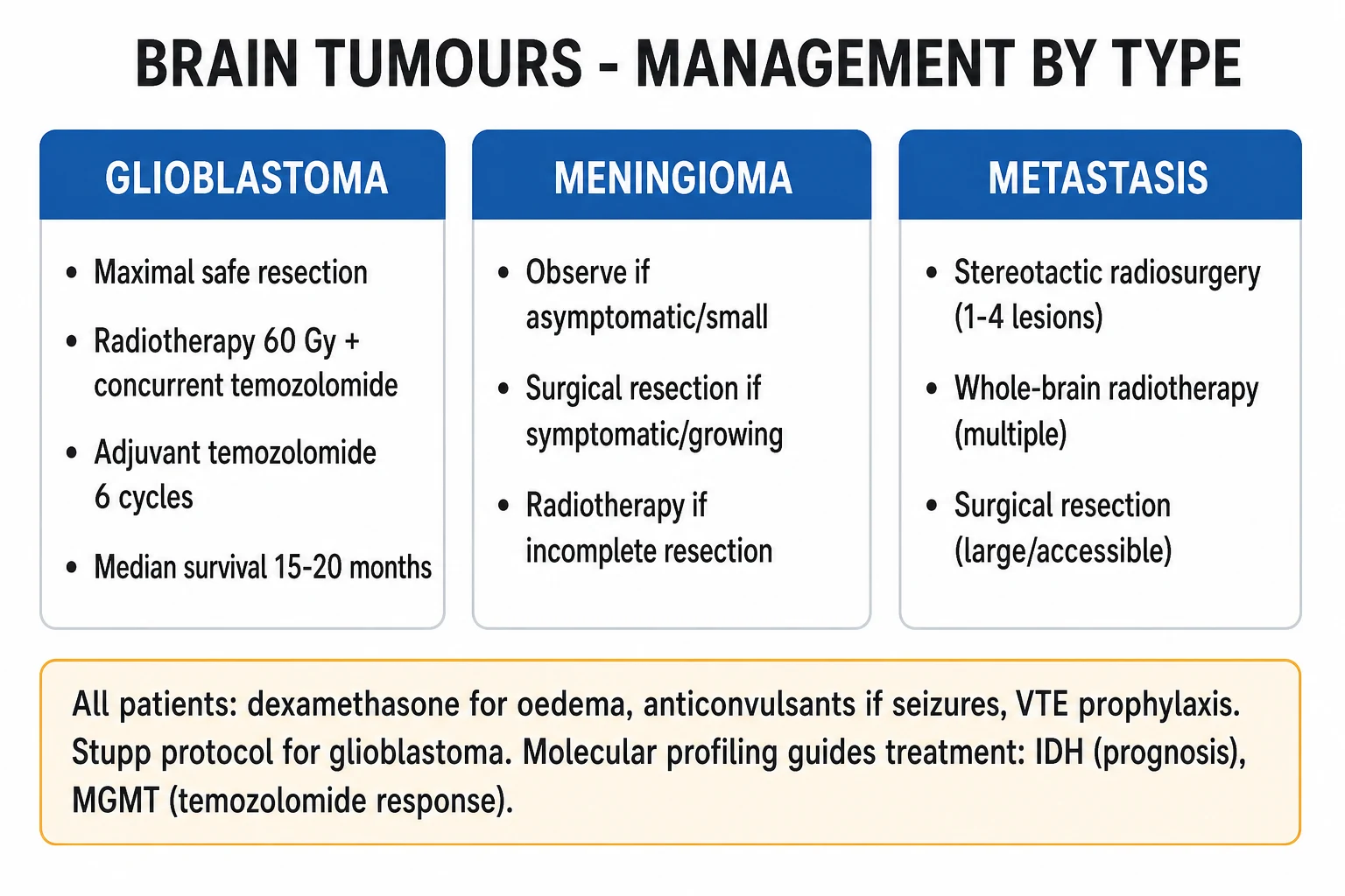
Definitive treatment is histology- and molecular-specific, delivered by a multidisciplinary team (neurosurgeon, neuro-oncologist, radiation oncologist, neurologist, specialist nurse, neuropsychologist, palliative care). The framework is: control symptoms, establish the diagnosis, resect what can be safely resected, then give adjuvant therapy according to the integrated diagnosis. [1]
Glioblastoma, IDH-wildtype — the Stupp protocol
The standard of care for fit adults with newly diagnosed glioblastoma is maximal safe surgical resection (the greater the extent of resection, the better the survival, while preserving eloquent function — awake craniotomy with direct cortical mapping is used for tumours in language, motor or sensory cortex) followed by the Stupp protocol:[2]
The Stupp protocol — glioblastoma standard of care (Stupp 2005)
This regimen more than doubled two-year survival compared with radiotherapy alone (27 percent versus 10 percent in the original EORTC/NCIC trial) and remains the global standard twenty years on. Tumour-treating fields (TTF) — low-intensity alternating electric fields delivered via scalp transducer arrays — added to maintenance temozolomide prolonged median survival to about 21 months in the EF-14 trial and are incorporated into many guidelines.[4] MGMT promoter methylation identifies the patients who benefit most from temozolomide — methylated tumours respond far better than unmethylated ones, a fact examined explicitly.[3]
For elderly or frail patients (typically over 65 to 70 years with good performance status), the Perry trial showed that short-course radiotherapy (40 Gy in 15 fractions) with concomitant and adjuvant temozolomide outperformed short-course radiotherapy alone; in patients with MGMT-methylated tumours, temozolomide alone is a reasonable option, while those with unmethylated tumours derive less benefit from temozolomide.[6]
Astrocytoma and oligodendroglioma, IDH-mutant
For astrocytoma, IDH-mutant (grades 2 to 4) and oligodendroglioma, IDH-mutant, 1p/19q codeleted (grades 2 to 3), maximal safe resection is followed by risk-stratified adjuvant therapy. High-risk low-grade glioma (subtotal resection, age over 40, IDH-wildtype features) and grade 3 to 4 IDH-mutant astrocytoma receive radiotherapy plus temozolomide or radiotherapy plus PCV chemotherapy (procarbazine, lomustine, vincristine). Oligodendroglioma with 1p/19q codeletion has the best prognosis of any diffuse glioma — median survival often exceeds 10 years — and responds well to radiotherapy plus PCV or temozolomide, the codeletion being a marker of chemosensitivity.[5]
Meningioma
The EANO meningioma guideline governs management.[8] WHO grade 1 meningioma with complete Simpson grade I resection (tumour, dural attachment and involved bone) is often cured by surgery alone — observation with serial MRI follows. Subtotal resection, recurrent, atypical (grade 2) or anaplastic (grade 3) meningioma warrants adjuvant radiotherapy — fractionated external-beam or stereotactic radiosurgery (gamma knife) for small residual or recurrent disease. Meningiomas are typically slow-growing, and small asymptomatic ones in the elderly can simply be surveilled.[8]
[1]Pituitary adenoma
Pituitary macroadenomas causing visual field compromise (bitemporal hemianopia) or hormonal hypersecretion are managed by transsphenoidal surgery — the endoscopic endonasal approach is now standard. Prolactinoma is the exception: cabergoline 0.25 to 1 mg twice weekly (a dopamine agonist) is first-line and often shrinks the tumour, reserving surgery for those intolerant or resistant. Acromegaly is treated with transsphenoidal surgery followed, if not cured, by somatostatin analogues (octreotide, lanreotide), the GH-receptor antagonist pegvisomant, or dopamine agonists. Cushing disease is managed surgically with steroidogenesis inhibitors (ketoconazole, metyrapone) and radiotherapy as adjuncts. Radiotherapy (fractionated or stereotactic) is reserved for residual, recurrent or medically refractory disease.[5]
Primary CNS lymphoma
Primary CNS lymphoma is managed completely differently from other brain tumours, and this is heavily examined. The cardinal rules: stereotactic biopsy only (surgical resection confers no benefit and may harm), do NOT give steroids before biopsy (glucocorticoids lyse lymphoma cells and can abolish the diagnostic tissue within hours), and treat with high-dose methotrexate-based chemotherapy (methotrexate 3 to 8 g/m² every 2 to 4 weeks) — often with rituximab, cytarabine and, in younger fit patients, consolidation with autologous stem-cell transplant. Whole-brain radiotherapy is effective but neurotoxic, especially in older patients, and is now usually deferred or omitted. In HIV-associated disease, combined antiretroviral therapy is essential alongside chemotherapy.[5]
Brain metastases
Management is guided by number, size, location, performance status and control of the primary. Single accessible metastasis with controlled primary and good performance status — surgical resection followed by stereotactic radiosurgery (SRS, gamma knife) to the cavity, which improves local control and survival compared with whole-brain radiotherapy alone. Limited metastatic burden (typically up to 3 to 4 small lesions) — SRS alone, sparing the cognitive cost of whole-brain radiotherapy. Multiple or extensive metastases, poor performance status, uncontrolled primary — whole-brain radiotherapy (WBRT, 20 Gy in 5 fractions or 30 Gy in 10 fractions) for palliation, plus systemic therapy appropriate to the primary. Melanoma, non-small-cell lung cancer (especially EGFR-mutant and ALK-rearranged) and HER2-positive breast cancer increasingly respond to targeted agents and immunotherapy (immune checkpoint inhibitors), which can achieve intracranial responses. Leptomeningeal disease is treated with intrathecal chemotherapy (methotrexate, cytarabine, thiotepa) plus systemic targeted therapy where the primary has a druggable driver.[5]
Symptom control and supportive care
Beyond definitive therapy, several agents are near-universal. Dexamethasone 4 to 16 mg daily controls peritumoural oedema, weaned to the lowest effective dose with PPI cover. Anticonvulsants — levetiracetam 500 to 1500 mg twice daily is preferred (no enzyme induction, no interaction with chemotherapy) — are given only to patients who have had a seizure; routine prophylactic anticonvulsants are not recommended in patients without a seizure history, a point examiners test. Venous thromboembolism is common in brain-tumour patients (especially glioma); prophylactic low-molecular-weight heparin is balanced against the small bleeding risk, and treatment doses are used for established VTE with a careful reassessment once any perioperative window has passed. Palliative care is involved early — symptom control, advance care planning, and attention to the psychological and social burden on patient and family.[5]
Specific Subtypes & Scenarios
Pilocytic astrocytoma (WHO grade 1, BRAF-altered) in children is often cured by complete surgical resection; BRAF inhibitors (dabrafenib plus trametinib) are emerging for unresectable or progressive disease. Medulloblastoma is treated with maximal safe resection, craniospinal irradiation, and chemotherapy (cisplatin, lomustine, vincristine); the molecular subgroup (WNT best, group 3 worst) now stratifies treatment intensity, and five-year survival is 70 to 80 percent overall. Diffuse midline glioma, H3 K27-altered (formerly DIPG) — biopsy for molecular confirmation is now standard where safe; focal radiotherapy provides transient symptomatic benefit; median survival remains under 12 months, and trials of targeted agents (ONC201) are ongoing. Craniopharyngioma — surgical resection plus radiotherapy for residual, with lifelong hormone replacement (hydrocortisone, levothyroxine, sex steroids, growth hormone, desmopressin for diabetes insipidus). Vestibular schwannoma — observation for small asymptomatic lesions, stereotactic radiosurgery or surgical resection (retrosigmoid or translabyrinthine) for growing or symptomatic tumours; bilateral is pathognomonic of NF2. Haemangioblastoma — resectable; multiple lesions suggest VHL syndrome.[1][5]
Recurrent glioblastoma has limited options: re-resection if feasible, re-irradiation in selected cases, bevacizumab (anti-VEGF monoclonal antibody) for symptomatic oedema and radiographic response (without proven overall survival benefit), retreatment with temozolomide (especially alternative dosing schedules), and clinical trial enrolment — which every patient should be offered if eligible. Tumour-treating fields can be continued or started at recurrence.[4][5]
Complications & Pitfalls
Disease- and treatment-related complications
The complications of a brain tumour and its treatment span neurological, surgical, oncological and systemic. Progressive neurological deficit from tumour growth; seizures (including status epilepticus); hydrocephalus (obstructive from posterior fossa or intraventricular tumour); cerebral herniation; tumoural haemorrhage (melanoma, renal cell, choriocarcinoma, glioblastoma); leptomeningeal spread; and paraneoplastic syndromes (rarely, with certain tumours). Venous thromboembolism is markedly more common in brain-tumour patients than in the general population, especially in glioma.[5]
Surgical complications include new focal deficit, haematoma, infection (wound, meningitis), CSF leak, and (with pituitary surgery) diabetes insipidus, CSF rhinorrhoea and hypopituitarism. Radiotherapy causes acute fatigue and skin reaction, early delayed somnolence syndrome (weeks), and late radiation necrosis (months to years — an enhancing mass that mimics recurrence, treated with bevacizumab or surgery). Whole-brain radiotherapy causes progressive cognitive decline, especially in survivors — a major reason SRS is preferred for limited metastases. Temozolomide causes myelosuppression (monitor FBC, especially lymphopenia and the small risk of PML) and nausea. PCV chemotherapy causes myelosuppression, infertility and (procarbazine) a disulfiram-like reaction with alcohol. Bevacizomab causes hypertension, proteinuria, bleeding and thrombosis, and bowel perforation. High-dose methotrexate causes nephrotoxicity, hepatotoxicity, mucositis and myelosuppression, requiring hydration, alkalinisation and folinic acid rescue.[5]
Pitfalls — the classic errors
The classic errors an examiner will probe: giving steroids before biopsy in suspected primary CNS lymphoma (destroys diagnostic tissue); performing lumbar puncture before imaging in a patient with a mass (risk of herniation); routinely prescribing prophylactic anticonvulsants (no benefit, real harm, drug interactions); treating a tumoural haemorrhage as a stroke without an MRI (missing the underlying lesion); failing to search for a systemic primary in a brain metastasis (with consequent denial of targeted therapy); and treating an IDH-mutant astrocytoma as glioblastoma (worse prognosis assumed, wrong prognostic conversation).[5]
Prognosis & Disposition
Prognosis varies more widely across brain tumours than in almost any other organ — from cure (pilocytic astrocytoma, grade 1 meningioma) to median survival measured in months (glioblastoma, DIPG). The key determinants are histology and integrated molecular diagnosis (IDH status and 1p/19q codeletion dominate glioma prognosis), age and performance status, extent of resection, MGMT promoter methylation (in glioblastoma), and the molecular subgroup in medulloblastoma.[2][5]
Median survival by tumour type
Disposition after definitive treatment: inpatient for surgery and acute complications; outpatient with serial MRI surveillance (typically every 3 to 6 months for two years, then 6 to 12 months) for low-grade and resected tumours; neurorehabilitation (physiotherapy, occupational therapy, speech and language, neuropsychology) for residual deficits; and palliative care integrated early for advanced or progressive disease, addressing symptoms (headache, seizures, cognitive change, dysphagia), advance care planning and family support.[5]
Special Populations
Pregnancy
Brain tumours may grow rapidly in pregnancy (haemodynamic and hormonal changes, fluid shifts), and presentation can be confounded with pre-eclampsia (headache, visual disturbance, papilloedema, seizures). MRI without gadolinium is safe in pregnancy; gadolinium is avoided unless essential. Management is by a combined neurosurgical, obstetric and anaesthetic team, balancing gestational age, tumour type and urgency — surgery can usually be deferred to the postpartum period unless the tumour is life-threatening. Pituitary adenomas may enlarge in pregnancy (prolactinoma risk), and visual fields are monitored.[5]
Children
Brain tumours are the commonest solid tumour and the leading cause of cancer death in children after leukaemia. The spectrum differs from adults: pilocytic astrocytoma, medulloblastoma, ependymoma, diffuse midline glioma (DIPG), craniopharyngioma. Children require paediatric neuro-oncology expertise, age-adapted treatment (craniospinal irradiation doses are reduced in the very young to spare neurocognition), and lifelong follow-up for late effects — endocrinopathy, cognitive decline, second malignancy, infertility. Surgical resection is often curative for pilocytic astrocytoma.[1]
Elderly
Glioblastoma is commonest in older adults, but age and performance status must temper therapy — the full Stupp protocol is demanding, and short-course radiotherapy with temozolomide (Perry) or temozolomide alone in MGMT-methylated tumours is appropriate for older or less fit patients. Meningioma is common and often indolent in the elderly, where observation is frequently the right choice for small asymptomatic tumours.[6]
Immunocompromised
In HIV and post-transplant immunosuppression, the differential of a brain mass shifts towards primary CNS lymphoma (EBV-driven) and cerebral toxoplasmosis. The classic approach to a solitary deep ring-enhancing lesion in HIV is to treat empirically for toxoplasmosis (sulfadiazine plus pyrimethamine) and reserve biopsy for non-responders; multiple lesions, a positive EBV CSF PCR, and single-photon emission CT thallium uptake favour lymphoma. Restoring immune function (antiretroviral therapy) is itself part of the lymphoma treatment.[5]
Inherited tumour syndromes
A new brain tumour in a young patient, a multifocal presentation, a family history of cancer, or a syndromic phenotype (cafe-au-lait patches, axillary freckling, Lisch nodules in NF1; bilateral acoustic neuromas in NF2; retinal and cerebellar haemangioblastomas in VHL) should prompt consideration of an inherited syndrome — see the FAMILIAL mnemonic. Surveillance of affected relatives is part of management.[1]
Evidence, Guidelines & Regional Differences
Foundational references
The framework used here rests on the WHO 2021 Classification of CNS Tumours (Louis 2021)[1] and the EANO guidelines on the diagnosis and treatment of diffuse gliomas of adulthood (Weller 2021)[5] for glioma, the EANO meningioma guideline (Goldbrunner 2021) for meningioma[8], and the CBTRUS statistical report (Price 2025) for epidemiology.[7]
Landmark trials
Glioblastoma — Stupp 2005
- EORTC/NCIC trial, NEJM (PMID 15758009)
- Concomitant + adjuvant temozolomide with 60 Gy radiotherapy
- Median survival 14.6 months vs 12.1 months with radiotherapy alone
- Became and remains the global standard of care (the Stupp protocol)
MGMT — Hegi 2005
- Companion analysis to Stupp, NEJM (PMID 15758010)
- MGMT promoter methylation predicts benefit from temozolomide
- Methylated tumours carry a better prognosis regardless of treatment
- MGMT testing now standard companion diagnostic in glioblastoma
Tumour-treating fields — EF-14 2017
- Stupp et al., JAMA (PMID 29260225)
- TTF plus maintenance temozolomide vs temozolomide alone
- Median survival about 21 months with TTF added
- TTF now incorporated into many guidelines for newly diagnosed glioblastoma
Elderly GBM — Perry 2017
- NEJM (PMID 28296618)
- Short-course radiotherapy (40 Gy/15) plus temozolomide in elderly patients
- Improved overall survival over short-course radiotherapy alone
- Temozolomide alone reasonable in MGMT-methylated elderly tumours
Regional deltas
[1] [1]In India (the NEET-PG/INICET context), the Stupp protocol and surgical principles are standard, but access to temozolomide for prolonged courses, tumour-treating fields, and advanced molecular profiling (IDH, 1p/19q, MGMT) varies by centre and ability to pay — government centres may provide temozolomide through access schemes, while molecular testing is increasingly available in tertiary centres. The Indian Council of Medical Research (ICMR) and the neuro-oncology sections of the national cancer control programme broadly align with WHO 2021 and EANO, with access-driven adaptations.
Current controversies
Active debates include the optimal management of IDH-mutant grade 2 glioma (observation versus early surgery and adjuvant therapy — the INDIVE and RTOG 9802 data support early adjuvant therapy in high-risk disease); the role and timing of temozolomide in elderly MGMT-unmethylated tumours; whether tumour-treating fields deliver enough benefit to justify the burden and cost outside trial settings; the place of targeted therapy and immunotherapy in primary brain tumours (largely disappointing to date, unlike in metastatic disease); and the integration of liquid biopsy (CSF and plasma cell-free DNA/tumour DNA) into surveillance. Recurrent glioblastoma remains an area of intense, as-yet-unfulfilled therapeutic innovation.[5]
Exam Pearls
- Metastases outnumber primary brain tumours five- to ten-fold; the order of primaries is lung, breast, melanoma, renal, colorectal — know it cold.
- New-onset seizure in an adult equals MRI with contrast — not CT, not EEG first.
- Morning headache, vomiting and papilloedema = raised ICP; a bilateral sixth-nerve palsy is a false-localising sign, not a brainstem lesion.
- Never LP before imaging if a mass is possible — risk of tonsillar herniation.
- Glioblastoma is IDH-wildtype by definition in WHO 2021; an IDH-mutant high-grade glioma is astrocytoma, IDH-mutant (grade 4), with a better prognosis.
- Oligodendroglioma requires both IDH mutation AND 1p/19q codeletion — and carries the best glioma prognosis.
- MGMT promoter methylation predicts temozolomide response (Hegi, PMID 15758010).
- The Stupp protocol: radiotherapy 60 Gy in 30 fractions plus concurrent temozolomide 75 mg/m² daily, then adjuvant temozolomide 150 to 200 mg/m² days 1 to 5 every 28 days for 6 to 12 cycles; median survival about 15 months.
- Primary CNS lymphoma: biopsy only, NO steroids before biopsy, high-dose methotrexate — never resect, never give steroids first.
- Pituitary adenoma: bitemporal hemianopia from chiasm compression; prolactinoma is treated medically with cabergoline, surgery for the rest.
- Vestibular schwannoma: cerebellopontine angle, unilateral hearing loss; bilateral = NF2.
- Simpson grade I meningioma resection is near-curative; subtotal resection or grade 2 to 3 needs radiotherapy.
- Dexamethasone 4 to 16 mg daily for peritumoural oedema; 10 mg IV bolus then 4 mg every 6 hours for acute raised ICP.
- Routine prophylactic anticonvulsants are NOT recommended in brain-tumour patients without seizures.
- Brain metastases: up to 3 to 4 small lesions get stereotactic radiosurgery (spares cognition); extensive disease gets whole-brain radiotherapy.
- Meningioma is the commonest primary brain tumour overall in adults; glioblastoma is the commonest primary malignant brain tumour. [1]
Exam application bank (NEET-PG / INICET)
One-line answer
Brain tumours are intracranial neoplasms — primary (arising from glia, meninges, cranial nerves, pituitary or germ cells) or secondary (metastatic, roughly five to ten times commoner than primary). The WHO 2021 classification defines them by integrated histology and molecular markers (IDH mutation, 1p/19q codeletion, MGMT methylation, BRAF, H3 K27). Presentation: raised ICP (morning headache, vomiting, papilloedema), seizures, progressive focal deficit. MRI with gadolinium is the imaging gold standard. Management: maximal safe resection, radiotherapy, chemotherapy (Stupp protocol for glioblastoma — radiotherapy 60 Gy plus concomitant and adjuvant temozolomide), dexamethasone for vasogenic oedema, anticonvulsants for seizures. Glioblastoma median survival is about 15 months with Stupp protocol.
Worked stems (answer without another resource)
Stem 1 — Classic presentation. Map symptoms to mechanism; name the first investigation and first treatment step with dose/route if drug therapy is standard. [1]
Stem 2 — Unstable / complicated. List red flags that force immediate resuscitation, theatre, ICU, antidote, or reperfusion — and what you do in the first 15 minutes. [1]
Stem 3 — Atypical group. Elderly, pregnancy, child, or immunocompromised: how presentation and thresholds change. [1]
Stem 4 — Differential trap. Name the three closest mimics and one discriminator for each. [1]
Stem 5 — Disposition. Who goes home with safety-netting, who is admitted, who needs HDU/ICU/theatre, and what follow-up is mandatory. [1]
Rapid viva checklist
- Definition + classification
- Pathophysiology chain
- Bedside signs / criteria
- Score with exact components (if any)
- Emergency bundle
- Definitive therapy with doses
- Complications of disease and of treatment
- Special populations
- Guideline/trial name if classic
- Three exam traps
Coverage self-check
If you cannot answer any stem above from this page alone, re-read the matching section — the page is intended to be self-sufficient for final-prof and NEET-PG/INICET questions on Brain Tumours.
References
- [1]Louis DN, Perry A, Wesseling P, et al. The 2021 WHO Classification of Tumors of the Central Nervous System: a summary Neuro Oncol, 2021.PMID 34185076
- [2]Stupp R, Mason WP, van den Bent MJ, et al. Radiotherapy plus concomitant and adjuvant temozolomide for glioblastoma N Engl J Med, 2005.PMID 15758009
- [3]Hegi ME, Diserens AC, Gorlia T, et al. MGMT gene silencing and benefit from temozolomide in glioblastoma N Engl J Med, 2005.PMID 15758010
- [4]Stupp R, Taillibert S, Kanner A, et al. Effect of Tumor-Treating Fields Plus Maintenance Temozolomide vs Maintenance Temozolomide Alone on Survival in Patients With Glioblastoma: A Randomized Clinical Trial JAMA, 2017.PMID 29260225
- [5]Weller M, van den Bent M, Preusser M, et al. EANO guidelines on the diagnosis and treatment of diffuse gliomas of adulthood Nat Rev Clin Oncol, 2021.PMID 33293629
- [6]Perry JR, Laperriere N, O'Callaghan CJ, et al. Short-Course Radiation plus Temozolomide in Elderly Patients with Glioblastoma N Engl J Med, 2017.PMID 28296618
- [7]Price M, Wrensch M, Berger MS, et al. CBTRUS Statistical Report: Primary Brain and Other Central Nervous System Tumors Diagnosed in the United States in 2018-2022 Neuro Oncol, 2025.PMID 41092086
- [8]Goldbrunner R, Minniti G, Preusser M, et al. EANO guideline on the diagnosis and management of meningiomas Neuro Oncol, 2021.PMID 34181733