Neurology · General Medicine
Concussion & Traumatic Brain Injury
Also known as Concussion · Traumatic brain injury · TBI · Head injury · Mild traumatic brain injury · mTBI · Diffuse axonal injury
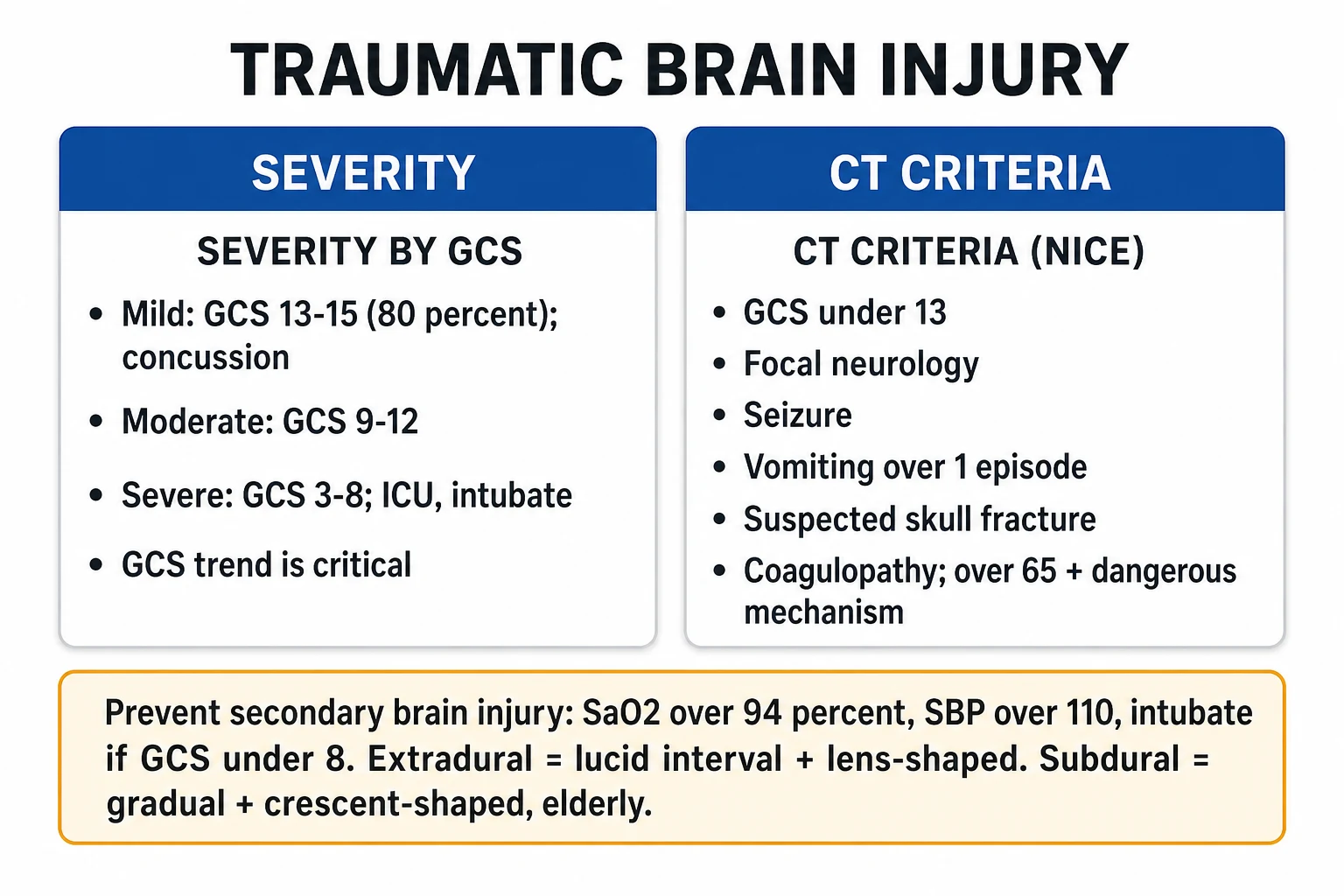
Traumatic brain injury (TBI) is a disruption of brain function from external mechanical force, graded by the Glasgow Coma Scale (GCS) into mild (GCS 13 to 15, about 80 percent — concussion), moderate (GCS 9 to 12, about 10 percent) and severe (GCS 3 to 8, about 10 percent). Injury is divided into primary (mechanical, instantaneous, largely irreversible — skull fracture, contusion, diffuse axonal injury) and secondary (delayed, PREVENTABLE — hypoxia, hypotension, raised intracranial pressure, ischaemia, infection); preventing secondary injury is the main target of treatment. Concussion produces transient headache, dizziness, confusion, nausea and amnesia without structural injury on imaging, and is managed with 24 to 48 hours of physical and cognitive rest followed by a graduated return. Moderate to severe TBI requires ABCDE resuscitation, urgent CT, ICP monitoring (target under 22 mmHg, CPP 60 to 70 mmHg), surgical evacuation of mass lesions and anticonvulsant prophylaxis. The extradural haematoma (lucid interval, biconvex/lens-shaped, middle meningeal artery, temporal) and subdural haematoma (crescent-shaped, cortical bridging veins, elderly/alcoholic) are the two neurosurgical emergencies every student must distinguish.
On this page & tools
Your progress
Saved locally on this device.
Exam tags
Red flags
Overview & Definition
Traumatic brain injury (TBI) is a disruption in brain function — or other evidence of brain pathology — caused by an external mechanical force. That force may be contact (a blow to the head, a fall, a projectile) or acceleration/deceleration (a whiplash or blast). The clinical spectrum runs from transient confusion that resolves in minutes (concussion) through to prolonged coma, mass lesions, and death (severe TBI and diffuse axonal injury). It is one of the commonest causes of death and disability in young adults worldwide, contributing to roughly 30 to 50 percent of all trauma deaths, and the discipline of managing it is built on a single principle: the brain cannot be repaired after the moment of impact, but the damage that follows in the next hours and days is largely preventable.[1][4]
Concussion — also termed mild TBI — is defined by the Concussion in Sport Group as a traumatic brain injury caused by direct or indirect biomechanical forces, producing a range of clinical symptoms that may or may not involve loss of consciousness, typically with no abnormalities on standard structural neuroimaging, and resolving sequentially over days to weeks. Two features are deliberately emphasised: loss of consciousness is not required for the diagnosis, and imaging is usually normal — concussion is a functional disturbance, not a structural one. This separates it from moderate and severe TBI, where structural injury (contusion, haematoma, axonal shearing) is demonstrable on imaging.[9][10]
The clinical task in TBI falls into three parts. First, triage correctly — decide who needs a CT scan, who can be discharged, and who needs the intensive care unit. Second, prevent secondary brain injury — the avoidable deaths from hypoxia, hypotension, raised intracranial pressure, and ischaemia that follow the primary insult. Third, manage complications — expanding haematomas, seizures, herniation, and the long tail of post-concussion syndrome, post-traumatic epilepsy and chronic traumatic encephalopathy. The GCS, taken serially, is the single observation that ties all three together: the trend matters more than any one value.[1][2]
Classification
TBI is classified along three independent axes — severity (by GCS), mechanism/morphology (focal vs diffuse, primary vs secondary), and pathology (skull fracture, contusion, the named haematomas). All three are recorded at first assessment because each predicts a different aspect of management and prognosis.[1][4]
Severity by the Glasgow Coma Scale
The Glasgow Coma Scale (GCS), introduced by Teasdale and Jennett in 1974, grades the depth of impaired consciousness from a maximum of 15 (alert and oriented) to a minimum of 3 (deep coma). It has three components — Eye opening (E1 to E4), Verbal response (V1 to V5) and Motor response (M1 to M6) — recorded separately and then summed. The motor response carries the most prognostic weight and is tested to a central painful stimulus. Severe TBI is defined as a GCS of 3 to 8, moderate as 9 to 12, and mild as 13 to 15; about 80 percent of all head injuries are mild, 10 percent moderate and 10 percent severe. The GCS is recorded serially because the trend is more important than any single value — a fall of 2 or more points is an emergency.[1]
| Score | Eye opening (E) | Verbal response (V) | Motor response (M) |
|---|---|---|---|
| 6 | — | — | Obeys commands |
| 5 | — | Oriented | Localises pain (purposeful) |
| 4 | Spontaneous | Confused conversation | Withdraws (flexion) to pain |
| 3 | To speech | Inappropriate words | Abnormal flexion (decorticate) |
| 2 | To pain | Incomprehensible sounds | Abnormal extension (decerebrate) |
| 1 | None | None | None[1] |
Practical note: if a patient is intubated, the verbal component cannot be tested and is recorded as "V1t" with a "t" suffix, giving a best possible score of 10t. Pupillary size and reactivity, and lateralisation of motor response, must always be recorded alongside the GCS — together they localise the lesion and detect early herniation.[1]
Mechanism and morphology
By mechanism, TBI is blunt (the overwhelming majority — falls, road traffic accidents, assaults) or penetrating (gunshot, shrapnel, stab — different management, higher epilepsy risk). It is closed (dura intact) or open (skull fracture with dural tear and CSF leak — a portal for infection, requiring antibiotics and surgical repair). By morphology, injury is focal (skull fracture, cortical contusion, extradural/subdural/intracerebral haematoma — a discrete lesion visible on CT) or diffuse (concussion, diffuse axonal injury, diffuse cerebral swelling — widespread dysfunction often out of proportion to the CT).[1]
The single most important conceptual division in TBI, however, is primary versus secondary injury — and examiners test it relentlessly.[1][4]
Primary injury
instantaneous, mechanical
- **Mechanical** event at the moment of impact — largely **fixed and irreversible**
- **Skull fracture** (linear, depressed, basilar), **cortical contusion** (coup and contrecoup), **laceration**
- **Diffuse axonal injury** from rotational/angular acceleration shearing axons
- **Mass lesions** — extradural, subdural, intracerebral haematoma, traumatic SAH
- Treatment cannot undo primary injury — only the surgeon can (evacuate a haematoma)
Secondary injury
delayed, PREVENTABLE
- Unfolds over **minutes to days** — the main target of all treatment
- **Hypoxia** (SaO2 under 90) and **hypotension** (SBP under 90/100) — each **doubles mortality**
- **Excitotoxicity** — glutamate release, calcium influx, free radicals, mitochondrial dysfunction, apoptosis
- **Cerebral oedema** raises ICP — CPP falls — ischaemia — **herniation syndromes**
- **Infection** (open fracture, CSF leak), **seizures**, **hyperglycaemia**, **fever** all worsen injury
Herniation syndromes
When a mass lesion or diffuse oedema raises the intracranial pressure, brain tissue is displaced from one compartment to another, producing the herniation syndromes — each a clinical emergency.[1]
| Syndrome | Mechanism | Cardinal clinical features |
|---|---|---|
| Uncal | Medial temporal lobe (uncus) herniates through the tentorial notch | Ipsilateral fixed dilated pupil (CN III compression) + contralateral hemiparesis (cerebral peduncle); sometimes ipsilateral hemiparesis (Kernohan's notch) |
| Central (transtentorial) | Diencephalon and midbrain pushed down through the tentorial notch | Bilateral small/reactive pupils, impairment of upward gaze, progressive drowsiness, decorticate then decerebrate posturing |
| Tonsillar | Cerebellar tonsils herniate through the foramen magnum, compressing the medulla | Cardiac and respiratory arrest — the pre-terminal event; neck stiffness; downbeat nystagmus |
| Subfalcine (cingulate) | Cingulate gyrus slips under the falx cerebri | Often silent early; anterior cerebral artery compression causing contralateral leg weakness |
| Upward | Posterior fossa mass pushes cerebellum upward through the tentorium | Obstructive hydrocephalus, coma, pin-point pupils |
| Transcallosal | Hemisphere shifts across midline under the falx | Bilateral cerebral dysfunction; seen radiologically as midline shift[1] |
Cushing's triad — the classic pre-terminal sign of markedly raised intracranial pressure — is hypertension, bradycardia and irregular respiration. It reflects medullary compression and demands immediate ICP-lowering therapy; its appearance is a warning that herniation is imminent or established.[1]
Epidemiology & Risk Factors
TBI is a leading cause of death and disability worldwide, with an estimated 50 to 60 million people sustaining a TBI each year. It is the leading cause of death and disability in those under 45 years of age, contributing to roughly 30 to 50 percent of trauma deaths overall. The global burden is uneven: low- and middle-income countries carry a disproportionate share, reflecting motorisation without parallel improvements in road safety and trauma systems.[1]
The leading causes of TBI are road traffic accidents (the dominant cause in young adults globally), falls (the leading cause in the elderly over 65 and in young children), assault (including non-accidental injury in children), sport (concussion), and blast injury in military populations. The aetiology shifts predictably with age, and examiners test that shift.[1]
The patients at highest risk of intracranial bleeding after apparently minor head injury — and therefore at a lower threshold for CT — are the elderly (cerebral atrophy stretches the bridging veins, making subdurals more likely from trivial trauma), the anticoagulated or antiplateleted (warfarin, DOACs, aspirin, clopidogrel — reverse urgently), alcoholics (chronic brain atrophy, falls, coagulopathy), and those with previous neurosurgery. Contact and collision sports (rugby, boxing, American football, ice hockey), cycling and equestrian activities, and military service carry the highest concussion risk. Repeated concussions matter because they are cumulative — prolonging recovery each time, raising the risk of second-impact syndrome (rare, catastrophic cerebral oedema when a second concussion is sustained before the first has resolved) and of chronic traumatic encephalopathy (a progressive tauopathy described in retired contact-sport athletes and military veterans).[2][9]
Pathophysiology
TBI injures the brain in two waves. Primary injury is the mechanical event at the moment of impact — skull fracture, cortical contusion (the coup at the site of impact and the contrecoup at the opposite pole as the brain accelerates within the skull), laceration, shearing of axons, and the tearing of blood vessels producing the named haematomas. It is largely fixed and irreversible: nothing the clinician does can un-stretch a torn axon or un-crush a contused cortex. Secondary injury unfolds over minutes to days and is preventable — this is the single idea that organises all of TBI management.[1][4]
The primary mechanical event
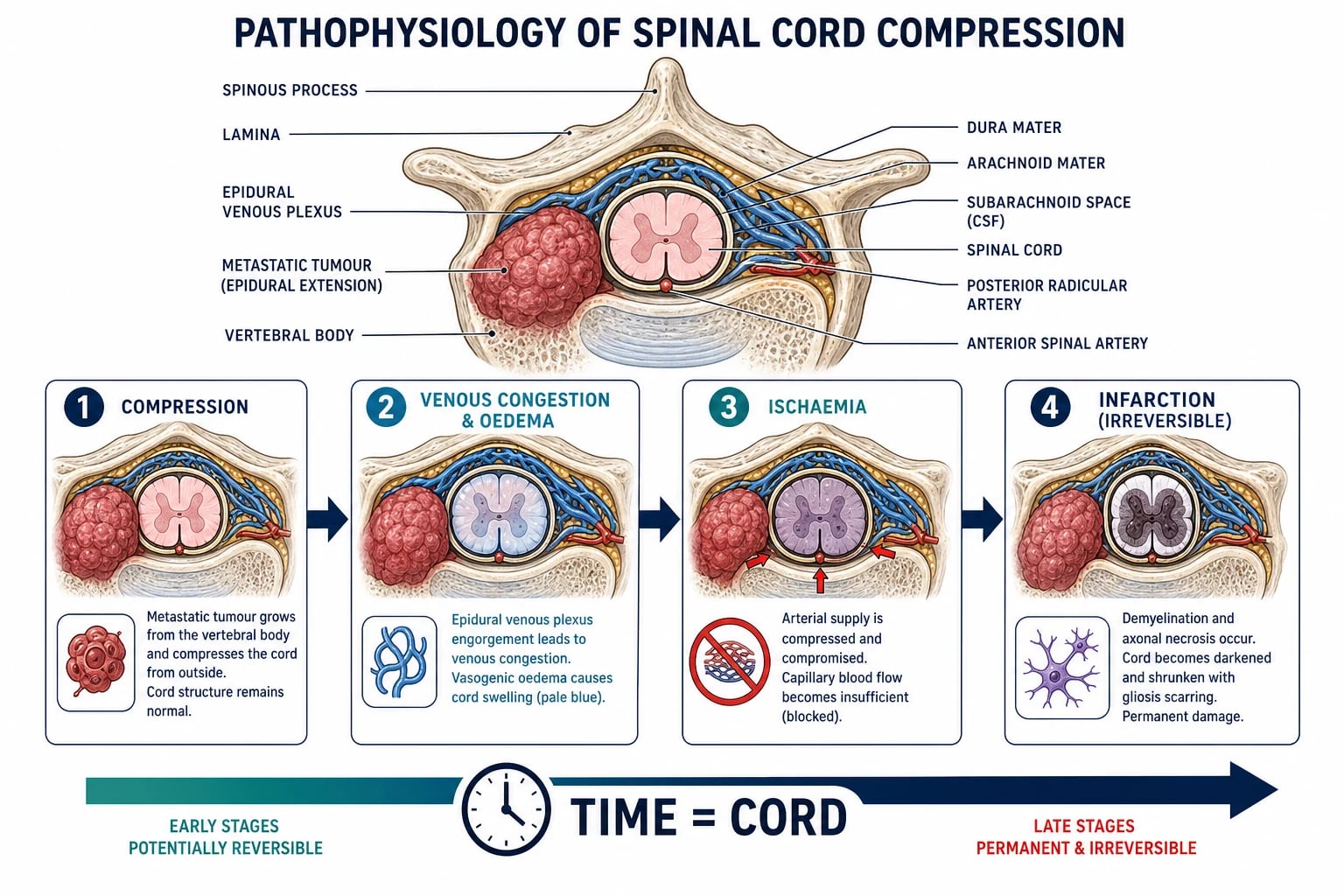
The mechanical insult operates through three mechanisms. Contact forces at the point of impact produce skull fracture, laceration and local cortical contusion (the coup lesion). Inertial (acceleration–deceleration) forces, especially rotational/angular acceleration, shear axons at points where grey and white matter have different densities — most prominently at the grey–white junction, in the corpus callosum, and in the dorsolateral brainstem — producing diffuse axonal injury (DAI). The same forces tear cortical bridging veins as they cross the subdural space on their way to the dural venous sinuses, producing the subdural haematoma; a fracture of the squamous temporal bone tears the middle meningeal artery, producing the extradural haematoma. Penetrating injuries lacerate brain and vessels directly and carry a high epilepsy risk.[1]
The secondary-injury cascade
After the primary impact, a biochemical cascade unfolds that is the target of every intensive-care intervention in TBI. Hypoxia (SaO2 under 90) and hypotension (SBP under 90) — the two most easily preventable insults — drive cellular energy failure: ATP depletion, failure of the sodium-potassium pump, and excitotoxicity as damaged neurons release glutamate. Glutamate opens NMDA receptors, producing a massive calcium influx into neurons; calcium activates proteases (calpains) and generates free radicals, which damage mitochondrial membranes, triggering mitochondrial dysfunction and apoptosis (programmed cell death) and necrosis. The same energy failure disables the membrane pumps, so intracellular sodium and water accumulate (cytotoxic oedema), while breakdown of the blood–brain barrier allows protein-rich fluid to leak into the extracellular space (vasogenic oedema). Cerebral oedema, combined with any expanding haematoma, raises the intracranial pressure (ICP).[1][4]
Why the secondary cascade kills
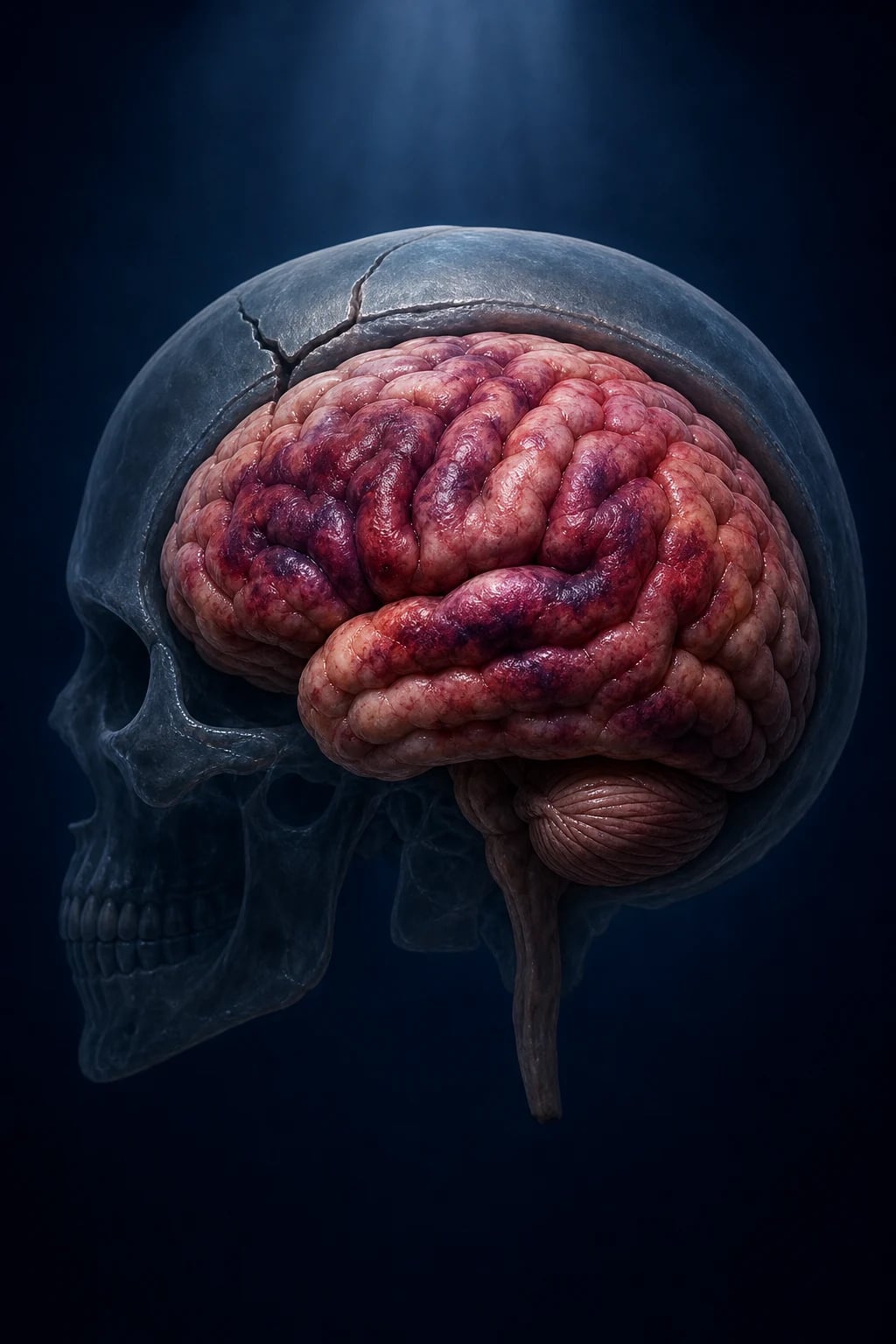
Because autoregulation is impaired, cerebral blood flow becomes pressure-passive — the brain can no longer protect itself against falls in blood pressure. A single episode of hypotension (SBP under 90) in severe TBI doubles mortality; a single episode of hypoxia (SaO2 under 90 or apnoea) does the same. As ICP rises, CPP falls and the brain is further ischaemic, generating more oedema and raising ICP further — a vicious circle that culminates in herniation and the Cushing response. Hyperglycaemia, fever, seizures and anaemia each independently worsen outcome by the same mechanism of increased metabolic demand or reduced oxygen delivery. This is why the entire management of severe TBI — intubation, oxygenation, blood-pressure support, head elevation, sedation, osmolar therapy, ICP monitoring, anticonvulsants, normoglycaemia and normothermia — exists to break the secondary cascade.[1][4]
Clinical Presentation
The presentation of TBI follows the severity grade, but examiners deliberately probe atypical presentations — especially in the elderly, the anticoagulated, and after seemingly minor trauma.[1][2]
Mild TBI (concussion)
Concussion produces a cluster of symptoms rather than signs: headache (the commonest), dizziness, nausea and vomiting, confusion or a "foggy" feeling, amnesia (typically retrograde for events before impact and anterograde for events after), photophobia and phonophobia, poor concentration and memory, fatigue, irritability and sleep disturbance, and a sensitivity to alcohol and exertion. Loss of consciousness occurs in only a minority of concussions — its absence does not exclude the diagnosis. Symptoms resolve over days to weeks in most patients; in 10 to 20 percent they persist beyond a month as post-concussion syndrome. The Sport Concussion Assessment Tool, 5th edition (SCAT5) and the newer SCAT6 (Amsterdam 2022) standardise the assessment of sport-related concussion — symptom checklist, cognitive assessment (orientation, immediate memory, concentration, delayed recall) and a neurological screen — and are the recognised sideline tools.[9][10]
Moderate and severe TBI
As severity deepens, the picture moves from subjective symptoms to objective signs of structural brain injury: a depressed or declining GCS, focal neurological deficits (hemiparesis, aphasia, visual field cut), pupil asymmetry or a fixed dilated pupil (uncal herniation), seizures, and Cushing's triad (hypertension, bradycardia, irregular respiration) signalling markedly raised ICP. Decorticate posturing (M3, flexion to pain — lesion above the midbrain) and decerebrate posturing (M2, extension to pain — brainstem/midbrain lesion) localise the level of the injury. A period of post-traumatic amnesia (the interval from injury to the recovery of continuous memory) longer than 30 minutes distinguishes more significant injury from simple concussion.[1]
The named haematomas — the classic stems
The two neurosurgical emergencies every student must distinguish produce a stereotyped clinical picture that examiners reward.[1]
Extradural (epidural) haematoma. A young patient takes a low-velocity blow to the temporal region (a cricket ball, a falling object, a punch). The classic sequence is brief loss of consciousness, then a lucid interval (the patient walks, talks, refuses help) — then rapid neurological deterioration as the rapidly expanding biconvex haematoma compresses the temporal lobe. The full triad on the side of the impact is an ipsilateral fixed dilated pupil (CN III compression from uncal herniation) and contralateral hemiparesis (cerebral peduncle compression). The lucid interval is the warning that deterioration is imminent — and it is the single most testable clinical stem in head injury. The source is the middle meningeal artery, torn by a fracture of the squamous temporal bone. This is a neurosurgical emergency: urgent CT and evacuation.[1]
Subdural haematoma. An elderly, alcoholic, or anticoagulated patient — often after trivial or forgotten trauma — presents with a fluctuating level of consciousness, headache, and progressive focal deficit over days to weeks. The cause is a torn cortical bridging vein, and the haematoma spreads diffusely over the cerebral convexity. The CT appearance is crescent-shaped, crosses suture lines, and does not cross the midline (under the falx). The chronic subdural may present weeks after the forgotten injury with cognitive decline mistaken for dementia, or with seizures.[1]
Diffuse axonal injury. A patient injured in a high-speed rotational mechanism (motorcycle crash, high fall) is comatose from the moment of impact, often with normal or near-normal early CT — the classic "coma out of proportion to the CT". Small petechial haemorrhages at the grey–white junction, in the corpus callosum, or in the dorsolateral brainstem may be visible on CT; MRI gradient-echo or susceptibility-weighted imaging (SWI) shows the characteristic microbleeds.[1]
Classic presentations
- **Concussion** — headache, dizziness, confusion, amnesia; LOC in a minority only
- **Extradural** — temporal blow, lucid interval, ipsilateral fixed pupil + contralateral weakness
- **Subdural** — elderly/alcoholic/anticoagulated, fluctuating, subacute, crescentic on CT
- **DAI** — high-speed rotational injury, coma out of proportion to a normal CT
- **Raised ICP** — headache, vomiting, drowsiness; **Cushing's triad** is pre-terminal
Atypical / easily missed
- **Elderly** — confusion, a fall, or cognitive decline instead of the classic picture; chronic subdural mimics dementia
- **Anticoagulated** — intracranial bleeding after trivial trauma; reverse urgently, low CT threshold
- **Children** — non-accidental injury (NAI) must be considered; inconsistent history, retinal haemorrhages
- **Alcoholic** — falls, late presentation, may be mislabelled as 'just drunk'
- **Lucid interval without prior LOC** — extradural can present without a witnessed initial blackout
Skull fractures
A linear skull fracture is usually clinically silent but raises the risk of an extradural haematoma (especially across the middle meningeal groove). A depressed skull fracture (a palpable bony step) carries a high risk of cortical laceration, late epilepsy, and infection — it requires surgical elevation if depressed by more than the thickness of the skull, if there is a dural tear, or if there is an underlying haematoma. A basal skull fracture is suggested by the classic peri-orbital and mastoid signs: raccoon eyes (periorbital bruising, bilateral, restricted to the orbital margins — distinct from direct periorbital trauma), Battle's sign (bruising over the mastoid process, appearing hours after injury), CSF rhinorrhoea or otorrhoea (a clear watery nasal or ear discharge — confirmed by the halo sign on filter paper, or beta-2 transferrin), haemotympanum, and CN VII or VIII palsy. A basal skull fracture is an open fracture: it requires antibiotics and is a contraindication to nasal instrumentation (NG tube, nasotracheal intubation).[1]
Differential Diagnosis
The differential of a reduced-GCS patient after head injury must always include non-intracranial causes of reduced consciousness — which is why every head-injured patient has a glucose and a temperature checked at the bedside. The mnemonic AEIOU-TIPs (Alcohol/Abuse of drugs, Endocrine/Encephalopathy, Insulin (hypoglycaemia), Oxygen lack, Uraemia/electrolytes; Trauma, Infection, Poisoning, Seizure/post-ictal) frames the search.[1]
Extradural vs subdural on CT
- **Extradural**: biconvex/lens-shaped; does NOT cross suture lines; crosses midline under falx; temporal, middle meningeal artery
- **Subdural**: crescentic; crosses suture lines; does NOT cross midline/falx; cortical bridging veins
- **Lucid interval** is classic for extradural; subdural is gradual and subacute
- Extradural in the young after low-velocity impact; subdural in elderly/alcoholic/anticoagulated
Reduced GCS — non-TBI causes
- **Hypoglycaemia** — finger-prick glucose in EVERY head-injured patient; treat with IV dextrose
- **Post-ictal state** — a seizure may have caused the fall; look for tongue-biting and incontinence
- **Alcohol or drug intoxication** — alcohol on the breath does not explain a reduced GCS; exclude TBI
- **Opioid overdose** — pin-point pupils; trial of naloxone; remember co-existing TBI
- **Meningitis/encephalitis** — fever, neck stiffness; an open skull fracture is a portal for infection
Traumatic SAH vs aneurysmal SAH
- **Traumatic SAH** — blood in the cortical sulci over the convexity, contusional pattern
- **Aneurysmal SAH** — blood in the basal cisterns/Sylvian fissure; thunderclap headache
- History of trauma is decisive — but a ruptured aneurysm can cause a fall, confusing the picture
- Both need urgent CT; nimodipine is reserved for aneurysmal SAH
Post-concussion syndrome mimics
- **Vestibular dysfunction** — persistent dizziness; Dix-Hallpike for BPPV
- **Cervicogenic headache** — referred from cervical spine injury; treat the neck
- **Depression/anxiety** — overlaps with post-concussion mood symptoms
- **Migraine** — photophobia/phonophobia overlap; a careful history separates them
The cervical spine is a special case. Roughly 10 percent of significant head injuries have a co-existing cervical spine injury, and an unrecognised C-spine fracture can render a stable patient quadriplegic. Every head-injured patient is assumed to have a C-spine injury until cleared — by clinical criteria (Canadian C-spine rule or NEXUS) or by CT — and inline manual cervical stabilisation is maintained throughout the primary survey and intubation.[1]
A drunk patient with a head injury has a GCS of 12 — is the alcohol enough to explain the score?
No. Alcohol alone, even at high blood levels, rarely reduces the GCS below 13. A reduced GCS in an intoxicated head-injured patient is presumed to be due to intracranial injury until a CT proves otherwise. Hypoglycaemia must also be excluded — alcohol intoxication causes hypoglycaemia, especially in children and malnourished patients.[1]
Clinical & Bedside Assessment
The focused head-injury examination combines the ATLS primary survey (ABCDE) with cervical spine control, a neurological assessment (GCS, pupils, limb power), and a search for external signs of skull fracture. The GCS is recorded at first contact and repeated every 15 to 30 minutes initially — the trend is more important than any single value, and a fall of 2 or more points is an emergency mandating urgent CT.[1]
Airway with cervical spine control comes first: the cervical spine is immobilised in a hard collar, sandbags, and tape (or by manual in-line stabilisation during intubation) until cleared. Breathing is assessed for rate, oxygen saturation (target 94 percent or above), and chest movement — TBI frequently co-exists with chest trauma. Circulation looks for external bleeding and the shock pattern (hypovolaemia from scalp lacerations, facial fractures, or associated injuries) — shock is rarely caused by isolated head injury in adults, and its presence should trigger a search for a thoracic, abdominal, pelvic, or long-bone source. Disability records the GCS, pupils (size, symmetry, reactivity), and a capillary glucose (hypoglycaemia is a rapidly reversible mimic). Exposure completes the survey and a full secondary survey follows once the patient is stable.[1]
Pupillary examination is the single most useful focal sign. A unilaterally dilated, poorly reactive pupil in a comatose patient is ipsilateral CN III compression from uncal herniation until proven otherwise — an expanding mass lesion on that side. Bilateral small pupils suggest pontine (opiate) lesions; bilateral fixed dilated pupils are a grave sign of brainstem failure. The motor response is tested to a central painful stimulus (supraorbital pressure or trapezius squeeze): obeys commands (M6), localises (M5), withdraws (M4), abnormal flexion/decorticate (M3), abnormal extension/decerebrate (M2), none (M1). Lateralised motor weakness points to a contralateral cerebral hemisphere lesion.[1]
External signs of a basal skull fracture are sought in every significant head injury: raccoon eyes, Battle's sign, CSF leak (test any clear nasal or ear discharge), haemotympanum, and CN VII/VIII signs. A palpable bony step suggests a depressed fracture; a boggy scalp swelling may overlie a fracture line. The scalp is inspected and palpated for lacerations (which can bleed profusely — a scalp laceration in a shocked patient is a real possibility).[1]
Neurological observations (GCS, pupils, vital signs) are recorded every 15 minutes initially, then less frequently as the patient stabilises. Continuous pulse oximetry, ECG, and blood-pressure monitoring are mandatory. The discipline of observation is to detect secondary deterioration early — an expanding extradural, a rising ICP, a delayed haematoma — before they cause irreversible harm.[1]
Investigations
First test — non-contrast CT brain
The non-contrast CT brain is the first and most important investigation in a significant head injury. It identifies acute blood (extradural lens, subdural crescent, intracerebral contusion, intraventricular and subarachnoid blood), mass effect and midline shift, skull fractures, pneumocephalus (an open fracture), hydrocephalus, and cerebral oedema (effaced basal cisterns, loss of grey–white differentiation). The CT determines who needs neurosurgery, who needs ICP monitoring, and who can be discharged.[1]
Because CT carries a small radiation dose and most head-injured patients have no intracranial injury, clinical decision rules were developed to identify the patients in whom CT is warranted. The two best validated are the Canadian CT Head Rule (CCHR), reproduced here, and the NICE criteria used in the UK. Both are highly sensitive (designed to rule out clinically important injury) and examiners expect their components to be stated exactly.[3]
The NICE Head Injury guidance (CG176, updated in NG232) lists similar CT indications for adults: GCS under 13 on initial assessment; GCS under 15 at 2 hours; suspected open, depressed, or basal skull fracture; post-traumatic seizure; focal neurological deficit; more than one episode of vomiting; dangerous mechanism; coagulopathy or anticoagulant use; age 65 or over; amnesia of events more than 30 minutes before impact; and previous brain surgery. The Brain Trauma Foundation 4th edition (2017, Carney) endorses early CT for severity grading and for selection of patients for ICP monitoring. Across UK (NICE), Canada (CCHR), US (CDC), and India (MoHFW), the principle is the same: scan early, scan the high-risk patient, and use the GCS trend to drive repeat scanning.[3][4]
MRI brain — for diffuse axonal injury
An MRI brain is indicated when the CT is normal but the clinical picture suggests significant injury — most often suspected diffuse axonal injury in a comatose patient after high-speed rotational trauma, persistent unexplained symptoms after concussion, or suspected non-accidental injury in children. Gradient-echo (GRE) and susceptibility-weighted imaging (SWI) sequences show the microbleeds of DAI at the grey–white junction, in the corpus callosum, and in the brainstem. MRI may also reveal small ischaemic strokes, contusions below the resolution of CT, and the diffuse oedema of second-impact syndrome. MRI is not a first-line test in the unstable patient.[1]
ICP monitoring
An intracranial pressure (ICP) monitor — an intraventricular catheter (external ventricular drain, EVD) or an intraparenchymal sensor — is indicated in severe TBI (GCS 3 to 8) with an abnormal CT (haematoma, contusion, oedema, compressed cisterns), or a normal CT with two or more of age over 40, motor posturing, or SBP under 90. The Brain Trauma Foundation 4th edition target is ICP under 22 mmHg (lowering above this threshold improves outcome) and CPP 60 to 70 mmHg. The ICP waveform, the pressure–volume index, and the relationship between ICP and MAP guide therapy.[4][7]
Bloods and adjuncts
Blood tests in every significant head injury include glucose (exclude hypoglycaemia — the rapidly reversible mimic), full blood count, urea and electrolytes, coagulation/INR (a coagulopathy may develop after injury and dramatically worsen a haematoma), blood group and crossmatch (for surgery), alcohol and drug levels, beta-hCG in women of childbearing age, and a trauma panel (amylase/lipase, lactate, venous blood gas). CT angiography is added if a vascular injury (carotid or vertebral dissection) is suspected. CT cervical spine is performed in all high-risk patients as part of the trauma pan-scan. Skull X-rays are now rarely indicated (CT is more sensitive for both fractures and intracranial injury) but may be used in settings without CT. An EEG is reserved for suspected seizures or unexplained depressed consciousness.[1]
Management — Resuscitation
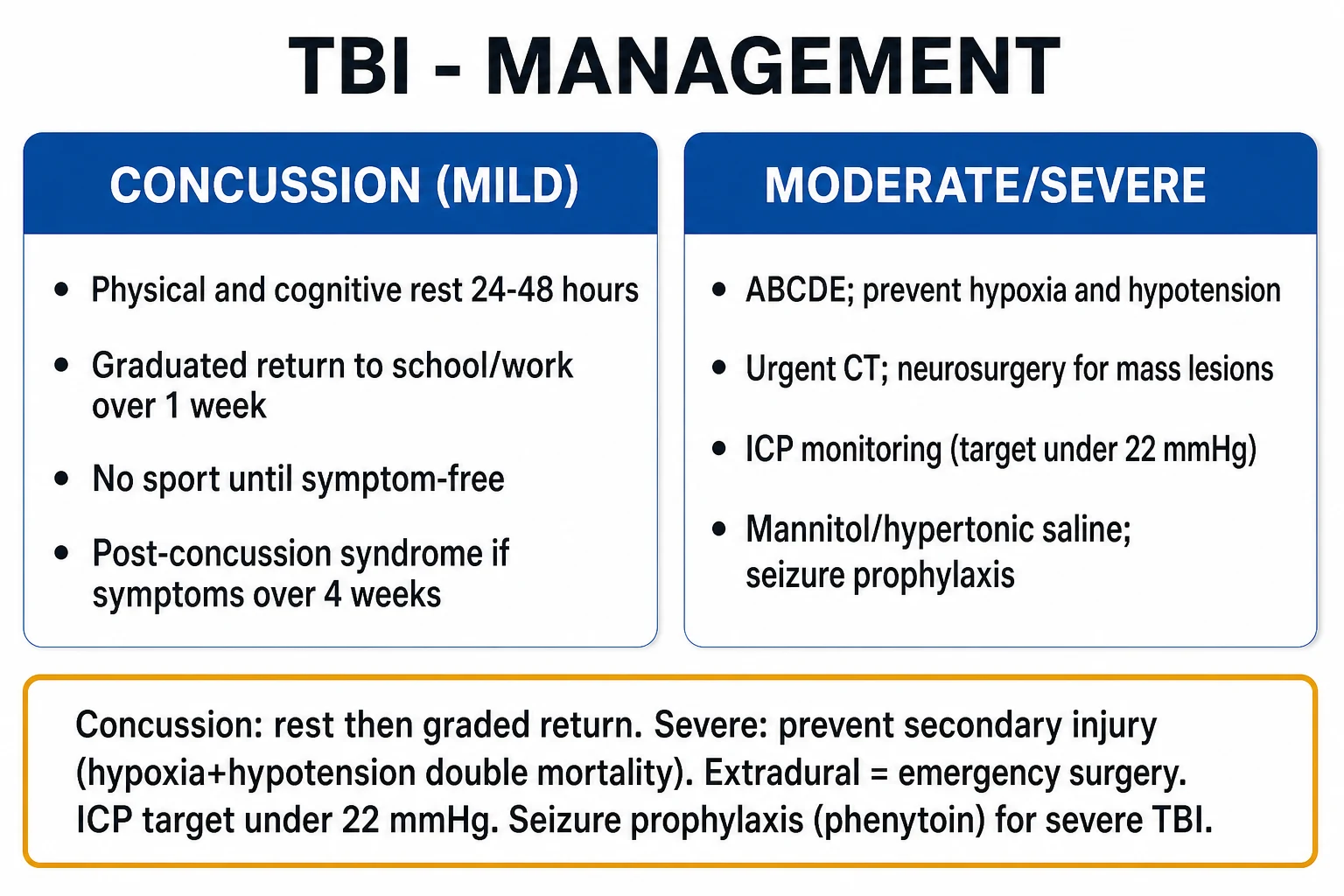
Resuscitation follows the ATLS primary survey (ABCDE) with cervical spine control, with a single overriding principle: the brain cannot tolerate hypoxia or hypotension, and a single episode of either doubles mortality in severe TBI. The discipline of resuscitation is therefore to secure the airway early, oxygenate aggressively, and maintain the blood pressure — while not missing a co-existing cervical spine injury.[1][4]
Resuscitation bundle in the first hour (ABCDE + ICP)
Airway + C-spine
Manual in-line stabilisation; hard collar/sandbags/tape. **Intubate (RSI) if GCS 8 or lower**, or for airway compromise, hypoxia, maxillofacial injury, or predicted long scan time. 10 percent of significant head injuries have a C-spine injury — assume it.
Breathing
High-flow oxygen to keep **SaO2 at least 94 percent / PaO2 over 11 kPa**. Avoid hypoxia — a single hypoxic episode doubles mortality. Ventilate to **normocapnia** (PaCO2 4.5 to 5.0 kPa); avoid prophylactic hyperventilation (it causes cerebral vasoconstriction and ischaemia).
Circulation
Control external bleeding (scalp lacerations bleed profusely); two large-bore cannulae; IV fluids or blood products to keep **SBP over 110 mmHg / MAP over 80**. **Shock is rarely caused by isolated head injury in adults** — search for a thoracic, abdominal, pelvic, or long-bone source. A single SBP under 90 episode doubles mortality.
Disability
Record **GCS, pupils, and capillary glucose**; treat hypoglycaemia (50 mL of 50% dextrose IV, or 25 g of 10% in children); treat seizures (IV lorazepam 0.1 mg/kg). Re-check GCS every 15 to 30 min — a fall of 2 or more points is an emergency.
Exposure + Environment
Full secondary survey; keep the patient **normothermic** (avoid hypothermia, treat fever — fever raises cerebral metabolic demand and worsens outcome). Log-roll to examine the back.
Raised ICP bundle
**Head of bed at 30 degrees and midline** (venous drainage); adequate sedation and analgesia; **osmolar therapy** if signs of raised ICP or herniation (**mannitol 0.25 to 1 g/kg IV** or **3% hypertonic saline 250 mL IV bolus**). Avoid prophylactic hyperventilation (use only briefly as a bridge to surgery for impending herniation).
Adjuncts + disposition
Crossmatch, coagulation/INR, trauma panel; urinary catheter; activate the **trauma team and neurosurgery**; document the time and mechanism of injury; **transfer to a neurosciences centre** as indicated (centralisation improves outcome).
Blood pressure is maintained with isotonic crystalloid or blood products; avoid hypotonic fluids (they lower serum osmolarity and worsen cerebral oedema). Vasopressors (noradrenaline) are used if fluids alone do not achieve the MAP target. Anticoagulation is reversed urgently: warfarin with vitamin K plus prothrombin complex concentrate; dabigatran with idarucizumab; anti-Xa DOACs with andexanet alfa; antiplatelets discussed with neurosurgery. Seizures are terminated with IV lorazepam 0.1 mg/kg (or IV diazepam), followed by a loading dose of an anticonvulsant. Hypoglycaemia is corrected with IV dextrose. The principle throughout is resuscitate before you image — an unstable patient should not be sent to the CT scanner.[1][4]
Management — Definitive & Stepwise
Definitive management separates cleanly into concussion (mild TBI) on one side and moderate-to-severe TBI on the other. The concussion pathway is a graded return to activity; the moderate-to-severe pathway is neurocritical care aimed at preventing secondary injury and surgically evacuating mass lesions.[4][9]
Concussion — the graded-return pathway
Concussion is managed conservatively — there is no role for routine CT in the asymptomatic patient who fulfils no high-risk criterion (per the Canadian CT Head Rule). Management has three components.[9][10]
1. Immediate removal from play. Any athlete with suspected concussion is removed from play and not returned the same day — "when in doubt, sit them out". Same-day return to play is the single behaviour that precipitates second-impact syndrome, a rare but catastrophic diffuse cerebral oedema that occurs when a second concussion is sustained before the first has fully resolved. The SCAT6 (Amsterdam 2022) is the recognised sideline tool.[9]
2. Physical and cognitive rest for 24 to 48 hours. Strict rest beyond 48 hours is no longer recommended — prolonged rest delays recovery, deconditions the patient, and worsens mood. After the initial rest period, the patient resumes light activity below symptom threshold.[10]
3. Graduated return to school/work and sport. The Concussion in Sport Group (Amsterdam 2022) six-stage protocol advances through symptom-limited activity, light aerobic exercise, sport-specific exercise, non-contact training drills, full-contact practice, and return to sport, spending a minimum of 24 hours at each stage and only progressing if symptom-free. The minimum time before return to full contact sport is approximately one week. If symptoms recur at any stage, the patient drops back to the previous asymptomatic level for 24 hours. Medical clearance is required before return to contact sport.[9][10]
| Stage | Aim | Activity | Progression |
|---|---|---|---|
| 1 | Symptom-limited activity | Daily activities that do not worsen symptoms | 24–48 h of relative rest |
| 2 | Light aerobic | Stationary cycling or walking at under 70 percent max HR; no resistance training | Move to stage 3 if symptom-free |
| 3 | Sport-specific exercise | Running drills; no head impact | Minimum 24 h symptom-free |
| 4 | Non-contact training drills | Harder training drills; resistance training may begin | Minimum 24 h symptom-free |
| 5 | Full-contact practice | Normal training activities | Medical clearance required |
| 6 | Return to sport | Normal game play | Minimum 1 week from injury[9][10] |
Moderate-to-severe TBI — the neurocritical-care bundle
The moderate-to-severe pathway builds on resuscitation with five pillars: ICP control, surgical evacuation, seizure prophylaxis, hyperosmolar therapy, and supportive ICU care. The Brain Trauma Foundation 4th edition (2017, Carney) is the international benchmark.[4]
1. ICP control. The head of bed is elevated to 30 degrees and kept midline to optimise venous drainage. Sedation and analgesia (propofol and fentanyl infusions) reduce the metabolic demand and the coughing/straining that raise ICP. Normocapnia is maintained (PaCO2 4.5 to 5.0 kPa) — avoid prophylactic hyperventilation, which causes cerebral vasoconstriction and ischaemia; brief hyperventilation is reserved as a bridge to surgery in impending herniation. An ICP monitor is placed for severe TBI with an abnormal CT, with a target ICP under 22 mmHg and CPP 60 to 70 mmHg.[4][7]
2. Hyperosmolar therapy. For an acutely raised ICP or impending herniation, osmotic agents draw water out of the brain and into the vascular space.[4]
Mannitol
Osmotic diuretic — first-line for acutely raised ICP / impending herniation
Dose
0.25 to 1 g/kg IV bolus over 10 to 15 minutes (typical adult 1 g/kg of 20% solution); repeat as guided by ICP and serum osmolarity
Hypertonic saline (3% or 23.4%)
Osmotic agent — alternative or adjunct to mannitol for raised ICP; preferred in hypovolaemia
Dose
3% NaCl 250 mL IV bolus over 10 to 15 minutes (or continuous infusion titrated to serum sodium 145 to 155 mmol/L); 23.4% NaCl 30 to 60 mL via central line for impending herniation
3. Surgical evacuation. A neurosurgical evacuation is indicated for an extradural or subdural haematoma more than 30 mL in volume or with a midline shift over 5 mm (lower thresholds in the posterior fossa), for any deteriorating patient with a mass lesion, for a depressed skull fracture depressed by more than the skull thickness or with an underlying injury, and for a large contusion with mass effect. The classic indications are summarised by the Bullock surgical guidelines: an acute subdural haematoma over 10 mm thick or with midline shift over 5 mm should be evacuated within 4 hours if the GCS is deteriorating; an extradural haematoma over 30 mL should be evacuated regardless of GCS; and a decompressive craniectomy is considered for refractory intracranial hypertension.[1][6][8]
4. Decompressive craniectomy for refractory raised ICP. When medical therapy fails to control the ICP, a decompressive craniectomy (removal of a large bone flap, often with a duraplasty) creates room for the swollen brain and lowers the ICP. The DECRA trial (Cooper 2011) showed that early bifrontal decompressive craniectomy for diffuse injury was associated with worse neurological outcome at 6 months — cautioning against early prophylactic craniectomy. The RESCUEicp trial (Hutchinson 2016) showed that craniectomy as a last-tier therapy for refractory intracranial hypertension lowered mortality compared with medical management, but at the cost of higher rates of vegetative state and severe disability in survivors. Together, these trials frame the current practice: decompressive craniectomy is reserved for refractory raised ICP, not used prophylactically.[6][8]
DECRA — Decompressive Craniectomy trial
N Engl J Med 2011 (Cooper DJ et al.)
Multicentre RCT of 155 adults with severe diffuse TBI and refractory intracranial hypertension, randomised to early bifrontal decompressive craniectomy vs standard care.
Key finding
Craniectomy lowered ICP and shortened ICU stay but was associated with a WORSE neurological outcome at 6 months (unfavourable outcome 70% vs 51%; p equals 0.02). Surprising and practice-changing.
Practice change
Early prophylactic bifrontal decompressive craniectomy for diffuse TBI is NOT recommended; decompression is reserved for refractory raised ICP as a last-tier therapy.
RESCUEicp — Trial of Decompressive Craniectomy for Traumatic Intracranial Hypertension
N Engl J Med 2016 (Hutchinson PJ et al.)
Multicentre RCT of 408 adults with severe TBI and refractory intracranial hypertension (ICP over 25 mmHg for 1 to 12 h despite tiered medical therapy), randomised to decompressive craniectomy vs continued medical therapy.
Key finding
Craniectomy LOWERED mortality at 6 months (26.9% vs 48.9%) but survivors had higher rates of vegetative state and lower-upper severe disability than the medical group.
Practice change
Decompressive craniectomy is an effective last-tier therapy for refractory ICP that saves lives, but the saved lives may be heavily dependent — a finding that mandates careful patient/family discussion before surgery.
5. Seizure prophylaxis. Early post-traumatic seizures (within 7 days) are prevented with a short course of an anticonvulsant — phenytoin or levetiracetam — in severe TBI, started within 24 hours and continued for 7 days. The Temkin 1990 randomised trial established that phenytoin reduces early seizures but does not prevent late epilepsy. Late post-traumatic epilepsy (after 7 days) is not prevented by prophylaxis and is treated as any other epilepsy if it develops. Prophylaxis is not indicated for mild TBI.[5]
Phenytoin (or levetiracetam) — 7-day early seizure prophylaxis
Prophylaxis against EARLY post-traumatic seizures (within 7 days) in severe TBI
Dose
**Phenytoin** — loading 15 to 20 mg/kg IV (max 1.5 g) at no more than 50 mg/min, then 5 mg/kg/day IV/PO for 7 days. OR **Levetiracetam** — 60 mg/kg load (max 4.5 g) then 1 g twice daily for 7 days.
6. Supportive ICU care. The general ICU bundle keeps the brain out of trouble while it recovers. Normoglycaemia (avoid hyperglycaemia with an insulin sliding scale — hyperglycaemia worsens outcome; avoid hypoglycaemia more strictly), normothermia (paracetamol and cooling for fever — every 1 degree rise in temperature increases cerebral metabolic demand by up to 10 percent), and treatment of anaemia (transfusion threshold generally a haemoglobin of 70 to 80 g/L). Venous thromboembolism prophylaxis begins with mechanical compression immediately, with low-molecular-weight heparin added after 48 hours once bleeding is controlled. Early enteral nutrition, stress-ulcer prophylaxis, glycaemic control, and infection surveillance (ventilator-associated pneumonia, line infections, urinary tract) complete the bundle. Early rehabilitation — physiotherapy, occupational therapy, speech and language, and neuropsychology — begins in the ICU.[4]
Steroids are contraindicated
The MRC CRASH trial (Edwards 2005), in a landmark finding, showed that high-dose corticosteroids increase mortality after significant head injury and must not be used. This is one of the most heavily tested facts in TBI.[11]
MRC CRASH — Corticosteroid Randomisation After Significant Head Injury
Lancet 2005 (Edwards P, CRASH Trial Collaborators)
International RCT of 10,008 adults with head injury and a GCS of 14 or lower, randomised to a 48-hour infusion of high-dose methylprednisolone vs placebo within 8 hours of injury.
Key finding
STEROIDS INCREASED 2-week mortality (21% vs 18%; relative risk 1.18; p equals 0.001) and 6-month death or severe disability.
Practice change
High-dose corticosteroids are CONTRAINDICATED in the management of acute significant head injury. The single most important drug class NOT to give in TBI.
Specific Subtypes & Scenarios
The named lesions each have a distinctive management pathway.[1]
Extradural (epidural) haematoma
- **Biconvex / lens-shaped** on CT; does NOT cross suture lines
- **Temporal**; **middle meningeal artery** (fracture of the squamous temporal bone)
- **Lucid interval** then deterioration; young patient after low-velocity impact
- **Neurosurgical emergency** — urgent CT and evacuation; mortality under 10 percent if evacuated promptly
Acute subdural haematoma
- **Crescentic** on CT; crosses suture lines; does NOT cross midline/falx
- **Cortical bridging veins**; **elderly, alcoholic, anticoagulated**
- **Fluctuating, subacute course**; may be chronic at presentation with cognitive decline
- Reverse anticoagulation; evacuate if over 30 mL, midline shift over 5 mm, or deteriorating
Intracerebral contusion / haematoma
- **Coup and contrecoup** — frontal and temporal poles, orbitofrontal cortex
- May 'blossom' (enlarge) over 24 to 72 h — re-scan if deterioration
- Operate if mass effect, midline shift over 5 mm, or uncontrolled ICP
- High risk of post-traumatic epilepsy
Diffuse axonal injury
- **Rotational/angular acceleration** shears axons (grey-white junction, corpus callosum, brainstem)
- Early CT often **normal or near-normal**; **MRI GRE/SWI** shows microbleeds
- **Coma out of proportion to CT** in high-speed trauma; prolonged ICU course
- Supportive ICU care; prognosis graded by Adams histological grade (I to III)
Skull base fracture
- **CSF rhinorrhoea/otorrhoea**, **Battle's sign**, **raccoon eyes**, haemotympanum, CN VII/VIII palsy
- **Open** fracture — antibiotics; do NOT instrument the nose (no NG tube, no nasotracheal intubation)
- Prophylactic antibiotics for CSF leak are NOT routine; treat meningitis if it occurs
- Persistent CSF leak over 7 days needs neurosurgical repair
Depressed skull fracture
- Palpable bony step; high risk of cortical laceration, infection, late epilepsy
- Surgical elevation if depressed by more than the skull thickness, dural tear, or underlying haematoma
- Open fracture — antibiotics and tetanus prophylaxis
Chronic subdural haematoma
The chronic subdural is a distinctive subtype of its own. It presents weeks to months after a forgotten or trivial head injury in an elderly, alcoholic, or anticoagulated patient, with a fluctuating level of consciousness, headache, focal deficit, cognitive decline (mistaken for dementia), or seizures. The CT shows a crescentic collection that may be iso- or hypodense relative to cortex (subacute/chronic blood). Management is burr-hole drainage for symptomatic collections; asymptomatic small collections may be managed conservatively. Anticoagulation is reversed before surgery. Outcomes are generally good, but recurrence is common.[1]
Sport-related concussion
Sport-related concussion deserves special attention because of the risk of second-impact syndrome and the cumulative brain damage associated with repeated concussions. Management is immediate and permanent removal from play ("when in doubt, sit them out"), no return to play the same day, and a graduated return-to-sport (CISG 6-stage protocol, minimum 24 hours per stage, symptom-free before progression, minimum approximately 1 week before full contact, medical clearance). The Amsterdam 2022 (6th) consensus refined the protocol to emphasise early light activity rather than prolonged strict rest, the use of the SCAT6 in adults and Child SCAT6 in children, and individualised return-to-school before return-to-sport.[9][10]
Complications & Pitfalls
The complications of TBI follow a predictable timeline from the moment of injury, and recognising each is a core skill.[1][2]
Complications timeline after TBI
Hypoxia, hypotension, expanding haematoma (extradural, subdural, contusion 'blossom'). The main preventable cause of death. Prevent by meticulous oxygenation, BP control and early CT.
Cerebral oedema, mass effect — Cushing's triad, pupillary changes, posturing. Treat with head elevation, sedation, osmotherapy, decompressive craniectomy for refractory cases.
Meningitis/ventriculitis (open or basal skull fracture, CSF leak, ICP monitor or EVD in situ); aspiration and ventilator-associated pneumonia. Treat aggressively; prophylactic antibiotics for CSF leak are not routine.
Early (within 7 days) — prevented by 7-day phenytoin/levetiracetam; late (after 7 days) — not prevented by prophylaxis, treated as chronic epilepsy if it develops.
Persistent headache, dizziness, poor concentration, irritability, sleep disturbance beyond 4 weeks. Affects 10 to 20 percent after concussion. Managed by reassurance, graded rehabilitation, and treatment of headache/vestibular/mood symptoms.
Progressive neurodegeneration (tauopathy) from repetitive head injury (contact sports, military). Presents with cognitive decline, mood and behaviour change, and eventually dementia. A diagnosis currently confirmed only at post-mortem.
Second-impact syndrome deserves special emphasis. A second concussion sustained before the first has fully resolved triggers catastrophic, often fatal, diffuse cerebral oedema within minutes, typically in young male athletes. It is rare but devastating, and entirely preventable by enforced rest and graduated return — which is the rationale for the strict "no return to play the same day" rule.[9]
Post-concussion syndrome — persistent headache, dizziness, poor concentration, irritability, fatigue, and sleep disturbance beyond 4 weeks — affects roughly 10 to 20 percent of patients after concussion. Risk factors include female sex, previous concussion, migraine, and a high symptom burden at presentation. Management is reassurance (most cases resolve within 3 months), graded aerobic rehabilitation (sub-symptom-threshold exercise), and targeted treatment of headache, vestibular symptoms, and mood. Cervicogenic headache and vestibular dysfunction are common, treatable contributors.[2]
Post-traumatic epilepsy is divided into early (within 7 days) and late (after 7 days). Early seizures are prevented by a 7-day course of phenytoin or levetiracetam (Temkin 1990); late epilepsy is not prevented by prophylaxis and is treated as chronic epilepsy if it develops. Features that raise the risk of late epilepsy include a penetrating injury, depressed skull fracture, intracerebral haematoma, early seizure, traumatic SAH, and a GCS under 10.[5]
Cranial nerve injuries (olfactory nerve anosmia, facial nerve palsy from temporal bone fracture, vestibulocochlear hearing loss), CSF leak and meningitis, hydrocephalus (communicating from subarachnoid blood, or obstructive from intraventricular clot), and vascular injury (carotid or vertebral dissection, traumatic pseudoaneurysm) are further recognised complications.[1]
Prognosis & Disposition
The prognosis of TBI follows the severity grade, modified by age, the depth and duration of coma, the pupillary response, the presence of hypotension or hypoxia, and the CT findings. The International Mission for Prognosis and Analysis of Clinical Trials (IMPACT) and CRASH prognostic models combine these variables to give calibrated 6-month outcome predictions.[1]
Mild TBI (concussion) recovers fully in most within days to weeks. About 10 to 20 percent develop post-concussion syndrome. Repeat injuries increase the risk of cumulative damage and chronic traumatic encephalopathy; the brain is more vulnerable to a second concussion for several weeks after the first (hence the second-impact syndrome risk).[2]
Moderate TBI leaves a significant minority with residual cognitive disability, fatigue, mood disturbance, and (in some) post-traumatic epilepsy.[1]
Severe TBI carries a mortality of 20 to 40 percent; of survivors, many have permanent cognitive, physical, and behavioural disability. The key prognostic factors are age, the initial GCS and its trend, pupillary reactivity (bilaterally fixed dilated pupils are a grave sign), the presence of hypotension or hypoxia, and the CT findings — midline shift, compressed or absent basal cisterns, traumatic SAH, and a mass lesion all predict worse outcome. The motor response of the GCS carries the most prognostic weight of any single component.[1]
Disposition follows severity. Mild TBI with a normal CT, normal GCS, and a responsible adult at home can be discharged with written head-injury advice ("return immediately if persistent/worsening headache, repeated vomiting, increasing drowsiness, weakness, seizures, or visual disturbance"). Moderate TBI is admitted for observation and CT. Severe TBI is admitted to a neurocritical-care ICU, with ICP monitoring and surgical evacuation as needed, followed by early multidisciplinary rehabilitation (physiotherapy, occupational therapy, speech and language therapy, neuropsychology). Driving is restricted for a period after concussion; alcohol is avoided; return to work and sport is graduated. Structured outpatient follow-up detects post-concussion syndrome, post-traumatic epilepsy, and mood disorder.[1]
Special Populations
Sport-related concussion — the priority is immediate removal from play, no return the same day, and a graduated return-to-sport over a minimum of approximately one week (CISG Amsterdam 2022). The SCAT6 is used for adults and the Child SCAT6 for children aged 8 to 12. Prolonged strict rest is no longer recommended — early sub-symptom-threshold light activity aids recovery. The diagnosis remains clinical; imaging is normal.[9][10]
Elderly and anticoagulated patients have a lower threshold for CT (the Canadian CT Head Rule and NICE criteria both include age 65 or older and anticoagulant use as high-risk), a high risk of subdural haematoma even from trivial trauma, and worse outcomes. Anticoagulation is reversed urgently: warfarin with vitamin K and prothrombin complex concentrate, dabigatran with idarucizumab, anti-Xa DOACs with andexanet alfa, and antiplatelets discussed with neurosurgery. The chronic subdural may present weeks after a forgotten injury with cognitive decline.[3][1]
Paediatric head injury uses a modified GCS (the verbal component is replaced by age-appropriate interactions in young children), wider CT indications (the PECARN paediatric criteria), and a lower threshold for admission. Non-accidental injury (NAI) must be considered in any infant with an unexplained head injury, an inconsistent history, retinal haemorrhages, or multiple injuries of different ages — and safeguarding procedures activated. A growing skull fracture (a diastatic fracture that enlarges as the brain pulsates through it) is a complication unique to children.[1]
Pregnant trauma patient — the mother is resuscitated first (the best resuscitation of the fetus is the optimal resuscitation of the mother). A left lateral tilt avoids aortocaval compression; beta-hCG is checked; imaging decisions balance radiation against risk (CT of the head is safe — the radiation dose to the fetus from a head CT is negligible). Multidisciplinary care with obstetrics is essential.[1]
Military / blast TBI is increasingly recognised. The mechanism includes the primary blast over-pressure wave (transmitted through the skull), and is associated with higher rates of PTSD, post-concussion syndrome, and chronic traumatic encephalopathy. The management principles are the same.[2]
Evidence, Guidelines & Regional Differences
The evidence base for TBI is anchored by several landmark trials and a small number of authoritative guidelines, which examiners reward knowing.[4]
The CRASH trial (Edwards 2005) is the single most important negative trial in TBI — it established that high-dose corticosteroids increase mortality and are contraindicated. The Temkin (1990) trial established that phenytoin prevents early post-traumatic seizures but not late epilepsy. The DECRA (2011) and RESCUEicp (2016) trials redefined the role of decompressive craniectomy — DECRA showed early prophylactic craniectomy worsens outcome, while RESCUEicp showed last-tier craniectomy for refractory ICP lowers mortality at the cost of higher disability. The BEST-TRIP (Chesnut 2012) trial showed that ICP-monitoring-guided therapy in a protocolised bundle was equivalent to imaging-and-examination-guided therapy in a region with limited neurocritical-care infrastructure — but should not be read as 'ICP monitoring does not matter'; rather, it emphasises the protocolised bundle.[5][6][7][8][11]
BEST-TRIP — Benchmark Evidence from South American Trials: Intracranial Pressure
N Engl J Med 2012 (Chesnut RM et al.)
Multicentre RCT of 324 patients with severe TBI in Bolivia/Ecuador, randomised to ICP-monitor-guided therapy vs imaging-and-examination-guided therapy without ICP monitoring.
Key finding
No difference in 6-month mortality or neurological outcome between the two groups.
Practice change
Often MISINTERPRETED as 'ICP monitoring does not matter'. The correct reading: in a setting with a strong protocolised neurocritical-care bundle, the addition of ICP monitoring did not by itself change outcome. ICP monitoring remains standard of care in well-resourced settings.
The Brain Trauma Foundation 4th edition (Carney 2017) is the current international benchmark for severe TBI. It targets ICP under 22 mmHg (a tightening from the 3rd edition's 20 mmHg on the basis of outcome data), CPP 60 to 70 mmHg, decompressive craniectomy for refractory raised ICP (not prophylactic), prophylactic anticonvulsant for 7 days, and no corticosteroids.[4]
Across the Brain Trauma Foundation 4th edition (US/international), NICE NG232 (UK) head-injury guidance, the Canadian CT Head Rule (Stiell 2001), the CDC Guideline for Mild TBI (US), and the Indian Ministry of Health TBI protocol, the universal principles converge: (1) prevent secondary brain injury (oxygenation, blood pressure, ICP control); (2) use clinical decision rules to drive CT (CCHR or NICE); (3) graded return for concussion (CISG Amsterdam 2022); (4) surgical evacuation for mass lesions; (5) no steroids; (6) decompressive craniectomy only for refractory raised ICP. Regional variation is mainly in access to neurosurgical and neurocritical-care services, the choice of clinical decision rule for CT (Canadian vs NICE vs PECARN in children), and the anticonvulsant of choice (phenytoin vs levetiracetam).[3][4][9]
Prevention is the highest-yield intervention of all. Bicycle and motorcycle helmets reduce head injury by up to 85 percent; seatbelts and airbags reduce mortality in road traffic accidents; fall prevention in the elderly (home safety assessments, medication review); sport-specific rule changes (head-high tackles, concussion substitution rules); and drink-driving legislation all act upstream of the emergency department.[1]
Exam Pearls
TBI — SECONDARY mnemonic for the preventable killers
SECONDARY
cerebral oedema raises ICP
post-traumatic seizures worsen injury
expanding haematoma (extradural/subdural)
hypoxia (SaO2 under 90) doubles mortality
hypotension (SBP under 90) doubles mortality
vascular injury, carotid/vertebral dissection
blood flow becomes pressure-passive
CPP equals MAP minus ICP; herniation if untreated
secondary injury is the target of treatment
The four numbers every student must know
- **GCS 8 or lower = intubate**
- **ICP target under 22 mmHg**; CPP 60 to 70 mmHg
- **SaO2 at least 94 percent; SBP over 110 mmHg** to prevent secondary injury
- **Phenytoin for 7 days** for early seizure prophylaxis in severe TBI
The high-yield associations
- **Temporal fracture + lucid interval = extradural** (middle meningeal artery)
- **Elderly + bridging veins + crescent = subdural**
- **Rotational acceleration + normal CT + coma = diffuse axonal injury**
- **CSF leak + Battle's sign + raccoon eyes = skull base fracture**
- **Hypertension + bradycardia + irregular respiration = Cushing's triad (raised ICP)**
- **Bilat fixed pupils + posturing = impending herniation — lower the ICP now**
The drugs and doses that decide a viva answer
- **Mannitol 0.25 to 1 g/kg IV** for raised ICP
- **3% hypertonic saline 250 mL IV bolus** for raised ICP
- **Phenytoin 15 to 20 mg/kg IV loading** (then 7 days) for early seizure prophylaxis
- **NEVER give corticosteroids** (CRASH trial — increased mortality)
Exam application bank (NEET-PG / INICET)
One-line answer
Traumatic brain injury (TBI) is a disruption of brain function from external mechanical force, graded by the Glasgow Coma Scale (GCS) into mild (GCS 13 to 15, about 80 percent — concussion), moderate (GCS 9 to 12, about 10 percent) and severe (GCS 3 to 8, about 10 percent). Injury is divided into primary (mechanical, instantaneous, largely irreversible — skull fracture, contusion, diffuse axonal injury) and secondary (delayed, PREVENTABLE — hypoxia, hypotension, raised intracranial pressure, ischaemia, infection); preventing secondary injury is the main target of treatment. Concussion produces transient headache, dizziness, confusion, nausea and amnesia without structural injury on imaging, and is managed with 24 to 48 hours of physical and cognitive rest followed by a graduated return. Moderate to severe TBI requires ABCDE resuscitation, urgent CT, ICP monitoring (target under 22 mmHg,
Worked stems (answer without another resource)
Stem 1 — Classic presentation. Map symptoms to mechanism; name the first investigation and first treatment step with dose/route if drug therapy is standard. [1]
Stem 2 — Unstable / complicated. List red flags that force immediate resuscitation, theatre, ICU, antidote, or reperfusion — and what you do in the first 15 minutes. [1]
Stem 3 — Atypical group. Elderly, pregnancy, child, or immunocompromised: how presentation and thresholds change. [1]
Stem 4 — Differential trap. Name the three closest mimics and one discriminator for each. [1]
Stem 5 — Disposition. Who goes home with safety-netting, who is admitted, who needs HDU/ICU/theatre, and what follow-up is mandatory. [1]
Rapid viva checklist
- Definition + classification
- Pathophysiology chain
- Bedside signs / criteria
- Score with exact components (if any)
- Emergency bundle
- Definitive therapy with doses
- Complications of disease and of treatment
- Special populations
- Guideline/trial name if classic
- Three exam traps
Coverage self-check
If you cannot answer any stem above from this page alone, re-read the matching section — the page is intended to be self-sufficient for final-prof and NEET-PG/INICET questions on Concussion & Traumatic Brain Injury.
References
- [1]Maas AI, Stocchetti N, Bullock R. Moderate and severe traumatic brain injury in adults Lancet Neurol, 2008.PMID 18635021
- [2]Zetterberg H, Smith DH, Blennow K. Biomarkers of mild traumatic brain injury in cerebrospinal fluid and blood Nat Rev Neurol, 2013.PMID 23399646
- [3]Stiell IG, Wells GA, Vandemheen K, et al. The Canadian CT Head Rule for patients with minor head injury Lancet, 2001.PMID 11356436
- [4]Carney N, Totten AM, O'Reilly C, et al. Guidelines for the Management of Severe Traumatic Brain Injury, Fourth Edition Neurosurgery, 2017.PMID 27654000
- [5]Temkin NR, Dikmen SS, Wilensky AJ, Keihm J, Chabal S, Winn HR. A randomized, double-blind study of phenytoin for the prevention of post-traumatic seizures N Engl J Med, 1990.PMID 2115976
- [6]Cooper DJ, Rosenfeld JV, Murray L, et al. (DECRA Trial Investigators; ANZICS Clinical Trials Group). Decompressive craniectomy in diffuse traumatic brain injury N Engl J Med, 2011.PMID 21434843
- [7]Chesnut RM, Temkin N, Carney N, et al. (Brain Trauma Foundation; Global Neurotrauma Research Group). A trial of intracranial-pressure monitoring in traumatic brain injury N Engl J Med, 2012.PMID 23234472
- [8]Hutchinson PJ, Kolias AG, Timofeev IS, et al. (RESCUEicp Trial Collaborators). Trial of Decompressive Craniectomy for Traumatic Intracranial Hypertension N Engl J Med, 2016.PMID 27602507
- [9]McCrory P, Meeuwisse W, Dvorak J, et al. Consensus statement on concussion in sport-the 5(th) international conference on concussion in sport held in Berlin, October 2016 Br J Sports Med, 2017.PMID 28446457
- [10]Patricios JS, Schneider KJ, Dvorak J, et al. Consensus statement on concussion in sport: the 6th International Conference on Concussion in Sport-Amsterdam, October 2022 Br J Sports Med, 2023.PMID 37316210
- [11]Edwards P, Arango M, Balica L, et al. (MRC CRASH Trial Collaborators). Final results of MRC CRASH, a randomised placebo-controlled trial of intravenous corticosteroid in adults with head injury-outcomes at 6 months Lancet, 2005.PMID 15936423