Neurology · Neurology
Dementia
Also known as Major neurocognitive disorder · Chronic cognitive impairment · Alzheimer disease · Presenile / senile dementia
Dementia is an acquired, persistent, global impairment of higher cortical functions (memory, language, visuospatial skills, executive function, personality) occurring in clear consciousness, sufficient to interfere with activities of daily living. It is not a single disease but a syndrome. The commonest cause is Alzheimer disease (AD) (60 to 70 percent), driven by amyloid-beta 42 plaque deposition and tau-hyperphosphorylated neurofibrillary tangles with a cholinergic deficit; followed by vascular dementia (VaD), dementia with Lewy bodies (DLB) and frontotemporal dementia (FTD). Presentation is progressive and insidious in AD, stepwise in vascular, fluctuating with visual hallucinations and parkinsonism in DLB, and behavioural/personality change in FTD. Always exclude reversible mimics — delirium (acute, fluctuating, clouded consciousness), depression (pseudodementia), normal ageing, and treatable metabolic causes (B12, folate, TSH, syphilis, HIV, NPH). Cognitive screening uses MMSE (under 24) or the more sensitive MoCA (under 26); confirm with structural and (where indicated) functional neuroimaging and CSF/PET biomarkers. Cholinesterase inhibitors (donepezil 5 to 10 mg, rivastigmine, galantamine) for mild-to-moderate AD; memantine (5 to 20 mg) for moderate-to-severe; lecanemab for early biomarker-confirmed AD. Avoid typical antipsychotics in DLB (life-threatening neuroleptic sensitivity). The 2020 Lancet Commission shows 40 percent of cases are potentially preventable by addressing 12 modifiable risk factors.
On this page & tools
Your progress
Saved locally on this device.
Practise this topic
Exam tags
Red flags
Overview & Definition
Dementia is best defined as an acquired, persistent and usually progressive global impairment of higher cortical function — memory, language, visuospatial skill, executive function, praxis, gnosis and personality — occurring in clear consciousness and severe enough to disrupt activities of daily living (ADLs).[2]
Three elements of the definition carry weight in a viva: [1]
- Acquired — distinguishes dementia from intellectual disability (developmental).
- In clear consciousness — distinguishes it from delirium, in which consciousness is clouded and the course is acute and fluctuating.
- Interferes with ADLs — distinguishes it from mild cognitive impairment (MCI), in which decline is present but independence is preserved. [1]
The DSM-5 (2013) replaced the word "dementia" with major neurocognitive disorder (major NCD) and reserved mild neurocognitive disorder (mild NCD) for the equivalent of MCI. "Dementia" remains the clinical term in everyday practice and in NICE and most national guidelines.[1]
The clinical skill in dementia is not recognising that a patient is cognitively impaired — it is (1) confirming the diagnosis is truly dementia and not a mimic, (2) identifying the underlying aetiology (because management, prognosis and genetic counselling differ by subtype), and (3) delivering a structured, multidomain plan — pharmacological, non-pharmacological, safety, legal and carer-focused.[1]
Classification
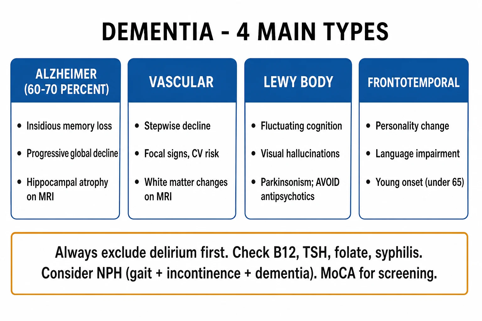
Dementia can be classified along several axes; all four are examinable. [1]
By aetiology (the classification that matters clinically): [1]
| Aetiology | Approximate share | Hallmark |
|---|---|---|
| Alzheimer disease | 60 to 70 percent | Amyloid-beta plaques, tau tangles, episodic memory loss |
| Vascular dementia | 15 to 20 percent | Stepwise decline, executive dysfunction, infarcts on imaging |
| Dementia with Lewy bodies | 5 to 10 percent | Alpha-synuclein, fluctuation, visual hallucinations, parkinsonism |
| Frontotemporal dementia | 5 to 10 percent | TDP-43 or tau, early personality/language change |
| Mixed (commonly AD + vascular) | 10 to 15 percent | Features of more than one subtype |
| Other (CJD, NPH, HIV, alcohol, prion) | small | Specific pathology |
Cortical dementia
- AD, FTD, DLB
- Memory, language, praxis, gnosis affected
- Cortical signs: aphasia, agnosia, apraxia
- MRI: temporal/parietal/frontal atrophy
Subcortical dementia
- Vascular small-vessel, Huntington, Parkinson, HIV, NPH
- Slowing (bradyphrenia), executive dysfunction, apathy
- Cortical signs (aphasia/agnosia) absent
- MRI: white-matter change, basal-ganglia/subcortical pathology
By reversibility (high-yield): [1]
- Irreversible (degenerative): Alzheimer, vascular, DLB, FTD, prion.
- Potentially reversible: the REVERSIBLE mnemonic (see Differential Diagnosis) — depression, drugs, metabolic/endocrine, eye/ear sensory deprivation, nutrition (B12), tumour/trauma, atherosclerosis/vascular, infection, alcohol. [1]
By tempo of decline: [1]
- Progressive / insidious — Alzheimer, FTD.
- Stepwise — vascular (multi-infarct).
- Fluctuating — dementia with Lewy bodies. [1]
The 1-year rule distinguishes dementia with Lewy bodies (DLB) from Parkinson disease dementia (PDD): cognitive symptoms arising at least one year before the motor features of parkinsonism = DLB; cognitive decline arising after established Parkinson disease = PDD. The pathology is essentially the same (alpha-synuclein); the distinction is clinical and matters for trial recruitment and drug selection.[4]
Epidemiology & Risk Factors
Dementia is the leading cause of disability in older adults worldwide. The WHO estimates about 55 million people live with dementia globally, with nearly 10 million new cases each year; numbers are projected to triple by 2050, driven by ageing populations, with the steepest rises in low- and middle-income countries (LMICs) including India.[3]
Prevalence doubles roughly every five years after age 65 — about 1 percent at 60 to 64, rising to over 30 percent after 90. Increasing age is the single strongest non-modifiable risk factor.[2]
Modifiable risk factors — the 2020 Lancet Commission[3] identified 12 modifiable factors responsible for about 40 percent of worldwide dementia that is theoretically preventable. They are grouped by life-stage:
- Early life — low educational attainment.
- Mid life — hearing loss, traumatic brain injury (TBI), hypertension, obesity, excessive alcohol.
- Later life — smoking, depression, social isolation, physical inactivity, diabetes, air pollution. [1]
Dementia — high-yield numbers
Genetics of Alzheimer disease (high-yield): [1]
- APOE epsilon-4 — the strongest genetic risk factor for sporadic, late-onset AD; one copy increases risk 2 to 3 times, two copies 8 to 12 times. APOE epsilon-2 is protective. APOE is a risk modifier, not deterministic.
- Autosomal-dominant (familial) early-onset AD — mutations in PSEN1 (chromosome 14), PSEN2 (chromosome 1) and APP (chromosome 21). Rare (under 1 percent of AD), onset typically 30 to 60 years, 50 percent chance of transmission to each child. APP on chromosome 21 explains the near-universal development of Alzheimer pathology in Down syndrome by the fourth to fifth decade. [1]
Other risk factors (rapid-recall): female sex (post-menopausal, longer survival), cardiovascular risk cluster (diabetes, AF, dyslipidaemia), smoking, low cognitive reserve (education, occupation, social engagement), sleep disorders (OSA), sensory loss (hearing), head trauma, midlife obesity.[2][3]
Pathophysiology
Dementia is a proteinopathy-driven neuronal loss; the protein that misfolds defines the disease.[2]
Alzheimer disease — the amyloid cascade hypothesis. Amyloid precursor protein (APP) (encoded on chromosome 21) is cleaved by beta- and gamma-secretases to produce amyloid-beta 42 (Abeta-42), an aggregation-prone peptide. Over-production or impaired clearance leads to extracellular deposition as senile (neuritic) plaques, especially in the neocortex and hippocampus. Abeta-42 oligomers are synaptotoxic, triggering hyperphosphorylation of the microtubule-associated protein tau. Normal tau stabilises axonal microtubules; hyperphosphorylated tau detaches and aggregates intraneuronally as paired helical filaments that form neurofibrillary tangles (NFTs). Tangles spread along a stereotyped anatomical path (Braak staging) — from the transentorhinal cortex, to the hippocampus and entorhinal cortex (causing the earliest episodic memory loss), then to the association neocortex (language, visuospatial, executive dysfunction).[2]
The earliest and most severe neurotransmitter deficit is cholinergic — degeneration of the nucleus basalis of Meynert (the basal forebrain cholinergic projection). This is the rationale for cholinesterase inhibitors. Noradrenergic (locus coeruleus) and serotonergic (raphe) loss follow, contributing to behavioural symptoms. [1]
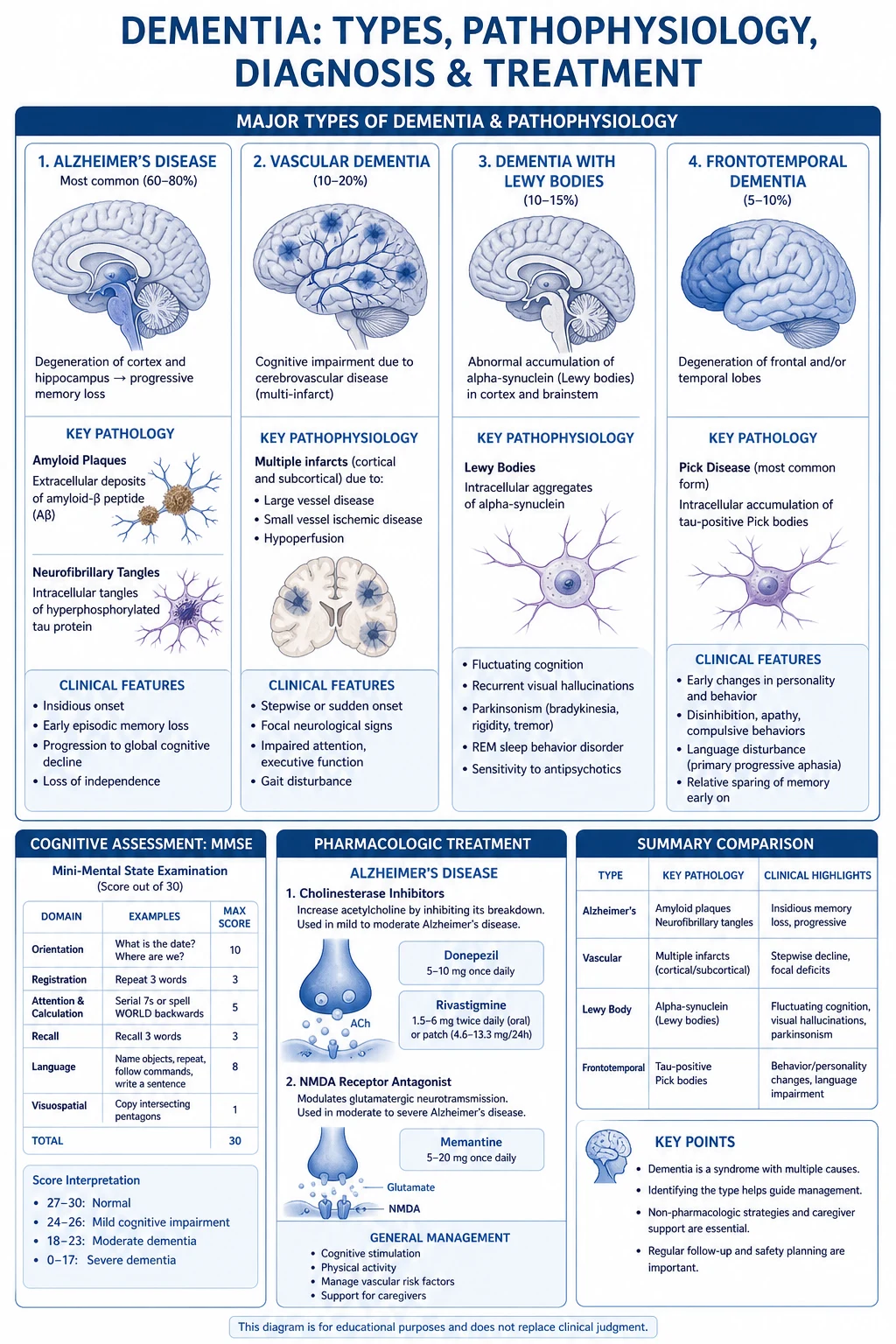
Vascular dementia. Cognitive loss from cerebrovascular disease operates through several mechanisms: (1) multi-infarct — cumulative damage from large cortical strokes; (2) single strategic infarct — a stroke in a cognition-critical region (thalamus, angular gyrus, basal forebrain, caudate); (3) small-vessel disease — lipohyalinosis of deep penetrating arteries producing lacunes and white-matter hyperintensities (leukoaraiosis), damaging fronto-subcortical circuits (hence the subcortical/executive profile); (4) Binswanger (subcortical arteriosclerotic encephalopathy) — severe white-matter change with hypertension. The cognitive profile reflects the disconnection of fronto-subcortical loops, hence prominent executive dysfunction and bradyphrenia with relative memory sparing.[2]
Dementia with Lewy bodies. Misfolding and intracellular accumulation of alpha-synuclein as Lewy bodies in cortical and subcortical (brainstem) neurons. The degeneration is both dopaminergic (substantia nigra → parkinsonism) and cholinergic (basal forebrain and pedunculopontine nucleus → cognitive and visual-hallucination features), which explains why DLB responds to cholinesterase inhibitors and why visual hallucinations are prominent and detailed.[4]
Frontotemporal dementia. Frontal and anterior temporal lobe degeneration with frontotemporal lobar degeneration (FTLD) pathology. The pathological substrate is most commonly TDP-43 (about half), then tau (mutations in the MAPT gene on chromosome 17; Pick bodies with tau-3R), and rarely FUS. The C9orf72 hexanucleotide repeat expansion (chromosome 9) causes familial FTD-ALS overlap. The anatomical selectivity (frontal and temporal) explains the behavioural and language presentation with relative memory sparing early on.[2]
Prion disease — sporadic Creutzfeldt-Jakob disease (sCJD). Misfolding of the prion protein (PrP) into the infectious PrP-scrapie (PrP-Sc) conformation, which templates conversion of normal PrP and accumulates as spongiform change with rapid (weeks-to-months) neuronal loss. The causative gene PRNP is on chromosome 20. [1]
Clinical Presentation
The shared core is progressive cognitive decline that crosses the threshold into functional impairment. Beyond that, each subtype has a recognisable signature.[2]
Alzheimer disease — the prototypical cortical dementia. The earliest and most prominent deficit is recent (episodic) memory — forgetting recent conversations, repeating questions, misplacing items — with remote memory and social graces preserved early on. The patient typically lacks insight (anosognosia). Language follows — anomia (word-finding) progressing to fluent aphasia — then visuospatial dysfunction (getting lost in familiar places) and executive dysfunction (poor judgement, inability to manage finances). Motor and sensory examination is typically normal until late. Cognitive testing: severely impaired delayed recall, preserved attention early on.[2]
Vascular dementia — classically stepwise deterioration, each step corresponding to a new infarct, with intervening plateaus. The cognitive profile is fronto-subcortical: executive dysfunction (planning, set-shifting), slowing (bradyphrenia), and processing-speed deficit, with memory relatively less affected than in AD. Clinical signs of the strokes are often present: gait disturbance, focal weakness, pseudobulbar signs (dysarthria, emotional lability, hyperreflexia, extensor plantars), and urinary urgency/incontinence. Vascular risk factors (hypertension, diabetes, AF) are typically present. [1]
Dementia with Lewy bodies — the four core clinical features[4]:
- Fluctuating cognition — marked variation in attention and alertness on a day-to-day or hour-to-hour basis (the patient may be lucid one day, drowsy and confused the next).
- Recurrent, well-formed, detailed visual hallucinations — often of people or animals, typically not frightening to the patient early on.
- Spontaneous parkinsonism features (rigidity, bradykinesia — tremor is less prominent than in idiopathic PD).
- Rapid eye movement (REM) sleep behaviour disorder (RBD) — loss of normal REM atonia, so the patient acts out dreams (shouting, thrashing) — often preceding the dementia by years. [1]
Supportive features: severe neuroleptic sensitivity (see Pitfalls), autonomic dysfunction (orthostatic hypotension, constipation, urinary incontinence), hypersomnia, depression, anosmia. Memory may be relatively better preserved than in AD early on, but visuospatial and attentional deficits are prominent (DLB patients do disproportionately badly on clock-drawing and trail-making).[4]
Frontotemporal dementia — onset is typically younger (45 to 65 years). Two broad syndromes: [1]
- Behavioural variant (bvFTD) — the commonest. Progressive personality change: disinhibition (socially inappropriate remarks, impulsivity), apathy/loss of empathy, repetitive/compulsive behaviours, dietary change (sweet tooth, hyperphagia), executive dysfunction with relative memory and visuospatial sparing. Insight is lost early. The cognitive profile is frontal (poor judgement, perseveration, concreteness).
- Primary progressive aphasia (PPA) — three subtypes: semantic (fluent but empty speech, loss of word meaning, anterior temporal atrophy, often TDP-43 type C), non-fluent/agrammatic (effortful halting speech, apraxia of speech, inferior frontal-insular atrophy, tau), and logopenic (slow, word-finding pauses, impaired repetition, posterior temporal/parietal atrophy — often an atypical AD variant).[2]
Normal-pressure hydrocephalus — the classic triad of gait apraxia (magnetic, shuffling, broad-based, "feet glued to the floor"), urinary incontinence, and cognitive slowing (subcortical). Potentially reversible — see Specific Subtypes. [1]
Atypical and masked presentations (examiners reward awareness): [1]
- In the elderly — dementia may present as falls, functional decline, anorexia, incontinence or behavioural change rather than a "memory complaint"; the family often first notices.
- In the hearing-impaired — apparent cognitive impairment may partly reflect sensory deprivation; correct hearing before testing cognition.
- In HIV (HAND, HIV-associated neurocognitive disorder) — a subcortical pattern (slowing, apathy, motor slowness) driven by HIV involvement of the basal ganglia and white matter, even with viral suppression.
- Young-onset (under 65) — behavioural change and language deficits predominate over amnesia; FTD and genetic/metabolic/prion causes are over-represented; diagnostic delay is common.
- In delirium superimposed on dementia (very common in hospital) — an acute worsening with fluctuating alertness in a patient with pre-existing dementia; do not assume "just the dementia". [1]
Differential Diagnosis
The diagnosis of dementia is always a diagnosis of exclusion of mimics. Four conditions must be excluded before committing to the label:[2]
Delirium
- ACUTE onset (hours-days)
- Fluctuating course, CLOUDED consciousness
- Reversible; look for precipitant (infection, drug, electrolyte, pain, faecal impaction)
- Inattention is the cardinal feature
- EEG: diffuse slowing
Depression (pseudodementia)
- Onset weeks-months, patient COMPLAINS of poor memory
- Answers 'I don't know' / no effort, poor motivation
- Mood change precedes cognitive change
- Diurnal variation; biological features of depression
- Reversible with treatment of depression
Normal ageing
- Benign forgetfulness - retrieval failure (cued recall helps)
- Slower processing but learning intact
- No functional impairment, insight preserved
- Naming and orientation preserved
Mild cognitive impairment (MCI)
- Objective cognitive decline beyond age norms
- INDEPENDENCE in ADLs preserved
- Not dementia; ~10-15 percent per year convert to AD
- Amnestic vs non-amnestic subtypes
Normal-pressure hydrocephalus
- Gait apraxia + urinary incontinence + subcortical dementia
- MRI: ventriculomegaly out of proportion to sulcal atrophy
- CSF tap/shunt test may improve symptoms
- Potentially REVERSIBLE with shunting
Distinguishing depression (pseudodementia) from true dementia — a recurring exam favourite: [1]
| Feature | Depression (pseudodementia) | Dementia |
|---|---|---|
| Onset | Subacute, dates it precisely | Insidious, dates vaguely |
| Patient's attitude | Complains bitterly, emphasises deficits | Minimises, confabulates, family brings |
| Effort on testing | Gives up — "I don't know" | Tries hard, gets it wrong |
| Consistency | Same deficit across visits | Variable, worse over time |
| Insight | Preserved | Impaired (anosognosia) |
| Course | Improves with antidepressant treatment | Progressive |
Potentially reversible causes — the REVERSIBLE mnemonic (must be actively excluded in every cognitive assessment): [1]
- Drugs (anticholinergics, benzodiazepines, opioids, anticonvulsants)
- Emotional — depression
- Metabolic/electrolyte (hypothyroidism, hyponatraemia, hypercalcaemia, renal/hepatic failure)
- Eye/Ear sensory deprivation
- Nutritional — vitamin B12, folate, thiamine (Wernicke-Korsakoff), niacin (pellagra)
- Tumour/trauma — subdural haematoma, meningioma, frontal-lobe tumour
- Atherosclerosis/vascular
- Infection — neurosyphilis, HIV, chronic meningitis
- Alcohol
- (the full mnemonic adds Endocrine and Structural) [1]
Other differentials worth naming: chronic subdural haematoma, paraneoplastic/limbic encephalitis (anti-NMDA, anti-LGI1, anti-CASPR2 — autoimmune, often responsive to steroids), progressive supranuclear palsy and corticobasal degeneration (atypical parkinsonian dementias), Huntington disease, Wilson disease (young), chronic traumatic encephalopathy. [1]
Clinical & Bedside Assessment
A structured cognitive assessment has three layers: cognitive testing, functional assessment, and behavioural/psychiatric assessment, all anchored to a collateral history from a reliable informant (the family knows first).[1]
Cognitive screening instruments: [1]
- Mini-Mental State Examination (MMSE, Folstein) — a 30-point bedside test sampling orientation (10), registration (3), attention/calculation (5 — serial 7s or spelling WORLD backwards), recall (3), language (8 — naming, repetition, 3-stage command, reading, writing) and visuoconstruction (1 — intersecting pentagons). A score under 24 out of 30 suggests impairment (cut-off adjusted for age and education; under 27 may be used in highly educated patients). Limitations: insensitive to mild/early disease and to executive dysfunction, biased by education and language, no timed/executive component, and it does not test frontal function well.[1]
- Montreal Cognitive Assessment (MoCA) — also 30-point, free to use for clinical purposes, includes trail-making, clock-drawing, cube-copying, naming, attention (target-letter tapping, serial 7s), sentence repetition, fluency, abstraction, delayed recall with cueing, and orientation. A cut-off under 26 out of 30 is abnormal; it is more sensitive than the MMSE for early/mild disease and executive dysfunction and is the preferred screen for MCI and FTD.[1]
- Clock-drawing test — ask the patient to draw a clock face with all the numbers and set the hands to "11:10" (or 10 past 11). A normal clock excludes most significant dementia; errors (omitted numbers, hand errors, perseveration, spatial disorganisation) suggest right parietal / visuospatial dysfunction — disproportionately affected in DLB and in posterior cortical atrophy (a visual variant of AD).[2]
- Addenbrooke's Cognitive Examination III (ACE-III) — a 100-point battery covering attention, memory, fluency, language and visuospatial domains; useful in specialist clinics to profile the subtype.
- Frontal Assessment Battery (FAB) — bedside frontal lobe testing (similarities, lexical fluency, motor series, conflicting instructions, go/no-go, prehension behaviour) for suspected FTD.
Functional scales: [1]
- ADLs (Basic — Barthel: feeding, transfer, toileting, bathing, dressing, continence, mobility) and IADLs (Lawton Instrumental: shopping, cooking, housekeeping, laundry, transport, phone use, medications, finances). Functional impairment is the threshold between MCI and dementia.[1]
Behavioural scales: Neuropsychiatric Inventory (NPI) for BPSD; Cornell Scale for depression in dementia; Cohen-Mansfield Agitation Inventory. [1]
Staging: FAST (Functional Assessment Staging Test, Reisberg) stages AD from normal (1) through to end-stage (7f); Global Deterioration Scale (GDS, Reisberg) uses seven clinical stages. GDS 4 = mild dementia, GDS 7 = end-stage. [1]
Bedside neurological examination in suspected dementia: look for focal signs (stroke, tumour), parkinsonism (DLB, PDD, PSP), gait apraxia and wide-based ataxic gait (NPH), primitive reflexes (frontal release — grasp, palmomental, snout) in FTD/advanced AD, myoclonus (CJD, severe AD), paratonia (gegenhalten). Always examine hearing and vision — sensory loss masquerades as dementia. [1]
Investigations
The goal of investigation is twofold: to exclude reversible causes and to identify the underlying aetiology.[1]
First-line blood tests (every patient): [1]
- Full blood count, ESR
- Urea and electrolytes, calcium, glucose, HbA1c, LFTs
- Thyroid function (TSH) — hypothyroidism is a classic reversible mimic
- Vitamin B12 and folate — deficiency is reversible
- Syphilis serology (treponemal and non-treponeminal) — neurosyphilis is treatable
- HIV serology (where risk or in young-onset)
- where relevant: drug levels, alcohol markers, copper/ceruloplasmin (young), thiamine. [1]
Structural neuroimaging — MRI brain (preferred) or CT if MRI contraindicated: [1]
- To exclude surgical/treatable lesions — tumour, subdural haematoma, abscess.
- To identify patterns supportive of a subtype: medial temporal / hippocampal atrophy (AD, rated on the MTA score — Scheltens scale, scored 0 to 4 per side, abnormal for age over 75 if score 3 or more); white-matter hyperintensities, lacunes, strategic infarcts (vascular); frontal/anterior temporal atrophy (FTD); disproportionate ventricular enlargement over sulcal atrophy (NPH); cortical ribboning/basal-ganglia hyperintensity on diffusion-weighted imaging (CJD). [1]
Functional and molecular neuroimaging: [1]
- FDG-PET / SPECT — regional hypometabolism. Temporoparietal hypometabolism in AD; occipital hypometabolism with relative occipital involvement and caudate hypometabolism in DLB; frontal/temporal hypometabolism in FTD. Useful when structural imaging and clinical picture are discordant.
- Amyloid PET (florbetapir, flutemetamol, pirquetapir) — in-vivo detection of cortical amyloid; supportive but not diagnostic alone (amyloid-positive asymptomatic elders exist).
- Dopamine transporter (DAT) SPECT — reduced striatal uptake supports DLB over AD (a supportive biomarker in the DLB criteria).[4]
CSF biomarkers (when the diagnosis is uncertain or a disease-modifying drug is being considered):[1]
- Low amyloid-beta 42 (and low Abeta-42/40 ratio) — reflects brain amyloid deposition.
- High total tau and high phospho-tau-181 — reflects neuronal injury and tau pathology.
- The Abeta-42 down / tau up pattern has high sensitivity and specificity for AD pathology even at the MCI stage. 14-3-3 protein in CSF supports prion disease (sCJD) (with typical EEG//DWI). [1]
EEG: typically normal or shows non-specific slowing in AD; periodic sharp-wave complexes at 1 to 2 Hz are characteristic of sporadic CJD; slowing with fluctuation in DLB. [1]
Genetic testing: indicated in family history of young-onset dementia — PSEN1, PSEN2, APP (autosomal-dominant AD), MAPT, GRN, C9orf72 (familial FTD), PRNP (familial prion disease). APOE genotyping is NOT a diagnostic test and should not be done routinely. [1]
Management — Resuscitation & Safety
There is no acute resuscitation bundle for chronic dementia as there is for stroke or sepsis — but every dementia encounter must address the safety domains that drive harm and admission.[1]
Safety assessment at every visit: [1]
- Driving — in many jurisdictions (including the UK DVLA) a diagnosis of dementia triggers a legal duty to notify the licensing authority; fitness is judged on functional ability and insight, not diagnosis alone.
- Cooking, smoking, heating — fire risk with forgotten pans / unattended cigarettes.
- Medication — ability to self-administer safely; consider a dosette box / blister pack.
- Wandering and getting lost — identification bracelet, GPS tracker, door alarms.
- Falls — home hazard assessment, night-lights, mobility aids.
- Financial safeguarding — power of attorney, scam/abuse risk. [1]
Capacity and best-interest decisions: assess mental capacity for each specific decision (treatment, finances, place of residence) using the test in the relevant capacity legislation (in England and Wales, the Mental Capacity Act 2005 — a two-stage functional and diagnostic test). When capacity is lacking, decisions are made in the patient's best interests, consulting family/attorneys; consider advance decisions (living wills) and Lasting Power of Attorney while capacity remains.[1]
Antipsychotics in BPSD — a resuscitation-grade caution: antipsychotics are not first-line in BPSD and carry an increased risk of stroke and mortality in elderly demented patients (boxed warning). When essential (severe distress/risk), use the lowest dose for the shortest time, review regularly, and document the discussion. Typical antipsychotics (haloperidol) are absolutely contraindicated in dementia with Lewy bodies because of life-threatening neuroleptic sensitivity reaction (severe rigidity, hyperthermia, autonomic instability, rhabdomyolysis, death).[4]
Management — Definitive & Stepwise
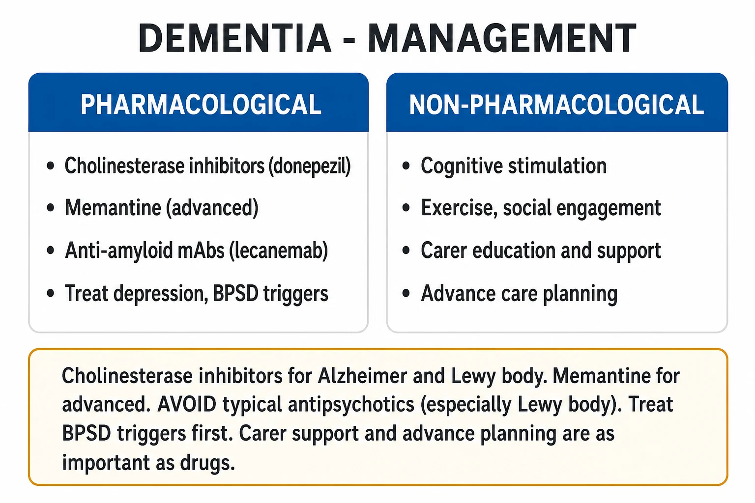
Management is multidomain and individualised — drugs are only one component. The plan addresses: (a) treatment of reversible contributors, (b) disease-modifying/symptomatic drugs for AD, (c) subtype-specific pharmacology, (d) non-pharmacological measures, (e) BPSD management, (f) carer support, (g) advance planning.[1]
Step 1 — Treat all reversible contributors (B12, hypothyroidism, infection, depression, drug review, NPH shunt where indicated). Address vascular risk factors aggressively in all subtypes (BP, lipids, glucose, AF anticoagulation, smoking) — this slows vascular contribution and is a central recommendation of the Lancet Commission.[3]
Step 2 — Cholinesterase inhibitors (ChEIs) — first-line for mild-to-moderate AD and for DLB.[1]
| Drug | Dose & titration | Route | Indication | Key cautions |
|---|---|---|---|---|
| Donepezil | Start 5 mg at night for 4 weeks, increase to 10 mg at night | Oral | Mild-moderate AD; DLB/PDD | Bradycardia, syncope, GI upset, vivid dreams; check ECG if cardiac disease |
| Rivastigmine | 1.5 mg twice daily, titrate every 2 weeks to 6 to 12 mg per day; transdermal patch 4.6 mg per 24 h, up to 13.3 mg per 24 h | Oral or transdermal patch | Mild-moderate AD; DLB/PDD | Highest GI adverse effect; patch improves tolerability |
| Galantamine | 4 mg twice daily (or 8 mg ER once daily), titrate to 8 to 12 mg twice daily (ER 16 to 24 mg once daily) | Oral (immediate / prolonged-release) | Mild-moderate AD | Nausea, anorexia, bradycardia |
Mechanism: inhibit acetylcholinesterase → increase synaptic acetylcholine, partially compensating for the cholinergic deficit. Effect size is modest — mean MMSE improvement of about 1 to 2 points, with stabilisation for 6 to 12 months rather than reversal. Review benefit at 3 months; continue if cognition/behaviour/function is stable or improved; stop if deterioration continues at the pre-treatment rate.[1]
Step 3 — Memantine — for moderate-to-severe AD (MMSE under about 20) and as add-on in moderate disease.[1]
- Dose: 5 mg once daily, titrate weekly in 5 mg steps to a target of 20 mg once daily (10 mg twice daily); reduce dose in moderate renal impairment (eGFR 5 to 29: 5 mg twice daily; max 10 mg).
- Mechanism: uncompetitive, low-affinity NMDA-receptor antagonist — modulates glutamatergic excitotoxicity without blocking normal neurotransmission.
- Side-effects: dizziness, headache, constipation, somnolence; rarely confusion.
- Combination ChEI + memantine is used in moderate-to-severe AD with modest added benefit over either agent alone. [1]
Step 4 — Anti-amyloid monoclonal antibodies — for early, biomarker-confirmed AD (MCI or mild dementia due to AD, amyloid-positive).[5]
- Lecanemab — anti-amyloid-beta protofibril IgG1. Clarity AD (van Dyck 2023, NEJM)[5] showed a 27 percent slowing of cognitive decline on the CDR-SB over 18 months (difference of about 0.45 points) versus placebo, with significant amyloid clearance on PET. Dose: 10 mg per kg intravenously every two weeks. Monitor for amyloid-related imaging abnormalities (ARIA — oedema ARIA-E and microhaemorrhage ARIA-H) with MRI at baseline and before the 5th, 7th and 14th infusions. APOE epsilon-4 homozygotes are at higher ARIA risk — genotype before treatment.
- Donanemab — anti-pyroglutamate amyloid IgG1; similar efficacy and ARIA profile, with target-to-clear stopping rules.
These are not cures — they modestly slow decline in carefully selected early patients; their place in routine practice, cost-effectiveness and infusion-burden are the subject of active guideline debate.[1]
Step 5 — BPSD management. Behavioural and psychological symptoms of dementia affect most patients at some stage. Stepwise approach:[1]
- Identify and treat precipitants — pain, constipation, urinary infection, hunger, thirst, sensory deprivation, medication side-effect, environmental change. (Pain is the single most under-treated driver.)
- Non-pharmacological first — person-centred care, structured routine, validation therapy, music, aromatherapy, sensory stimulation, avoid confrontation, modify the environment.
- Pharmacotherapy only if distress or risk is severe — SSRI (citalopram 10 to 20 mg for agitation in AD; some evidence), or short course of atypical antipsychotic (risperidone 0.25 to 0.5 mg, olanzapine, quetiapine — the latter preferred when parkinsonism/DLB coexists) at the lowest effective dose for the shortest time, with documented review. Avoid typicals. Memantine may help agitation in severe disease.
- Depression in dementia — SSRI (sertraline, citalopram); tricyclics worsened by anticholinergic load (avoid).
- Sleep disturbance — sleep hygiene, bright-light therapy; avoid benzodiazepines (deliriogenic); melatonin may help RBD. [1]
Step 6 — Non-pharmacological core measures (the foundation, not an add-on):[3][1]
- Cognitive stimulation therapy (CST) — group sessions, evidence-based benefit in mild-moderate AD.
- Regular physical exercise — improves cognition, mood, function.
- Social engagement — combats isolation.
- Hearing aids (in the hearing-impaired — the largest single modifiable mid-life risk factor).
- Diet — Mediterranean/MIND pattern associated with lower incidence.
- Multidomain interventions (FINGER model) — combined vascular, cognitive, exercise and dietary intervention. [1]
Step 7 — Carer-focused plan. Carer strain predicts institutionalisation. Offer psychoeducation, respite care, support groups, behavioural-management training, and screen carers for depression. Plan early for advance decisions, Lasting/Enduring Power of Attorney, finances, and future care preferences while capacity remains.[1]
Dementia — REVERSIBLE mnemonic for reversible causes
REVERSIBLE
anticholinergics, benzodiazepines, opioids, anticonvulsants
depression (pseudodementia)
multiple infarcts, strategic infarct, small-vessel
hypothyroidism, hyper/hypocalcaemia
sensory deprivation (eyes, ears)
subdural, tumour, NPH
neurosyphilis, HIV, chronic meningitis
and other nutritional — thiamine, niacin
hepatic/renal encephalopathy, hypoxia
alcohol-related brain disease (reversible component)
Specific Subtypes & Scenarios
Alzheimer disease — onset typically after 65 (early-onset under 65 if familial). Insidious, progressive episodic memory loss leading to global cortical dementia. MRI: medial temporal/hippocampal atrophy (MTA score raised). CSF: low Abeta-42, high tau/phospho-tau. Drugs: ChEI for mild-moderate; memantine for moderate-severe; lecanemab/donanemab for early amyloid-confirmed disease.[1][5]
Vascular dementia — subtype classification: (a) multi-infarct (cortical strokes); (b) strategic single infarct; (c) small-vessel / lacunar / subcortical (Binswanger); (d) hypoperfusion (e.g. after cardiac arrest); (e) haemorrhagic. Management emphasis: aggressive vascular risk-factor control (BP under 130/80 where tolerated, statin, diabetes, AF anticoagulation, smoking cessation) — this is the main lever. Donepezil has modest evidence in vascular dementia but ChEIs are less effective than in AD; memantine is sometimes used.[3]
Dementia with Lewy bodies — diagnostic criteria (4th DLB Consortium, 2017)[4]: probable DLB = 2 or more core clinical features, OR 1 core feature plus 1 indicative biomarker (reduced DAT-SPECT uptake, polysomnographic confirmation of REM sleep without atonia, or abnormal MIBG myocardial scintigraphy). ChEIs are particularly effective in DLB — rivastigmine has the strongest evidence — improving both cognition and hallucinations. Levodopa for parkinsonism (start low — DLB patients are sensitive). Avoid typical antipsychotics (neuroleptic sensitivity). Memantine may help. Prognosis worse than AD (median survival 5 to 7 years).[4]
Frontotemporal dementia — bvFTD and the three PPA variants (semantic, non-fluent/agrammatic, logopenic). Onset younger (45 to 65). ChEIs are NOT effective and may worsen behaviour. SSRIs (especially for disinhibition and compulsive behaviours — escitalopram, sertraline) are the mainstay for behavioural symptoms. Investigations: MRI frontal/anterior-temporal atrophy; FDG-PET frontal/temporal hypometabolism. Genetics: MAPT, GRN, C9orf72 in familial cases — offer genetic counselling. Prognosis median survival 6 to 11 years from diagnosis (faster if ALS-overlap).[2]
Creutzfeldt-Jakob disease — sporadic (85 percent, the commonest), variant (vCJD, bovine-origin, younger patients, psychiatric onset, tonsillar PrP-Sc), familial (PRNP) and iatrogenic (growth hormone, dura mater grafts). Clinically rapidly progressive dementia over weeks to months with myoclonus, cerebellar ataxia, visual/cortical disturbance. EEG: periodic sharp-wave complexes at 1 to 2 Hz (about two-thirds of sCJD). MRI: cortical ribboning and basal-ganglia/thalamic hyperintensity on DWI/FLAIR — the "hockey-stick" and "pulvinar" signs. CSF 14-3-3 protein and RT-QuIC (real-time quaking-induced conversion, highly specific) support the diagnosis. Invariably fatal, median survival about 4 to 6 months.[2]
Normal-pressure hydrocephalus — the reversible dementia that examiners love. Triad: gait apraxia, urinary incontinence, subcortical dementia. MRI: ventriculomegaly out of proportion to sulcal atrophy. CSF tap test (Fisher) or external lumbar drainage — clinical improvement (especially gait) after removing 30 to 50 mL CSF predicts response to ventriculoperitoneal shunting, which can reverse symptoms. Gait typically improves first and most.[1]
Complications & Pitfalls
Behavioural and psychological symptoms of dementia (BPSD) — agitation, aggression, wandering, repetitive questioning, sleep disturbance, depression, anxiety, apathy, psychosis (hallucinations, delusions), eating and sexual disinhibition — affect the majority at some stage, drive carer strain and institutionalisation, and are the commonest reason for emergency dementia presentations.[1]
Medical complications: falls and fractures (with delirium cascade), aspiration pneumonia (dysphagia in late disease — a leading terminal event), malnutrition and weight loss, dehydration, urinary and faecal incontinence (with UTI and pressure-area risk), seizures (late AD), infections (UTI, pneumonia, cellulitis), pressure ulcers in immobile patients, deep vein thrombosis/pulmonary embolism. Delirium is frequently superimposed and is both a complication and a cause of accelerated decline.[2]
Drug-related pitfalls: [1]
- Anticholinergic burden — many over-the-counter and prescription drugs (oxybutynin, tricyclics, antihistamines, hyoscine) worsen cognition; review and deprescribe.
- Benzodiazepines — deliriogenic; avoid.
- Typical antipsychotics in DLB — neuroleptic sensitivity reaction: severe rigidity, hyperthermia, autonomic instability, rhabdomyolysis, can be fatal after a single dose.[4]
- Antipsychotics in all-cause dementia — increase stroke and mortality risk (regulatory boxed warning); lowest dose, shortest time, documented indication.
- ChEIs — bradycardia, syncope (and thus falls and pacemaker consideration), GI upset, vivid dreams, urinary incontinence; contraindicated in sick sinus syndrome and caution with QT-prolonging drugs.
Diagnostic pitfalls: [1]
- Missing delirium in a demented elderly patient — assume a new acute change has a precipitant until proven otherwise.
- Labelling depression as dementia (pseudodementia) — undertreats a treatable illness.
- Missing NPH in a "dementia + falls + incontinence" patient.
- Over-diagnosing dementia from a low MMSE without considering education, language, sensory loss, anxiety or delirium.
- Under-investigating young-onset dementia — the differential is broader (genetic, prion, autoimmune, Wilson, NF-leucodystrophy) and reversible/heritable causes are over-represented. [1]
Caregiver burnout and elder abuse — recognise carer strain (Zarit Burden Interview), offer respite, and have a low threshold to safeguard when neglect, financial abuse or physical abuse is suspected. [1]
Prognosis & Disposition
Dementia is progressive and incurable in the degenerative subtypes; the trajectory and median survival differ by aetiology.[2]
- Alzheimer disease — median survival 8 to 10 years from diagnosis (longer from symptom onset); some live 15 to 20 years.
- Vascular dementia — variable, determined by stroke burden and comorbidity; median 3 to 5 years but with wide spread.
- Dementia with Lewy bodies — 5 to 7 years, often faster than AD; high mortality from falls, aspiration and neuroleptic sensitivity.
- Frontotemporal dementia — 6 to 11 years, faster with ALS-overlap.
- Creutzfeldt-Jakob disease — median 4 to 6 months; invariably fatal. [1]
Predictors of rapid decline: younger onset, lower baseline cognition, presence of parkinsonism or falls, behavioural disturbance, low CSF Abeta-42 with high tau, hippocampal atrophy on MRI, and APOE epsilon-4 homozygosity (AD). [1]
Institutionalisation is driven less by cognition than by behavioural disturbance (especially aggression), incontinence, wandering, carer strain, and loss of ADLs — discharge planning and community-support packages delay or prevent care-home placement. [1]
End-of-life considerations: dementia is now recognised as a terminal illness. Advance care planning should begin early — while capacity allows — covering future care preferences, resuscitation status, hospital vs home management of infections, and artificial nutrition/hydration. As the disease advances, focus shifts to comfort, dignity, symptom control (pain, agitation, secretions, dyspnoea) and family support — often through palliative care / hospice input. Aspiration pneumonia and sepsis are the usual terminal events. [1]
Special Populations
- Young-onset dementia (under 65) — about 5 percent of all dementia; FTD, genetic AD, prion disease, autoimmune and metabolic causes are over-represented. Lower threshold for MRI, CSF biomarkers, genetic testing and autoimmune encephalitis panels. Greater diagnostic delay, more diagnostic uncertainty, profound impact on employment and young families, and stronger genetic counselling needs.
- Down syndrome (trisomy 21) — the extra APP gene (chromosome 21) leads to near-universal Alzheimer pathology by age 40 to 50, with clinical dementia usually emerging in the 40s to 50s; diagnose with baseline-confirmed decline, using Dementia Questionnaire for Individuals with Intellectual Disabilities (DLD) rather than MMSE.
- Dementia in Parkinson disease (PDD) — common; up to 80 percent of PD patients develop dementia over the disease course, typically after more than one year of motor symptoms (the 1-year rule). Visuospatial and executive deficits predominate. ChEIs (rivastigmine) are first-line. Beware anticholinergics and amantadine (worsen cognition/hallucinations); reduce these before adding antipsychotics. Quetiapine or clozapine (with FBC monitoring) are the antipsychotics of choice when needed; pimavanserin is approved in some regions for PD psychosis.[4]
- Post-stroke cognitive impairment — up to a third of stroke survivors develop cognitive impairment within a year; early screening (MoCA at 6 months) and vascular secondary prevention are essential.
- HIV-associated neurocognitive disorder (HAND) — subcortical pattern (slowing, executive, motor) even in virologically suppressed patients; the most common neurological complication of HIV. Distinguish from opportunistic processes (toxoplasmosis, PML, cryptococcal meningitis).
- Learning disability / intellectual disability — baseline decline from premorbid function is the key diagnostic shift; use informant-based tools.
Evidence, Guidelines & Regional Differences
NICE NG97 (2018, UK)[1] — the UK national guideline: care by a memory-assessment service; ChEIs (donepezil, rivastigmine, galantamine) for mild-moderate AD (the three agents are clinically equivalent — choice by tolerability/comorbidity); memantine for moderate-severe AD or where ChEIs are not tolerated; ChEIs reviewed at 3 months then 6-monthly; group cognitive stimulation therapy for mild-moderate; structured non-pharmacological measures first for BPSD, with antipsychotics only short-term for severe distress and reviewed every 3 months; offer carers psychoeducation and support. NICE does not currently recommend routine amyloid PET or anti-amyloid antibodies outside trials/specialist commissioning.
NIA-AA Research Framework (2018) — recast AD biologically (not clinically) as a biomarker-defined entity using the AT(N) system:[1]
- A — Amyloid (low CSF Abeta-42 or positive amyloid PET)
- T — Tau (high CSF phospho-tau or positive tau PET)
- (N) — Neurodegeneration (high CSF total tau, atrophy on MRI, hypometabolism on FDG-PET) [1]
A biomarker-confirmed diagnosis requires A-positive (with T for "AD"), irrespective of clinical stage. This framework underpins the eligibility criteria for the new anti-amyloid antibodies. [1]
European intersocietal biomarker recommendations (Frisoni 2024)[1] — codify when to use blood, CSF and PET biomarkers, including the emerging plasma p-tau217 blood test as a triage tool before confirmatory CSF/PET.
2020 Lancet Commission (Livingston)[3] — the key prevention paper: 12 modifiable risk factors account for about 40 percent of dementia (up from 35 percent in the 2017 report, after adding excessive alcohol, TBI and air pollution). Be ambitious about prevention: what is good for the heart is good for the brain.
Clarity AD (van Dyck 2023, NEJM)[5] — phase 3 trial of lecanemab in early AD: 27 percent relative slowing of decline on CDR-SB (about 0.45 absolute points over 18 months); 17 percent relative reduction in amyloid on PET; ARIA-E in 12.6 percent and ARIA-H in 17.3 percent (symptomatic in about a quarter of these, with three treatment-related deaths in extension data reported). The first rigorously positive disease-modifying trial in AD.
Other landmark evidence: AD2000 (donepezil modest, not disease-modifying); DOMINO (donepezil continuation beneficial even in moderate-severe AD); MEM-MD-02 (memantine); FINGER (multidomain lifestyle intervention improves cognition in at-risk elders). [1]
[1]Regional differences: In India, prevalence is rising with longevity; the vascular burden is higher (driven by hypertension and diabetes), so mixed AD-vascular is common. Cultural norms favour family-based home care, with state-funded memory clinics still uneven; the Indian Consortium on Dementia and AIIMS / NIMHANS guidelines adapt international evidence to local resources. Self-medication and polypharmacy drive high anticholinergic burden. Genetic counselling is less commonly sought than in high-income settings but is increasingly relevant for young-onset disease. [1]
Exam Pearls
- Alzheimer disease = amyloid-beta plaques + tau neurofibrillary tangles + cholinergic deficit; episodic memory loss is the earliest feature; nucleus basalis of Meynert degenerates first.[2]
- MMSE under 24 (or MoCA under 26) = impairment; MoCA is more sensitive for early disease and executive dysfunction.
- Always exclude reversible causes in every cognitive assessment: B12, folate, TSH, calcium, glucose, syphilis (RPR/VDRL), HIV; NPH and depression.
- Donepezil (5 to 10 mg), rivastigmine, galantamine (cholinesterase inhibitors) for mild-moderate AD; memantine (5 to 20 mg) for moderate-severe.
- DLB = fluctuating cognition + detailed visual hallucinations + parkinsonism + REM sleep behaviour disorder. ChEIs (rivastigmine) help; typical antipsychotics are absolutely contraindicated (fatal neuroleptic sensitivity).[4]
- Vascular dementia = stepwise decline + executive dysfunction + imaging infarcts. Aggressive vascular risk-factor control is the main lever.
- FTD = early personality/behaviour or language change with relative memory sparing; ChEIs do NOT help; SSRIs do.
- NPH triad = gait apraxia + urinary incontinence + subcortical dementia; reversible with ventriculoperitoneal shunt.
- Delirium = acute, fluctuating, clouded consciousness, reversible — do not mislabel as dementia.
- Depression (pseudodementia) = "I don't know" answers, complains of memory loss, diurnal variation, biological features, preserved insight.
- CJD = rapidly progressive dementia + myoclonus + periodic sharp waves on EEG + cortical ribboning on DWI + CSF 14-3-3/RT-QuIC; fatal in months.
- APOE epsilon-4 = risk; APOE epsilon-2 = protective. Familial early-onset AD = PSEN1, PSEN2, APP (autosomal dominant).
- Down syndrome → AD by age 40 to 50 (extra APP on chromosome 21).
- Lecanemab / donanemab (anti-amyloid antibodies) slow (not cure) early amyloid-confirmed AD; monitor for ARIA.[5]
- 2020 Lancet Commission: 40 percent of dementia is preventable via 12 modifiable risk factors.[3]
- 1-year rule: dementia before parkinsonism = DLB; dementia after established PD = PDD.
- Anticholinergic burden worsens cognition — always review the drug chart.
Exam application bank (NEET-PG / INICET)
One-line answer
Dementia is an acquired, persistent, global impairment of higher cortical functions (memory, language, visuospatial skills, executive function, personality) occurring in clear consciousness, sufficient to interfere with activities of daily living. It is not a single disease but a syndrome. The commonest cause is Alzheimer disease (AD) (60 to 70 percent), driven by amyloid-beta 42 plaque deposition and tau-hyperphosphorylated neurofibrillary tangles with a cholinergic deficit; followed by vascular dementia (VaD), dementia with Lewy bodies (DLB) and frontotemporal dementia (FTD). Presentation is progressive and insidious in AD, stepwise in vascular, fluctuating with visual hallucinations and parkinsonism in DLB, and behavioural/personality change in FTD. Always exclude reversible mimics — delirium (acute, fluctuating, clouded consciousness), depression (pseudodementia), normal ageing, and
Worked stems (answer without another resource)
Stem 1 — Classic presentation. Map symptoms to mechanism; name the first investigation and first treatment step with dose/route if drug therapy is standard. [1]
Stem 2 — Unstable / complicated. List red flags that force immediate resuscitation, theatre, ICU, antidote, or reperfusion — and what you do in the first 15 minutes. [1]
Stem 3 — Atypical group. Elderly, pregnancy, child, or immunocompromised: how presentation and thresholds change. [1]
Stem 4 — Differential trap. Name the three closest mimics and one discriminator for each. [1]
Stem 5 — Disposition. Who goes home with safety-netting, who is admitted, who needs HDU/ICU/theatre, and what follow-up is mandatory. [1]
Rapid viva checklist
- Definition + classification
- Pathophysiology chain
- Bedside signs / criteria
- Score with exact components (if any)
- Emergency bundle
- Definitive therapy with doses
- Complications of disease and of treatment
- Special populations
- Guideline/trial name if classic
- Three exam traps
Coverage self-check
If you cannot answer any stem above from this page alone, re-read the matching section — the page is intended to be self-sufficient for final-prof and NEET-PG/INICET questions on Dementia.
References
- [1]Frisoni GB, Festari C, Massa F, et al. European intersocietal recommendations for the biomarker-based diagnosis of neurocognitive disorders Lancet Neurol, 2024.PMID 38365381
- [2]Schneider JA. Neuropathology of Dementia Disorders Continuum (Minneap Minn), 2022.PMID 35678405
- [3]Livingston G, Huntley J, Sommerlad A, et al. Dementia prevention, intervention, and care: 2020 report of the Lancet Commission Lancet, 2020.PMID 32738937
- [4]McKeith IG, Boeve BF, Dickson DW, et al. Diagnosis and management of dementia with Lewy bodies: Fourth consensus report of the DLB Consortium Neurology, 2017.PMID 28592453
- [5]van Dyck CH, Swanson CJ, Aisen P, et al. Lecanemab in Early Alzheimer's Disease N Engl J Med, 2023.PMID 36449413