Neurology · Neurology
Epilepsy
Also known as Seizure disorder · Epilepsy · Focal epilepsy · Generalised epilepsy · Idiopathic generalised epilepsy · Juvenile myoclonic epilepsy · Childhood absence epilepsy · Temporal lobe epilepsy
Epilepsy is a chronic brain disorder defined by an enduring predisposition to generate unprovoked epileptic seizures. Operationally (ILAE 2014): two or more unprovoked seizures more than 24 hours apart, OR one unprovoked seizure with at least a 60 percent recurrence risk over ten years, OR a recognised epilepsy syndrome. Seizures are classified by the ILAE 2017 framework into focal onset (aware or impaired awareness), generalised onset (motor or non-motor/absence), and unknown onset. Syndrome-specific AED choice is decisive: focal — lamotrigine or levetiracetam; idiopathic generalised — valproate (avoid in women of childbearing age); childhood absence — ethosuximide; juvenile myoclonic epilepsy — valproate or levetiracetam, lifelong. Convulsive status epilepticus is a seizure lasting 5 minutes or more: IV lorazepam 4 mg then IV levetiracetam 60 mg/kg or fosphenytoin 20 mg PE/kg then, if refractory, ICU anaesthetic infusion.
On this page & tools
Your progress
Saved locally on this device.
Exam tags
Red flags
Overview & Definition
A seizure is the transient occurrence of signs or symptoms produced by abnormal excessive or synchronous neuronal activity in the brain. A provoked (acute symptomatic) seizure is one occurring in close temporal relationship with a transient systemic or brain insult — hypoglycaemia, hyponatraemia, alcohol withdrawal, fever, head trauma, CNS infection, or a high-dose stimulant drug — and does not, by itself, constitute epilepsy. An unprovoked seizure is one without an identified immediate precipitant, occurring either out of the blue or in the context of an enduring predisposition. Epilepsy is the chronic disorder of enduring predisposition to generate unprovoked seizures, with its neurobiological, cognitive, psychological, and social consequences.[1]
The ILAE 2014 practical clinical definition of epilepsy is met by any one of three criteria: (1) at least two unprovoked seizures occurring more than 24 hours apart; (2) a single unprovoked seizure with at least a 60 percent probability of further seizures over the next ten years (this recurrence risk applies, for example, after a single seizure with a structural lesion on imaging and epileptiform abnormalities on EEG, after a stroke, or after certain remote insults such as severe traumatic brain injury); or (3) the diagnosis of a recognised epilepsy syndrome. The definition is deliberately operational — it is meant to guide the decision to treat, not to make an absolute pronouncement about a patient's identity.[1]
This distinction matters because it drives management. A provoked seizure — a convulsion in a hypoglycaemic, hyponatraemic, or alcohol-withdrawing patient, or a febrile seizure in a child — is treated by correcting the precipitant, not by starting an antiepileptic drug (AED). Starting an AED after a single truly unprovoked seizure with low recurrence risk is not mandatory; after a single unprovoked seizure with high recurrence risk, or after two seizures, treatment is the norm. The error to avoid is conflating any convulsion with epilepsy. [1]
Classification
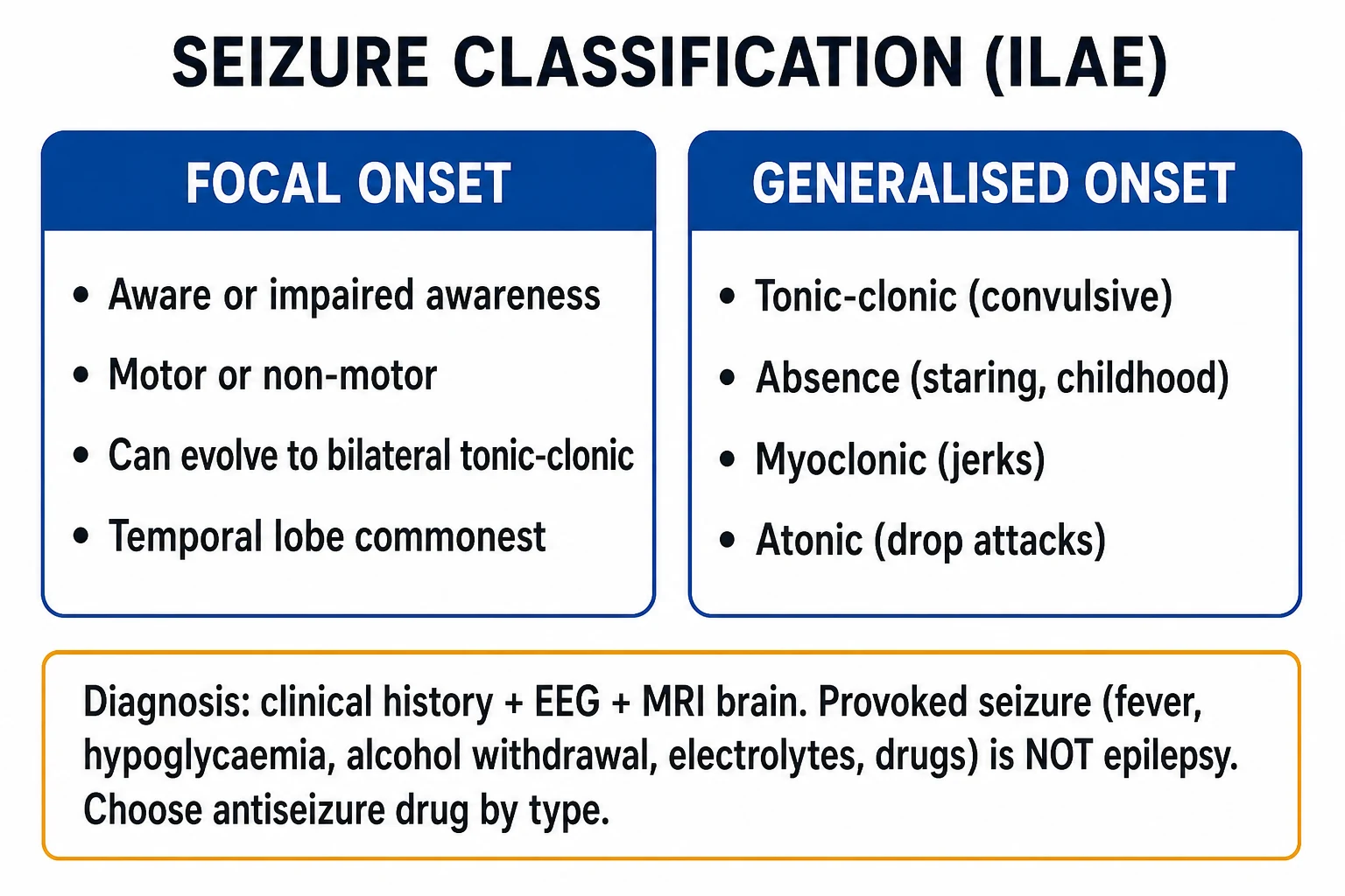
Seizure classification is not an academic exercise — it dictates AED choice, prognosis, and the imaging and genetic work-up. The ILAE 2017 operational classification of seizure types divides seizures by where in the brain they begin, and by whether awareness is preserved.[2]
Focal onset
- Originates within networks limited to ONE hemisphere — was 'partial' seizure
- Subdivided by awareness: focal AWARE (formerly 'simple partial') vs focal IMPAIRED AWARENESS (formerly 'complex partial')
- Further described by motor (clonic, myoclonic, tonic, atonic, automatisms, hyperkinetic) or non-motor (sensory, autonomic, emotional/cognitive, psychic) features
- May evolve to FOCAL-TO-BILATERAL TONIC-CLONIC (formerly 'secondary generalisation')
- First-line AEDs: lamotrigine, levetiracetam, carbamazepine, oxcarbazepine; MRI epilepsy protocol to find a lesion (e.g., mesial temporal sclerosis, tumour, dysplasia, neurocysticercosis)
Generalised onset
- Originates in and rapidly engages BILATERALLY DISTRIBUTED networks — both hemispheres at onset
- Motor: tonic-clonic, clonic, tonic, myoclonic, atonic, epileptic spasms
- Non-motor (absence): typical (childhood absence, 3-Hz spike-wave), atypical, myoclonic absence, eyelid myoclonia
- First-line AEDs: valproate (most effective; avoid in women of childbearing age), levetiracetam, lamotrigine, ethosuximide (absence only)
- Carbamazepine and phenytoin can WORSEN absence and myoclonic seizures — the classic wrong-drug trap
Unknown onset
- Used when onset cannot be classified — unwitnessed, nocturnal-only, or already in status on arrival
- May be motor (tonic-clonic, epileptic spasms) or non-motor
- Re-classify once additional history or video-EEG clarifies the onset
- First-line AED: a broad-spectrum agent (levetiracetam or valproate in non-childbearing patients)
The 2017 framework also classifies the epilepsies themselves along three axes — seizure type, aetiology (the six ILAE categories), and syndrome (a cluster of age of onset, seizure types, EEG signature, and prognosis that together name a recognisable entity such as childhood absence epilepsy or juvenile myoclonic epilepsy).[3] Aetiology matters because structural and metabolic causes may be treatable; genetic causes carry counselling implications; immune and infectious causes direct the work-up.
Structural
- A lesion visible on imaging that causally relates to seizures
- Examples: mesial temporal sclerosis, stroke, traumatic brain injury, brain tumour, cerebral malformation (focal cortical dysplasia), vascular malformation
Genetic
- A known or presumed genetic cause directly contributing to seizures
- Examples: Dravet syndrome (SCN1A), childhood absence epilepsy, juvenile myoclonic epilepsy, tuberous sclerosis complex, ring chromosome 20
Infectious
- The commonest identified cause of epilepsy worldwide
- Examples: neurocysticercosis (Taenia solium), bacterial meningitis sequelae, viral encephalitis (HSV), cerebral malaria, tuberculosis
Metabolic
- A distinct metabolic disorder causing seizures
- Examples: hypoglycaemia, hyponatraemia, hypocalcaemia, hypomagnesaemia, uraemia, hepatic encephalopathy, pyridoxine-dependent seizures (neonate)
Immune
- Autoimmune inflammation of the CNS as the cause
- Examples: anti-NMDA-receptor, anti-LGI1, anti-GAD65, anti-GABA-A/B receptor encephalitis; often limbic encephalitis with faciobrachial dystonic seizures
Unknown
- The cause of the epilepsy is not yet identified — the category that shrinks as work-up improves
- Was 'cryptogenic' or 'idiopathic' under older terminology
Epidemiology & Risk Factors
Epilepsy is one of the commonest serious neurological disorders, affecting roughly 1 in 100 to 1 in 200 people at any time — a lifetime prevalence of around 1 percent and an incidence of about 50 to 120 per 100,000 per year. The age distribution is bimodal: a peak in early childhood (driven by genetic and perinatal causes, febrile seizures, and cerebral palsies) and a larger peak in older adults (driven by stroke and neurodegeneration), with a trough in young adults.[12]
The dominant cause of epilepsy shifts with age, and an examiner will test this directly. In children, the commonest causes are perinatal injury (hypoxic-ischaemic encephalopathy, neonatal hypoglycaemia), genetic/generalised epilepsy syndromes, congenital brain malformations, and fever-related presentations (febrile seizures, with a minority progressing to epilepsy). In young and middle-aged adults, the commonest identifiable causes are traumatic brain injury, CNS infection (in the developing world, neurocysticercosis from Taenia solium is the leading cause, acquired by ingesting tapeworm eggs in faecally-contaminated food or water), brain tumour, and the lasting scar of mesial temporal sclerosis. In older adults, stroke (ischaemic and haemorrhagic) and neurodegeneration (especially Alzheimer disease) dominate, with brain tumour and metabolic disturbance contributing.[12]
Epilepsy — the numbers worth memorising
Recognised risk factors for developing epilepsy mirror the causes: perinatal injury, significant traumatic brain injury (the more severe and the longer the post-traumatic amnesia, the higher the risk), CNS infection with residual scarring, stroke, brain tumour, a history of febrile seizures (especially complex or prolonged, or with a family history of epilepsy), family history of epilepsy, neurodevelopmental disorders (cerebral palsy, intellectual disability, autism), and chronic alcohol misuse (both withdrawal seizures and, less commonly, structural injury). [1]
Sudden unexpected death in epilepsy (SUDEP) is the leading cause of epilepsy-related premature mortality in adults, with an incidence of roughly 1 per 1,000 adults with epilepsy per year overall — and much higher (up to 1 in 100 per year) in those with frequent generalised tonic-clonic seizures, nocturnal seizures, drug-resistant epilepsy, intellectual disability, or non-adherence. The mechanism is likely postictal cerebral shutdown producing respiratory and cardiac arrest. The single most effective risk-reduction strategy is seizure freedom through adherence to effective therapy.[12]
Pathophysiology
A seizure is fundamentally an imbalance between excitation and inhibition in cortical (and, for generalised seizures, thalamocortical) networks, tipping the system into a state of abnormal, hypersynchronous neuronal firing. The two principal molecular players are glutamate — the dominant cortical excitatory neurotransmitter, acting on AMPA, kainate, and NMDA ionotropic receptors and on metabotropic receptors — and gamma-aminobutyric acid (GABA), the dominant inhibitory neurotransmitter, acting on GABA-A (chloride-channel, fast inhibition) and GABA-B (G-protein-coupled, slow inhibition) receptors.[2]
In a focal seizure, a localised group of neurons (the epileptic focus) loses its normal restraint and begins to fire in synchronised bursts. The cellular signature is the paroxysmal depolarising shift: a giant excitatory postsynaptic potential, mediated by glutamatergic input through NMDA and AMPA channels, that brings the neuron to threshold repeatedly and recruits its neighbours through recurrent collateral excitation. The discharge may stay localised (a focal aware seizure), spread into adjacent cortex producing a Jacksonian march of motor or sensory symptoms, or recruit the wider network and evolve into a focal-to-bilateral tonic-clonic seizure.[2]
In a generalised seizure from onset, the abnormal activity engages bilaterally distributed thalamocortical networks essentially simultaneously. The classic example is typical absence epilepsy, where a circuit between cortical pyramidal neurons and thalamic relay neurons firing through T-type calcium channels generates the characteristic 3-Hz spike-and-wave discharge on EEG and the brief blank-stare of the seizure. T-type calcium channels in thalamic neurons are the molecular target of ethosuximide, the drug of choice for absence — a piece of mechanism that explains a piece of pharmacology. Juvenile myoclonic epilepsy and the other idiopathic generalised epilepsies are largely genetic channelopathies — mutations in voltage-gated sodium, calcium, potassium, or GABA-A channel subunits produce a neuronal membrane that is too excitable or insufficiently inhibited. Examples include SCN1A in Dravet syndrome (a loss-of-function sodium-channel mutation, paradoxically treated by avoiding sodium-channel blockers), and GABA-A receptor subunit mutations in genetic generalised epilepsy. [1]
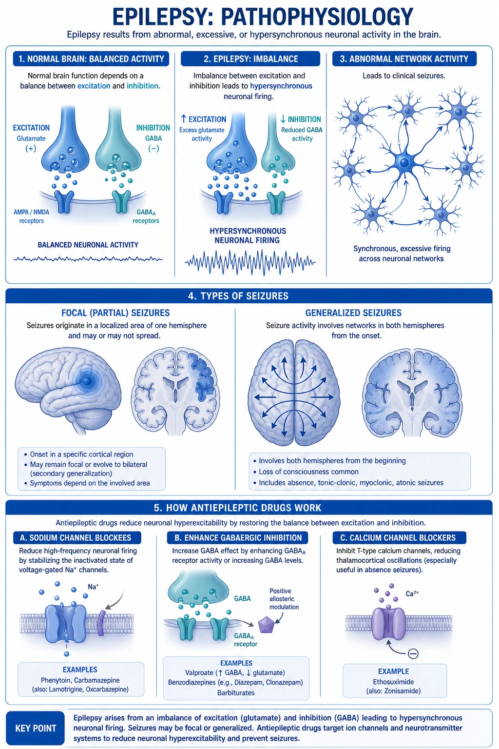
The molecular targets of the major AED classes map onto this physiology, and remembering this correspondence earns examiner credit: [1]
Na+ channel blockers
- Stabilise the inactivated state of voltage-gated sodium channels, slowing rapid repetitive firing
- Carbamazepine, oxcarbazepine, phenytoin, lamotrigine, lacosamide, eslicarbazepine
- Best for focal-onset and generalised tonic-clonic seizures; AVOID in absence and myoclonus (carbamazepine, phenytoin can worsen)
T-type Ca2+ blockers
- Block T-type calcium channels in thalamic neurons that generate the 3-Hz spike-and-wave
- Ethosuximide (drug of choice for childhood absence)
- Treats absence ONLY — does NOT protect against tonic-clonic seizures, so often combined with valproate if both seizure types coexist
GABAergic drugs
- Enhance inhibitory neurotransmission through GABA-A or GABA-B
- Benzodiazepines (positive allosteric modulators of GABA-A) — first-line for status epilepticus; barbiturates; vigabatrin (irreversible GABA-transaminase inhibitor); clobazam; valproate (also increases GABA synthesis)
SV2A binders
- Bind the synaptic vesicle protein 2A, modulating neurotransmitter release
- Levetiracetam, brivaracetam — broad-spectrum, favourable pharmacokinetics, low interaction profile, first-line for focal and generalised tonic-clonic seizures
AMPA antagonists
- Block AMPA-type glutamate receptors
- Perampanel — a non-competitive AMPA antagonist, used as adjunctive therapy for focal and generalised tonic-clonic seizures; can cause psychiatric side-effects (irritability, aggression)
The postictal state — confusion, headache, sleepiness, and sometimes focal neurological deficit — reflects transient neuronal exhaustion, ion-gradient depletion, local neurotransmitter depletion, and possibly a brief postictal spreading depolarisation or transient hypometabolism in the involved cortex. Postictal (Todd's) paralysis is a transient focal weakness lasting hours (occasionally a day or two) after a focal or generalised seizure, caused by transient cortical dysfunction in the motor strip; it is clinically indistinguishable from an acute stroke at first encounter, which is why a witnessed seizure or a history of epilepsy is decisive. The mechanism is not infarction — the deficit resolves completely — but transient inhibition of the involved cortex. [1]
Clinical Presentation
The semiology (the clinical appearance) of a seizure is the single most diagnostic piece of information, and a structured eyewitness account is the cornerstone of the diagnosis. The major seizure types have recognisable patterns that the examiner will expect you to describe. [1]
Focal aware seizures (formerly simple partial) produce symptoms the patient remains conscious of and can describe — these are often an aura, which is in fact a small focal seizure: a rising epigastric sensation, déjà vu or jamais vu, an olfactory hallucination (classic for temporal lobe, classically the burnt-rubber smell of an uncinate fit), a visual distortion, or unprovoked fear. Motor focal seizures produce clonic movements that may march across a limb (the Jacksonian march, from hand to arm to face, reflecting orderly spread across the motor homunculus). Sensory focal seizures produce tingling or numbness in the same distribution. Autonomic and psychic symptoms — fear, depersonalisation, déjà vu — localise to the temporal lobe. [1]
Focal impaired-awareness seizures (formerly complex partial) — typically of temporal or frontal origin — produce an alteration of awareness with automatisms: lip-smacking, chewing, fumbling with buttons, picking at clothes, wandering, or verbal automatisms. The patient is not in contact, may respond only partially, and has postictal confusion. Frontal-lobe seizures tend to be brief, nocturnal, with bizarre hypermotor movements that are easily mistaken for a sleep disorder or even psychogenic episodes. [1]
Generalised tonic-clonic seizures (the grand mal of older terminology) follow a stereotyped four-phase sequence. A brief aura or warning (if focal onset with secondary generalisation) is followed by the tonic phase — sudden loss of consciousness, a forced expiratory cry or vocalisation as thoracic muscles contract, falling, and tonic stiffening for roughly 30 to 60 seconds, often with central cyanosis. The clonic phase follows: rhythmic symmetrical jerking of the limbs for another one to two minutes, with the jerks slowing in frequency as the seizure ends. The postictal phase — flaccidity, deep unconsciousness, irregular breathing, tongue biting (typically the lateral border of the tongue), and sometimes urinary or faecal incontinence — gives way to confusion and drowsiness over minutes to hours. The stereotyped physical signs — lateral tongue bite, incontinence, and postictal cyanosis — are diagnostically valuable when an eyewitness reports them.[2]
Tonic phase
- 30-60 seconds
- Loss of consciousness, fall, sustained muscle contraction
- Vocalisation ('epileptic cry') from forced expiration against closed glottis
- Cyanosis, dilated pupils, tachycardia
Clonic phase
- 1-2 minutes
- Rhythmic bilateral limb jerking, gradually slowing in frequency
- Tongue biting (lateral border), urinary incontinence
- Respiration irregular, frothy saliva may be blood-tinged
Postictal phase
- Minutes to hours
- Flaccid unconsciousness, then confusion, headache, drowsiness, sleep
- Possible Todd's paralysis (transient focal weakness)
- Patient often has no memory of the event
Typical absence seizures (childhood absence epilepsy, age 4 to 12) are striking: a brief (5 to 10 seconds), abrupt-onset, abrupt-offset blank stare with arrest of activity, sometimes with subtle eyelid flickering, with no aura, no fall, and no postictal confusion. The child resumes the interrupted activity as if nothing had happened and may have dozens of these per day, often mistaken for day-dreaming at school and associated with academic decline. The EEG signature is pathognomonic: 3-Hz spike-and-wave discharges, generalised, provoked by hyperventilation. Atypical absence seizures (Lennox-Gastaut syndrome) are longer, with incomplete loss of awareness and a slower (1.5 to 2.5 Hz) spike-and-wave pattern. [1]
Juvenile myoclonic epilepsy (JME) is the examinable classic of adolescence. It presents between 12 and 18 years with early-morning myoclonic jerks of the arms (the patient drops the breakfast or shaving mug), infrequent generalised tonic-clonic seizures also typically on waking, and often photosensitivity. The EEG shows polyspike-and-wave discharges, generalised, often photically induced. Valproate is first-line and the response is excellent — but JME is lifelong and seizures recur in the great majority if the AED is withdrawn, even after years of freedom. Carbamazepine and phenytoin can worsen the myoclonus — the wrong-drug pitfall again. [1]
Atypical presentations the examiner tests deliberately: in the elderly, focal seizures (often from stroke, tumour, or neurodegeneration) may present as confusion, episodic staring, falls, or fluctuating cognition rather than classical convulsions — and postictal Todd's paralysis can mimic an acute stroke so closely that only the history of a preceding seizure distinguishes them. In the diabetic, a focal deficit from hypoglycaemia can mimic a stroke or a focal seizure. In the pregnant patient with new seizures, eclampsia must be considered alongside epilepsy. In the immunocompromised patient (HIV, transplant), toxoplasmosis, lymphoma, or opportunistic infection are the structural causes. [1]
Febrile seizures are a special case in children aged 6 months to 5 years. A simple febrile seizure is a brief (under 15 minutes), generalised seizure with fever in a child with no CNS infection, occurring once in 24 hours. A complex febrile seizure has at least one of: duration 15 minutes or more, focal features, or recurrence within 24 hours. Simple febrile seizures carry only a slightly elevated risk of later epilepsy; complex febrile seizures, febrile status epilepticus, a family history of epilepsy, and pre-existing neurodevelopmental abnormality all predict a higher risk of subsequent epilepsy. Febrile seizures are provoked — they are not epilepsy and are not usually treated with chronic AEDs.[11]
Differential Diagnosis
Not every paroxysmal event is a seizure, and the epilepsy clinic is filled with patients mislabelled as having epilepsy who in fact have syncope, a movement disorder, or a psychiatric mimic. The clinical cost of misdiagnosis is high — patients are exposed to teratogenic and side-effect-laden drugs unnecessarily, deprived of driving licences, and stigmatised — so the differential is examined rigorously. At least three mimics with discriminating features are required.[2]
Syncope
- Cardiac output drops (vasovagal, orthostatic, arrhythmia, carotid sinus hypersensitivity); brain perfusion falls
- Preceding pallor, sweating, nausea, dimming or tunnel vision, lightheadedness — a clear PRODROME
- Brief loss of tone, limp fall, rapid recovery (seconds) without postictal confusion
- May have brief myoclonic jerks (CONVULSIVE SYNCOPE) — do not mistake these for a seizure; pallor and rapid recovery point to syncope
- Cardiac syncope (long-QT, arrhythmia) needs an ECG — long-QT masquerading as epilepsy is a fatal diagnostic error
Psychogenic non-epileptic seizures (PNES / dissociative)
- Look like seizures but are NOT associated with abnormal cortical electrical discharges
- Out-of-phase, asynchronous, pelvic thrusting, side-to-side head movement, eyes tightly shut, resistance to eye opening, retained awareness, weeping
- NO postictal confusion, NORMAL prolactin, NORMAL interictal and ictal EEG
- Video-EEG telemetry is the gold-standard diagnostic test
- Treatment is psychological therapy, NOT AEDs — prescribing AEDs is harmful
Cardiac syncope / long-QT
- Arrhythmia producing abrupt cerebral hypoperfusion — classically long-QT (congenital or drug-induced)
- Triggered by exertion, fright, sudden noise, or swimming (long-QT type 1)
- Family history of sudden death, deafness (Jervell-Lange-Nielsen)
- An ECG with QTc (corrected) over 440 ms in men or 460 ms in women is the red flag — every first-seizure work-up should include an ECG
Transient ischaemic attack (TIA)
- Sudden focal neurological deficit lasting under 24 hours (often minutes), no convulsion, no loss of consciousness, no postictal phase
- Negative symptoms (weakness, loss of sensation, loss of vision) rather than the POSITIVE symptoms (jerking, tingling, hallucination) of a seizure
- ABCD2 score risk-stratifies for early stroke risk
Migraine aura
- Gradual spread over minutes (march), visual scintillating scotoma or sensory march, headache following
- Positive visual phenomena that GROW slowly (over 5-60 min), unlike a seizure's rapid onset
- Family history of migraine
Sleep disorders (narcolepsy, parasomnias)
- Cataplexy (sudden loss of tone with emotion) and excessive daytime sleepiness of narcolepsy
- Non-REM parasomnias (sleepwalking, night terrors) and REM sleep behaviour disorder can mimic nocturnal seizures
- Distinguished by polysomnography and video-EEG — timing within the sleep cycle matters
Breath-holding, panic, hypoglycaemia
- Breath-holding in young children (cyanotic, precipitated by upset); panic attacks (intact consciousness, hyperventilation)
- Hypoglycaemia can produce a true provoked seizure or focal deficit — bedside glucose excludes it
The decisive questions an examiner expects you to ask when distinguishing these: Was there a prodrome? (syncope, migraine yes; seizure often no). What was the colour? (pallor and sweating — syncope). Was consciousness truly lost, and for how long? (syncope seconds, seizure minutes, PNES variable). Was there postictal confusion, tongue bite, incontinence? (seizure yes; syncope and PNES no). Did it occur on waking in the morning? (JME). Was it triggered by exertion, fright, or noise? (long-QT). A normal ictal EEG with convulsive semiology points to PNES or cardiac syncope — never idiopathic epilepsy. [1]
Clinical & Bedside Assessment
The eyewitness account is the single most important diagnostic tool in epilepsy. The patient is usually unconscious during the event and remembers nothing; a structured interview with whoever saw it — often best collected by asking them to demonstrate what they saw, or by showing them video clips of different seizure types — is more valuable than any investigation. Ask specifically about the onset (focal symptoms, an aura, a cry, a fall), the progression (motor pattern, side-to-side vs synchronous, eyes open or shut, automatisms), the duration (seizures are typically 1 to 2 minutes; PNES often longer and fluctuating), and the recovery (rapid vs postictal confusion; tongue bite; incontinence). A home video from a smartphone is now routine and frequently diagnostic. [1]
After a suspected seizure, examine for the supportive physical signs: a lateral tongue bite (highly specific for a generalised tonic-clonic seizure), urinary incontinence, a shoulder dislocation or other fall injury, and postictal confusion. Examine for the underlying cause: neurocutaneous stigmata (café-au-lait macules and axillary freckling in neurofibromatosis; hypopigmented ash-leaf macules, facial angiofibromas, and Shagreen patches in tuberous sclerosis), a focal neurological deficit pointing to a structural lesion, signs of sepsis or meningitis (especially in the febrile fitting patient), developmental delay or cognitive impairment suggesting a neurodevelopmental syndrome, and signs of chronic alcohol misuse (which raises both withdrawal seizures and structural risk). [1]
Level of consciousness at the bedside is graded with the Glasgow Coma Scale (GCS) — eye opening (4 spontaneous, 3 to speech, 2 to pain, 1 none), verbal response (5 oriented, 4 confused, 3 inappropriate words, 2 incomprehensible sounds, 1 none), motor response (6 obeys, 5 localises, 4 withdraws, 3 abnormal flexion, 2 abnormal extension, 1 none), summed to a range of 3 to 15. The postictal patient should be GCS-tracked over time; failure to recover as expected should prompt reassessment for ongoing non-convulsive status, a structural lesion, or a missed precipitant. [1]
The bedside capillary glucose is the one investigation that must be performed in any fitting or postictal patient. Hypoglycaemia is both the most important reversible seizure precipitant and a perfect mimic of stroke and focal seizures; failing to check it and instead treating the patient for epilepsy is a classic and dangerous error. Other bedside checks in the fitting patient: temperature (a febrile fitting adult is meningitis or encephalitis until proven otherwise), blood pressure, oxygen saturation, and — if intravenous access is available — bloods for electrolytes including sodium, calcium, and magnesium, AED levels, and toxicology. [1]
Investigations
The diagnostic work-up of a first seizure or new-onset epilepsy has three purposes: to confirm the event was epileptic, to classify the seizure type and syndrome, and to find the cause. The order matters and is examined. [1]
Electroencephalography (EEG) is the first-line investigation to classify seizures. An interictal EEG (recorded between seizures) detects epileptiform discharges — spikes, sharp waves, spike-and-wave complexes — in roughly half of patients with epilepsy on a single recording, rising to over 90 percent with repeated recordings and provocative manoeuvres such as sleep deprivation, hyperventilation, and photic stimulation. A single normal EEG never excludes epilepsy. A sleep-deprived EEG is more sensitive, and video-EEG telemetry (prolonged inpatient recording with concurrent video, used to capture a typical event) is the gold standard for definitive classification and for distinguishing epileptic seizures from PNES. The EEG signatures worth memorising: typical absence — generalised 3-Hz spike-and-wave; juvenile myoclonic epilepsy — polyspike-and-wave with photosensitivity; mesial temporal lobe epilepsy — anterior temporal spikes; Lennox-Gastaut syndrome — slow (1.5 to 2.5 Hz) spike-and-wave. [1]
Brain imaging is required for any adult with new-onset focal epilepsy, any adult with new-onset seizures at all (because focal features may be subtle), and any child with focal features or an abnormal developmental or neurological examination. MRI brain with a dedicated 3-Tesla epilepsy protocol (thin coronal slices through the hippocampus, FLAIR and T2 sequences) is the modality of choice: it detects mesial temporal sclerosis, focal cortical dysplasia, low-grade tumours such as ganglioglioma or dysembryoplastic neuroepithelial tumour (DNET), vascular malformations, and post-traumatic or post-stroke scarring. A non-contrast CT brain is the modality of choice in the acute/emergency setting (a first seizure in the ED with focal features, head trauma, persisting altered consciousness, or suspected haemorrhage) or when MRI is contraindicated — its job is to exclude a surgical emergency (haemorrhage, mass effect) rather than to find an epilepsy lesion. [1]
Blood tests on a first seizure or in the ED fitting patient: capillary glucose (always, first), sodium, potassium, calcium, magnesium, urea and creatinine, liver function tests, full blood count, toxicology (especially in the young adult), AED levels if the patient is on treatment (to assess adherence or toxicity), inflammatory markers and blood cultures if febrile. Prolactin, drawn within 10 to 20 minutes of a suspected generalised tonic-clonic seizure, may be modestly elevated; it is not reliable enough to be a routine test but a normal prolactin after a convulsive event of uncertain nature supports PNES over epilepsy. [1]
Electrocardiography (ECG) is mandatory in the seizure work-up and is the single most under-performed test in this setting. Long-QT syndrome and other arrhythmias can produce syncope with secondary anoxic convulsive movements and be mislabelled as epilepsy for years — a fatal diagnostic error, because the treatment is a beta-blocker, an implantable cardioverter-defibrillator, and avoidance of QT-prolonging drugs (including some AEDs), not an AED. An ECG reading QTc over 440 ms in men or 460 ms in women warrants cardiology assessment. [1]
Lumbar puncture is indicated when a CNS infection is suspected — a febrile fitting patient, a febrile status epilepticus, or the immunocompromised patient with new seizures — and only after neuroimaging has excluded raised intracranial pressure or a mass. Autoimmune encephalitis antibody panels on CSF and serum are indicated when limbic encephalitis is suspected (subacute onset of focal seizures, memory loss, psychiatric features, faciobrachial dystonic seizures). [1]
Management — Resuscitation
An actively convulsing patient in the emergency department is managed by the ABCDE approach with three time-critical parallel aims: protect the airway, terminate the seizure, and find and treat the precipitant. Place the patient in the lateral recovery position to protect the airway, give high-flow oxygen by mask, secure intravenous access, attach monitoring, and — most important — check a bedside capillary glucose and treat hypoglycaemia immediately (25 to 50 mL of 50 percent dextrose IV in adults, or 5 mL/kg of 10 percent dextrose in children). In the alcohol-misusing patient, give thiamine (Pabrinex or 100 mg IV) before or alongside glucose to prevent Wernicke encephalopathy — never give glucose alone to a chronically malnourished alcoholic. [1]
The first-line drug to abort an ongoing convulsion is a benzodiazepine: IV lorazepam 4 mg (0.1 mg/kg), repeatable once after 10 minutes; or, if no IV access, IM midazolam 10 mg (the RAMPART trial showed intramuscular midazolam was at least as effective as intravenous lorazepam in prehospital convulsive status, and it is faster to administer when an IV cannot be obtained).[10] Alternative routes include buccal midazolam and rectal diazepam. Always have resuscitation equipment ready: benzodiazepines depress respiration, particularly after repeated doses.
A seizure becomes status epilepticus — a continuous neurological emergency — when it lasts 5 minutes or more, or when recurrent seizures occur without recovery in between. Why 5 minutes? Because most self-limiting seizures stop within 2 minutes; seizures that have not stopped by 5 minutes are statistically unlikely to stop spontaneously, mortality and morbidity rise steeply with duration, and benzodiazepine receptors internalise (down-regulate) over time making the drugs less effective the longer treatment is delayed. Status is a clinical diagnosis at the bedside — do not wait for EEG. [1]
Status epilepticus ladder — remember STOP
STOP
ABCDE, recovery position, oxygen, IV access, check and treat glucose, give thiamine in alcohol misuse
IV lorazepam 4 mg (repeat once at 10 min), or IM midazolam 10 mg if no IV access
IV levetiracetam 60 mg/kg OR IV fosphenytoin 20 mg PE/kg OR IV valproate 40 mg/kg OR IV phenytoin 20 mg/kg
ICU: intubate, anaesthetic infusion (propofol, midazolam, or thiopentone) with continuous EEG, treat the precipitant
Management — Definitive & Stepwise
Definitive management of epilepsy rests on antiepileptic drugs (AEDs) and, for the drug-resistant minority, surgery, neuromodulation, or diet. The guiding principle is monotherapy, syndrome-appropriate, lowest effective dose, titrated to response. The single most important step in choosing an AED is matching the drug to the syndrome — a broad-spectrum drug for generalised epilepsy, a focal drug for focal epilepsy, and avoiding the drugs that worsen certain syndromes. [1]
When to start an AED after a first seizure: not after every first seizure. The decision balances the recurrence risk against the harms of treatment. Treatment is usually recommended when the recurrence risk is high — a structural lesion on imaging, an abnormal EEG with epileptiform discharges, a remote neurological insult, a nocturnal seizure, or a clear syndrome — and after two unprovoked seizures in any case. A first seizure with low recurrence risk (no lesion, normal EEG, clear provocative trigger) may be managed by reassurance and review. [1]
Focal epilepsy first-line (per SANAD and SANAD-II): lamotrigine or levetiracetam — both effective, well-tolerated, and broad-spectrum — with carbamazepine or oxcarbazepine as alternatives, though carbamazepine causes more rashes and interactions.[4][5] Typical oral maintenance doses: lamotrigine 100 to 400 mg daily (must be titrated slowly — see below), levetiracetam 500 to 1500 mg twice daily, carbamazepine 600 to 1800 mg daily in divided doses, oxcarbazepine 600 to 2400 mg daily. For women of childbearing age, lamotrigine or levetiracetam are preferred over the enzyme-inducers and over valproate.
Idiopathic (genetic) generalised epilepsy first-line: valproate is the most effective single drug for generalised tonic-clonic seizures and JME. Typical oral maintenance dose: 600 to 2500 mg daily in divided doses. However, valproate is teratogenic (neural tube defects, cardiac and facial anomalies) and lowers offspring IQ in a dose-dependent fashion (the NEAD study); it is therefore contraindicated in women and girls of childbearing potential unless a Pregnancy Prevention Programme is in place.[7] In that population, levetiracetam or lamotrigine is preferred. JME is lifelong — seizures almost always recur on withdrawal, even after years of freedom — and patients should be counselled accordingly.
Childhood absence epilepsy first-line: ethosuximide, the drug of choice, because it specifically blocks the thalamic T-type calcium channels that drive the 3-Hz spike-and-wave. Typical oral maintenance: 15 to 40 mg/kg/day in divided doses. Ethosuximide treats absence only — it does not protect against tonic-clonic seizures, so if a child also has or develops generalised tonic-clonic seizures, valproate (which treats both) is preferred. Childhood absence typically remits by adolescence; juvenile absence epilepsy is less likely to. [1]
Key AED adverse effects and pitfalls the examiner expects you to know: [1]
Lamotrigine
- Skin RASH — must be titrated SLOWLY to avoid Stevens-Johnson syndrome / toxic epidermal necrolysis
- Risk of rash HIGHER when combined with valproate (which inhibits lamotrigine metabolism) — halve the titration rate
- Broad-spectrum, well-tolerated, first-line for focal and generalised tonic-clonic; may WORSEN myoclonus in some patients
- Levels FALL in pregnancy (increased clearance) — monitor levels and increase dose
Valproate
- TERATOGENIC — neural tube defects, cardiac defects; lowers offspring IQ; CONTRAINDICATED in women of childbearing potential unless on a Pregnancy Prevention Programme
- Tremor, weight gain, hair thinning, thrombocytopenia, HYPERAMMONAEMIA and hepatotoxicity (especially in young children and in metabolic disorders)
- Most effective single agent for idiopathic generalised epilepsy; broad-spectrum
Levetiracetam
- Behavioural side-effects — irritability, aggression, depression, rarely psychosis
- Favourable pharmacokinetics: no enzyme induction, no interactions, renal excretion, reduce dose in renal impairment
- Broad-spectrum; first-line for focal and generalised tonic-clonic; available IV
Carbamazepine
- HYPONATRAEMIA (SIADH), AGRANULOCYTOSIS / aplastic anaemia, rash (HLA-B*1502 in Han Chinese and Thai — test before starting to avoid Stevens-Johnson)
- Potent enzyme INDUCER — accelerates metabolism of the oral contraceptive pill (failure), warfarin, and many other drugs; auto-induces its OWN metabolism
- AVOID in absence and myoclonic epilepsies — can worsen them
Phenytoin
- Zero-order (non-linear) pharmacokinetics — small dose changes produce large level changes; therapeutic drug monitoring essential
- Gum hypertrophy, hirsutism, coarse facies, acne, neuropathy, cerebellar ataxia in toxicity, nystagmus
- Narrow therapeutic index; IV causes hypotension and arrhythmias (use fosphenytoin instead, which can be given faster and IM)
Ethosuximide
- GI upset, nausea, hiccups, headache, behavioural disturbance
- Drug of CHOICE for childhood absence — does not treat tonic-clonic seizures
Topiramate
- Cognitive slowing, word-finding difficulty, weight LOSS (paradoxical vs valproate), kidney stones, acute angle-closure glaucoma
- Teratogenic (oral clefts)
Perampanel
- AMPA antagonist; irritability, aggression, somnolence, dizziness, psychiatric warnings
The status epilepticus ladder (detailed in the Resuscitation section above and in the Mnemonic) is reproduced with doses and routes in the figure and is examined in depth.[8][9]
Convulsive status epilepticus — the staged protocol
Recognition: seizure 5 min or more, or recurring without recovery. Stabilise: ABCDE, recovery position, oxygen, IV access, bedside glucose (treat if low), thiamine in alcohol misuse. Treat febrile cause with paracetamol and cooling; treat sepsis.
Benzodiazepine: IV LORAZEPAM 4 mg (0.1 mg/kg), repeat once at 10 min if still fitting. Or, no IV access — IM MIDAZOLAM 10 mg (adults). Alternatives: IV diazepam 10 mg, buccal midazolam, rectal diazepam.
Second-line agent, ANY ONE: IV LEVETIRACETAM 60 mg/kg (max 4500 mg) over 5 min; OR IV FOSPHENYTOIN 20 mg PE/kg (max 1500 mg PE) at up to 150 mg PE/min; OR IV VALPROATE 40 mg/kg (max 3000 mg); OR IV PHENYTOIN 20 mg/kg (slower, hypotension risk). The ESETT trial showed these three (levetiracetam, fosphenytoin, valproate) were EQUIVALENT as second-line.
ICU: intubate and ventilate; anaesthetic infusion — IV PROPOFOL, IV MIDAZOLAM, or IV THIOPENTONE — titrated to burst-suppression or seizure-suppression on CONTINUOUS EEG. Vasopressors as needed. Treat the precipitant (AED non-adherence, infection, alcohol withdrawal, metabolic disturbance, stroke, tumour, anoxia).
The ESETT trial established that levetiracetam, fosphenytoin, and valproate were equally effective as second-line agents for established convulsive status epilepticus, ending the default use of phenytoin and informing the modern protocol.[6]
Drug-resistant epilepsy — failure of two appropriate and tolerated AEDs (used as monotherapy or in combination) to achieve seizure freedom — affects roughly 30 percent of people with epilepsy. These patients should be referred early to a specialist epilepsy centre for surgical evaluation: temporal lobectomy for drug-resistant mesial temporal lobe epilepsy (Wiebe's landmark randomised trial showed surgery was far superior to continued medical therapy), lesionectomy for a resectable structural lesion, corpus callosotomy for drop attacks, hemispherectomy for catastrophic paediatric syndromes, and — for non-resectable cases — vagus nerve stimulation (VNS), deep brain stimulation, responsive neurostimulation (RNS), or the ketogenic diet (especially in children with drug-resistant epilepsy and certain metabolic syndromes such as GLUT1 deficiency).[13]
Specific Subtypes & Scenarios
Juvenile myoclonic epilepsy (JME) — onset age 12 to 18, early-morning myoclonic jerks of the arms, infrequent generalised tonic-clonic seizures on waking, photosensitivity; EEG shows polyspike-and-wave. First-line valproate (or levetiracetam in women of childbearing age); seizures are usually fully controlled, but JME is lifelong — relapse on withdrawal is the rule. Lifestyle triggers matter: sleep deprivation, alcohol, and flickering light are common precipitants. [1]
Childhood absence epilepsy — onset age 4 to 12, brief frequent blank spells with subtle eyelid flickering, no postictal state; EEG shows 3-Hz spike-and-wave provoked by hyperventilation. First-line ethosuximide (drug of choice for absence alone) or valproate (if tonic-clonic seizures coexist). The majority remit by adolescence; juvenile absence epilepsy is similar but onset is later and remission is less likely. [1]
Mesial temporal lobe epilepsy — the commonest focal epilepsy in adults. The cause is hippocampal (mesial temporal) sclerosis, often with a childhood febrile seizure history. Semiology: epigastric aura, déjà vu, olfactory hallucination, focal impaired-awareness seizure with oral or manual automatisms, postictal confusion. MRI (3-T epilepsy protocol) shows hippocampal atrophy and increased T2/FLAIR signal with ipsilateral temporal horn dilatation. Drug-resistant cases are surgically curable with anterior temporal lobectomy.[13]
Febrile seizures (children 6 months to 5 years) — simple (brief, generalised, once in 24 h) vs complex (long, focal, or recurrent in 24 h). Simple febrile seizures need no neuroimaging, no EEG, no AED — parental reassurance and antipyretics are the management, with rescue benzodiazepine (rectal diazepam or buccal midazolam) if a seizure lasts over 5 minutes. Complex febrile seizures warrant evaluation for meningitis (especially if the child looks unwell or meningeal), neuroimaging if focal or persistent deficit, and carry a higher risk of later epilepsy.[11]
Women with epilepsy is an examinable scenario in its own right:
- Contraception: enzyme-inducing AEDs (carbamazepine, phenytoin, phenobarbitone, primidone, topiramate, rufinamide, perampanel) reduce the efficacy of the combined oral contraceptive pill — use a higher-dose oestrogen or a non-hormonal method. Levetiracetam, lamotrigine, valproate, ethosuximide, and zonisamide do not.
- Pre-conception counselling: aim for monotherapy at the lowest effective dose, switch off valproate, start high-dose folic acid 5 mg daily pre-conception to reduce neural tube defect risk, and stabilise the regimen before conception.
- Pregnancy: continue AEDs — the risk to mother and baby of uncontrolled seizures exceeds the teratogenic risk. Lamotrigine clearance rises markedly in pregnancy (oestrogen-induced glucuronidation) — monitor levels and increase the dose, then reduce rapidly postpartum. Avoid valproate (the Pregnancy Prevention Programme). Seizure frequency is unchanged in most, increases in some, decreases in a few. AED levels should be monitored in the third trimester.
- Labour and delivery: AEDs are continued; an additional dose of IV levetiracetam or lamotrigine may be needed if oral intake is interrupted; backup rescue benzodiazepine available.
- Breastfeeding: most AEDs are compatible and breastfeeding is generally encouraged — the benefits outweigh the small drug exposure; lamotrigine and levetiracetam are excreted in milk but at low levels. [1]
Complications & Pitfalls
Sudden unexpected death in epilepsy (SUDEP) is defined as sudden, unexpected, non-traumatic, non-drowning death in a person with epilepsy, with or without evidence of a seizure and without a structural or toxicological cause at post-mortem. The mechanism is thought to be postictal central apnoea and cardiac arrhythmia following a generalised tonic-clonic seizure. Annual incidence is about 1 per 1,000 adults with epilepsy overall — but the risk is concentrated in those with generalised tonic-clonic seizures, nocturnal seizures, frequent seizures, drug-resistant epilepsy, intellectual disability, and non-adherence. Risk reduction: seizure freedom through adherence, nocturnal supervision where appropriate (especially for those with nocturnal tonic-clonic seizures), and discussing SUDEP with every patient (guidelines recommend this be done, early and sensitively).[12]
Physical complications: injury from falls (fractures, including vertebral compression fractures from the tonic phase; shoulder dislocation; head injury), aspiration and aspiration pneumonia (always assess swallow before oral intake), drowning (patients must never swim alone or bathe unobserved — bathroom drowning is a recognised cause of death in epilepsy), oral and dental trauma from tongue biting or falls, and status epilepticus itself. Psychiatric comorbidity is substantial: depression (suicide risk is elevated several-fold in epilepsy), anxiety, interictal and postictal psychosis, and the cognitive impact of recurrent seizures and some AEDs. [1]
[1]The diagnostic pitfalls the examiner tests are predictable:
- Misdiagnosing PNES as epilepsy, exposing the patient to years of futile, side-effect-laden AEDs — video-EEG is the discriminator.
- Misdiagnosing cardiac syncope (long-QT) as epilepsy — always do an ECG; the cost of missing long-QT is sudden death.
- Choosing the wrong AED for the syndrome — carbamazepine or phenytoin for absence or myoclonic epilepsy can worsen the seizures. Always classify first, then prescribe.
- Missing a treatable structural cause — a low-grade tumour, a resectable cavernoma, neurocysticercosis, autoimmune encephalitis — that needs surgery or specific therapy, not a third AED.
- Confusing postictal Todd's paralysis with stroke — the history of a preceding seizure is the key.
- Confusing a provoked seizure (hypoglycaemia, hyponatraemia, alcohol withdrawal) with epilepsy — treat the precipitant, do not start an AED. [1]
Prognosis & Disposition
About 70 percent of people with epilepsy achieve seizure freedom on the first or second monotherapy AED; the remaining 30 percent are drug-resistant and should be referred for surgical evaluation. Good prognostic features: an identifiable benign syndrome (childhood absence remits), good response to the first AED, no structural lesion, normal imaging and EEG after treatment. Poor prognostic features: a structural cause (mesial temporal sclerosis, tumour, malformation), multiple seizure types, intellectual disability, abnormal neurological examination, psychiatric comorbidity, and non-adherence. [1]
Withdrawal of AED therapy can be considered in a patient who has been seizure-free for at least 2 years (longer for higher-risk syndromes), with a normal neurological examination and ideally a normal EEG, in a low-risk syndrome, after counselling about recurrence risk (which is roughly 40 percent at 2 years after withdrawal) and the implications for driving (which is the practical issue that most often stops a patient withdrawing). Withdrawal must be slow — over at least 2 to 3 months, longer for benzodiazepines and barbiturates — and one drug at a time. JME, structural epilepsy, and symptomatic causes generally argue against withdrawal. [1]
Lifestyle counselling: driving (in India, the RTO requires the patient to be seizure-free — typically at least 1 year for a private licence, with regional variation; in the UK, group 1 driving requires at least 1 year seizure-free or 1 year of sleep-only seizures; commercial/group 2 driving is more restrictive); bathing/showering (use a shower, not a bath, to avoid drowning; leave the door unlocked); heights and water (no unsupervised swimming, no working at heights); cooking (use the back hobs, avoid carrying hot liquids); alcohol (moderation — heavy intake and withdrawal both lower seizure threshold); and photosensitivity (for JME, avoid flickering lights and screen exposure in those affected). [1]
Surgical outcome in temporal lobe epilepsy is best predicted by a clear unilateral hippocampal sclerosis on MRI, concordant EEG and PET/SPECT, and intact contralateral memory on neuropsychology — Wiebe's trial showed roughly 60 to 70 percent seizure freedom after anterior temporal lobectomy, far better than continued medical therapy in this group.[13]
Special Populations
Women of childbearing age: the cardinal issues are contraception, pre-conception counselling, pregnancy, delivery, and breastfeeding, summarised in the Specific Subtypes section. The headline: avoid valproate, give high-dose folic acid 5 mg daily pre-conception, aim for monotherapy at the lowest effective dose, monitor lamotrigine levels through pregnancy, and encourage breastfeeding. [1]
Pregnancy (region-specific guidance): in the UK, the MHRA Valproate Pregnancy Prevention Programme (PPP) requires valproate to be prescribed to a woman or girl of childbearing potential ONLY if a Pregnancy Prevention Programme is in place — she is on highly effective contraception, has had a negative pregnancy test, is seen annually to review the need, and has signed an Acknowledgement of Risk form. Equivalent restrictions exist across Europe and Australia.[7]
[1]The elderly: the commonest cause of new-onset epilepsy is stroke or neurodegeneration. AEDs should be started at lower doses and titrated more slowly than in younger adults because of reduced clearance, increased sensitivity to side-effects, polypharmacy, and comorbidity. Levetiracetam and lamotrigine are preferred for their favourable pharmacokinetics and low interaction profile; enzyme-inducing AEDs (carbamazepine, phenytoin) complicate anticoagulation, statins, and antiarrhythmics through drug interactions and should generally be avoided where possible. Renal clearance falls with age — reduce levetiracetam dose in renal impairment. [1]
Children: recognise the epilepsy syndromes (childhood absence, JME, West syndrome / infantile spasms with hypsarrhythmia, Lennox-Gastaut, Dravet, benign rolandic epilepsy of childhood), use weight-based dosing, treat febrile seizures appropriately, and refer catastrophic or drug-resistant syndromes early for epilepsy surgery — surgery in children with a resectable lesion can be transformative. [1]
The immunocompromised patient (HIV, post-transplant, on immunosuppression): always consider CNS infection (toxoplasmosis, cryptococcus, CMV, PML), lymphoma (including EBV-driven primary CNS lymphoma), and immune-mediated causes (limbic encephalitis) as the cause of new seizures. A contrast MRI and, after imaging, a lumbar puncture are part of the work-up. [1]
Evidence, Guidelines & Regional Differences
The ILAE 2017 classification of seizure types and of the epilepsies is the international framework that guides AED choice, imaging, genetic work-up, and surgical evaluation; it replaced the older 1981 and 1989 classifications and is now the global standard.[2][3] The ILAE 2014 practical clinical definition of epilepsy operationalised the diagnosis (two unprovoked seizures, or one with high recurrence risk, or a syndrome) to guide treatment decisions.[1]
SANAD (2007) was a landmark UK pragmatic trial establishing lamotrigine as preferred first-line for focal epilepsy (better tolerability than carbamazepine) and valproate as first-line for generalised epilepsy.[4] SANAD-II (2021) confirmed that levetiracetam was non-inferior to lamotrigine for focal epilepsy, with a better side-effect profile in some patients, and is now a recommended first-line option alongside lamotrigine.[5]
ESETT (2019) — the Established Status Epilepticus Treatment Trial — randomised patients with established convulsive status epilepticus (still fitting after benzodiazepines) to levetiracetam, fosphenytoin, or valproate and found all three equally effective (each stopped the seizure in about half). This ended the default use of phenytoin and is the basis of the modern second-line choice in the status protocol.[6]
RAMPART (2012) — the prehospital status trial — showed intramuscular midazolam was at least as effective as intravenous lorazepam for status epilepticus, and faster when IV access could not be obtained; it is the basis for IM midazolam as first-line when there is no IV access.[10]
The NEAD study (NEJM 2009, Lancet Neurology 2013) prospectively followed children exposed in utero to AEDs and showed that valproate lowered offspring IQ in a dose-dependent fashion (by roughly 6 to 9 points at age 3 and 6 years compared to lamotrigine), with the strongest effect at higher doses; this evidence underpins the modern restrictions on valproate in pregnancy.[7]
Wiebe et al. (NEJM 2001) — the randomised trial of surgery for temporal-lobe epilepsy — showed that anterior temporal lobectomy was far superior to continued medical therapy in drug-resistant temporal lobe epilepsy (about 64 percent seizure-free vs 8 percent), establishing surgery as the standard of care for the well-selected drug-resistant patient.[13]
Driving rules vary regionally and are commonly examined. India (RTO): typically requires the patient to be seizure-free for at least 1 year to hold a private driving licence, with regional variation. UK (DVLA, group 1): at least 1 year seizure-free, OR at least 1 year of established sleep-only seizures; group 2 (HGV/PCV) requires 10 years seizure-free off all medication. The role of nocturnal-only seizures: a patient with exclusively nocturnal seizures for at least 1 year may retain a private (group 1) licence in the UK — a specific, examinable exception. [1]
Exam Pearls
- 3-Hz spike-and-wave on EEG = typical absence (childhood absence epilepsy); polyspike-and-wave with photosensitivity = juvenile myoclonic epilepsy.
- Ethosuximide is the drug of CHOICE for absence; it does NOT treat tonic-clonic seizures — combine with valproate if both types coexist.
- Carbamazepine and phenytoin can WORSEN absence and myoclonic seizures — the classic wrong-drug pitfall. Always classify first, then prescribe.
- Valproate is the most effective single drug for idiopathic generalised epilepsy — but is teratogenic and lowers offspring IQ; avoid in women of childbearing age.
- Lamotrigine must be titrated slowly to avoid Stevens-Johnson syndrome; risk is higher when combined with valproate.
- Status epilepticus = seizure 5 minutes or more: lorazepam → levetiracetam (or fosphenytoin or valproate) → anaesthesia.
- JME is LIFELONG — seizures recur on withdrawal; counsel the patient.
- Discuss SUDEP with every patient; the best prevention is seizure freedom through adherence.
- Always do an ECG in a seizure work-up — long-QT masquerading as epilepsy is a fatal diagnostic error.
- Never stop an AED abruptly — withdrawal seizures and status can result.
- Hypoglycaemia is the first precipitant to exclude at the bedside in any fitting or postictal patient.
- Driving (India): typically seizure-free at least 1 year for a private licence.
- Todd's paralysis mimics stroke — a witnessed preceding seizure is the discriminator.
- The ILAE definition of epilepsy: two unprovoked seizures more than 24 hours apart, OR one with at least 60 percent ten-year recurrence risk, OR a recognised syndrome. [1]
The wrong-drug trap in absence/myoclonus — AVOID
AVOID
Absence and myoclonic epilepsies are WORSENED by carbamazepine and phenytoin
Valproate is first-line for idiopathic generalised epilepsy (avoid in women of childbearing age)
Ethosuximide is the drug of CHOICE for childhood absence — and only for absence
Always classify by ILAE 2017 BEFORE prescribing
Never stop an AED abruptly — taper over months
Quick check — which AED is the drug of choice for childhood absence epilepsy?
Ethosuximide — because it blocks the thalamic T-type calcium channels that generate the 3-Hz spike-and-wave. If tonic-clonic seizures coexist, use valproate (which treats both).
Exam application bank (NEET-PG / INICET)
One-line answer
Epilepsy is a chronic brain disorder defined by an enduring predisposition to generate unprovoked epileptic seizures. Operationally (ILAE 2014): two or more unprovoked seizures more than 24 hours apart, OR one unprovoked seizure with at least a 60 percent recurrence risk over ten years, OR a recognised epilepsy syndrome. Seizures are classified by the ILAE 2017 framework into focal onset (aware or impaired awareness), generalised onset (motor or non-motor/absence), and unknown onset. Syndrome-specific AED choice is decisive: focal — lamotrigine or levetiracetam; idiopathic generalised — valproate (avoid in women of childbearing age); childhood absence — ethosuximide; juvenile myoclonic epilepsy — valproate or levetiracetam, lifelong. Convulsive status epilepticus is a seizure lasting 5 minutes or more: IV lorazepam 4 mg then IV levetiracetam 60 mg/kg or fosphenytoin 20 mg PE/kg then, if [1]
Worked stems (answer without another resource)
Stem 1 — Classic presentation. Map symptoms to mechanism; name the first investigation and first treatment step with dose/route if drug therapy is standard. [1]
Stem 2 — Unstable / complicated. List red flags that force immediate resuscitation, theatre, ICU, antidote, or reperfusion — and what you do in the first 15 minutes. [1]
Stem 3 — Atypical group. Elderly, pregnancy, child, or immunocompromised: how presentation and thresholds change. [1]
Stem 4 — Differential trap. Name the three closest mimics and one discriminator for each. [1]
Stem 5 — Disposition. Who goes home with safety-netting, who is admitted, who needs HDU/ICU/theatre, and what follow-up is mandatory. [1]
Rapid viva checklist
- Definition + classification
- Pathophysiology chain
- Bedside signs / criteria
- Score with exact components (if any)
- Emergency bundle
- Definitive therapy with doses
- Complications of disease and of treatment
- Special populations
- Guideline/trial name if classic
- Three exam traps
Coverage self-check
If you cannot answer any stem above from this page alone, re-read the matching section — the page is intended to be self-sufficient for final-prof and NEET-PG/INICET questions on Epilepsy.
References
- [1]Fisher RS, Acevedo C, Arzimanoglou A, et al. ILAE official report: a practical clinical definition of epilepsy Epilepsia, 2014.PMID 24730690
- [2]Fisher RS, Cross JH, French JA, et al. Operational classification of seizure types by the International League Against Epilepsy: Position Paper of the ILAE Commission for Classification and Terminology Epilepsia, 2017.PMID 28276060
- [3]Scheffer IE, Berkovic S, Capovilla G, et al. ILAE classification of the epilepsies: Position paper of the ILAE Commission for Classification and Terminology Epilepsia, 2017.PMID 28276062
- [4]Marson AG, Al-Kharusi AM, Alwaidh M, et al. The SANAD study of effectiveness of carbamazepine, gabapentin, lamotrigine, oxcarbazepine, or topiramate for treatment of partial epilepsy: an unblinded randomised controlled trial Lancet, 2007.PMID 17382827
- [5]Marson A, Burn S, Davie B, et al. The SANAD II study of the effectiveness and cost-effectiveness of levetiracetam, zonisamide, or lamotrigine for newly diagnosed focal epilepsy: an open-label, non-inferiority, multicentre, phase 4, randomised controlled trial Lancet, 2021.PMID 33838757
- [6]Kapur J, Elm J, Chamberlain JM, et al. Randomized Trial of Three Anticonvulsant Medications for Status Epilepticus N Engl J Med, 2019.PMID 31774955
- [7]Meador KJ, Baker GA, Browning N, et al. Fetal antiepileptic drug exposure and cognitive outcomes at age 6 years (NEAD study): a prospective observational study Lancet Neurol, 2013.PMID 23352199
- [8]Trinka E, Cock H, Hesdorffer D, et al. A definition and classification of status epilepticus--Report of the ILAE Task Force on Classification of Status Epilepticus Epilepsia, 2015.PMID 26336950
- [9]Glauser T, Shinnar S, Gloss D, et al. Evidence-Based Guideline: Treatment of Convulsive Status Epilepticus in Children and Adults: Report of the Guideline Committee of the American Epilepsy Society Epilepsy Curr, 2016.PMID 26900382
- [10]Silbergleit R, Durkalski V, Lowenstein D, et al. Intramuscular versus intravenous therapy for prehospital status epilepticus N Engl J Med, 2012.PMID 22335736
- [11]Steering Committee on Quality Improvement and Management, Subcommittee on Febrile Seizures, American Academy of Pediatrics. Febrile seizures: clinical practice guideline for the long-term management of the child with simple febrile seizures Pediatrics, 2008.PMID 18519501
- [12]Thurman DJ, Logroscino G, Beghi E, et al. The burden of premature mortality of epilepsy in high-income countries: A systematic review from the Mortality Task Force of the International League Against Epilepsy Epilepsia, 2017.PMID 27888514
- [13]Wiebe S, Blume WT, Girvin JP, Eliasziw M. A randomized, controlled trial of surgery for temporal-lobe epilepsy N Engl J Med, 2001.PMID 11484687