Neurology · General Medicine
Guillain-Barré Syndrome
Also known as Guillain-Barré syndrome · GBS · Acute inflammatory demyelinating polyneuropathy · AIDP
Guillain-Barré syndrome (GBS) is an acute, monophasic, immune-mediated polyradiculoneuropathy producing symmetric, ascending flaccid paralysis with areflexia, progressing to a nadir within four weeks and often preceded by an infection one to six weeks earlier. The commonest subtype is acute inflammatory demyelinating polyradiculoneuropathy (AIDP, 85 to 90 percent in Western countries), with axonal motor (AMAN) forms commoner in Asia. The antecedent trigger is classically Campylobacter jejuni gastroenteritis, but cytomegalovirus, Epstein-Barr virus, Mycoplasma pneumoniae, influenza and Zika are also recognised. Diagnosis is clinical, supported by CSF albuminocytological dissociation (high protein, normal cell count) and nerve conduction studies showing demyelination or axonal loss. Twenty to thirty percent of patients need mechanical ventilation, and autonomic instability is the leading cause of death. The two equally effective first-line treatments are intravenous immunoglobulin (IVIg, 2 g/kg over five days) and plasma exchange (5 sessions over one to two weeks); corticosteroids are NOT effective. Mortality is 3 to 7 percent and about one in five patients has residual disability.
On this page & tools
Your progress
Saved locally on this device.
Practise this topic
Exam tags
Red flags
Overview & Definition
Guillain-Barre syndrome (GBS) is the commonest cause of acute generalized neuromuscular paralysis in the developed world, and it is a neurological emergency. It is a monophasic, immune-mediated polyradiculoneuropathy: the immune system, provoked by a recent infection, attacks the peripheral nerves and spinal nerve roots, producing symmetric weakness that ascends from the legs, almost always with loss of tendon reflexes, reaching its worst point (the nadir) within four weeks before plateauing and recovering.[1][2]
The danger in GBS is not the weakness itself but its two complications: respiratory failure (the diaphragm and intercostal muscles weaken in 20 to 30 percent) and autonomic instability (cardiac arrhythmia and blood-pressure swings — the commonest cause of sudden death). The discipline of managing GBS is therefore to recognise the ascending pattern early, admit for serial respiratory monitoring, and treat with IVIg or plasma exchange while providing meticulous supportive care in a high-dependency or intensive-care setting. Most patients recover well, but the experience is life-changing and around one in five is left with significant residual disability.[2]
[1]Classification
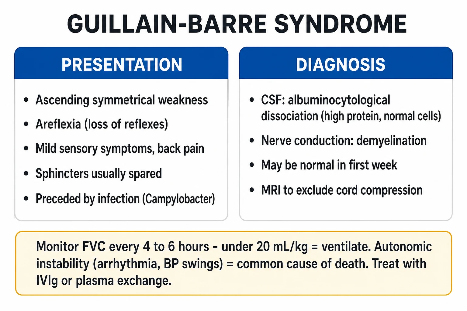
GBS is an umbrella term covering several distinct clinicopathological subtypes. They share the temporal profile (acute, monophasic, nadir within four weeks, post-infectious) but differ in the target of immune attack (myelin versus axon), the pattern of weakness, and the specific ganglioside antibody that drives the disease. Recognising the subtype matters because it predicts the electrophysiological pattern, the antecedent trigger, and the prognosis.[4]
AIDP
- 85 to 90 percent in the West
- Demyelination on NCS: prolonged distal latency, conduction block, slow velocity, temporal dispersion, prolonged F-wave
- Antibody: mixed/complement; often no specific ganglioside
- Most recover well
AMAN
- Pure motor (no sensory loss)
- Reduced CMAP amplitudes, normal sensory studies, normal velocity
- Strongly linked to Campylobacter jejuni
- Anti-GM1 / anti-GD1a positive
- Commoner in Asia; may have worse prognosis
AMSAN
- Axonal loss affecting motor and sensory fibres
- Severe weakness, prominent sensory loss and pain
- Slow recovery
- Like AMAN but with sensory involvement
MFS
- Triad: ophthalmoplegia + ataxia + areflexia
- Anti-GQ1b positive in over 90 percent
- Ataxia is sensory (proprioceptive), not cerebellar
- May overlap with GBS
- Same treatment as AIDP
BBE
- Ophthalmoplegia + ataxia + altered consciousness (drowsiness, coma)
- Hyperreflexia or Babinski sign
- Anti-GQ1b positive
- Overlaps MFS and GBS
- MRI may show brainstem signal change
PCB
- Weakness of pharynx, neck, and arms; legs spared
- Mimics myasthenia or brainstem stroke
- Often anti-GT1a positive
- Ptosis and ophthalmoplegia can occur
In India, China, Japan and Bangladesh, AMAN is as common or commoner than AIDP because of high background exposure to Campylobacter jejuni in the water and food supply. In Europe and North America, AIDP dominates at 85 to 90 percent. Examiners use this delta to test whether you recognise that the commonest subtype depends on geography.
Epidemiology & Risk Factors
GBS is rare but not negligible: it strikes about 100,000 people worldwide every year. Incidence rises steadily with age, doubling for every decade after the second, so the disease is commoner in older adults (with a smaller peak in young adults). There is a slight male predominance. The incidence can rise sharply during outbreaks — the Zika epidemics in French Polynesia (2013) and Latin America (2015 to 2016) increased GBS incidence by 5- to 20-fold in affected regions.[2][10]
The hallmark of GBS is the antecedent infection, occurring one to six weeks (usually two to three weeks) before the weakness begins. About 70 percent of patients recall a respiratory or gastrointestinal infection in that window. Identifying the trigger is more than academic — it predicts the subtype and the antibody.[4]
Campylobacter jejuni
- Commonest antecedent (25 to 30 percent of GBS)
- Preceding diarrhoea, often bloody
- Drives AMAN subtype
- Anti-GM1 / anti-GD1a
- Worse prognosis
Cytomegalovirus (CMV)
- 10 to 15 percent of GBS
- Young women, respiratory prodrome
- Often severe, sensory and cranial-nerve involvement
- Anti-GM2
Mycoplasma pneumoniae
- Especially in children
- Respiratory prodrome
- Anti-GQ1b / GalNAc-GD1a
EBV
- Infectious mononucleosis prodrome
- Anti-GM1 / anti-GM2
Influenza virus
- Respiratory prodrome
- More often AIDP phenotype
Zika virus
- Outbreaks 2013 (Polynesia), 2015 (Americas)
- AIDP-like phenotype
- 5- to 20-fold rise in GBS incidence
Hepatitis E
- Increasingly recognised trigger
- Often in travellers
Pathophysiology
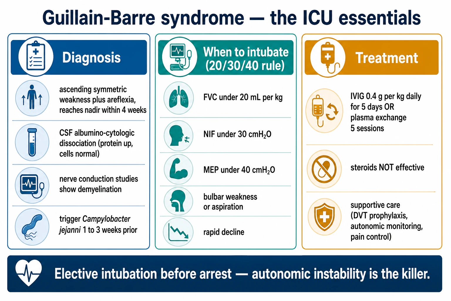
The unifying mechanism in GBS is molecular mimicry: an antecedent pathogen expresses surface molecules that structurally resemble components of the peripheral nerve, so the antibodies raised to clear the infection cross-react with the nerve. The clearest example is Campylobacter jejuni, whose lipo-oligosaccharide bears epitopes that mimic GM1 and GD1a gangliosides concentrated at the motor node of Ranvier — producing the AMAN phenotype. In Miller Fisher syndrome, the target is GQ1b (concentrated in oculomotor myelin), giving the ophthalmoplegia.[2][4]
The downstream effector cascade differs by subtype. In AIDP, antibodies and complement deposit membrane attack complex (C5b-9) on the outermost myelin lamellae of the spinal nerve roots and the peripheral nerves. Macrophages are recruited, strip the myelin, and produce segmental demyelination. Because the axon is largely preserved, conduction block predominates and is potentially reversible — which is why AIDP recovery tends to be faster and more complete.[4]
In AMAN, the antibodies bind the nodal and paranodal axolemma of the motor nerve, complement deposits, and macrophages invade the periaxonal space, detaching myelin from the axon and producing Wallerian-like axonal degeneration. The myelin sheath is structurally spared, but the axon degenerates — so recovery requires axonal regrowth at about 1 millimetre per day, which is slow and often incomplete. AMSAN applies the same mechanism to motor and sensory fibres.[4]
Three consequences of the location of attack explain the clinical picture. First, because the longest nerves are affected first, weakness ascends from the feet to the legs, then to the arms and trunk — the cardinal feature. Second, because the inflammation is in the spinal nerve root rather than the meninges, the blood-nerve barrier (and the blood-CSF barrier at the root) leaks albumin into the CSF without a cellular response — producing the albuminocytological dissociation that is the CSF signature. Third, because autonomic fibres travel in the same roots, they are damaged alongside motor and sensory fibres, producing the autonomic instability that is the leading cause of sudden death.[1]
[1]Clinical Presentation
GBS typically unfolds in four phases: a prodromal infectious illness (one to six weeks earlier), a progressive phase of weakness (days to four weeks), a plateau (days to weeks), and recovery (weeks to months, sometimes years). The cardinal clinical features of classic AIDP follow a recognizable pattern.[1][2]
Weakness is the defining feature: symmetric, ascending (begins in the feet and legs, then climbs to the thighs, trunk, arms, and face), flaccid (reduced tone), and reaching nadir within four weeks (most reach it within one to two weeks). The patient notices difficulty climbing stairs, rising from a chair, or gripping objects; severe cases become quadriparetic within days. Areflexia is early and characteristic: the deep tendon reflexes are lost (absent ankle and knee jerks, then upper-limb reflexes) and distinguishes GBS from myopathy (where reflexes are preserved early) and spinal cord disease (where they are brisk after spinal shock). Sensory symptoms are mild by comparison with motor weakness — distal paraesthesia in the hands and feet, often with prominent neuropathic back and thigh pain (sometimes the presenting complaint). Objective sensory loss is usually minor.[1]
Cranial nerve involvement is common: bilateral facial (VII) palsy in up to half of patients, bulbar weakness (dysphagia, dysarthria, weak cough — an aspiration risk), and, in Miller Fisher, ophthalmoplegia. Sphincter function is usually spared early — a key discriminator from cauda equina and acute spinal cord compression, where bladder and bowel involvement are early. Autonomic dysfunction is dangerous and under-recognised: sinus tachycardia or bradycardia, blood-pressure swings (both hypertension and hypotension), cardiac arrhythmia, paralytic ileus, urinary retention, sweating disturbance, and SIADH. It is the commonest cause of sudden death in GBS.[1][2]
Respiratory involvement — weakness of the diaphragm and intercostals — develops in 20 to 30 percent and is the indication for mechanical ventilation. Bulbar weakness adds aspiration risk. The bedside warning signs are tachypnoea, use of accessory muscles, weak cough, inability to count to 10 in one breath, and a falling serial vital capacity.[1]
Pain is under-recognised and frequently the presenting symptom before weakness becomes obvious. Patients describe deep aching back and thigh pain, painful paraesthesiae in the calves, and, in ventilated patients, pain that is difficult to localise. The pain is neuropathic (from root and nerve inflammation) and musculoskeletal (from immobility) and is often the dominant complaint in the early phase. Autonomic involvement extends beyond the cardiovascular system: paralytic ileus with abdominal distension, urinary retention (a distended bladder on admission is easily mistaken for a primary bladder problem), patchy sweating, and SIADH producing hyponatraemia. The last is common enough that serum sodium should be checked daily in the first week. [1]
Atypical presentations are deliberate examiner traps. Pure motor GBS (no sensory symptoms, often anti-GM1 positive) is distinguished from a myopathy by the rapid tempo, areflexia, and CSF. Pharyngeal-cervical-brachial GBS presents with arm and pharyngeal weakness and relative leg-sparing, mimicking brainstem stroke or myasthenia gravis. Miller Fisher syndrome may present with ataxia and diplopia alone, with no limb weakness, and can be misdiagnosed as a cerebellar or brainstem event until the reflexes are found to be absent. Bickerstaff brainstem encephalitis adds drowsiness and hyperreflexia, mimicking a posterior circulation stroke. In the elderly, GBS can present as a fall or functional decline rather than clear weakness, and in children the presenting complaint is often refusal to walk, leg pain, or ataxia — frequently misattributed to a viral illness or transient synovitis before the ascending pattern becomes clear. [1]
Classic AIDP
- Ascending symmetric weakness with areflexia
- Mild sensory symptoms, back/thigh pain
- Cranial nerve and autonomic involvement variable
- Sphincters spared early
Atypical presentation
- Pure motor GBS (no sensory loss, often anti-GM1)
- Pharyngeal-cervical-brachial variant (arms and pharynx first)
- Miller Fisher presenting with ataxia and diplopia rather than weakness
- Bickerstaff with drowsiness, hyperreflexia
- Paediatric GBS: refusal to walk, pain, ataxia
Differential Diagnosis
The differential of acute ascending flaccid paralysis is broad, and examiners test the discriminating features. The structure is: anything that causes rapidly progressive limb weakness with reduced tone must be considered — and then distinguished by pattern, tempo, reflexes, and a few targeted tests.[1][4]
Acute spinal cord lesion
- Sensory LEVEL on the trunk
- Early sphincter involvement
- Reflexes brisk (after spinal shock)
- Pyramidal signs; upgoing plantar
- MRI spine diagnostic
Botulism
- DESCENDING paralysis (cranial first)
- Dilated pupils, dry mouth
- Parasympathetic failure
- History of wound / honey / food
- Repetitive nerve stimulation increments
Tick paralysis
- Ascending weakness with areflexia — MIMICS GBS
- Tick attached (scalp, skin)
- Removal of tick cures
- Children, outdoors
Hypokalaemic periodic paralysis
- Recurrent attacks
- Painless, no sensory loss
- Areflexic during attack
- Low serum K, resolves with K replacement
- Family history
Myasthenia gravis
- Fatigable, fluctuating weakness
- Ocular predominant
- NO sensory loss, reflexes preserved
- Worse through the day
- AChR / MuSK antibody
CIDP
- Progresses BEYOND 8 weeks
- Relapsing or chronic progressive
- Long-term immunotherapy needed
- Often more sensory
Other mimics
- Vasculitic neuropathy (mononeuritis multiplex pattern)
- Porphyria (abdominal pain, seizures)
- Diphtheritic polyneuropathy (palatal palsy first)
- Lead toxicity (wrist drop)
- Organophosphate poisoning (cholinergic)
The two cannot-miss mimics are acute spinal cord compression (a surgical emergency that GBS is not) and tick paralysis (which is cured by removing the tick). The clinical discriminator from cord compression is the combination of areflexia, ascending pattern, no sharp sensory level, and sphincter sparing — if any of these is violated, image the spine.[1] Several discriminating reasoning chains are worth memorising. Acute transverse myelitis and GBS both produce leg weakness and the patient may be unable to void — but transverse myelitis has a sharp sensory level on the trunk, early bladder involvement, upgoing plantars, and initial spinal shock with flaccidity that evolves into spasticity and hyperreflexia within days, whereas GBS has no sensory level, sphincter sparing, and persistent areflexia. Botulism is the great descending-paralysis mimic: weakness begins in the cranial nerves (ptosis, diplopia, dysarthria, dysphagia) and descends symmetrically, with dilated unreactive pupils and a dry, parasympathetic-deficient picture — the opposite direction to GBS. Tick paralysis is almost indistinguishable from GBS at first (ascending areflexic weakness) and the diagnostic step is to undress the patient and search the scalp, behind the ears, and the axilla and groin for an attached tick; removal of the tick reverses the paralysis within hours to days. Hypokalaemic periodic paralysis produces painless, areflexic weakness over minutes to hours (not days), is often recurrent with a family history, has a low serum potassium during the attack, and resolves rapidly with potassium replacement.
A practical bedside rule: in any patient with rapidly progressive limb weakness, examine for a sensory level and test the plantars and sphincter tone first — these three findings alone separate the spinal cord emergencies (which need immediate MRI) from the polyneuropathies (which need CSF and NCS). An upgoing plantar, a sharp sensory level, or a distended flaccid bladder points away from GBS and toward the cord. [1]
Clinical & Bedside Assessment
The focused assessment of a suspected GBS patient has three aims: confirm the clinical pattern, quantify the weakness, and assess respiratory and bulbar reserve — because the decision to admit to ICU and to intubate is made at the bedside on serial measurements, not on a single value.[1]
Begin with ABCDE: airway (bulbar function — cough, gag, swallow), breathing (rate, depth, accessory muscle use, ability to count to ten in one breath), circulation (heart rate, blood pressure — both directions of autonomic swing), disability (GCS, pupils), and exposure (look for a tick, especially behind the ears and in the scalp). Examine the cranial nerves (bilateral facial palsy, ophthalmoplegia, palatal movement), tone and power graded with the MRC scale (0 to 5) in four limbs, reflexes (the loss of ankle and knee jerks is early and characteristic), plantar responses (usually flexor in GBS — an upgoing plantar should prompt reconsideration), sensory examination (look for a sharp level, which would argue against GBS), and sphincters (bladder volume, anal tone).[1]
FVC-DROP
The bedside respiratory measurements are the most important numbers you will take in GBS. Perform serial FVC, peak expiratory flow (PEF), and negative inspiratory force (NIF) every four to six hours in any patient with weakness progressing or affecting the arms, face, or bulbar muscles. The thresholds that demand ICU transfer and preparation for intubation are: FVC under 20 mL/kg (or under 1 L in an adult), NIF weaker than minus 30 cmH2O, a fall in FVC of more than 30 percent over 24 hours, and the clinical markers of bulbar weakness, weak cough, tachypnoea, tachycardia, or exhaustion.[1][8]
The Erasmus GBS Respiratory Insufficiency Score (EGRIS), validated internationally in the IGOS cohort, uses seven admission variables — days from onset to admission, ability to lift the head, lift the arms, lift the legs, peak flow, MRC sum score, and the presence of a cranial nerve deficit — to predict the probability of needing mechanical ventilation within one week. It does not replace serial FVC, but it flags the high-risk patient at the door.[8][9]
Investigations
GBS is a clinical diagnosis, supported by three pillars: the clinical pattern, the CSF, and nerve conduction studies. No single test is necessary to make the diagnosis, but together they confirm it, define the subtype, and exclude mimics.[1]
CSF
- Protein elevated (over 0.55 g/L; can exceed 2 g/L)
- White cells normal (under 5 per microlitre)
- May be NORMAL in the first week — repeat
- Over 50 cells suggests HIV, Lyme, meningoradiculitis
Nerve conduction
- AIDP: prolonged distal latency, conduction block, slow velocity, temporal dispersion, prolonged F-wave
- AMAN: reduced CMAP amplitude, normal sensory studies, normal velocity
- AMSAN: axonal, motor and sensory
- MFS: often normal or mild demyelination
Antibodies
- Anti-GQ1b — Miller Fisher (sensitivity over 90 percent)
- Anti-GM1, anti-GD1a — AMAN
- Not routinely needed for classic AIDP
MRI spine
- Sensory level, early sphincter involvement, pyramidal signs
- GBS may enhance the cauda equina on post-contrast MRI
Bloods
- FBC, U&E, LFT, glucose, CK (exclude myopathy)
- ECG (autonomic)
- beta-hCG in women
- Campylobacter / CMV / Mycoplasma serology
- HIV (HIV-associated GBS can have CSF pleocytosis)
The Brighton Collaboration diagnostic criteria standardise GBS diagnosis across clinical trials, vaccine-safety surveillance, and global epidemiology. They are not for everyday clinical use, but examiners use them in vaccine-safety and epidemiology stems:[3]
Level 1
- Bilateral flaccid paralysis
- PLUS nerve conduction studies consistent with GBS
- PLUS CSF albuminocytological dissociation
- PLUS detection of an antecedent infection (serology or PCR)
Level 2
- Bilateral flaccid paralysis
- PLUS CSF albuminocytological dissociation OR nerve conduction evidence of GBS
Level 3
- Bilateral flaccid paralysis (clinical syndrome alone)
- No confirmatory CSF or NCS available
Nerve conduction studies do more than confirm GBS — they define the subtype and predict the prognosis. In AIDP, the hallmarks are prolonged distal motor latencies, conduction block, slowed motor conduction velocity, temporal dispersion, and prolonged or absent F-waves — all reflecting demyelination. In AMAN, the compound muscle action potential (CMAP) amplitudes are reduced with normal sensory studies and normal conduction velocities, reflecting axonal loss. F-wave abnormalities (prolonged latency or absence) are among the earliest NCS changes because they test the proximal root, which is inflamed first.[4]
Management — Resuscitation
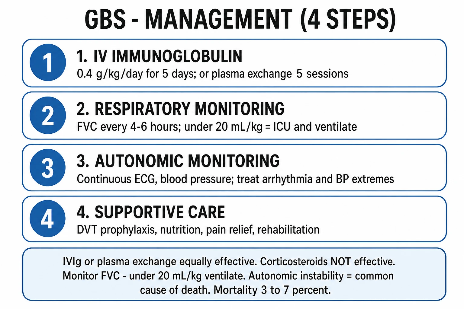
The resuscitation of GBS has one overriding principle: the patient is admitted to a monitored bed (HDU or ICU) the moment there is any respiratory, bulbar, or autonomic involvement, because the trajectory is unpredictable and 20 to 30 percent will need ventilation.[1]
The time-critical bundle on admission comprises: continuous cardiac monitoring, secure intravenous access, baseline and serial FVC, NIF and PEF every four to six hours, a plan for IVIg or plasma exchange to be started within 2 to 4 weeks of onset, DVT prophylaxis (enoxaparin 40 mg subcutaneously daily, plus compression stockings), pressure-area care, a urinary catheter if in retention, and early nasogastric feeding for nutrition and medication. Place the patient in a monitored bed; the safety-net of continuous ECG and a low threshold for vasopressor or pacing support is what prevents the sudden autonomic death.[1]
Autonomic resuscitation is the second pillar. Treat symptomatic bradycardia with atropine or isoprenaline (have a transcutaneous pacer ready); treat severe hypertension with a short-acting beta-blocker or nitrate (avoid rapid swings, which can precipitate hypotension or arrhythmia); treat paralytic ileus with nasogastric decompression; catheterise for urinary retention; and correct SIADH-related hyponatraemia with fluid restriction. Atropine and resuscitation drugs should be at the bedside throughout the admission.[1]
Aspiration prevention in bulbar GBS means nil by mouth, nasogastric or orogastric feeding, head-up positioning, and a low threshold for early intubation for airway protection. A weak cough is itself a reason to consider the ICU.[1] When intubation is needed, plan it as an elective procedure in a controlled environment. Use a rapid-sequence technique with the smallest haemodynamic swing possible: pre-oxygenase fully, choose propofol or thiopentone at reduced dose (autonomic instability makes these patients cardiovascularly sensitive), and a short-acting neuromuscular blocker such as rocuronium (with sugammadex available for reversal) or suxamethonium — note that suxamethonium carries a hyperkalaemia risk in any denervating illness after the first week, so prefer rocuronium in late-presenting GBS. Have atropine, a vasopressor (metaraminol or noradrenaline) and a transcutaneous pacer immediately available, because laryngoscopy can precipitate profound bradycardia or asystole in the dysautonomic patient. Tracheostomy is considered when ventilation is prolonged beyond 10 to 14 days; the Walgaard model predicts prolonged ventilation and can guide timing, but the decision is individualised.[1][8]
Haemodynamic and fluid management must be deliberately gentle. Aggressive fluid boluses precipitate pulmonary oedema in the dysautonomic patient; abrupt positional changes provoke blood-pressure swings; and any noxious stimulus (suctioning, turning) can trigger bradycardia. Analgesia is needed for the neuropathic pain and for procedural discomfort — fentanyl is preferred over morphine for less haemodynamic effect. Deep-vein thrombosis prophylaxis (enoxaparin 40 mg subcutaneously daily, dose-adjusted for renal function, plus graduated compression stockings) is started on admission because immobility and the prothrombotic state of inflammation combine to make PE a leading cause of death.[1]
Management — Definitive & Stepwise
The two equally effective first-line disease-modifying treatments are intravenous immunoglobulin (IVIg) and plasma exchange (plasmapheresis). The Cochrane meta-analyses confirm that each hastens recovery to the same degree, that combining them adds no benefit, and that corticosteroids alone do not work.[5][6][7]
IVIg (first-line)
- Total 2 g/kg over 5 days (0.4 g/kg/day for 5 days)
- Equally effective as plasma exchange
- Simpler — no central line, no large-volume shifts
- Preferred in haemodynamic instability
- Adverse effects: thromboembolism (stroke, MI, DVT), aseptic meningitis, AKI (sucrose formulations), flu-like
- Caution: IgA deficiency (anaphylaxis), renal impairment, hyperviscosity
Plasma exchange
- 5 exchanges (1 plasma volume, about 50 mL/kg each)
- Over 1 to 2 weeks, alternate days
- Started within 4 weeks of onset
- Adverse effects: central-line complications, haemodynamic instability, hypocalcaemia (citrate), bleeding, infection
- Avoid in sepsis, haemodynamic instability, severe coagulopathy
Corticosteroids
- Cochrane 2016: NO benefit as monotherapy
- IVIg plus methylprednisolone shows only a small, non-durable benefit
- Do NOT use — delays appropriate disease-modifying therapy
Indications for IVIg or plasma exchange: any patient unable to walk independently (MRC sum score under 40), or with rapidly progressive weakness, respiratory involvement, bulbar weakness, or autonomic instability — started within 2 to 4 weeks of onset. Mild cases who can still walk may be observed, but most clinicians treat any patient whose trajectory suggests they will not be able to walk within days.[1]
Pain management is often under-treated. First-line is gabapentin (300 to 600 mg three times daily, titrated) or pregabalin (75 to 150 mg twice daily); carbamazepine (100 to 200 mg twice daily) for paroxysmal neuropathic pain; opioids (tramadol 50 to 100 mg, morphine 2.5 to 5 mg) for severe pain. NSAIDs alone are inadequate for neuropathic pain. Treat the pain actively — it is a major contributor to distress and immobility.[1]
The multidisciplinary rehabilitation pathway begins in the acute phase: physiotherapy (range of movement, splinting to prevent foot drop and contractures, mobilisation as weakness recovers), occupational therapy (activities of daily living, aids, home assessment), speech and language therapy (swallow assessment, communication), dietetics (nutrition — nasogastric early, PEG if prolonged), psychology (anxiety, depression, PTSD are common), and early discharge planning for community rehabilitation.[1]
Specific Subtypes & Scenarios
Each subtype carries its own traps. Miller Fisher syndrome (MFS) presents with the triad of ophthalmoplegia, ataxia, and areflexia; the ataxia is sensory (proprioceptive) rather than cerebellar (no dysarthria or nystagmus), and anti-GQ1b is positive in over 90 percent. MFS may progress to overlap with GBS (limb weakness) and is treated the same way — IVIg or plasma exchange. Bickerstaff brainstem encephalitis (BBE) overlaps MFS but adds altered consciousness (drowsiness, coma) and hyperreflexia or a Babinski sign; the same anti-GQ1b antibody is positive, MRI may show brainstem signal change, and treatment is identical.[2][4]
AMAN (acute motor axonal neuropathy) is pure motor (no sensory loss), shows reduced CMAP amplitudes with normal sensory studies and normal velocities on NCS, is strongly linked to Campylobacter jejuni and anti-GD1a / anti-GM1 antibodies, and is commoner in Asia. Because the target is the axon rather than the myelin, recovery depends on axonal regrowth and is slower and sometimes less complete than AIDP. AMSAN applies the same axonal mechanism to motor and sensory fibres, producing severe weakness with prominent sensory loss and neuropathic pain.[4]
The pharyngeal-cervical-brachial variant produces weakness of the pharynx, neck, and arms with relative sparing of the legs; it can mimic myasthenia gravis or brainstem stroke, often has anti-GT1a antibodies, and may include ptosis and ophthalmoplegia. Pure motor GBS (no sensory loss, often anti-GM1 positive) overlaps AMAN phenotypically but may show demyelination; differentiation is by NCS.[4]
Paediatric GBS is commoner in children aged 1 to 10 years and presents with refusal to walk, leg pain, ataxia, and weakness — often misdiagnosed initially as a limp or viral myositis. Recovery is usually faster and more complete than in adults; treatment is IVIg at 2 g/kg total (0.4 g/kg/day for 5 days), weight-based as in adults.[1]
GBS in pregnancy is managed unchanged — IVIg is safe in pregnancy and plasma exchange is also possible. The peripartum period carries additional DVT risk warranting prophylactic LMWH (enoxaparin 40 mg daily), and there should be multidisciplinary planning with obstetrics and anaesthetics. The fetus is not directly affected.[1]
Treatment-related fluctuation (TRF) — a relapse after initial improvement within 8 weeks — occurs in up to 10 percent. A repeat course of IVIg is reasonable. If fluctuations continue beyond 8 weeks, investigate for CIDP, which requires long-term immunotherapy.[1]
Complications & Pitfalls
The complications of GBS are what kill and disable patients, and most are preventable with meticulous supportive care. Respiratory failure requiring ventilation develops in 20 to 30 percent; mean ventilation duration is 2 to 3 weeks. Autonomic dysfunction (arrhythmia, blood-pressure swings, paralytic ileus, urinary retention, SIADH) is the leading cause of sudden death — continuous ECG monitoring is mandatory. Venous thromboembolism (DVT, PE) from immobility requires prophylactic LMWH and compression stockings. Aspiration pneumonia from bulbar weakness needs airway protection and early feeding via nasogastric tube. Critical illness polyneuropathy and myopathy can compound GBS in the ICU. Pressure sores, contractures, and foot drop from prolonged immobility are prevented by pressure-area care, splinting, and physiotherapy. Hyponatraemia from SIADH is common and needs fluid restriction. Depression, anxiety, and PTSD are under-recognised and warrant psychological support. About 20 percent of patients are left unable to walk independently at six months.[1][2]
The classic pitfalls deserve direct naming:[1]
Normal first-week CSF
- Does NOT exclude GBS
- Albuminocytological dissociation in only 50 to 66 percent in week 1
- Repeat the LP after 1 to 2 weeks
FVC alone
- A patient with bulbar weakness, weak cough, or rapid progression may need intubation even with FVC just above 20 mL/kg
- Trend and clinical picture matter — intubate early and electively
Corticosteroids
- Not effective in GBS
- Delay appropriate disease-modifying therapy
- Do NOT use
Cauda equina mimic
- GBS has areflexia, ascending weakness, sphincter sparing
- Cauda equina has sacral sensory level and sphincter tone loss
- Emergency MRI spine differentiates
IVIg thromboembolism
- Stroke, MI, DVT, PE — especially in elderly and cardiovascular risk
- Slow infusion, hydration, monitor
HIV-associated GBS
- May show CSF pleocytosis
- Can occur at seroconversion or late disease
- Exclude CMV polyradiculopathy in advanced HIV
Prognosis & Disposition
Most patients reach nadir within 2 to 4 weeks, then enter a plateau of days to weeks, then recover over weeks to months (sometimes years for full recovery). About 80 percent recover fully or with minor residual deficits; about 20 percent have significant residual disability (unable to walk independently at 6 months). Mortality is 3 to 7 percent even with modern care, and the leading causes are autonomic dysfunction, respiratory failure, sepsis, and pulmonary embolism.[1][2]
The poor prognostic factors are the variables that load the EGOS and mEGOS: older age, antecedent diarrhoea (Campylobacter, often AMAN), severe weakness at nadir (MRC sum score under 40), need for mechanical ventilation, axonal physiology on NCS, delayed treatment, and the EGRIS-predicted need for ventilation. The modified Erasmus GBS Outcome Score (mEGOS) at 7 days uses age, antecedent diarrhoea, MRC sum score, and its change over the first week to predict the inability to walk at four weeks and six months — the most useful bedside counselling tool.[8][9]
Disposition follows the trajectory. Any patient with respiratory involvement, rapid progression, severe weakness, bulbar weakness, or autonomic instability goes to HDU or ICU. Stable, mild cases (still walking, slow progression) may be managed on a neurology ward with serial FVC. After the acute phase, a rehabilitation unit supports recovery of mobility and function; community rehabilitation continues for residual deficits.[1]
The safety-net and follow-up includes serial FVC during recovery, monitoring for treatment-related fluctuation (re-treat with IVIg if relapse within 8 weeks), evaluation for CIDP if symptoms continue beyond 8 weeks, early physiotherapy, and vaccination advice: avoid the specific trigger vaccine if it preceded onset (within 6 weeks); otherwise vaccination, including influenza, remains safe and recommended.[1]
Special Populations
Paediatric
- Aged 1 to 10; presents with refusal to walk, pain, ataxia
- Recovery usually faster and more complete
- IVIg 2 g/kg total (0.4 g/kg/day x 5 days), weight-based
Pregnancy
- IVIg is safe in pregnancy
- Management unchanged
- Prophylactic LMWH (enoxaparin 40 mg daily) for DVT risk
- Multidisciplinary with obstetrics, anaesthetics
Elderly
- Worse prognosis, higher mortality
- Higher rates of respiratory failure and autonomic complications
- Early ICU referral; monitor renal function with IVIg (AKI risk)
HIV / immunocompromised
- HIV-associated GBS may show CSF pleocytosis
- Can occur at seroconversion or late disease
- Exclude CMV polyradiculopathy in advanced HIV
- Treat the same way
Anticoagulated
- Caution with LMWH DVT prophylaxis (adjust for anticoagulant)
- Plasma exchange needs careful coagulation management
- IVIg preferred
Prior GBS
- Lifetime recurrence risk about 3 to 7 percent
- Avoid the specific trigger vaccine if identified
- IVIg safe for any recurrence
Evidence, Guidelines & Regional Differences
The landmark evidence base for GBS is dominated by three Cochrane meta-analyses and the Brighton criteria. The Cochrane IVIg review (Hughes 2012) showed that IVIg hastens recovery as much as plasma exchange, with the advantage of simpler administration and fewer lines. The Cochrane plasma exchange review (Raphael 2012) confirmed that plasma exchange improves outcome versus supportive care alone, with benefit strongest when started within two to four weeks. The Cochrane corticosteroid review (Hughes 2016) is the definitive evidence that corticosteroids alone are not effective — the combination of IVIg plus methylprednisolone showed only a small, non-durable benefit that does not justify routine use.[5][6][7]
The Brighton Collaboration diagnostic criteria (Fokke 2014) were developed to standardise GBS diagnosis across clinical trials, vaccine-safety surveillance, and global epidemiology, and were validated prospectively in a large Dutch cohort. They are not for everyday clinical use, but examiners use them in vaccine-safety and epidemiology stems.[3]
The PSVE study (Plasma Exchange / Sandoglobulin GBS Trial) and subsequent meta-analyses established the equivalence of IVIg and plasma exchange — choice depends on availability, comorbidity, and centre preference. The combination of IVIg plus methylprednisolone showed only a small, non-reproduced, non-durable short-term benefit and is not recommended routinely.[5][7]
The controversial second course of IVIg (SID-GBS and I-SID GBS trials) is not routinely recommended; it is reserved for patients with a poor prognosis and ongoing deterioration after the first course, guided by the mEGOS at 7 days.[4]
The International GBS Outcome Study (IGOS), a global prospective cohort, has validated the EGRIS and mEGOS scores across continents and refined the understanding of subtype distribution worldwide.[9]
In India, China, Japan and Bangladesh, AMAN is commoner than AIDP because of high Campylobacter exposure. In the West, AIDP dominates at 85 to 90 percent. IVIg availability and cost are practical barriers in low-resource settings, where plasma exchange (cheaper per session but needs a central line and a machine) may be the realistic first-line — both are equally effective. Examiner stems use this delta to test cost-awareness and global neurology.
The weak evidence for complement-inhibitor therapies (eculizumab, ravulizumab) in severe GBS — early-phase studies only, not standard care — is mentioned for completeness; do not invoke these as first-line.[4]
Exam Pearls
The six facts that decide a GBS answer
- GBS = acute, monophasic, immune-mediated polyradiculoneuropathy; symmetric ascending flaccid paralysis with areflexia; reaches nadir within 4 weeks; preceded by infection in 70 percent (Campylobacter jejuni commonest).
- CSF = albuminocytological dissociation (high protein, normal cells); may be normal in the first week — repeat.
- NCS defines the subtype: AIDP = demyelination; AMAN = axonal motor, reduced CMAP, normal sensory.
- Antibodies: anti-GQ1b = Miller Fisher; anti-GM1 / anti-GD1a = AMAN.
- Treat: IVIg (0.4 g/kg/day for 5 days, total 2 g/kg) OR plasma exchange (5 sessions); equally effective, never combine; corticosteroids do NOT work.
- Monitor serial FVC; under 20 mL/kg or under 1 L = intubate. Autonomic instability is the commonest cause of death.
4-WEEK WALL
Exam application bank (NEET-PG / INICET)
One-line answer
Guillain-Barré syndrome (GBS) is an acute, monophasic, immune-mediated polyradiculoneuropathy producing symmetric, ascending flaccid paralysis with areflexia, progressing to a nadir within four weeks and often preceded by an infection one to six weeks earlier. The commonest subtype is acute inflammatory demyelinating polyradiculoneuropathy (AIDP, 85 to 90 percent in Western countries), with axonal motor (AMAN) forms commoner in Asia. The antecedent trigger is classically Campylobacter jejuni gastroenteritis, but cytomegalovirus, Epstein-Barr virus, Mycoplasma pneumoniae, influenza and Zika are also recognised. Diagnosis is clinical, supported by CSF albuminocytological dissociation (high protein, normal cell count) and nerve conduction studies showing demyelination or axonal loss. Twenty to thirty percent of patients need mechanical ventilation, and autonomic instability is the leading c
Worked stems (answer without another resource)
Stem 1 — Classic presentation. Map symptoms to mechanism; name the first investigation and first treatment step with dose/route if drug therapy is standard. [1]
Stem 2 — Unstable / complicated. List red flags that force immediate resuscitation, theatre, ICU, antidote, or reperfusion — and what you do in the first 15 minutes. [1]
Stem 3 — Atypical group. Elderly, pregnancy, child, or immunocompromised: how presentation and thresholds change. [1]
Stem 4 — Differential trap. Name the three closest mimics and one discriminator for each. [1]
Stem 5 — Disposition. Who goes home with safety-netting, who is admitted, who needs HDU/ICU/theatre, and what follow-up is mandatory. [1]
Rapid viva checklist
- Definition + classification
- Pathophysiology chain
- Bedside signs / criteria
- Score with exact components (if any)
- Emergency bundle
- Definitive therapy with doses
- Complications of disease and of treatment
- Special populations
- Guideline/trial name if classic
- Three exam traps
Coverage self-check
If you cannot answer any stem above from this page alone, re-read the matching section — the page is intended to be self-sufficient for final-prof and NEET-PG/INICET questions on Guillain-Barré Syndrome.
References
- [1]Leonhard SE, Mandarakas MR, Gondim FAA, et al. Diagnosis and management of Guillain-Barré syndrome in ten steps Nat Rev Neurol, 2019.PMID 31541214
- [2]Willison HJ, Jacobs BC, van Doorn PA. Guillain-Barré syndrome Lancet, 2016.PMID 26948435
- [3]Fokke C, van den Berg B, Drenthen J, Walgaard C, van Doorn PA, Jacobs BC. Diagnosis of Guillain-Barré syndrome and validation of Brighton criteria Brain, 2014.PMID 24163275
- [4]van den Berg B, Walgaard C, Drenthen J, Fokke C, Jacobs BC, van Doorn PA. Guillain-Barré syndrome: pathogenesis, diagnosis, treatment and prognosis Nat Rev Neurol, 2014.PMID 25023340
- [5]Hughes RA, Swan AV, van Doorn PA. Intravenous immunoglobulin for Guillain-Barré syndrome Cochrane Database Syst Rev, 2012.PMID 22786476
- [6]Raphael JC, Chevret S, Hughes RA, Annane D. Plasma exchange for Guillain-Barré syndrome Cochrane Database Syst Rev, 2012.PMID 22786475
- [7]Hughes RA, Brassington R, Gunn AA, van Doorn PA. Corticosteroids for Guillain-Barré syndrome Cochrane Database Syst Rev, 2016.PMID 27775812
- [8]Walgaard C, Lingsma HF, Ruts L, et al. Prediction of respiratory insufficiency in Guillain-Barré syndrome Ann Neurol, 2010.PMID 20517939
- [9]Doets AY, Walgaard C, Lingsma HF, et al.; IGOS Consortium. International Validation of the Erasmus Guillain-Barré Syndrome Respiratory Insufficiency Score Ann Neurol, 2022.PMID 35106830
- [10]Walteros DM, Soares J, Styczynski AR, et al.; Sejvar JJ. Long-term outcomes of Guillain-Barré syndrome possibly associated with Zika virus infection PLoS One, 2019.PMID 31369576