Neurology · Neurology
Meningitis and Encephalitis
Also known as Bacterial meningitis · Viral meningitis · Aseptic meningitis · Herpes simplex encephalitis · Meningococcal meningitis · TB meningitis · Cryptococcal meningitis · Anti-NMDA receptor encephalitis
Meningitis is inflammation of the meninges, most often infective; encephalitis is inflammation of the brain parenchyma, and meningoencephalitis is both together. Bacterial meningitis is a time-critical emergency — fever plus headache plus neck stiffness plus altered mental status is bacterial meningitis until proven otherwise, and empirical therapy (ceftriaxone 2 g IV q12h plus vancomycin plus dexamethasone 10 mg IV q6h, add ampicillin if over 50 or immunocompromised) must start within one hour, before lumbar puncture or imaging. Herpes simplex encephalitis is the treatable encephalitis not to miss — fever plus altered mental status plus seizures needs empirical IV aciclovir 10 mg/kg q8h for 14 to 21 days, without waiting for PCR. CSF analysis (cell count, glucose, protein, Gram stain, culture, PCR) distinguishes bacterial, viral, tuberculous and fungal causes.
On this page & tools
Your progress
Saved locally on this device.
Practise this topic
Exam tags
Red flags
Overview & Definition
Meningitis is inflammation of the meninges — the three protective membranes (dura mater, arachnoid mater, pia mater) that envelop the brain and spinal cord — most often caused by infection but occasionally by malignancy, drugs, or autoimmunity. Encephalitis is inflammation of the brain parenchyma itself, again usually infective (predominantly viral) or autoimmune. When both the meninges and the parenchyma are inflamed together — common in severe viral disease — the syndrome is termed meningoencephalitis, and the clinical features of meningism and encephalopathy overlap.[1][4]
The reason these two diagnoses dominate emergency neurology is that they are common, lethal, and treatable. Acute bacterial meningitis kills 10 to 30 percent of adults within days and leaves another 30 to 50 percent of survivors with permanent neurological sequelae — sensorineural hearing loss, epilepsy, cognitive impairment, and motor deficit. The single most important determinant of survival is the time to antibiotics: every hour of delay in the first few hours after presentation measurably increases mortality. For this reason the discipline of practice is to treat first and investigate second. The classical phrase "meningitis until proven otherwise" exists to make clinicians act before the cerebrospinal fluid (CSF) is back.[1][4][7]
Viral meningitis, in contrast, is usually a self-limiting, aseptic illness (enteroviruses cause over 80 percent) that needs supportive care only. Viral encephalitis, however, is dangerous: herpes simplex virus type 1 (HSV-1) accounts for about 10 to 15 percent of all encephalitis and is the single most important treatable cause. Untreated HSV encephalitis kills over 70 percent of patients; with prompt aciclovir, mortality falls to under 30 percent. The combination of a high pre-treatment mortality, a cheap and effective drug, and a treatable window makes HSV the central fact of the encephalitis workup — every patient with suspected encephalitis receives empirical aciclovir until HSV PCR returns negative.[3][6]
Classification
Central nervous system (CNS) infections are classified along two axes — the anatomical compartment involved (meninges versus parenchyma) and the aetiological agent (bacterial, viral, fungal, parasitic, or non-infectious). The anatomical axis determines the clinical syndrome; the aetiological axis determines the drug. [1]
Bacterial meningitis
the emergency
- *Streptococcus pneumoniae* — 50 to 70 percent of adult community-acquired cases
- *Neisseria meningitidis* — 10 to 20 percent; adolescents, young adults, meningococcal belt
- *Listeria monocytogenes* — 5 to 10 percent; over 50 years, immunocompromised, neonate, pregnant
- Group B streptococcus and *E. coli* — neonates (K1 capsular type)
- *Haemophilus influenzae* type b — now rare where conjugate vaccination is routine
Viral (aseptic) meningitis
usually self-limiting
- Enteroviruses (coxsackie, echovirus) — over 80 percent, summer/autumn
- HSV-2 (Mollaret meningitis), VZV, EBV, CMV, HHV-6
- HIV (acute seroconversion illness)
- Mumps (uncommon with MMR), arboviruses (West Nile, dengue)
Viral encephalitis
HSV is the treatable one
- HSV-1 — 10 to 15 percent; treatable with aciclovir; temporal lobe predilection
- VZV, EBV, enteroviruses, CMV (immunocompromised)
- Arboviruses — Japanese encephalitis (Asia), West Nile (North America), tick-borne (Europe), Zika/dengue
- Rabies — universally fatal once symptomatic
- Nipah, Hendra, Eastern/Western equine — region-specific
Fungal / TB / parasitic
subacute, immunocompromised
- *Cryptococcus neoformans/gattii* — HIV with low CD4, transplant
- *Mycobacterium tuberculosis* — subacute basilar meningitis
- *Coccidioides*, *Histoplasma* — endemic regions
- Cerebral malaria (*Plasmodium falciparum*) — travellers
- Toxoplasmosis — HIV with CD4 under 100
Non-infectious / autoimmune
do not forget
- Anti-NMDA receptor encephalitis — young women, ovarian teratoma
- Anti-LGI1, anti-CASPR2, anti-GAD, anti-GABA-A/B, anti-AMPAR
- Carcinomatous / lymphomatous meningitis (malignant)
- Drug-induced (NSAIDs, TMP-SMX, IVIG, vaccines — aseptic)
- Neuro sarcoidosis, SLE, Behcet, Vogt-Koyanagi-Harada
The relative frequency of pathogens is region-dependent. In the meningococcal belt of sub-Saharan Africa (the Sahel), N. meningitidis serogroup A (now W and X) causes dry-season epidemics with attack rates up to 1,000 per 100,000. In South and Southeast Asia, Streptococcus suis (pig contact) and Japanese encephalitis virus are important. In the United States, West Nile virus is the leading arboviral encephalitis in late summer. In northern Europe and Russia, tick-borne encephalitis (TBE) follows forest exposure. Always take a travel, animal, and exposure history.
Epidemiology & Risk Factors
Bacterial meningitis has an annual incidence of roughly 1 to 5 per 100,000 in high-income countries and far higher where vaccination is incomplete. After the introduction of conjugate Haemophilus influenzae type b (Hib), pneumococcal conjugate (PCV13) and meningococcal vaccines into childhood immunisation schedules, the epidemiology has shifted: Hib meningitis has nearly disappeared in children, the median age of bacterial meningitis has risen, and S. pneumoniae has become the dominant adult pathogen. Meningococcal disease peaks in adolescents and young adults (15 to 24 years) and in the meningococcal belt during the dry season (December to June).[4][5]
Viral meningitis is far commoner than bacterial (tens of thousands of cases per year), is dominated by enteroviruses in summer and autumn, and carries an excellent prognosis. Viral encephalitis has an annual incidence of roughly 5 to 10 per 100,000; HSV is the most frequently identified cause in adults year-round, while arboviruses are seasonal and geographically delimited. Autoimmune encephalitis, particularly anti-NMDA receptor, is increasingly recognised and in young people may exceed any single viral cause.[6][10]
The modifiable and host risk factors cluster into groups that mirror the empirical antibiotic choice: [1]
Host susceptibility
- Extremes of age — neonate (immature immunity), elderly (waned immunity)
- Immunocompromise — HIV, transplant, chemotherapy, asplenia, alcohol use disorder, diabetes, malignancy
- Pregnancy — increased Listeria risk
- Complement deficiency (C5–C9), properdin deficiency, hypogammaglobulinaemia — recurrent meningococcal/pneumococcal
- Cochlear implant — especially pneumococcal
Anatomical breach
- CSF leak — skull base fracture, post-neurosurgery, CSF shunt
- Otic or sinus source — otitis media, mastoiditis, sinusitis, dental infection
- Dural defect after trauma or surgery
- Congenital dermal sinus, meningomyelocele
Exposure / contact
- Meningococcal — household, dormitory, kissing, Hajj pilgrimage
- Listeria — soft cheese, unpasteurised milk, deli meats, smoked fish
- Enterovirus — daycare, summer outbreaks
- TB — household/contact
- Arbovirus — mosquito/tick bite, travel; rabies — animal bite
Pathophysiology
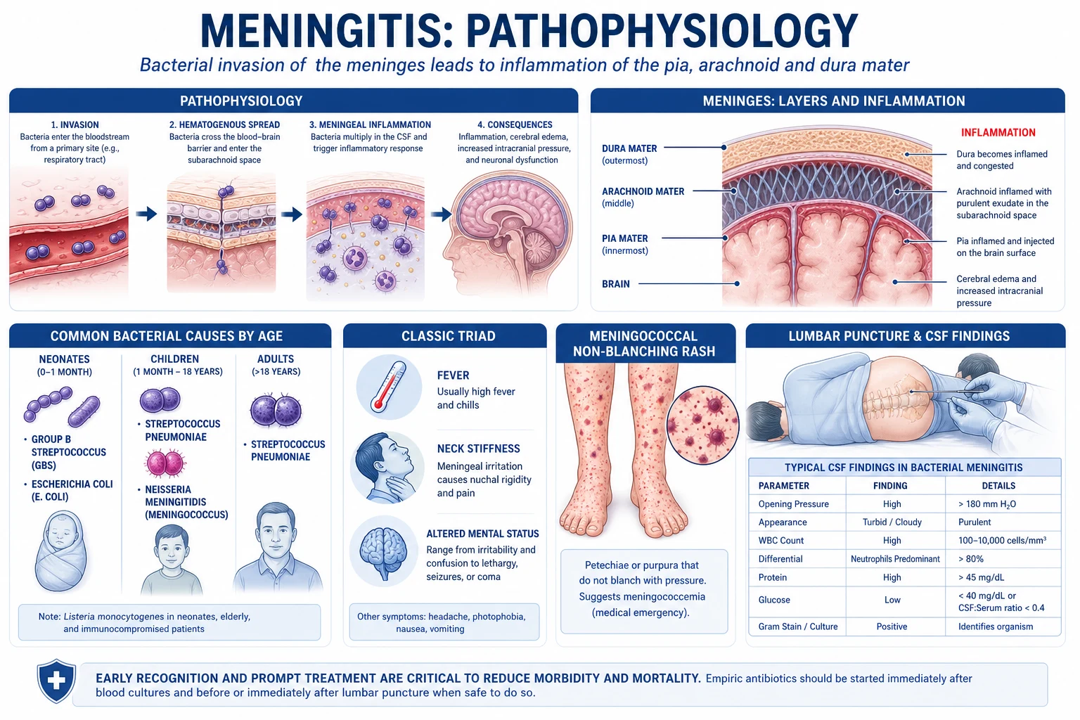
Bacteria reach the subarachnoid space by three principal routes: haematogenous spread (the dominant pathway — colonisation of the nasopharynx followed by invasion across the mucosa, bacteraemia, and crossing the blood–brain barrier at the choroid plexus or cerebral capillaries); direct contiguous extension from sinusitis, otitis media, mastoiditis, or a dental focus; and direct inoculation from skull fracture, neurosurgery, lumbar puncture, or a CSF shunt. Once in the subarachnoid space, bacteria multiply rapidly because the CSF has low concentrations of complement and immunoglobulin and no effective cellular immune surveillance — a privileged niche that becomes their growth medium.[1][4]
The clinical damage is driven less by the bacteria themselves than by the host inflammatory response. Bacterial cell-wall components — pneumococcal lipoteichoic acid and pneumolysin, meningococcal and Gram-negative lipopolysaccharide (endotoxin) — activate Toll-like receptors (TLR-2, TLR-4) on meningeal macrophages and brain microglia. This releases a cytokine cascade — tumour necrosis factor-alpha (TNF-alpha), interleukin-1 (IL-1), interleukin-6 (IL-6), interleukin-8 (IL-8) — that recruits neutrophils, upregulates adhesion molecules, opens the blood–brain barrier, and increases vascular permeability. The consequences are neutrophilic exudate in the subarachnoid space, cerebral oedema (vasogenic and cytotoxic), raised intracranial pressure, vasculitis of perforating vessels producing cerebral infarction, impairment of CSF reabsorption at the arachnoid granulations producing hydrocephalus, and direct neuronal injury. This single cascade is why dexamethasone (which dampens the cytokine response) works — and works only if given before or with the first antibiotic dose, before the inflammatory cascade is fully triggered.[1][4][7]
HSV-1 reaches the brain by a different and elegant route. After primary infection (usually orolabial), the virus establishes latency in the trigeminal ganglion. Reactivation periodically sends virus in a retrograde axonal transport along the trigeminal and olfactory nerves to their cortical terminations — the frontotemporal cortices and limbic structures. This neuroanatomy explains the striking temporal lobe predilection of HSV encephalitis, the haemorrhagic necrosis that is its pathology, and the clinical features (aphasia, behaviour change, complex partial seizures, memory disturbance) that result. Untreated, the virus destroys the temporal cortices bilaterally and is uniformly fatal; aciclovir halts viral DNA polymerase and rescues tissue that has not yet necrosed.[3]
Tuberculous and cryptococcal meningitis follow a subacute tempo because their pathogens replicate slowly and elicit a lymphocytic, low-grade, basilar inflammation. TB organisms lodge in the basal meninges and form a thick gelatinous exudate that encases the cranial nerves and obstructs CSF flow at the basal cisterns, producing cranial nerve palsies and communicating hydrocephalus, and causing a vasculitis of the lenticulostriate and thalamoperforating vessels with basal ganglia infarcts. Cryptococcus reaches the brain via the capsule of yeasts crossing the choroid plexus and accumulates in the Virchow–Robin spaces as "soap-bubble" lesions; it secretes capsular polysaccharide that impairs phagocytosis and produces a strikingly high CSF opening pressure.[4][9]
Clinical Presentation
The clinical syndromes of meningitis and encephalitis differ in one decisive feature: the level of consciousness and the presence of parenchymal signs. Meningitis (especially bacterial) preserves higher function until late; encephalitis disturbs it early. [1]
Bacterial meningitis
The classic triad of fever, neck stiffness and altered mental status is present in only about 40 to 50 percent of adults at presentation; adding headache raises sensitivity to roughly 95 percent, so that almost every patient has at least two of fever, headache, neck stiffness or confusion. The full syndrome comprises:[1][4]
- Fever — present in 80 to 95 percent (may be absent in the elderly, immunocompromised, or after self-administered antibiotics).
- Headache — severe, often holocranial, the most sensitive individual symptom (over 90 percent).
- Neck stiffness — resistance to passive flexion; meningism appears over hours.
- Photophobia and phonophobia.
- Altered mental status — drowsiness, confusion, agitation; at the severe end, coma.
- Nausea and vomiting — from raised intracranial pressure or meningeal irritation.
- Seizures — focal or generalised, in 15 to 30 percent, commoner with S. pneumoniae.
- Focal neurological deficits — cranial nerve palsies (especially III, IV, VI, VII, VIII), hemiparesis from vasculitic infarction.
- Rash — petechial or purpuric, non-blanching, in 50 to 75 percent of meningococcal cases; a vital sign that mandates immediate treatment. [1]
Meningococcal septicaemia
the rash saves lives
- Petechial or purpuric rash — non-blanching on glass test; check skin, conjunctiva, palate
- Rapid progression to purpura fulminans and Waterhouse–Friderichsen (adrenal haemorrhage)
- Septic shock, DIC, multi-organ failure within hours
- Treat immediately; notify public health; chemoprophylaxise close contacts
Pneumococcal meningitis
highest mortality
- Often follows otitis media, sinusitis, pneumonia, or CSF leak
- Highest case-fatality (20 to 30 percent or more)
- Dexamethasone benefit greatest here; vancomycin added for resistant strains
- Frequent seizures and cranial nerve palsies; hearing loss in survivors
Listeria monocytogenes
rhombencephalitis pattern
- Over 50 years, pregnant, immunocompromised, alcohol use, neonate
- May cause brainstem rhombencephalitis with cranial nerve palsies and ataxia
- Inherently resistant to all cephalosporins — MUST add ampicillin empirically
- Food-borne (soft cheese, deli meats, unpasteurised milk)
Encephalitis
Encephalitis presents with fever plus altered consciousness plus seizures plus focal neurological deficit. The altered mental status is the cardinal feature distinguishing encephalitis from meningitis — a patient with pure viral meningitis is uncomfortable but lucid; a patient with encephalitis is confused, obtunded, or comatose. Seizures are commoner than in meningitis. The focal signs reflect the site of parenchymal involvement.[3][6]
HSV encephalitis classically produces a temporal lobe syndrome: aphasia (dominant hemisphere), behaviour and personality change, anosmia, complex visual or olfactory hallucinations, focal seizures arising from the temporal lobe, and memory disturbance (Korsakoff-like state in survivors). Anti-NMDA receptor encephalitis (more common in young women, often with an ovarian teratoma) follows a characteristic sequence: a viral-like prodrome, then psychiatric symptoms (psychosis, agitation, paranoia), then seizures, dyskinesias (orofacial, choreoathetoid), autonomic instability (hyperthermia, tachycardia, hypertension, hypoventilation) and decreased consciousness, often progressing to require prolonged ICU care.[10]
Atypical presentations (examiners deliberately test these)
Neonate
- Non-specific: fever or hypothermia, irritability, lethargy, poor feeding, vomiting
- Bulging fontanelle late; neck stiffness often ABSENT
- Apnoea, seizures, jaundice; rapid deterioration
- Pathogens: group B strep, E. coli K1, Listeria; treat with ampicillin plus cefotaxime or gentamicin
Elderly
- Subtle: confusion or falls without fever; meningeal signs often absent
- Listeria and pneumococcus predominate; broader empirical cover needed
- Comorbidity masks the picture; high threshold to LP and low threshold to add ampicillin
- Worse outcomes; delirium may be the only sign
Immunocompromised
- HIV with low CD4: Cryptococcus, TB, toxoplasmosis, CMV, syphilis
- Transplant: Listeria, Cryptococcus, HHV-6, West Nile
- Neutropenic: Gram-negative bacilli, *Pseudomonas*, *Listeria*
- Symptoms blunted; CSF may be remarkably bland — diagnosis requires PCR and antigen tests
Partially treated
- Antibiotics before LP reduce Gram stain and culture yield to under 50 percent
- Pneumococcal antigen and bacterial PCR still detect organisms
- Clinical picture persists; do not be reassured by a negative CSF culture alone
Differential Diagnosis
The differential of fever plus headache plus altered mental status is broad and contains several mimics that demand specific treatment. The high-yield framework separates infectious CNS disease from non-infectious causes. [1]
Other CNS infections
- **Brain abscess** — focal ring-enhancing lesion; often with sinus/ear/dental source; LP is CONTRAINDICATED (herniation)
- **Subdural / epidural empyema** — focal, post-sinusitis or post-surgery; needs MRI and drainage
- **Cerebral malaria** — travel to endemic area; blood film; artesunate
- **Cerebral toxoplasmosis** — HIV with ring-enhancing lesions; toxo serology
Vascular / structural
- **Subarachnoid haemorrhage** — thunderclap headache; CT blood in basal cisterns; xanthochromia
- **Cerebral venous sinus thrombosis** — headache, seizures, focal deficit; procoagulant state
- **Stroke** — ischaemic or haemorrhagic; PRES; RCVS
- **Cerebral vasculitis**, **reversible posterior leucoencephalopathy**
Metabolic / toxic
- **Metabolic encephalopathy** — hepatic, uraemic, hyponatraemic, hypoglycaemic, hypoxic
- **Sepsis-associated encephalopathy** — systemic infection without CNS invasion
- **Drug toxicity** — neuroleptic malignant syndrome, serotonin syndrome, anticholinergic, alcohol withdrawal
- **Wernicke encephalopathy** — give thiamine before glucose
Autoimmune / other
- **Autoimmune encephalitis** — anti-NMDAR, anti-LGI1, anti-CASPR2, anti-GABA, anti-AMPAR, anti-GAD, anti-Ma2
- **Paraneoplastic limbic encephalitis** — anti-Hu, anti-Ma2, anti-Yo
- **ADEM** (acute disseminated encephalomyelitis) — post-infectious, demyelinating
- **Neuro-sarcoidosis, SLE cerebritis, Behcet, Hashimoto encephalopathy**
The decisive can't-miss mimics in the acute setting are subarachnoid haemorrhage (CT), brain abscess (CT/MRI — never LP if a mass lesion is present), cerebral malaria (blood film in the traveller), and autoimmune encephalitis (CSF antibody panels, MRI). All patients with suspected encephalitis should also receive empirical antibiotics until bacterial meningitis is excluded, and all patients with suspected HSV encephalitis should receive aciclovir until PCR returns negative.[3][6]
Clinical & Bedside Assessment
The focused examination in suspected CNS infection has four aims: to confirm the syndrome (meningism, encephalopathy, rash), to search for a focus (ear, sinus, skin, heart), to identify red flags mandating CT before LP, and to stage severity (GCS, sepsis) for disposition. [1]
Named meningeal signs (low sensitivity 5 to 30 percent, but high specificity when present): [1]
MENINGES
- Kernig sign — with the patient supine and the hip and knee flexed to 90 degrees, attempted extension of the knee meets resistance or produces pain in the back/hamstrings (stretch on inflamed meninges/nerve roots).
- Brudzinski sign (nape of the neck) — passive flexion of the neck produces involuntary flexion of the hips and knees.
- Brudzinski sign (contralateral / cheek) — flexion of one hip causes flexion of the contralateral leg; pressure on the cheek produces forearm flexion.
- Jolt accentuation — exacerbation of an existing headache on horizontal rotation of the head twice per second; more sensitive than Kernig/Brudzinski for meningitis. [1]
A full examination must include: vital signs (fever, tachycardia, hypotension, tachypnoea, oxygenation, capillary refill — septic shock), GCS, pupils, fundoscopy for papilloedema (a red flag for raised ICP — do not LP), cranial nerves, motor and sensory exam, coordination, skin (rash, bite, chancre, erythema migrans, Janeway lesions), ears/nose/throat (otitis, mastoid tenderness, sinus tenderness, parotid), neck stiffness, chest (pneumonia, murmur of endocarditis), abdomen (hepatosplenomegaly, splenomegaly of asplenia absent), lymph nodes, joints, and signs of immunocompromise (oral candidiasis, Kaposi sarcoma, wasting, needle marks). In suspected meningococcal disease, repeat the skin examination every 15 minutes — the rash can evolve rapidly.[1][4]
Investigations
Bedside and bloods
Bloods: full blood count (leucocytosis in bacterial; lymphopenia in viral), C-reactive protein and procalcitonin (high procalcitonin supports a bacterial cause and helps guide empiric de-escalation), urea and electrolytes (hyponatraemia from SIADH or cerebral salt wasting), glucose (must be measured alongside CSF glucose to calculate the ratio), liver function, coagulation, arterial blood gas (lactate, acid–base), blood cultures (two sets, before antibiotics if possible but never delaying therapy), HIV serology, malaria film in the traveller, and (in viral syndromes) throat and rectal swabs for enterovirus PCR. Chest X-ray looks for pneumonia, TB, or a focus; echocardiography if endocarditis is suspected.[1][3]
Lumbar puncture — the key investigation, but only if safe
The lumbar puncture (LP) is the cornerstone of diagnosis. It is performed at the L3–L4 or L4–L5 interspace (adult spinal cord ends at L1–L2; Tuffier's line through the iliac crests crosses L4). The patient is positioned lateral decubitus, knees to chest, fully flexed. Opening pressure is measured with a manometer attached to the needle (normal 8 to 20 cm CSF in adults; elevated in bacterial, TB, and cryptococcal meningitis). CSF is collected into four numbered tubes sent for: (1) cell count and differential, (2) glucose and protein, (3) Gram stain, culture and sensitivity, (4) cell count (to compare with tube 1 and detect a 'traumatic tap') and reserve for special tests (PCR, antigen, cytology). A fifth tube is sent for viral PCR (HSV-1/2, VZV, enterovirus) and, where indicated, TB PCR/GeneXpert, cryptococcal antigen, and autoimmune antibody panels.[1][2]
When to perform CT before LP — the red flags
A mass lesion with raised intracranial pressure can herniate when CSF is drained from below. CT must be performed before LP if any of the following are present (the IDSA criteria): [1]
CT before LP — absolute indications
- **Immunocompromised** state (HIV, transplant, chemotherapy, malignancy)
- **New-onset seizures** (focal or generalised) within 1 week
- **Focal neurological deficit** (cranial nerve, motor, sensory, cerebellar)
- **Papilloedema** on fundoscopy or **abnormal level of consciousness** (GCS under 12)
- **On anticoagulation** or **coagulopathy** (platelets under 50, INR over 1.4) — risk of spinal haematoma
Practical note
- If CT is required, **give antibiotics and dexamethasone first** — do not delay treatment for imaging
- A normal CT does NOT guarantee normal ICP — LP can still precipitate herniation if clinically raised
- Antibiotics before LP reduce culture yield; bacterial **antigen and PCR** still identify the organism
CSF interpretation — the patterns
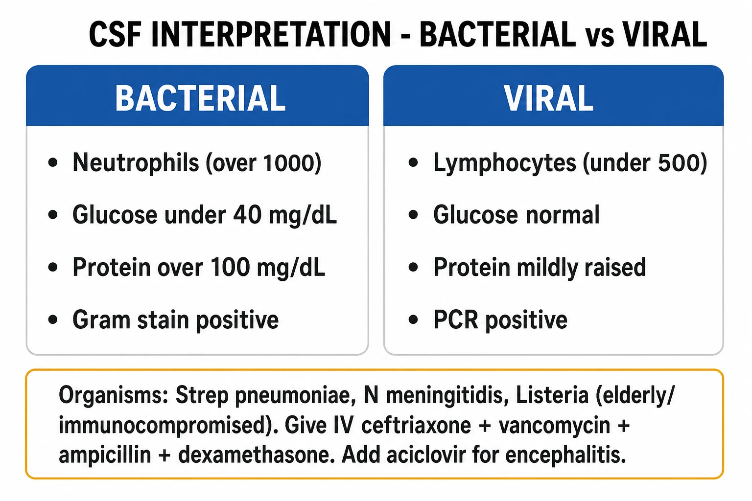
The CSF parameters are among the most heavily examined facts in clinical medicine. Each pattern reflects the underlying pathophysiology (neutrophils for bacteria; lymphocytes for viruses, TB, and fungi; low glucose when organisms consume it or infiltrate the meninges; high protein when the blood–CSF barrier is breached). [1]
Bacterial meningitis
- **Opening pressure** — elevated (over 20 cm CSF)
- **White cells** — markedly raised, 100 to 5,000; neutrophil predominance (over 80 percent)
- **Glucose** — low, under 2.2 mmol/L or **CSF-to-serum ratio under 0.4**
- **Protein** — high, over 1.0 g/L (often 1.5 to 5 g/L)
- **Gram stain** positive in 60 to 90 percent (less after antibiotics); **culture** positive in 70 to 85 percent
- **Lactate** over 3.5 mmol/L (helps distinguish bacterial from viral)
Viral (aseptic) meningitis
- **Opening pressure** — normal or mildly raised
- **White cells** — mildly raised, 10 to 500; **lymphocyte** predominance (neutrophils early, then lymphocytes)
- **Glucose** — normal (usually over 2.5 mmol/L and over 0.6 of serum)
- **Protein** — mildly raised, 0.5 to 1.0 g/L
- **Gram stain and culture negative** (aseptic); **PCR** identifies HSV, VZV, enterovirus
- **Lactate** under 3.5 mmol/L
Tuberculous meningitis
- **Opening pressure** — high; **white cells** — 50 to 500, lymphocyte predominance (neutrophils early)
- **Glucose** — low (under 2.2 mmol/L), often very low; **protein** — markedly high (1 to 5 g/L)
- **AFB smear** positive in only 10 to 20 percent; **culture** takes weeks; **GeneXpert MTB/RIF** faster
- **Adenosine deaminase (ADA)** elevated (over 10 U/L) — supportive
- Cobweb/fibrin web; basilar enhancement on MRI
Cryptococcal / fungal
- **Opening pressure** — often very high (manage with serial therapeutic LP)
- **White cells** — lymphocyte predominance (may be very low in profound immunosuppression)
- **Glucose** — low; **protein** — high
- **India ink** positive in 50 to 75 percent; **cryptococcal antigen** over 95 percent sensitive
- **Culture** on Sabouraud; MRI shows 'soap-bubble' gelatinous pseudocysts in basal ganglia
HSV encephalitis
- **Opening pressure** — often raised; **white cells** — lymphocytic, often with **red cells** (haemorrhagic necrosis)
- **Glucose** — usually normal; **protein** — mildly to moderately raised
- **HSV-1 PCR** — over 95 percent sensitive and specific; may be negative in the first 24 to 48 hours — **repeat at 3 to 7 days if suspicion persists**
- **Oligoclonal bands** may be present; do NOT stop aciclovir on a single early negative PCR
Imaging
CT brain (non-contrast) is performed before LP in patients with red flags (above), to exclude a mass lesion, hydrocephalus, or cerebral oedema. In bacterial meningitis the CT is often normal or shows only meningeal enhancement after contrast; it may show sinusitis, mastoiditis, or a focus. MRI brain with contrast is more sensitive and is the modality of choice for encephalitis and subacute meningitis: HSV encephalitis shows T2/FLAIR hyperintensity and restricted diffusion in the temporal lobes (often bilateral, asymmetric), TB meningitis shows basal meningeal enhancement, hydrocephalus, and basal ganglia infarcts, cryptococcosis shows gelatinous pseudocysts, and autoimmune encephalitis often shows medial temporal lobe T2 hyperintensity (limbic encephalitis). Electroencephalogram (EEG) is highly characteristic in HSV encephalitis — periodic lateralising epileptiform discharges (PLEDs) over a temporal lobe — though MRI and PCR have largely supplanted its diagnostic role.[3][6]
Management — Resuscitation
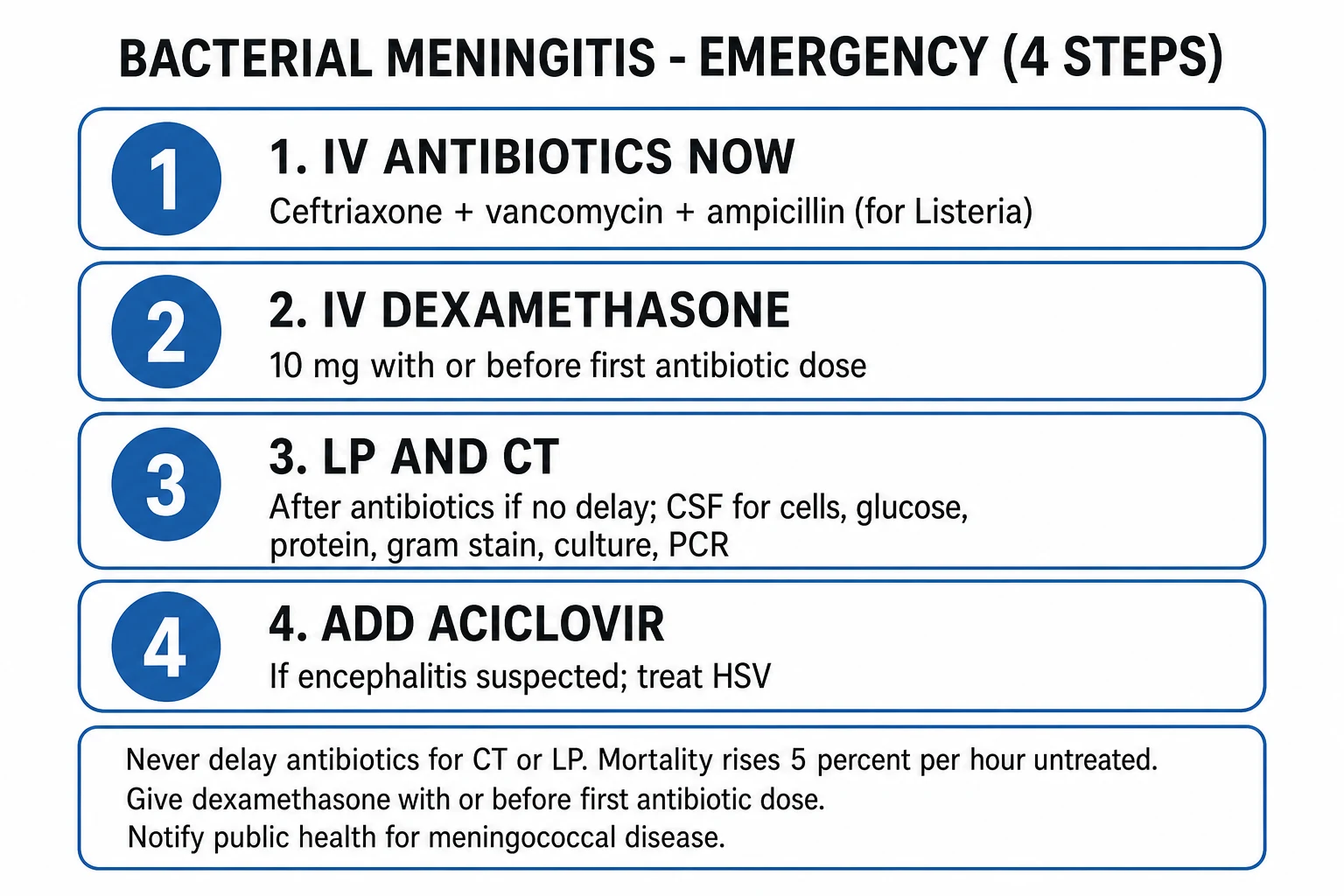
Suspected bacterial meningitis is a time-critical emergency. The discipline of practice is: assess, take blood cultures, give empirical antibiotics and dexamethasone within one hour, then perform LP if safe and image if needed. Treatment is never delayed for LP or imaging. [1]
Empirical antibiotic bundle (adult, community-acquired)
The empirical regimen is chosen to cover the three dominant adult pathogens (pneumococcus, meningococcus, Listeria) and to penetrate the CSF at meningeal doses. Third-generation cephalosporins achieve excellent CSF penetration in inflamed meninges; vancomycin covers penicillin-resistant pneumococci; ampicillin covers Listeria, which is intrinsically resistant to all cephalosporins.[1][2][4]
Standard empirical regimen
- **Ceftriaxone 2 g IV every 12 hours** (or cefotaxime 2 g IV every 4 to 6 hours) — covers pneumococcus, meningococcus, H. influenzae, group B strep
- **PLUS vancomycin 30 mg/kg IV loading then 15 to 20 mg/kg every 12 hours** (target trough or AUC) — for penicillin-resistant pneumococcus
- **PLUS ampicillin 2 g IV every 4 hours** — add if over 50, immunocompromised, pregnant, or alcoholic, to cover Listeria
- **PLUS dexamethasone 10 mg IV every 6 hours for 4 days** — given BEFORE or WITH the first antibiotic dose; benefit mainly in pneumococcal
If immediate LP is unsafe / delayed
- Give the antibiotic bundle first; LP can be performed after CT
- Antibiotics reduce culture yield but PCR and antigen tests remain positive for hours to days
- Do NOT withhold dexamethasone if pneumococcal is suspected
Suspected HSV encephalitis
- **Add aciclovir 10 mg/kg IV every 8 hours (over 1 hour infusion)** — renal-dose adjusted
- Continue for **14 to 21 days** for confirmed HSV; do NOT wait for PCR before starting
- Cover bacterial meningitis in parallel until CSF excludes it
Severe penicillin / cephalosporin allergy
- **Moxifloxacin 400 mg IV daily plus vancomycin** as cephalosporin substitute (with infectious diseases input)
- **Trimethoprim-sulfamethoxazole** for Listeria in cephalosporin-allergic patients
- Consider skin testing / desensitisation in true anaphylaxis
Adjunctive and supportive care
Airway / breathing / circulation
- Protect the airway if GCS under 8 — intubate
- Oxygen to keep SpO2 over 94 percent; avoid hypotension and hypoxia (secondary brain injury)
- IV fluids — use **isotonic** crystalloid (avoid hypotonic, which worsens cerebral oedema); resuscitate septic shock aggressively
- Vasopressors (noradrenaline) for septic shock; aim MAP over 65 mmHg and CPP over 60 mmHg
Raised intracranial pressure
- Head of bed elevated 30 degrees; midline neck position
- **Mannitol 0.25 to 1 g/kg IV (20 percent) over 20 minutes**, or **hypertonic saline 3 percent 250 mL bolus**
- Avoid hypoxia, hypercapnia, hypotension; consider neurosurgical decompression for focal oedema
- Seizure prophylaxis if seizures present; treat status epilepticus with lorazepam then levetiracetam
Electrolytes and seizures
- Correct hyponatraemia cautiously — cerebral salt wasting needs **hypertonic saline and volume**, NOT fluid restriction (unlike SIADH)
- **Lorazepam 4 mg IV** for seizures, then **levetiracetam 60 mg/kg loading** or **fosphenytoin 20 mg/kg**
- Continuous EEG if non-convulsive status suspected (especially in encephalitis)
Public health and contacts
- Notify meningococcal and TB meningitis to public health
- Chemoprophylaxis for close contacts of meningococcal case (see Prevention)
- Isolate the index case with droplet precautions for the first 24 hours of antibiotics
Management — Definitive & Stepwise
Once the organism and sensitivities are known, antibiotics are narrowed and continued for the appropriate total duration. Dexamethasone is continued for four days in confirmed pneumococcal disease; in meningococcal or Hib disease it is given if started empirically but offers little additional benefit. In suspected or confirmed Listeria, dexamethasone is not continued (no benefit, possible harm).[1][2]
Pathogen-specific duration
- *S. pneumoniae* — ceftriaxone 2 g IV q12h (add vancomycin if resistant) for **10 to 14 days**
- *N. meningitidis* — ceftriaxone 2 g IV q12h for **7 days** (penicillin or ceftriaxone both effective)
- *H. influenzae* type b — ceftriaxone for **7 days**
- *L. monocytogenes* — **ampicillin 2 g IV q4h PLUS gentamicin 5 mg/kg/day** for at least **21 days** (gentamicin for first week)
- Group B streptococcus — ceftriaxone or penicillin for **14 to 21 days**
- Gram-negative bacilli — ceftriaxone or cefepime for **21 days**
Repeat LP
- Repeat LP at 48 to 72 hours if **clinical deterioration** or persistent fever, especially with resistant pneumococcus
- Sterilisation of CSF by 24 to 48 hours predicts good outcome
- Persistent fever may indicate subdural empyema, ventriculitis, brain abscess, or drug fever
De-escalation and oral switch
- Switch to oral therapy only when the patient is afebrile, clinically improved, and an oral agent with good CSF penetration is available (e.g. fluoroquinolone, linezolid)
- Complete the full duration; counsel about vaccination and hearing follow-up
HSV encephalitis
HSV encephalitis is treated with IV aciclovir 10 mg/kg every 8 hours (infused over one hour, renal-adjusted) for 14 to 21 days. Treatment is started empirically in every patient with suspected encephalitis and continued until HSV PCR returns negative; a single early negative PCR does not exclude HSV (sensitivity is low in the first 24 to 48 hours) and the LP should be repeated at 3 to 7 days. With aciclovir, mortality falls from over 70 percent to 20 to 30 percent; survivors often have residual cognitive and behavioural deficits. Repeat MRI at the end of therapy is reasonable; clinical relapse occurs in a minority and warrants re-treatment.[3][6]
Tuberculous meningitis
TB meningitis is treated with four-drug intensive therapy — isoniazid (with pyridoxine), rifampicin, pyrazinamide, ethambutol (HRZE) for two months, followed by isoniazid plus rifampicin (HR) for a total of 12 months (some authorities extend to 18 months). Adjunctive corticosteroids improve survival (dexamethasone or prednisolone, tapering over 6 to 8 weeks) — the landmark Thwaites trial in NEJM established this. Thalidomide and infliximab are reserved for refractory cases. Monitor for hepatotoxicity, optic neuritis (ethambutol), and peripheral neuropathy (isoniazid). Communicating hydrocephalus may need a shunt.[8]
Cryptococcal meningitis
Cryptococcal meningitis (HIV-related or not) follows the 2010 IDSA three-phase regimen: induction with amphotericin B deoxycholate 0.7 to 1 mg/kg/day IV PLUS flucytosine 100 mg/kg/day orally in four divided doses for at least two weeks; consolidation with fluconazole 400 mg (6 mg/kg) daily for eight weeks; and maintenance (in HIV) with fluconazole 200 mg daily until immune reconstitution (CD4 over 100 for at least 6 months on antiretroviral therapy). Manage raised intracranial pressure with serial therapeutic lumbar punctures daily until pressure normalises; a lumbar drain or shunt may be needed. Liposomal amphotericin is preferred where toxicity (renal, electrolyte, anaemia) is a concern.[9]
Autoimmune encephalitis
Anti-NMDA receptor encephalitis and other autoimmune encephalitides are treated with immunotherapy: first-line — methylprednisolone 1 g IV daily for 3 to 5 days, intravenous immunoglobulin 0.4 g/kg/day for 5 days, and/or plasma exchange (5 sessions); second-line for non-response — rituximab and/or cyclophosphamide. A teratoma search (CT chest/abdomen/pelvis, transvaginal ultrasound, MRI pelvis) is mandatory in young women with anti-NMDAR — resection of the tumour is the most effective single intervention. Recovery is slow (months), and prolonged ICU support for autonomic instability is often required. Anti-LGI1 encephalitis responds well to first-line immunotherapy and classically presents with faciobrachial dystonic seizures.[10]
Specific Subtypes & Scenarios
Meningococcal meningitis and septicaemia
- Ceftriaxone 2 g IV immediately, then q12h for 7 days; benzylpenicillin 2.4 g IV q4h acceptable once sensitivities known
- Aggressive fluid resuscitation and ICU for septic shock; activated protein C and haemofiltration in severe DIC
- Notify public health; chemoprophylaxis for household and intimate 'kissing' contacts within 24 hours
Pneumococcal meningitis
- Dexamethasone 10 mg IV q6h for 4 days — benefit greatest; give BEFORE antibiotic
- Ceftriaxone plus vancomycin; review for resistance; repeat LP at 48 hours if not improving
- Search for and treat the focus (otitis, sinusitis, pneumonia, endocarditis, CSF leak); vaccination afterwards
Listeria monocytogenes meningitis / rhombencephalitis
- Ampicillin 2 g IV q4h PLUS gentamicin 5 mg/kg/day for at least 21 days
- Inherently cephalosporin-resistant — add ampicillin empirically in those over 50, immunocompromised, pregnant, alcoholic
- May cause brainstem rhombencephalitis with cranial nerve palsies, ataxia, nystagmus
HSV encephalitis
- Aciclovir 10 mg/kg IV q8h for 14 to 21 days; do NOT stop on a single early negative PCR — repeat at 3 to 7 days
- MRI: T2/FLAIR temporal lobe hyperintensity; EEG: temporal PLEDs; CSF: lymphocytic with red cells
- Long-term cognitive and behavioural sequelae common; consider neuropsychology follow-up
Anti-NMDA receptor encephalitis
- Young women, ovarian teratoma; psychiatric prodrome then seizures, dyskinesias, autonomic instability
- First-line immunotherapy (steroid + IVIG + plasmapheresis); tumour resection; second-line rituximab
- Prolonged ICU; reversible with early aggressive immunotherapy
Neonatal meningitis
- Group B strep, E. coli K1, Listeria — **ampicillin plus gentamicin** or **ampicillin plus cefotaxime**
- Weight-based dosing; Duration 14 to 21 days; CSF may need repeat LP
- High morbidity — hearing loss, seizures, neurodevelopmental delay
CSF shunt / post-neurosurgical meningitis
- *Staphylococcus epidermidis* and *S. aureus*, Gram-negative bacilli, *P. acnes*
- **Vancomycin** (often intrathecal/intraventricular); shunt externalisation or removal
- Cefepime or meropenem if Gram-negative; infectious diseases input mandatory
Complications & Pitfalls
Acute neurological
- **Cerebral oedema and raised ICP** — herniation, death
- **Seizures and status epilepticus** — focal or generalised; non-convulsive in encephalitis
- **Cerebral infarction** — vasculitis of perforating vessels; hemiparesis
- **Hydrocephalus** — communicating (impaired CSF reabsorption) or obstructive (intraventricular clot)
- **Subdural empyema / brain abscess / ventriculitis** — focal collections needing drainage
Systemic
- **Septic shock and multi-organ failure** (especially meningococcal)
- **DIC and purpura fulminans**; **Waterhouse–Friderichsen** (adrenal haemorrhage, adrenal crisis)
- **SIADH or cerebral salt wasting** — hyponatraemia; distinguish and treat differently
- **Disseminated intravascular coagulation**; limb loss from purpura
Long-term sequelae
- **Sensorineural hearing loss** in roughly 10 percent of bacterial meningitis — audiometry at discharge and 6 weeks; commonest with pneumococcus
- **Cognitive impairment**, behavioural change, memory disturbance (especially HSV)
- **Epilepsy** — post-infective focus; driving restrictions
- **Cranial nerve palsies** (especially VIII), **motor deficit**, **ataxia**, **visual loss**
- **Cerebral palsy and developmental delay** in neonates
The classic pitfalls that examiners test — and that cause avoidable harm — are: delaying antibiotics for LP or CT (the single biggest error); omitting ampicillin in the over-50 or immunocompromised patient, missing Listeria; giving dexamethasone after the antibiotic (no benefit); performing LP in the presence of papilloedema or a mass lesion (herniation); stopping aciclovir on a single early negative HSV PCR (HSV may be missed); fluid-restricting hyponatraemic patients with cerebral salt wasting (worsens cerebral perfusion); and failing to arrange chemoprophylaxis for meningococcal contacts.[1][3]
Prognosis & Disposition
Bacterial meningitis carries a case-fatality of 10 to 30 percent, rising with age (over 60), immunocompromise, septicaemic shock, low GCS, seizures, focal deficit, delayed antibiotics, and pneumococcal aetiology. Among survivors, 30 to 50 percent have some neurological sequelae, most often sensorineural hearing loss (about 10 percent), cognitive impairment, epilepsy, or motor deficit. HSV encephalitis, even with aciclovir, kills 20 to 30 percent and leaves half of survivors with cognitive and behavioural deficits that may only become fully apparent months later. TB meningitis mortality is 20 to 60 percent and is heavily stage-dependent (Medical Research Council grade I vs III). Cryptococcal meningitis mortality in HIV depends on immune reconstitution and intracranial pressure control. Autoimmune encephalitis has a good prognosis with early immunotherapy and tumour removal, but a poor prognosis with delay.[1][3][4]
Poor prognostic factors
- Age over 60 or under 1 month; immunocompromise
- Septicaemic shock; low admission GCS (under 10)
- Seizures; focal neurological deficit; rapid onset
- Pneumococcal aetiology; penicillin-resistant organism
- Delayed antibiotics (over 1 to 3 hours); delayed presentation
- Low CSF leucocyte count with high bacterial load (inadequate host response)
Disposition
- **ICU** — GCS under 12, seizures, shock, need for ventilation, raised ICP
- **HDU/step-down** — moderate severity, close neurological observation
- **Ward** — mild viral meningitis or improving bacterial meningitis
- **Outpatient** — viral meningitis once improving; arrange hearing test and follow-up for bacterial
Special Populations
Pregnancy
- Increased risk of **Listeria** (food-borne — soft cheese, deli meats, unpasteurised milk)
- Varicella and influenza pneumonia severe; HSV may be transmitted
- Treat with **ampicillin for Listeria**, **aciclovir for HSV/VZV**, **oseltamivir for influenza**
- Ceftriaxone, aciclovir, vancomycin are acceptable in pregnancy
Neonate
- Pathogens: **group B strep, E. coli (K1), Listeria**
- Empirical: **ampicillin PLUS gentamicin** or **ampicillin PLUS cefotaxime** (avoid ceftriaxone — displaces bilirubin)
- Weight-based dosing; non-specific presentation; high morbidity
Elderly
- Listeria and pneumococcus predominate; broader empirical cover (add ampicillin)
- Comorbidity masks presentation; delirium may be the only sign
- Worse outcomes; cautious fluid balance; consider dexamethasone risk–benefit
Immunocompromised
- HIV: **Cryptococcus, TB, toxoplasmosis, CMV, syphilis, JC virus**
- Transplant/neutropenic: **Listeria, Gram-negatives, HHV-6, Cryptococcus**
- Broader empirical and diagnostic panel; PCR and antigen tests essential
- Consider immune reconstitution inflammatory syndrome (IRIS) after starting ART
Travellers
- Meningococcal belt (sub-Saharan Africa, dry season) — vaccine; **rifampicin/ciprofloxacin prophylaxis**
- **Japanese encephalitis** (Asia), **West Nile** (Americas), **tick-borne encephalitis** (Europe/Russia)
- **Cerebral malaria** — blood film, **IV artesunate**; rabies — exposure history
- Schistosomiasis, cysticercosis, amoebic meningoencephalitis in specific exposures
Dose anchors examiners expect in special groups. Neonates: never default to adult ceftriaxone monotherapy — use ampicillin + gentamicin/cefotaxime with neonatal weight-based charts. Adults with Listeria risk (age >50, pregnancy, cell-mediated immunosuppression): ampicillin 2 g IV every 4 hours added to a third-generation cephalosporin. HSV encephalitis: aciclovir 10 mg/kg IV every 8 hours (adjust in renal impairment) until PCR negative or full course completed. Cryptococcal meningitis in HIV: induction with amphotericin B + flucytosine (guideline regimen), then fluconazole consolidation — LP for raised pressure is therapeutic as well as diagnostic. Contact prophylaxis after meningococcus: ciprofloxacin 500 mg PO once (adult) or ceftriaxone 250 mg IM once in pregnancy. [1]
Evidence, Guidelines & Regional Differences
The contemporary evidence base for bacterial meningitis rests on two landmark randomised trials of adjunctive dexamethasone. The European Dexamethasone in Adulthood Bacterial Meningitis Study (de Gans and van de Beek, NEJM 2002) randomised adults with suspected bacterial meningitis to dexamethasone 10 mg every six hours for four days versus placebo, given before or with the first antibiotic. Mortality fell from 25 to 15 percent overall, and from 30 to 14 percent in pneumococcal meningitis, with a striking reduction in sensorineural hearing loss. Benefit was confined to pneumococcal disease and only when dexamethasone was given before the antibiotic. The Thwaites trial (NEJM 2004) showed that dexamethasone similarly improved survival in tuberculous meningitis.[7][8]
Landmark evidence
- **de Gans 2002 (NEJM)** — dexamethasone before antibiotic reduces mortality and hearing loss in pneumococcal meningitis
- **Thwaites 2004 (NEJM)** — dexamethasone improves survival in TB meningitis
- **Brouwer 2010 (Clin Microbiol Rev)** — comprehensive review of epidemiology and treatment
- **Dalmau 2011 (Lancet Neurol)** — anti-NMDAR encephalitis clinical spectrum
- **Venkatesan 2013 (Clin Infect Dis)** — international encephalitis case definitions
Guidelines
- **ESCMID 2016** (van de Beek) — European bacterial meningitis diagnosis and treatment
- **IDSA 2004** (Tunkel) — bacterial meningitis management (US)
- **ABN / British Infection Association 2012** (Solomon) — adult viral encephalitis
- **IDSA 2010** (Perfect) — cryptococcal disease
In Australia and New Zealand, empiric therapy mirrors the UK pattern with ceftriaxone 2 g IV q12h plus vancomycin, adding ampicillin for Listeria risk groups. Australia has a funded MenACWY adolescent programme and MenB (Bexsero) for at-risk groups. Indigenous Australian children carry a higher burden of pneumococcal and Hib disease (historically); PCV13 and Hib vaccination are prioritised.
In low- and middle-income settings, the burden is far higher, empiric therapy may include chloramphenicol where cephalosporins are unavailable, and WHO prioritises pentavalent (Hib) and pneumococcal conjugate vaccination through Gavi. The African meningitis belt has been transformed by MenAfriVac (serogroup A), but serogroups W, X, and C persist.
Prevention
Prevention of bacterial meningitis rests on vaccination and chemoprophylaxis. The conjugate vaccines have changed the epidemiology in countries with routine programmes. [1]
Vaccines in routine use
- **Hib conjugate** — *H. influenzae* type b; given in infancy; near-eliminated Hib meningitis
- **Pneumococcal conjugate PCV13** — 13 serotypes, in infancy; **PPSV23** polysaccharide for high-risk adults and over 65
- **Meningococcal ACWY conjugate** — adolescents, university entrants, asplenia, complement deficiency, Hajj, travel to belt
- **Meningococcal B (Bexsero, Trumenba)** — adolescents at risk, outbreaks
- **BCG** — reduces disseminated and TB meningitis in children; **MMR** — mumps meningitis
Chemoprophylaxis (meningococcal contacts)
- **Rifampicin 600 mg orally every 12 hours for 2 days (4 doses)** — household and 'kissing' contacts
- **Ciprofloxacin 500 mg orally as a SINGLE dose** — adult alternative (preferred in pregnancy: single-dose ceftriaxone 250 mg IM)
- **Ceftriaxone 250 mg IM single dose** — preferred in pregnancy
- Give within 24 hours of index case diagnosis; vaccinate if serogroup covered by vaccine
Haemophilus influenzae type b contacts
- Rifampicin 20 mg/kg (max 600 mg) daily for 4 days to all household contacts if an unvaccinated child under 4 years is present
- Vaccinate under-immunised contacts
Exam Pearls & High-Yield Minutiae
BACTERIA
- Classic triad of bacterial meningitis — fever, neck stiffness, altered mental status — is present in only about 40 to 50 percent; the presence of two of fever, headache, neck stiffness, confusion is over 95 percent sensitive.
- Meningococcal rash — petechial or purpuric, non-blanching; check conjunctiva and palate; can evolve hourly.
- CSF bacterial: neutrophils, glucose under 2.2 mmol/L or CSF-to-serum ratio under 0.4, protein over 1 g/L.
- CSF viral: lymphocytes, normal glucose, mildly raised protein.
- CSF TB: lymphocytes, very low glucose, very high protein, cobweb, ADA elevated.
- CSF HSV: lymphocytes with red cells (haemorrhagic necrosis); PCR over 95 percent sensitive but may be negative in first 24 to 48 hours — repeat.
- Dexamethasone 10 mg IV q6h for 4 days, before or with the first antibiotic, for suspected pneumococcal meningitis.
- Aciclovir 10 mg/kg IV q8h for 14 to 21 days for HSV encephalitis — do NOT wait for PCR.
- Vancomycin added for penicillin-resistant pneumococcus; ampicillin for Listeria (cephalosporin-resistant).
- Kernig and Brudzinski signs — low sensitivity (under 30 percent) but high specificity.
- CT before LP if immunocompromised, new seizures, focal deficit, papilloedema, GCS under 12, or anticoagulated.
- Prophylaxis — rifampicin 600 mg BD x2 days or ciprofloxacin 500 mg single dose for meningococcal contacts.
- Listeria — food-borne (soft cheese, deli meats, unpasteurised milk); over 50, pregnant, immunocompromised.
- Waterhouse–Friderichsen — adrenal haemorrhage in meningococcaemia; adrenal crisis.
- Neonate — group B strep, E. coli, Listeria; treat with ampicillin plus gentamicin/cefotaxime.
- Hearing loss in about 10 percent — audiometry at discharge and 6 weeks. [1]
Self-test: a 22-year-old university student with fever, headache and a petechial rash
This is meningococcal meningitis/septicaemia until proven otherwise. Immediate management: airway, breathing, circulation; ceftriaxone 2 g IV immediately (do not wait for LP); blood cultures; fluid resuscitation for shock; ICU if shocked; notify public health; chemoprophylaxis for household and kissing contacts (rifampicin 600 mg BD x2 days or ciprofloxacin 500 mg stat). LP is performed once stable, with CT first if any red flag. Droplet precautions for the first 24 hours.
Self-test: a 65-year-old with fever, confusion and a focal seizure
Consider HSV encephalitis and Listeria meningitis. Give empirical ceftriaxone 2 g IV plus vancomycin plus ampicillin (Listeria cover, age over 50) plus dexamethasone 10 mg IV plus aciclovir 10 mg/kg IV. CT first (new seizure, focal deficit = red flag). LP after CT: send for cells, glucose, protein, Gram stain, culture, HSV PCR, and bacterial PCR/antigen. MRI temporal lobes. Continue aciclovir until HSV PCR negative; narrow antibiotics once the organism is identified.
Exam application bank (NEET-PG / INICET)
One-line answer
Meningitis is inflammation of the meninges, most often infective; encephalitis is inflammation of the brain parenchyma, and meningoencephalitis is both together. Bacterial meningitis is a time-critical emergency — fever plus headache plus neck stiffness plus altered mental status is bacterial meningitis until proven otherwise, and empirical therapy (ceftriaxone 2 g IV q12h plus vancomycin plus dexamethasone 10 mg IV q6h, add ampicillin if over 50 or immunocompromised) must start within one hour, before lumbar puncture or imaging. Herpes simplex encephalitis is the treatable encephalitis not to miss — fever plus altered mental status plus seizures needs empirical IV aciclovir 10 mg/kg q8h for 14 to 21 days, without waiting for PCR. CSF analysis (cell count, glucose, protein, Gram stain, culture, PCR) distinguishes bacterial, viral, tuberculous and fungal causes. [1]
Worked stems (answer without another resource)
Stem 1 — Classic presentation. Map symptoms to mechanism; name the first investigation and first treatment step with dose/route if drug therapy is standard. [1]
Stem 2 — Unstable / complicated. List red flags that force immediate resuscitation, theatre, ICU, antidote, or reperfusion — and what you do in the first 15 minutes. [1]
Stem 3 — Atypical group. Elderly, pregnancy, child, or immunocompromised: how presentation and thresholds change. [1]
Stem 4 — Differential trap. Name the three closest mimics and one discriminator for each. [1]
Stem 5 — Disposition. Who goes home with safety-netting, who is admitted, who needs HDU/ICU/theatre, and what follow-up is mandatory. [1]
Rapid viva checklist
- Definition + classification
- Pathophysiology chain
- Bedside signs / criteria
- Score with exact components (if any)
- Emergency bundle
- Definitive therapy with doses
- Complications of disease and of treatment
- Special populations
- Guideline/trial name if classic
- Three exam traps
Coverage self-check
If you cannot answer any stem above from this page alone, re-read the matching section — the page is intended to be self-sufficient for final-prof and NEET-PG/INICET questions on Meningitis and Encephalitis.
References
- [1]van de Beek D, Cabellos C, Dzupova O, et al. ESCMID guideline: diagnosis and treatment of acute bacterial meningitis Clin Microbiol Infect, 2016.PMID 27062097
- [2]Tunkel AR, Hartman BJ, Kaplan SL, et al. Practice guidelines for the management of bacterial meningitis Clin Infect Dis, 2004.PMID 15494903
- [3]Solomon T, Michael BD, Smith PE, et al. Management of suspected viral encephalitis in adults--Association of British Neurologists and British Infection Association National Guidelines J Infect, 2012.PMID 22120595
- [4]McGill F, Heyderman RS, Panagiotou S, Tunkel AR, van de Beek D. Acute bacterial meningitis in adults Lancet, 2016.PMID 27265346
- [5]Brouwer MC, Tunkel AR, van de Beek D. Epidemiology, diagnosis, and antimicrobial treatment of acute bacterial meningitis Clin Microbiol Rev, 2010.PMID 20610819
- [6]Venkatesan A, Tunkel AR, Bloch KC, et al. Case definitions, diagnostic algorithms, and priorities in encephalitis: consensus statement of the international encephalitis consortium Clin Infect Dis, 2013.PMID 23861361
- [7]de Gans J, van de Beek D, European Dexamethasone in Adulthood Bacterial Meningitis Study Investigators. Dexamethasone in adults with bacterial meningitis N Engl J Med, 2002.PMID 12432041
- [8]Thwaites GE, Nguyen DB, Nguyen HD, et al. Dexamethasone for the treatment of tuberculous meningitis in adolescents and adults N Engl J Med, 2004.PMID 15496623
- [9]Perfect JR, Dismukes WE, Dromer F, et al. Clinical practice guidelines for the management of cryptococcal disease: 2010 update by the infectious diseases society of america Clin Infect Dis, 2010.PMID 20047480
- [10]Dalmau J, Lancaster E, Martinez-Hernandez E, Rosenfeld MR, Balice-Gordon R. Clinical experience and laboratory investigations in patients with anti-NMDAR encephalitis Lancet Neurol, 2011.PMID 21163445