Neurology · General Medicine
Motor Neuron Disease (ALS)
Also known as Motor neuron disease · MND · Amyotrophic lateral sclerosis · ALS · Lou Gehrig disease
Motor neuron disease (MND/ALS) is a progressive neurodegenerative disorder that destroys BOTH upper motor neurons (UMN: spasticity, hyperreflexia, Babinski sign) AND lower motor neurons (LMN: weakness, wasting, fasciculations) while sparing sensation, eye movements and sphincter function. Annual incidence is about 2 per 100,000, peak onset 55 to 65 years, male-to-female ratio 1.5 to 1. About 10 percent is familial (C9orf72, SOD1, TARDBP, FUS). Diagnosis is clinical (Gold Coast 2020 criteria), supported by EMG (active denervation with chronic reinnervation across regions) and MRI to exclude mimics. Treatment is multidisciplinary: riluzole 50 mg twice daily extends median survival by about 2 to 3 months, non-invasive ventilation when FVC is 50 percent or less (or SNIP 40 cmH2O) extends survival by about 7 months and improves quality of life, gastrostomy maintains nutrition, and multidisciplinary clinic care extends survival by about 7 to 9 months. Median survival from symptom onset is 3 to 5 years; about 10 percent live more than 10 years. Death is usually from respiratory failure. Frontotemporal dementia coexists in about 15 percent. There is no cure.
On this page & tools
Your progress
Saved locally on this device.
Practise this topic
Exam tags
Red flags
Overview & Definition
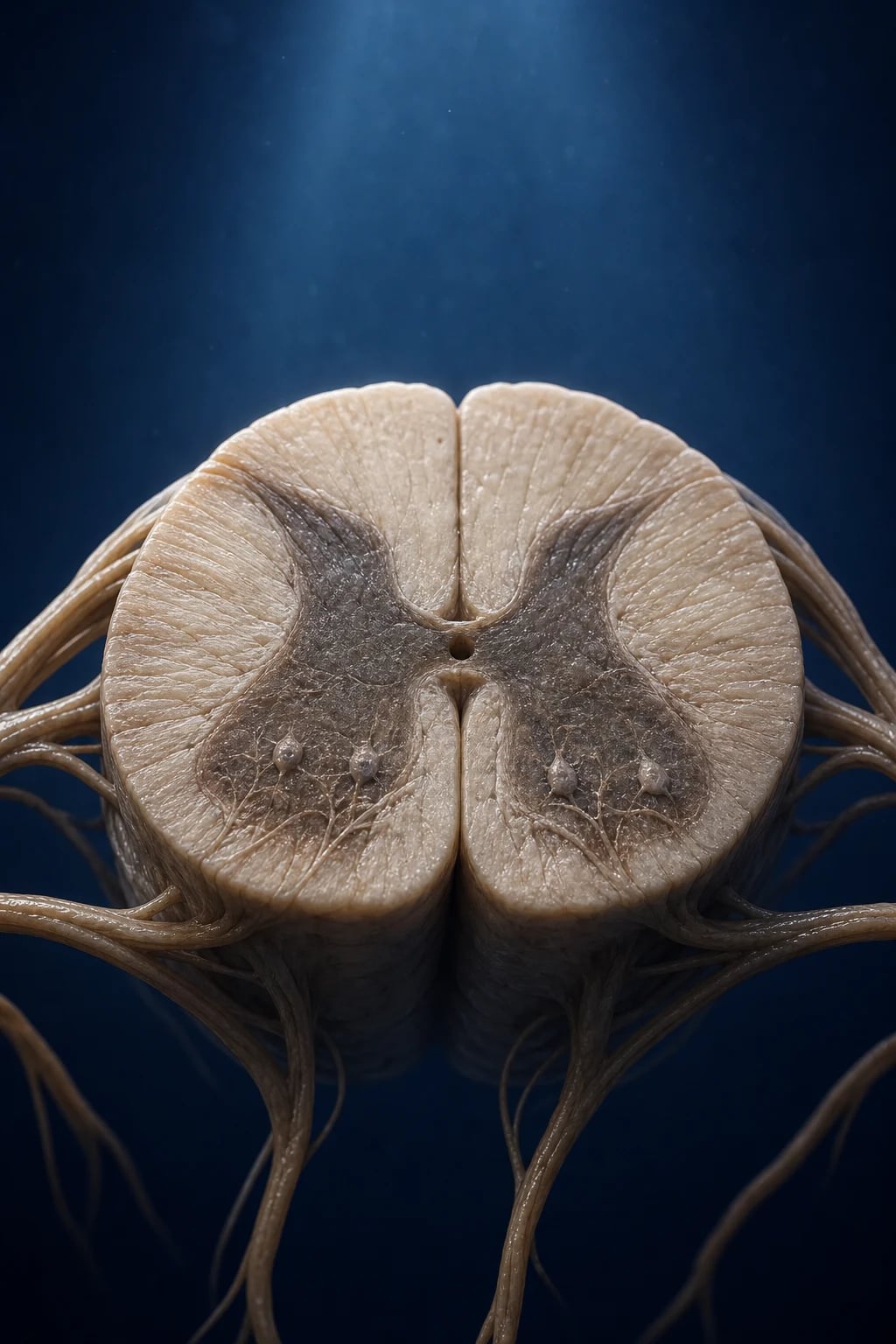
Motor neuron disease (MND), of which amyotrophic lateral sclerosis (ALS) is the commonest and most important form, is a relentlessly progressive neurodegenerative disorder that destroys both the upper motor neurons (the Betz cells of the motor cortex and the corticospinal tract) and the lower motor neurons (the brainstem motor nuclei and the anterior horn cells of the spinal cord). The name encodes the pathology: amyotrophic (a-, without; myo, muscle; trophos, nourishment) describes the muscle wasting caused by lower motor neuron death, and lateral sclerosis describes the firm, scarred (sclerosed) lateral corticospinal tracts felt at autopsy in the lateral columns of the spinal cord.[1]
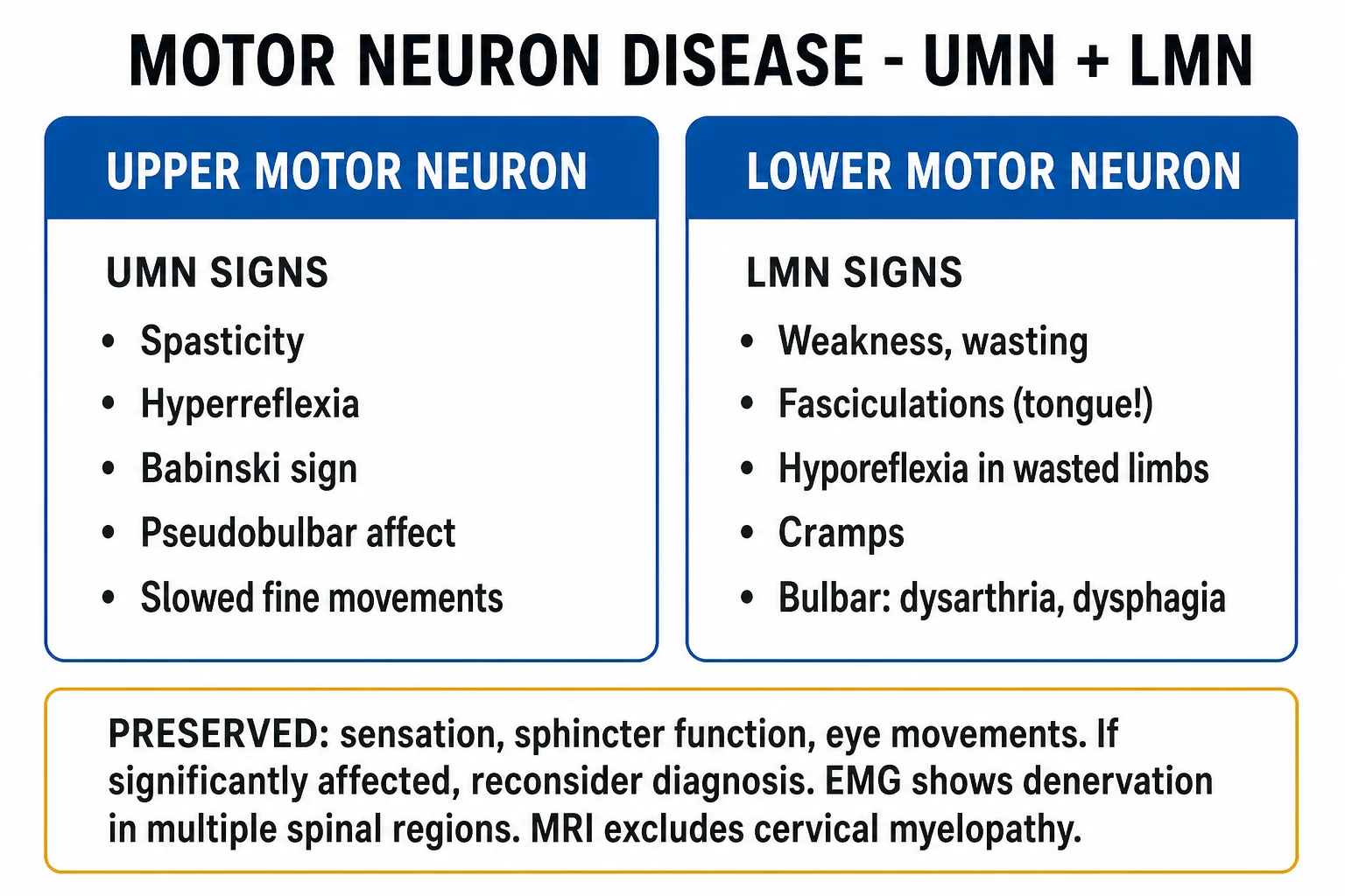
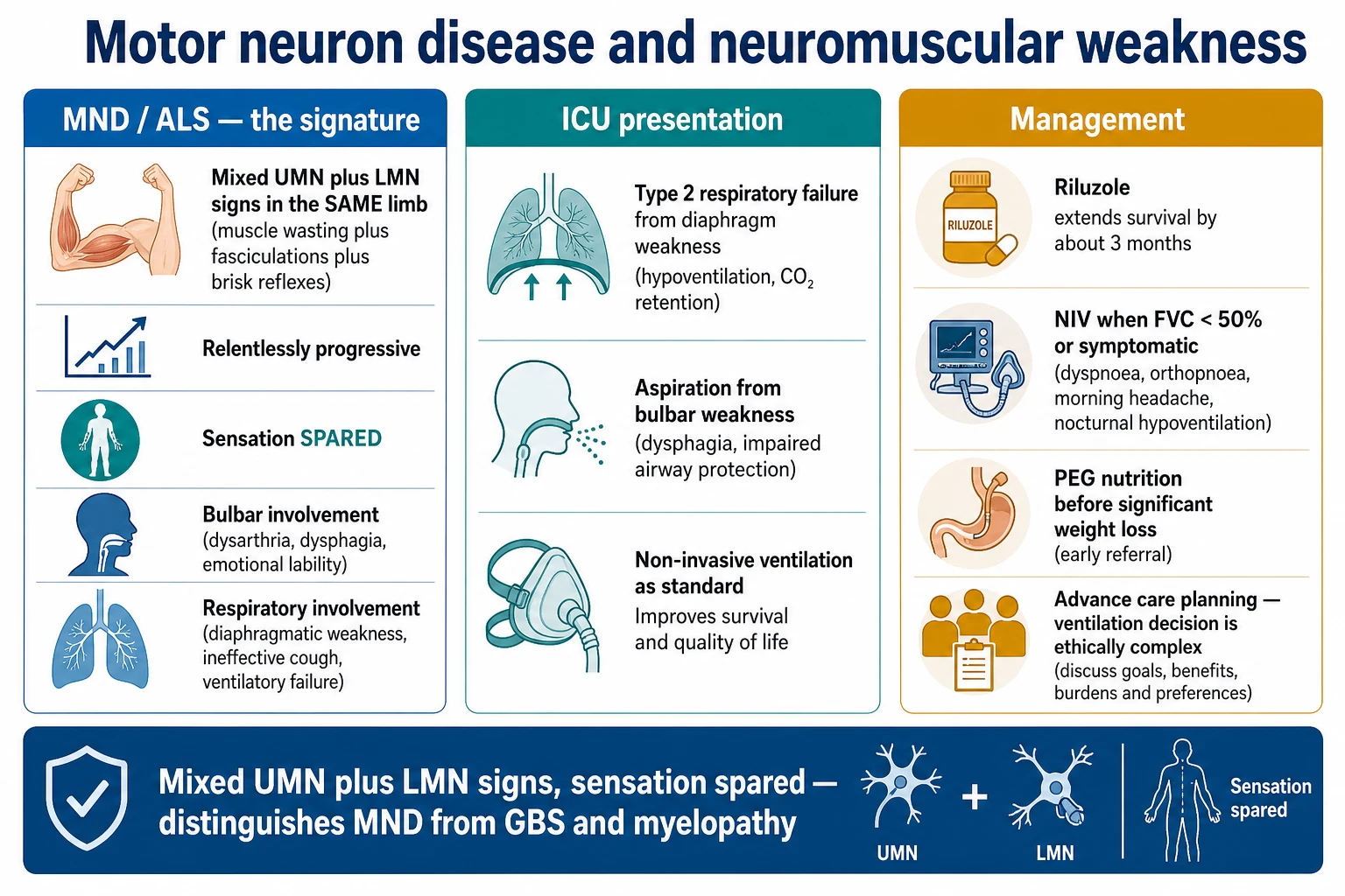
The clinical signature, and the single fact that decides most exam answers, is that UMN and LMN signs coexist in the same limb, in the same patient, with sensation and sphincter function preserved. A wasted, weak, fasciculating hand that is at the same time areflexia-resisting — brisk-finger-jerk, spastic on tone testing, with an upgoing plantar — is MND until proven otherwise. The disease progresses to involve the bulbar muscles and the diaphragm, and death is overwhelmingly from respiratory failure, usually within 3 to 5 years of symptom onset.[1][2]
Classification
MND is a clinical spectrum. The four major subtypes differ in which motor neuron population predominates, in the region of onset, and — most usefully for the viva — in prognosis. [1]
Classic ALS
85 percent of cases
- Both UMN and LMN signs
- Limb onset in about 70 percent, bulbar in 25 percent
- Median survival 3 to 5 years from symptom onset
Progressive bulbar palsy (PBP)
~20 percent, usually older women
- Predominantly bulbar UMN and LMN signs
- Dysarthria, dysphagia, tongue wasting with fasciculations, pseudobulbar affect
- Fastest progressing — median survival often under 2 years
Progressive muscular atrophy (PMA)
~5 to 10 percent
- Pure lower motor neuron phenotype
- Limb onset, asymmetric wasting and weakness
- Often slower; many eventually show UMN signs, converting to ALS
Primary lateral sclerosis (PLS)
~1 to 5 percent
- Pure upper motor neuron phenotype
- Slowly progressive spastic paraparesis or tetraparesis
- Near-normal life expectancy; minority convert to ALS over years
Two further phenotypes deserve exam-level recognition. ALS-FTD describes patients who meet ALS criteria AND show overt frontotemporal dementia; these two diseases share the TDP-43 pathological substrate and frequently cosegregate in C9orf72 expansion carriers.[8][9] The flail arm (man-in-a-barrel) and flail leg regional syndromes have slower progression and longer survival than classical ALS and are worth naming in a viva because they confound prognostication.
Epidemiology & Risk Factors
ALS is uncommon but not rare. The headline numbers reward memorisation. [1]
ALS — the numbers examiners want
About 90 percent of ALS is sporadic with no family history; 10 percent is familial, transmitted most often in an autosomal-dominant pattern. Of familial ALS, the C9orf72 GGGGCC hexanucleotide repeat expansion on chromosome 9p21 is the single commonest cause (about 30 to 40 percent of familial ALS, and 5 to 7 percent of apparently sporadic ALS), followed by mutations in SOD1 (15 to 20 percent of familial), TARDBP and FUS.[1][8] The C9orf72 expansion is also the strongest genetic link between ALS and frontotemporal dementia — carriers often present with either disease or with the combined ALS-FTD syndrome.[8]
Established environmental risk factors are smoking (modest, dose-dependent, stronger in women) and possibly prior military service (the 2008 US Institute of Medicine report found a small but consistent excess). Proposed but unconfirmed associations include head trauma, rural living, exposure to pesticides and BMAA (beta-methylamino-L-alanine, from cyanobacteria — the leading hypothesis for the western Pacific foci). The famous high-incidence foci — Guam, the Kii peninsula of Japan, and West New Guinea — had ALS, parkinsonism and dementia co-occurring at 50 to 100 times the global rate (the ALS-PDC complex); incidence has fallen sharply with westernisation, supporting an environmental trigger.[1]
Pathophysiology
Motor neuron degeneration in ALS is the net result of multiple convergent mechanisms acting on a population of cells that are, for reasons still poorly understood, selectively vulnerable. No single pathway explains every case; the disease is best understood as a final common pathway of motor neuron death. [1]
Glutamate excitotoxicity
the riluzole rationale
- Loss of the astrocytic glutamate transporter EAAT2 raises synaptic glutamate
- Excess glutamate over-activates NMDA and AMPA receptors
- Calcium influx triggers motor neuron death — the rationale for riluzole, a glutamate-release inhibitor
TDP-43 aggregation
the pathological hallmark
- TDP-43 mislocates from nucleus to cytoplasm, forming ubiquitinated inclusions
- Found in 97 percent of ALS cases — the unifying pathological protein
- Encoded by TARDBP; loss of nuclear function plus toxic cytoplasmic gain
Mitochondrial dysfunction
energy failure
- Impaired oxidative phosphorylation and ATP generation
- Defective calcium buffering sensitises motor neurons
- SOD1 mutants directly damage mitochondria — familial mechanism
Oxidative stress & neuroinflammation
the bystander damage
- Reactive oxygen species damage lipids, proteins, DNA
- Activated microglia release pro-inflammatory cytokines (TNF, IL-1)
- Edaravone, a free-radical scavenger, targets this pathway
The anatomical targets are the Betz cells of layer V of the motor cortex (UMN) and the motor neurons of the brainstem nuclei (trigeminal, facial, hypoglossal, nucleus ambiguus) and the spinal anterior horn cells (LMN). The corticospinal tract degenerates from above downward; the anterior horn cells degenerate in a focal, often asymmetric, spreading pattern. Sensation is preserved because the sensory neurons in the dorsal root ganglia and the dorsal columns are unaffected; eye movements and sphincters are preserved (until very late) because the oculomotor nuclei and Onuf's nucleus in the sacral cord are resistant. [1]
The genetic frontier is moving fast. Tofersen, an antisense oligonucleotide targeting SOD1 mRNA, was approved by the FDA (2023) for SOD1-mutant ALS after the VALOR/ATLAS trials showed reduced neurofilament and slowed progression. C9orf72-targeted antisense and ATXN2-modulating therapies are in trials. Genetic testing is now recommended for all people with ALS (regardless of family history) by the AAN, EFNS and NICE — a shift from the era when testing was reserved for familial cases.
Clinical Presentation
The presentation is dictated by which region is affected first, but the signature — UMN and LMN signs together — is constant. [1]
Limb onset (about 70 percent)
The patient notices painless asymmetric weakness, often in one hand (the patient drops objects, has difficulty with buttons, develops wasted thenar muscles) or in one foot (a foot drop, frequent tripping). On examination, the affected limb shows LMN signs (wasting, weakness, fasciculations — look especially at thenar muscles, intrinsic hand muscles, and the tongue) coexisting with UMN signs in the same limb — a brisk finger jerk or jaw jerk, a spastic catch on tone testing, an upgoing plantar. The "split-hand" sign (the abductor pollicis brevis and first dorsal interosseous wasted disproportionately to the abductor digiti minimi) is a high-yield ALS pearl.[1] Reflexes are typically brisk in a wasted limb — a finding that should always prompt the question "could this be MND?".
Bulbar onset (about 25 percent)
Bulbar disease presents with dysarthria (a mixed spastic-strangled and flaccid-nasal quality), dysphagia (initially for liquids, progressing to solids), and characteristic tongue wasting with fasciculations — the single most rewarding exam finding. Sialorrhoea (drooling from poor swallowing rather than excess saliva) and pseudobulbar affect (inappropriate, uncontrollable laughter or crying) are common and distressing. Bulbar onset is more common in older women and carries the worst prognosis.[2]
Respiratory onset (rare, under 5 percent)
A small minority present with diaphragmatic weakness: orthopnoea (breathless lying flat), morning headaches, daytime somnolence, poor sleep, a weak cough. These patients often reach the neurologist via the respiratory or sleep clinic, having been misdiagnosed with asthma, COPD or sleep apnoea. Symptomatic hypoventilation with elevated bicarbonate on venous gas is the clue. [1]
Cognitive and behavioural features
ALS is no longer considered a purely motor disease. Frontotemporal dysfunction is detectable on formal testing in up to 50 percent, and overt frontotemporal dementia (ALS-FTD) develops in about 15 percent — more often in C9orf72 carriers.[8][9] Look for executive impairment, behavioural change (apathy, disinhibition, perseveration, dietary change) and language disturbance. Cognitive impairment reduces survival (through impaired adherence to NIV and PEG) and complicates capacity and end-of-life decisions.
What is deliberately spared
The clinician must explicitly test and document the spared systems, because their preservation is what distinguishes ALS from its mimics: [1]
Sensation
- Pinprick, vibration, joint position preserved throughout
- Sensory loss demands reconsideration — cervical myelopathy, MMN, B12
Eye movements
- Oculomotor nuclei are resistant — preserved until very terminal
- Eye-movement disorder suggests myasthenia or MND-mimic
Sphincters
- Onuf's nucleus in the sacral cord is resistant
- Urinary urgency or incontinence early argues against ALS
Cognition (usually)
- Frontal executive function preserved in about 50 percent
- Overt FTD in 15 percent — part of the disease spectrum, not a separate problem
Differential Diagnosis
The differential is the highest-yield viva ground in MND, because the diagnosis is one of exclusion and because two of the mimics — multifocal motor neuropathy and cervical myelopathy — are treatable and catastrophic to miss. [1]
Multifocal motor neuropathy (MMN)
the treatable mimic
- Pure LOWER motor neuron signs, asymmetric, often upper limb
- Conduction block on nerve conduction studies; anti-GM1 antibodies in 50 percent
- RESPONDS to intravenous immunoglobulin — must not be missed
Cervical myelopathy + radiculopathy
the surgical mimic
- UMN signs below the lesion (legs), LMN signs at the level (arms)
- Sensory level or sensory loss, sphincter disturbance, neck pain
- MRI cervical spine is mandatory — decompression can halt progression
Kennedy disease (SBMA)
the genetic mimic
- X-linked CAG repeat in androgen receptor — males, family history
- Bulbar and LMN signs, BUT with gynaecomastia, sensory neuropathy, tremor
- Slowly progressive; test creatinine low, CK high, genetic test
Post-polio syndrome
the historical mimic
- Remote history of paralytic poliomyelitis
- New weakness in previously affected muscles, decades later
- Pure LMN, slowly progressive, no UMN signs
Syringomyelia / syringobulbia
the central cord mimic
- Dissociated sensory loss (pain and temperature)
- LMN signs at the level, UMN below
- MRI demonstrates the syrinx
Benign fasciculation syndrome
the reassurance mimic
- Fasciculations WITHOUT weakness, wasting, or UMN signs
- Normal EMG (no denervation)
- Reassure and observe
The features that should make you reconsider an ALS diagnosis and search for a mimic are: significant sensory loss, sphincter disturbance, an eye-movement disorder, demonstrable conduction block on nerve conduction studies, a sensory neuropathy on NCS, long-standing strict asymmetry without spread, and purely LMN or purely UMN disease over many years without progression to the other population. [1]
[1]Clinical & Bedside Assessment
The MND examination has one purpose: to demonstrate UMN and LMN signs in multiple body regions while documenting the spared systems. Examine by region — bulbar, cervical (upper limbs), thoracic (trunk), lumbosacral (lower limbs). [1]
Named UMN signs to elicit and name: Babinski (upgoing plantar — stimulation of the lateral sole), Hoffman (flick the distal phalanx of the middle finger; thumb flexion is positive), spastic catch on passive extension of the elbow or flexion of the knee, slowed rapid alternating movements (dysdiadochokinesia from spasticity), jaw jerk (a brisk jaw jerk indicates bilateral UMN lesion above the pons — a frontal release in the bulbar region), and pseudobulbar affect (involuntary laughter or crying disproportionate to mood). [1]
Named LMN signs: muscle wasting (thenar eminence, intrinsic foot and hand muscles, tongue), fasciculations (look especially at the tongue — ask the patient to rest it gently in the floor of the mouth, never to protrude, because everyone fasciculates on protrusion), weakness (typically proximal and distal), and hyporeflexia or areflexia in a wasted limb (note the paradox of a brisk reflex in a wasted limb — that is ALS). [1]
Bedside respiratory assessment is mandatory at every visit, because respiratory failure is the leading cause of death and is silently progressive. Ask about orthopnoea (breathlessness lying flat — sensitive for diaphragmatic weakness), morning headaches (carbon-dioxide retention overnight), daytime somnolence, and poor sleep. Measure forced vital capacity (FVC) in the lying AND standing positions (a postural drop of greater than 25 percent indicates diaphragmatic weakness), sniff nasal inspiratory pressure (SNIP) (a sensitive measure of inspiratory muscle strength, normal above 70 cmH2O in men and 60 cmH2O in women), peak cough flow (reflects expiratory and bulbar muscle strength), and arrange overnight oximetry if there is any suspicion of nocturnal hypoventilation.[2][7]
Bedside cognitive screen: use the ALS Cognitive Behavioural Screen (ALS-CBS) or the Edinburgh Cognitive and Behavioural Screen (ECAS) — both designed for ALS and validated for the executive, behavioural and language domains affected. A decline in cognition is part of the disease and should be sought, not assumed absent. [1]
The 4 regions of the El Escorial criteria — examine each one
BTCL
tongue, facial and masticator muscles, speech and swallow
trunk, paraspinal, intercostal; check for head drop, truncal weakness
upper limbs — thenar eminence, intrinsic hand, deltoid, biceps
lower limbs — quadriceps, tibialis anterior (foot drop), gastrocnemius
Investigations
ALS is a clinical diagnosis supported by EMG and by exclusion of mimics. There is no single confirmatory blood test, scan or biopsy. The diagnostic pathway is: clinical pattern recognition (Gold Coast criteria) → EMG to confirm widespread LMN degeneration → MRI brain and spine to exclude mimics → blood tests to exclude treatable mimics → genetic testing if familial or young-onset. [1]
Diagnostic criteria: El Escorial → Awaji → Gold Coast
The diagnostic criteria have evolved over 30 years. The original El Escorial criteria (1994)[4] were revised at Airlie House (1998) and refined at Awaji (2008). The Gold Coast criteria (2020) are the current standard — simpler and more sensitive than their predecessors.[3]
The older revised El Escorial categories — definite (UMN + LMN in 3 regions), probable (in 2 regions), probable-laboratory-supported (LMN on EMG plus UMN clinically), possible (in 1 region) — remain in the literature and in trial entry criteria, and the Awaji-Shima consensus added that fasciculations count as denervation alongside fibrillations and positive sharp waves. Examiners may test either; Gold Coast is the modern answer.[3][4]
Electromyography and nerve conduction studies
The EMG in ALS shows active denervation PLUS chronic reinnervation in muscles of at least two of the four body regions (bulbar, cervical, thoracic, lumbosacral).[1][2]
Active denervation (LMN dying now)
- Fibrillations and positive sharp waves at rest
- Fasciculations (now count as denervation per Awaji)
- Found in at least 2 of the 4 body regions
Chronic reinnervation (collateral sprouting)
- Large-amplitude, long-duration, polyphasic motor unit potentials
- Reduced recruitment pattern (single-fibre or discrete)
- Confirms the process is chronic and neurogenic, not acute
Nerve conduction studies (NCS)
- Sensory conduction NORMAL (key — excludes neuropathy)
- Motor amplitudes reduced in affected myotomes
- NO conduction block (conduction block = MMN, not ALS)
The thoracic region is sampled most usefully from the paraspinal muscles at one or two levels (denervation here is highly specific). The most important role of NCS is excluding conduction block — its presence mandates reclassification as multifocal motor neuropathy and a trial of intravenous immunoglobulin. [1]
Imaging and laboratory exclusion
MRI brain and cervical spine is mandatory. The MRI serves two purposes: it excludes cervical myelopathy (the surgical mimic — a cervical cord compression with myelopathy above and radiculopathy below can mimic combined UMN and LMN) and may show corticospinal tract hyperintensity on T2 and FLAIR (a supportive but not diagnostic sign, often visible in the posterior limb of the internal capsule and the cerebral peduncles). [1]
Blood tests to exclude treatable mimics and look for associated disease: creatine kinase (often mildly raised in ALS — a markedly raised CK suggests MND-mimic or PMA overlap), full blood count, electrolytes, glucose, renal and liver function, thyroid-stimulating hormone, vitamin B12 with methylmalonic acid (deficiency can cause combined UMN and LMN signs), calcium, copper (deficiency causes a myeloneuropathy resembling ALS), HIV and HTLV-1 (retroviral-associated motor neuron syndromes), anti-GM1 and anti-MAG antibodies if MMN suspected, syphilis serology, and immunoelectrophoresis. Cerebrospinal fluid is normal or shows only mildly raised protein — requested when CIDP or an inflammatory mimic is in the differential. [1]
Genetic testing is now recommended for all ALS patients (not just familial), given the rise of gene-targeted therapy — at minimum a C9orf72 repeat-prime PCR plus sequencing of SOD1, with broader panel testing (TARDBP, FUS, UBQLN2, ATXN2, VCP, OPTN) where resources allow.[1][8]
Management — Resuscitation
There is no resuscitative "golden hour" in MND as there is in stroke or sepsis, but four scenarios demand urgent recognition and treatment because they are the immediate threats to life and dignity. [1]
Management — Definitive & Stepwise
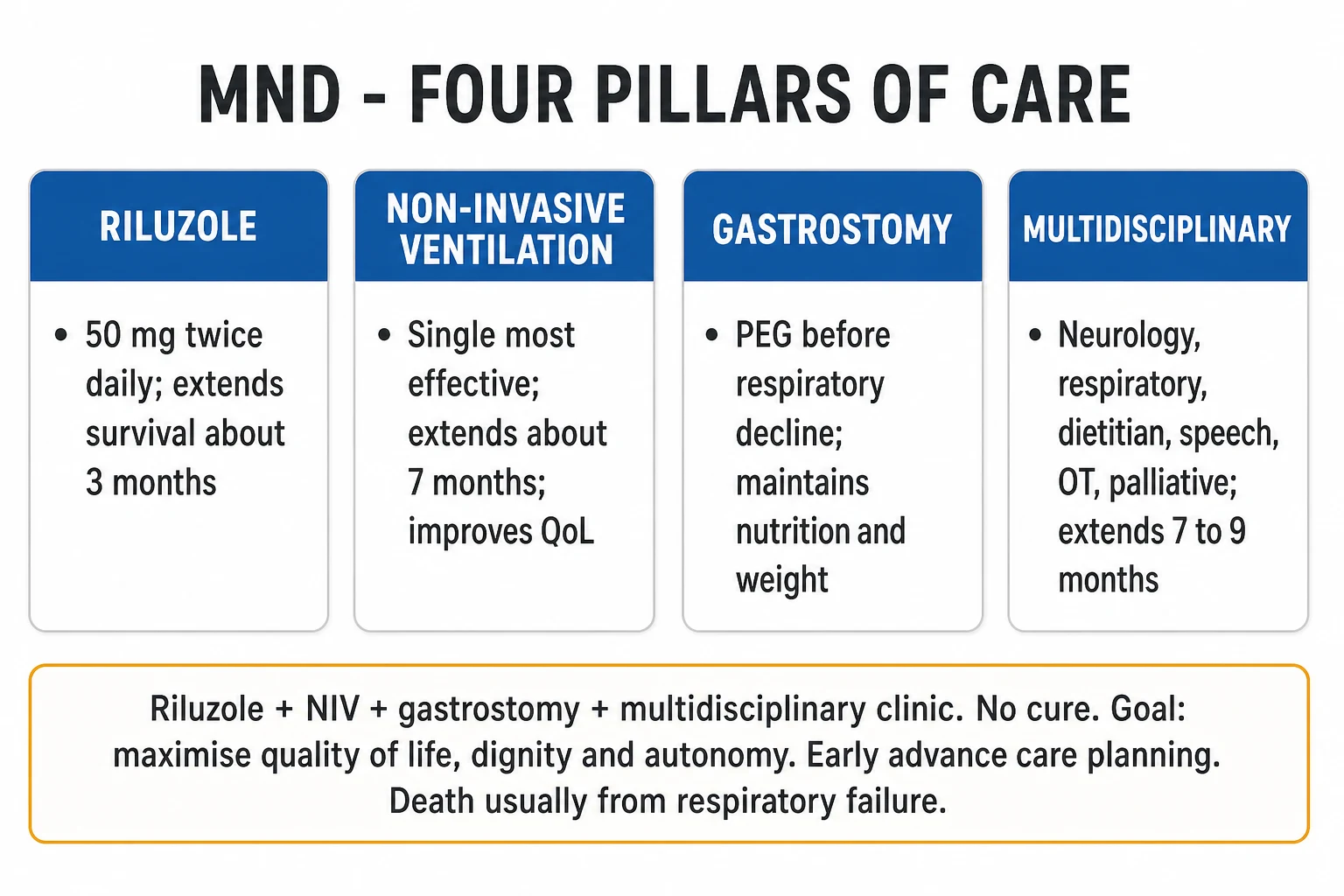
Treatment is multidisciplinary and symptomatic, with two disease-modifying drugs (riluzole, edaravone) that modestly extend survival or function, and three life-extending interventions (NIV, gastrostomy, multidisciplinary clinic) with substantially larger benefit. The goal is to maximise quality of life, dignity and autonomy, with early advance care planning. [1]
Disease-modifying drug therapy
Disease-modifying drugs — dose, evidence, what it does
Riluzole is the cornerstone. The Cochrane meta-analysis of four trials (1477 patients) found a hazard ratio of 0.80 (95 percent confidence interval 0.70 to 0.99) for tracheostomy-free survival, equivalent to prolonging median survival by about 2 to 3 months.[5] The standard dose is 50 mg orally twice daily, started at diagnosis. Contraindications are significant hepatic impairment (Child-Pugh C), pregnancy (caution; manufacturer advises avoid), and concomitant hepatotoxic drugs. Monitor ALT and AST at baseline, monthly for the first three months, then three-monthly; discontinue if transaminases exceed five times the upper limit of normal. Leucopenia is rare but warrants a full blood count if fever develops.
Edaravone is a free-radical scavenger given intravenously. The pivotal trial (Writing Group, Lancet Neurology 2017) randomised 137 selected patients (early stage, definite or probable ALS by revised El Escorial, FVC 80 percent or more, disease duration 2 years or less) to 60 mg intravenous edaravone or placebo for six cycles of 4 weeks each (2 weeks on, 2 weeks off). The edaravone group declined 2.49 ALSFRS-R points less over 24 weeks (p equals 0.0013) than placebo — a modest benefit in a highly selected subgroup.[6]
[1]Non-invasive ventilation — the single biggest survival intervention
Non-invasive ventilation (NIV) is the most effective intervention in ALS, more impactful than riluzole. The Bourke 2006 randomised trial (Lancet Neurology) showed that, in patients with normal or only mildly impaired bulbar function, NIV improved both survival (median benefit about 205 days, roughly 7 months) and quality of life; in those with severe bulbar impairment, NIV improved sleep-related symptoms but did not extend survival.[7]
NIV thresholds — when to start
Initiate NIV with a bilevel device, a nasal or orofacial mask titrated to comfort, starting at night and extending into the day as weakness progresses. Most patients tolerate NIV well once acclimatised; bulbar weakness and excess secretions are the main barriers. The decision to escalate to tracheostomy invasive ventilation is profound — it does prolong survival indefinitely but at the cost of locked-in dependence; this must be discussed in advance as part of goals-of-care planning.[2][7]
Nutritional support — gastrostomy timing
Weight loss in ALS reflects hypermetabolism, dysphagia and the catabolic state of progressive disease, and is itself an independent adverse prognostic factor. The ProGas study and subsequent guidelines recommend percutaneous endoscopic gastrostomy (PEG) or radiologically inserted gastrostomy (RIG) when there is symptomatic dysphagia, weight loss of 10 percent or more, or declining respiratory function — and ideally before FVC drops below 50 percent predicted, because procedural sedation risk rises sharply once respiratory function is impaired.[2]
Gastrostomy does not necessarily mean abandoning oral intake — most patients continue to eat for pleasure and supplement via the tube. A RIG is preferred when bulbar weakness makes endoscopy unsafe. High-calorie feeds, supplemental vitamins and minerals, and a careful fluid balance complete nutritional support. [1]
Symptomatic therapy — the daily life of an MND clinic
Symptom control is the bulk of MND care and is what determines day-to-day quality of life. Every symptom has a ladder. [1]
Spasticity
- Baclofen 5 to 25 mg three times daily (oral)
- Tizanidine 2 to 8 mg three times daily
- Intrathecal baclofen pump for severe spasticity
Cramps
- Magnesium, stretching, adequate hydration
- Gabapentin 100 to 300 mg at night
- Quinine sulphate has been WITHDRAWN for cramps (cardiotoxic)
Sialorrhoea (drooling)
- Hyoscine hydrobromide 0.4 mg daily as transdermal patch
- Glycopyrrolate 1 mg once to twice daily
- Botulinum toxin into submandibular and parotid glands (refractory)
Thick secretions
- Carbocisteine 750 mg three times daily
- Adequate hydration
- Mechanical suction device
Pseudobulbar affect
- Dextromethorphan-quinidine 20 mg/10 mg BD (where available)
- Amitriptyline 25 to 75 mg at night
- SSRIs (sertraline 50 to 100 mg daily)
Pain
- Paracetamol, then NSAIDs for nociceptive pain
- Opioids (oral morphine) for severe pain and terminal dyspnoea
- Neuropathic agents (gabapentin, pregabalin) where indicated
Communication
- Speech therapy early; voice banking before bulbar decline
- Augmentative and alternative communication (AAC)
- Eye-tracking devices for advanced disease
Constipation
- Laxatives (macrogol, senna)
- Adequate hydration, mobility
- Treat opioid-induced constipation proactively
Multidisciplinary clinic care — the framework
Attendance at a specialist multidisciplinary ALS clinic extends survival by about 7 to 9 months and improves quality of life, in two large observational studies (Van den Berg 2005; Traynor 2003, cited in Hardiman 2017).[1] The team is large by design:
The MND multidisciplinary team
NEURO-TEAM
leads the clinic, manages riluzole, monitors progression
FVC/SNIP, NIV initiation and weaning, infection management
swallow assessment, voice banking, AAC devices
weight monitoring, PEG feeds, supplements
aids and adaptations, home modifications, wheelchairs
stretching, range of motion, mobility aids
benefits, carer support, community services
advance care planning, symptom relief, end-of-life
depression, anxiety, ALS-FTD support, carer burden
Advance care planning — start at diagnosis, not at the end
The conversation about goals of care, place of death, ventilation preferences and symptom relief should begin early — at diagnosis, ideally, and revisited at each milestone. Key decisions: tracheostomy ventilation (yes/no — usually declined given locked-in outcome), place of death (home, hospice, hospital), advance directives and lasting power of attorney, and a symptom-relief plan for the terminal phase. The Gold Standards Framework (UK) and the AAN palliative care parameters converge on early, structured goals-of-care discussion.[2]
Specific Subtypes & Scenarios
Progressive bulbar palsy (PBP)
Predominant bulbar UMN and LMN signs in an older patient (often female), presenting with dysarthria, dysphagia and tongue wasting with fasciculations. PBP carries the worst prognosis of the MND subtypes — median survival is often under 2 years, because respiratory involvement develops early and aspiration is frequent. NIV is offered but is less effective in this group; PEG is almost always needed.[1][2]
Progressive muscular atrophy (PMA)
Pure lower motor neuron phenotype, limb onset, asymmetric wasting and weakness, with no UMN signs on examination. Often slower progression than classical ALS. The diagnostic trap is that many PMA patients eventually develop UMN signs, converting the diagnosis to ALS — PMA may simply be ALS presenting with its LMN component first.[1]
Primary lateral sclerosis (PLS)
Pure upper motor neuron phenotype — slowly progressive spastic paraparesis or tetraparesis with bulbar UMN signs (spastic dysarthria, brisk jaw jerk), no LMN signs, no wasting, no fasciculations. Near-normal life expectancy; however, a minority (perhaps 20 percent) develop LMN signs over years and convert to ALS. Differentiating PLS from hereditary spastic paraparesis and from a chronic cervical myelopathy is the diagnostic challenge.[1]
ALS-FTD
Combined ALS and frontotemporal dementia, frequently C9orf72-related, with reduced survival and complex care needs. Behavioural variant FTD (apathy, disinhibition, dietary change, loss of empathy) is commoner than language-predominant (PNFA) in ALS-FTD. The cognitive impairment impairs adherence to NIV and PEG and complicates capacity and end-of-life decisions.[8][9]
Flail arm and flail leg syndromes
Regional phenotypes with disproportionately slower progression and longer survival. The flail arm syndrome (man-in-a-barrel) presents with symmetric upper-limb weakness and wasting, sparing the legs and bulbar muscles for years. The flail leg syndrome presents with progressive lower-limb weakness. Both deserve explicit naming in the viva because they confound prognostication — patients survive far longer than the textbook 3 to 5 years. [1]
Juvenile ALS and rare childhood motor neuron disorders
Juvenile ALS is rare and usually genetic (often ALS2/alsin, SETX, FUS mutations). The riboflavin transporter deficiency syndromes (Brown-Vialetto-Van Laere, Fazio-Londe) cause childhood bulbar and LMN weakness and respond dramatically to high-dose riboflavin — a treatable mimic worth knowing. [1]
Complications & Pitfalls
The complications of ALS are the natural history of untreated disease, and they are the targets of symptomatic care. [1]
Respiratory failure
- The leading cause of death in ALS
- Surveil with FVC, SNIP, overnight oximetry at every visit
- Treat with NIV at FVC 50 percent or less or SNIP 40 cmH2O or less
Aspiration pneumonia
- Bulbar weakness pools secretions and food
- Prevent with PEG, posture, secretion management
- Treat early with antibiotics; recurrent aspiration worsens survival
Malnutrition and weight loss
- Hypermetabolism plus dysphagia
- Independent adverse prognostic factor
- Maintain weight with supplements and PEG
Venous thromboembolism
- From immobility; assess risk and prophylax
- Low-molecular-weight heparin in the immobilised patient
Pain
- Immobility, spasticity, cramps, constipation
- Ladder from paracetamol to morphine
- Treat the cause, not just the symptom
Depression and anxiety
- Common; address proactively
- SSRIs for depression and pseudobulbar affect
- Suicidal ideation demands urgent psychiatric input
Frontotemporal dementia
- 15 percent overt; impacts capacity and care
- Cognitive and behavioural support for patient and carer
- Affects adherence to life-extending interventions
The classic diagnostic pitfalls are: (1) missing cervical myelopathy coexisting with a peripheral neuropathy (always image the cervical spine); (2) mislabelling MMN as ALS (always look for conduction block); (3) ignoring Kennedy disease in a male with bulbar and LMN signs — check for gynaecomastia and sensory neuropathy, and send the genetic test; (4) overdiagnosing ALS on benign fasciculations — fasciculations alone, without weakness, wasting or EMG denervation, are benign; (5) treating "ALS-FTD" as a separate problem rather than recognising the cognitive impairment as part of the disease.[1][2]
Prognosis & Disposition
ALS is uniformly fatal. The headline prognostic numbers: [1]
Prognosis — the numbers that decide the viva
The favourable prognostic factors are: younger age at onset (under 50), limb onset (especially lower limb), longer diagnostic delay (a slow diagnostic interval implies a slow disease), PLS or PMA phenotype (versus classic ALS), slow ALSFRS-R decline (under 0.5 points per month), normal or near-normal vital capacity at diagnosis, and preserved weight.[1] The unfavourable factors are: bulbar onset (especially in older women), older age, respiratory onset, short diagnostic interval, frontotemporal dysfunction, low vital capacity at diagnosis, and rapid weight loss.
The end-of-life phase is signalled by rising respiratory symptoms despite NIV (or refusal/withdrawal of NIV), recurrent aspiration, increasing weakness, and declining cognition. The palliative priorities in this phase are: symptom relief (opioids for dyspnoea and pain, midazolam for anxiety, anticholinergics for secretions), withdrawal of NIV if requested (with concurrent opioid and benzodiazepine for comfort), support for the family, and placement (home, hospice or hospital per the advance plan). Prognosis once NIV is withdrawn is days to weeks.[2]
Special Populations
Familial ALS
About 10 percent of ALS is familial, transmitted most often autosomal dominantly. The C9orf72 hexanucleotide repeat expansion is the commonest cause (30 to 40 percent of familial ALS, 5 to 7 percent of sporadic ALS), followed by SOD1, TARDBP, FUS, UBQLN2, VCP, OPTN, ATXN2. Genetic counselling and predictive testing are now standard for at-risk relatives — particularly given the rise of gene-targeted therapy (tofersen for SOD1).[1][8]
Young-onset ALS
Onset under 40 is more often genetic, frequently slower, and more often limb onset. Send a broad gene panel (C9orf72, SOD1, FUS, TARDBP, ATXN2 at minimum) and counsel about family implications. Tofersen for SOD1-mutant ALS has changed the prognosis for this subgroup. [1]
Elderly ALS
Bulbar onset is more common, comorbidity complicates NIV and PEG decisions, and polypharmacy increases the burden of riluzole hepatic monitoring. The threshold for invasive interventions is calibrated to overall frailty and the patient's expressed goals. [1]
Pregnancy in ALS
Rare and generally not advised, because progression is usually rapid and care burden is high. Where pregnancy occurs, multidisciplinary planning (obstetrics, anaesthetics, neurology, neonatology) is essential; riluzole is contraindicated in pregnancy (animal teratogenicity).[2]
Coexisting disease
ALS may coexist with cancer (a paraneoplastic motor neuron syndrome is reported but rare), diabetes, and other major disease. The management principle is to treat the comorbidity and the ALS in parallel, and to recognise that comorbidity modifies the threshold for invasive ALS interventions. [1]
Evidence, Guidelines & Regional Differences
The evidence base for ALS therapy rests on a small number of pivotal randomised trials, supplemented by large observational cohorts and consensus guidelines. [1]
Riluzole
- Bensimon 1995 NEJM — the original positive trial (155 patients)
- Miller 2012 Cochrane — HR 0.80, prolongs survival by about 2 to 3 months
- Globally endorsed; the only universally recommended disease-modifying drug
Edaravone
- Writing Group 2017 Lancet Neurology — 2.49 ALSFRS-R point benefit at 24 weeks
- Selected subgroup only (early, definite or probable, FVC 80 percent or more)
- EMA-refused in Europe on benefit-cost grounds; FDA-approved USA
Non-invasive ventilation
- Bourke 2006 Lancet Neurology — survival benefit ~7 months in non-bulbar subgroup
- Quality of life improved across measures
- The single most effective intervention in ALS
Multidisciplinary care
- Traynor 2003; Van den Berg 2005 — clinic attendance extends survival by about 7 to 9 months
- Recommended by all guidelines
- The framework on which riluzole, NIV and PEG are layered
Genetic therapy (frontier)
- Tofersen (SOD1 antisense) — VALOR trial; FDA-approved 2023
- Reduces neurofilament light chain and slows progression
- C9orf72 and ATXN2-targeted therapies in trials
Regional guideline deltas
[1]Exam Pearls
Exam application bank (NEET-PG / INICET)
One-line answer
Motor neuron disease (MND/ALS) is a progressive neurodegenerative disorder that destroys BOTH upper motor neurons (UMN: spasticity, hyperreflexia, Babinski sign) AND lower motor neurons (LMN: weakness, wasting, fasciculations) while sparing sensation, eye movements and sphincter function. Annual incidence is about 2 per 100,000, peak onset 55 to 65 years, male-to-female ratio 1.5 to 1. About 10 percent is familial (C9orf72, SOD1, TARDBP, FUS). Diagnosis is clinical (Gold Coast 2020 criteria), supported by EMG (active denervation with chronic reinnervation across regions) and MRI to exclude mimics. Treatment is multidisciplinary: riluzole 50 mg twice daily extends median survival by about 2 to 3 months, non-invasive ventilation when FVC is 50 percent or less (or SNIP 40 cmH2O) extends survival by about 7 months and improves quality of life, gastrostomy maintains nutrition, and multidiscip [1]
Worked stems (answer without another resource)
Stem 1 — Classic presentation. Map symptoms to mechanism; name the first investigation and first treatment step with dose/route if drug therapy is standard. [1]
Stem 2 — Unstable / complicated. List red flags that force immediate resuscitation, theatre, ICU, antidote, or reperfusion — and what you do in the first 15 minutes. [1]
Stem 3 — Atypical group. Elderly, pregnancy, child, or immunocompromised: how presentation and thresholds change. [1]
Stem 4 — Differential trap. Name the three closest mimics and one discriminator for each. [1]
Stem 5 — Disposition. Who goes home with safety-netting, who is admitted, who needs HDU/ICU/theatre, and what follow-up is mandatory. [1]
Rapid viva checklist
- Definition + classification
- Pathophysiology chain
- Bedside signs / criteria
- Score with exact components (if any)
- Emergency bundle
- Definitive therapy with doses
- Complications of disease and of treatment
- Special populations
- Guideline/trial name if classic
- Three exam traps
Coverage self-check
If you cannot answer any stem above from this page alone, re-read the matching section — the page is intended to be self-sufficient for final-prof and NEET-PG/INICET questions on Motor Neuron Disease (ALS).
References
- [1]Hardiman O, Al-Chalabi A, Chio A, Corr EM, Logroscino G, Robberecht W, Shaw PJ, Simmons Z, van den Berg LH Amyotrophic lateral sclerosis Nat Rev Dis Primers, 2017.PMID 28980624
- [2]Radunovic A, Mitsumoto H, Leigh PN Clinical care of patients with amyotrophic lateral sclerosis Lancet Neurol, 2007.PMID 17884681
- [3]Shefner JM, Al-Chalabi A, Baker MR, Cui LY, de Carvalho M, Eisen A, et al. A proposal for new diagnostic criteria for ALS Clin Neurophysiol, 2020.PMID 32387049
- [4]Brooks BR El Escorial World Federation of Neurology criteria for the diagnosis of amyotrophic lateral sclerosis. Subcommittee on Motor Neuron Diseases/Amyotrophic Lateral Sclerosis of the World Federation of Neurology Research Group on Neuromuscular Diseases and the El Escorial Clinical limits of amyotrophic lateral sclerosis workshop contributors J Neurol Sci, 1994.PMID 7807156
- [5]Miller RG, Mitchell JD, Moore DH Riluzole for amyotrophic lateral sclerosis (ALS)/motor neuron disease (MND) Cochrane Database Syst Rev, 2012.PMID 22419278
- [6]Writing Group, Edaravone (MCI-186) ALS 19 Study Group Safety and efficacy of edaravone in well defined patients with amyotrophic lateral sclerosis: a randomised, double-blind, placebo-controlled trial Lancet Neurol, 2017.PMID 28522181
- [7]Bourke SC, Tomlinson M, Williams TL, Bullock RE, Shaw PJ, Gibson GJ Effects of non-invasive ventilation on survival and quality of life in patients with amyotrophic lateral sclerosis: a randomised controlled trial Lancet Neurol, 2006.PMID 16426990
- [8]Renton AE, Majounie E, Waite A, Simon-Sanchez J, Rollinson S, Gibbs JR, et al. A hexanucleotide repeat expansion in C9ORF72 is the cause of chromosome 9p21-linked ALS-FTD Neuron, 2011.PMID 21944779
- [9]Neumann M, Sampathu DM, Kwong LK, Truax AC, Micsenyi MC, Chou TT, et al. Ubiquitinated TDP-43 in frontotemporal lobar degeneration and amyotrophic lateral sclerosis Science, 2006.PMID 17023659