Neurology · Neurology
Multiple Sclerosis
Also known as MS · Disseminated sclerosis · Relapsing-remitting MS · RRMS · Primary progressive MS · PPMS
Multiple sclerosis (MS) is a chronic, immune-mediated, inflammatory demyelinating disease of the central nervous system (CNS) defined by episodes of neurological dysfunction (disseminated in time and space) that, untreated, progress to irreversible disability. It is the commonest non-traumatic cause of neurological disability in young adults. Onset is typically between 20 and 40 years, female predominance about 3:1. About 85% present with relapsing-remitting MS (RRMS); 10 to 15% have primary progressive MS (PPMS) from onset. Diagnosis is clinical and anchored on the McDonald 2017 criteria — MRI showing periventricular, juxtacortical, infratentorial and spinal-cord plaques (Dawson fingers), with CSF oligoclonal bands (intrathecal IgG) substituting for dissemination in time. Acute relapse = IV methylprednisolone 1000 mg daily for 3 to 5 days; plasma exchange if steroid-refractory. Disease-modifying therapy (DMT) ranges from interferon-beta / glatiramer, oral agents (fingolimod, dimethyl fumarate, teriflunomide) to monoclonal antibodies (natalizumab, ocrelizumab) — ocrelizumab is the only approved DMT for PPMS (ORATORIO). Always exclude NMOSD (AQP4) and MOGAD first — interferon worsens NMOSD.
On this page & tools
Your progress
Saved locally on this device.
Practise this topic
Exam tags
Red flags

Overview & Definition
Multiple sclerosis (MS) is a chronic, immune-mediated, inflammatory demyelinating disease of the central nervous system in which autoreactive lymphocytes attack oligodendrocyte-derived myelin, producing discrete inflammatory plaques scattered throughout the brain, optic nerves and spinal cord (the peripheral nervous system is spared — Schwann-cell myelin is not targeted).[1][3]
Two unifying principles define the disease and underpin every diagnostic criterion since Charcot: [1]
- Dissemination in space (DIS) — lesions occur in at least two of four typical CNS regions (periventricular, juxtacortical/cortical, infratentorial, spinal cord).
- Dissemination in time (DIT) — lesions occur at different points in time (a gadolinium-enhancing and a non-enhancing lesion, a new lesion on follow-up MRI, or CSF oligoclonal bands). [1]
The clinical skill in MS is not naming the disease — it is (i) recognising the typical syndromes of a young adult (optic neuritis, transverse myelitis, brainstem/cerebellar syndromes); (ii) applying the McDonald 2017 criteria to confirm the diagnosis at the first attack wherever possible; (iii) excluding mimics, especially NMOSD and MOGAD — because the wrong DMT (interferon in NMOSD) worsens disease; and (iv) choosing and monitoring a disease-modifying therapy (DMT) with its specific risks (PML with natalizumab, autoimmunity with alemtuzumab, teratogenicity with teriflunomide).[2][4]
Classification
MS is classified by its clinical course. The four classic phenotypes were refined in 2013 by Lublin and Reingold into a two-axis descriptor — adding activity (relapses or new MRI lesions) and progression (worsening independent of relapse) on top of the relapsing/progressive backbone.[1]
Clinically isolated syndrome (CIS)
- First clinical episode of CNS demyelination (optic neuritis, transverse myelitis, brainstem) monophasic, under 24 h to weeks
- High risk of conversion to MS if MRI shows silent lesions — the 2017 McDonald criteria can diagnose MS at this stage if DIS + DIT are met
- Not yet MS by definition until dissemination in time is demonstrated
Relapsing-remitting MS (RRMS)
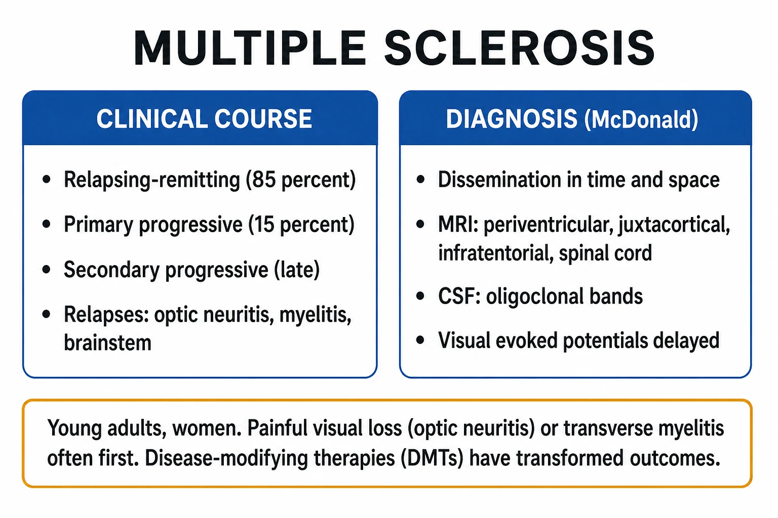
- About 85% of patients at onset
- Clearly defined attacks (relapses) with full or partial recovery, separated by periods of disease stability (remission)
- No progression between attacks
- Histologically: active gadolinium-enhancing inflammatory lesions
Secondary progressive MS (SPMS)
- Follows an initial relapsing-remitting course
- Gradual neurological worsening begins, with or without superimposed relapses
- Median time to transition historically 10 to 20 years from onset (longer in the DMT era)
- Recognised by insidious progression over at least 6 months independent of relapses
Primary progressive MS (PPMS)
- About 10 to 15% — gradual worsening from disease onset, no distinct relapses
- Older onset (around 40 years), more equal sex ratio, more spinal-cord predominant
- Worse prognosis; ocrelizumab is the only approved DMT (ORATORIO)
- Histologically: less inflammation, more smouldering neurodegeneration and meningeal lymphoid aggregates
Radiologically isolated syndrome (RIS)
- Incidental MRI white-matter abnormalities consistent with MS in an asymptomatic person
- Not by itself MS; treated only if clinical conversion occurs
- CSF oligoclonal bands or spinal-cord lesions raise conversion risk

The 2013 Lublin descriptors in full:[1]
- Clinical course — RRMS, SPMS, PPMS (and CIS, RIS).
- Activity — active (relapses, gadolinium-enhancing lesions or new/enlarging T2 lesions on MRI over a defined period) vs not active.
- Progression (progressive phenotypes only) — progressing (objectively worsening disability independent of relapse over a defined period) vs not progressing. [1]
When does RRMS convert to SPMS? The transition is insidious and often retrospectively diagnosed: a sustained increase in disability (rising EDSS) over at least 6 to 12 months that is independent of relapses, often with reducing relapse frequency, more cognitive and progressive-motor burden, and fewer gadolinium-enhancing lesions. Median time from onset was 10 to 20 years in the pre-DMT era and is lengthening with early high-efficacy therapy.[4]
Epidemiology & Risk Factors
MS affects about 2.8 million people worldwide and is the commonest non-traumatic cause of neurological disability in young adults. Typical onset is 20 to 40 years (mean around 30); female-to-male ratio in RRMS is about 3:1; PPMS has a more equal sex ratio (about 1:1) and later onset.[1][3]
The latitude gradient — prevalence rises sharply with distance from the equator (e.g. under 5 per 100,000 near the equator vs over 200 per 100,000 in northern Europe, Canada and the northern US). Migration studies show that the age at migration matters: those who move from high- to low-risk regions before puberty acquire the lower risk of their new home, while those who migrate after age 15 retain the higher risk of their birthplace — strongly implicating an adolescent environmental exposure (chiefly Epstein-Barr virus and vitamin D / UV sunlight).[3][4]
MS — key numbers
Genetic and environmental risk factors: [1]
- Genetic — strongest association is HLA-DRB1*15:01 (the major MS susceptibility allele); over 200 other non-HLA loci (mostly immune genes) contribute small effects. Family history raises risk (general population about 0.1%, rising to about 2 to 4% with one affected first-degree relative, and to about 25 to 30% in a monozygotic twin of an MS patient).
- Epstein-Barr virus (EBV) — infection is almost universal in MS; the risk after infectious mononucleosis is multi-fold higher than after asymptomatic primary EBV. EBV infection is now considered a near-necessary trigger for MS.
- Vitamin D / sunlight — low UV exposure and low 25-hydroxy-vitamin D levels (and the latitude gradient) are independent risk factors.
- Smoking — raises the risk of developing MS, accelerates conversion from CIS to MS, and accelerates progression to SPMS.
- Adolescent obesity — independent risk factor, especially in girls.
- Female sex — strong risk factor for relapsing disease. [1]
What twin concordance tells you: monozygotic concordance (about 25 to 30%) is much higher than dizygotic (about 3 to 5%), confirming a genetic contribution, but the fact that it is far below 100% proves MS is not a simple Mendelian disease — an environmental trigger (EBV, vitamin D, smoking) is required on a susceptible genetic background. [1]
Pathophysiology
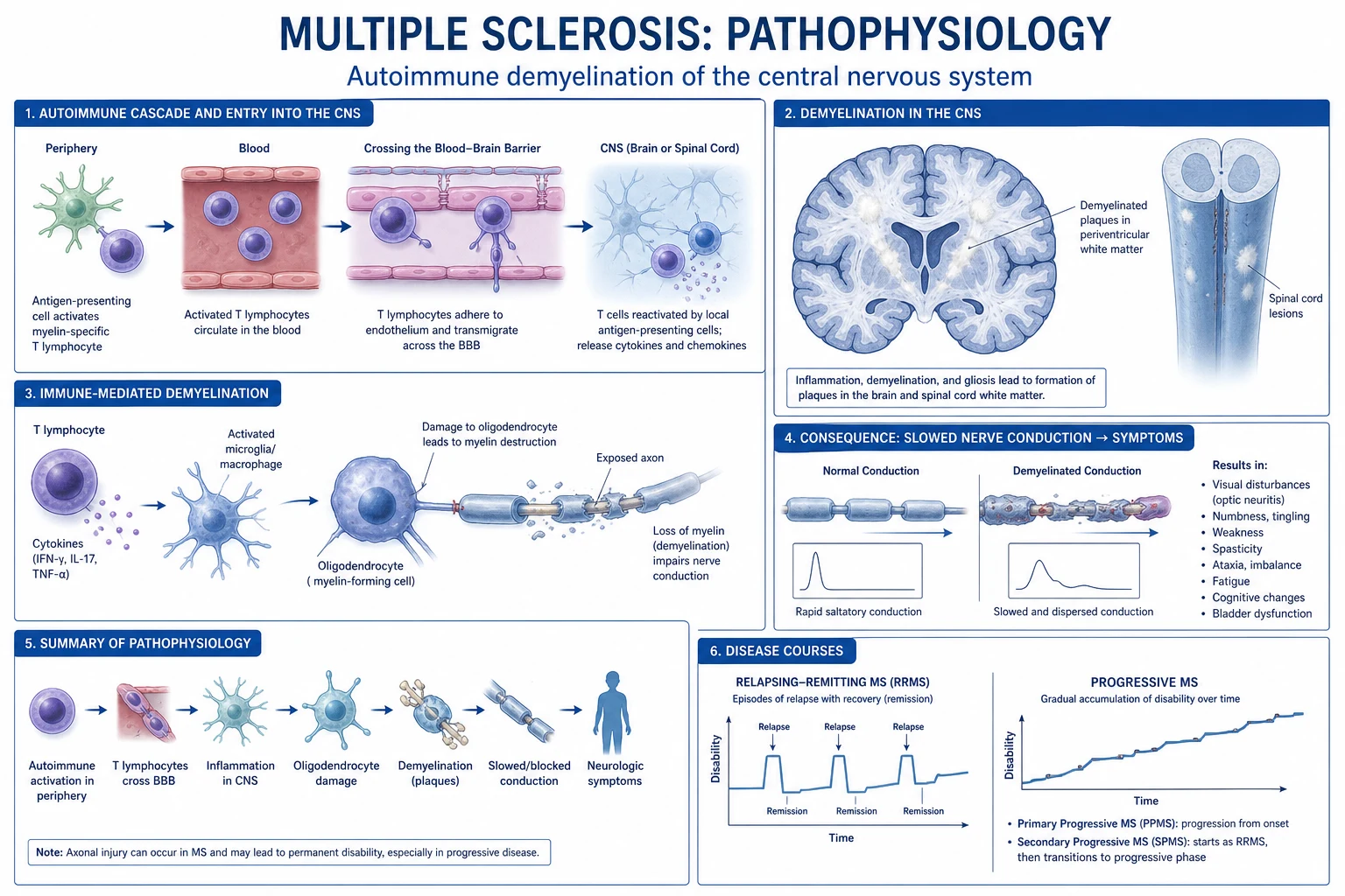
MS is immune-mediated: a breakdown of self-tolerance to myelin antigens (myelin basic protein, proteolipid protein, myelin oligodendrocyte glycoprotein) triggers an autoreactive T-cell response that attacks the CNS.[1][3]
The immunopathological sequence of an acute MS plaque: [1]
- Loss of self-tolerance in a genetically susceptible host (HLA-DRB1*15:01) after an environmental trigger (EBV, low vitamin D).
- Autoreactive CD4+ T-helper cells — chiefly Th1 (IFN-gamma) and Th17 (IL-17) subsets — are activated in the periphery and cross the blood-brain barrier (BBB) (whose disruption, with gadolinium leakage, is the radiological hallmark of an active lesion).
- Inside the CNS, T-cells are re-stimulated by antigen-presenting cells (microglia, macrophages, dendritic cells) presenting myelin peptides on HLA class II.
- Macrophage- and antibody-mediated myelin destruction: CD8+ cytotoxic T-cells, macrophages and antibodies (with complement) strip myelin from axons. Oligodendrocytes (the myelinating cells) are lost; CD4+ T-cells also recruit B-cells and plasma cells that produce oligoclonal IgG within the CNS.
- Acute plaque histology — perivenular inflammatory infiltrate (T-cells, macrophages) with demyelination but relative preservation of axons (the lesion is demyelinating, not primarily axonal); oedema and gadolinium enhancement.
- Chronic plaque histology — inflammation resolves; gliosis, astrogliosis and permanent axonal loss dominate; the plaque becomes a sharply demarcated grey-pink glial scar (sclerosis). Shadow plaques (thinly remyelinated axons) indicate partial remyelination. [1]
Two overlapping disease processes operate across the MS course:[1][4]
- Focal inflammatory relapses — driven by autoreactive T- and B-cells, BBB breakdown, gadolinium-enhancing lesions. This dominates early RRMS and is the target of relapse steroids and most DMTs.
- Diffuse smouldering neurodegeneration — meningeal lymphoid aggregates, microglial activation, mitochondrial injury, chronic axonal loss and brain atrophy. This dominates progressive MS (PPMS, SPMS) and is the substrate for the irreversible disability that current DMTs only partly address. [1]
How demyelination produces symptoms (the four mechanisms — high-yield for viva): [1]
- Conduction block — demyelinated axons lose their saltatory conduction; impulses fail to propagate. This is the mechanism of an acute relapse deficit (e.g. weakness in transverse myelitis, visual loss in optic neuritis).
- Ectopic discharges — demyelinated axons fire spontaneously, producing positive symptoms: Lhermitte's sign (electric-shock sensation down the spine on neck flexion from a cervical-cord plaque) and paroxysmal symptoms (trigeminal neuralgia, tonic spasms).
- Heat sensitivity (Uhthoff's phenomenon) — small rises in body temperature (exercise, hot bath, fever) further impair conduction in marginally demyelinated axons, transiently worsening pre-existing deficits.
- Axonal loss — irreversible; explains why some deficits never recover and why progressive disability accumulates. The axon is the unit of permanent disability, which is why protecting axons (early, high-efficacy DMT) is the modern therapeutic goal. [1]
Clinical Presentation
MS classically presents in a young woman (20 to 40 years) with episodic neurological dysfunction that comes on over hours to days, persists for over 24 hours, and then partially or completely recovers over weeks. The presentation reflects the location of the plaque.[1][4]
Three classic first-presentation syndromes (know these cold): [1]
1. Optic neuritis — the single most common first event. [1]
- Painful, unilateral, subacute visual loss — periocular pain exacerbated by eye movement.
- Reduced colour vision (dyschromatopsia) — red desaturation (reds look washed-out) is a sensitive early sign; test with each eye using a red pin.
- Relative afferent pupillary defect (RAPD) — the swinging-flashlight test shows a dilating pupil when light swings to the affected eye (afferent limb defect; optic nerve lesion).
- Visual field defect — typically a central scotoma; diffuse depression also common.
- Fundus — papillitis (swollen disc) if the lesion is anterior (intraocular); retrobulbar (normal disc, "patient sees nothing, doctor sees nothing") if posterior. Optic atrophy develops later (pale disc).
- Recovery begins within weeks; over 90% recover useful vision. Severe bilateral optic neuritis with poor recovery should prompt NMOSD/MOGAD exclusion. [1]
2. Partial transverse myelitis — a focal spinal-cord plaque. [1]
- Paraparesis or monoparesis with upper motor neuron (UMN) signs (spasticity, hyperreflexia, extensor plantar/Babinski response); an acute lesion may transiently produce flaccidity and areflexia ("spinal shock").
- Sensory level — a band of altered sensation at the level of the lesion with loss below.
- Sphincter disturbance — bladder urgency, hesitancy, retention; constipation.
- Lhermitte's sign if the lesion is in the cervical cord. [1]
3. Brainstem / cerebellar syndromes: [1]
- Internuclear ophthalmoplegia (INO) — weakness of adduction on horizontal gaze (medial longitudinal fasciculus lesion) with nystagmus in the abducting eye; bilateral INO in a young adult is highly suggestive of MS. Convergence is preserved.
- Diplopia, vertigo, facial sensory loss, lower-motor-neuron facial palsy.
- Cerebellar signs — ataxia, intention tremor, dysmetria (finger-nose), dysdiadochokinesia, scanning dysarthria (explosive, broken-up speech). [1]
Two eponymous phenomena (high-yield — always asked): [1]
- Lhermitte's sign — an electric-shock-like sensation radiating down the spine and sometimes into the limbs on neck flexion; mechanism is mechanical irritation of demyelinated axons in a cervical cord plaque. (It is a sign, not a symptom to be asked about — demonstrate.)
- Uhthoff's phenomenon — a transient worsening of existing neurological symptoms (especially vision) on raising body temperature (exercise, hot bath, fever); mechanism is heat-induced conduction block in demyelinated axons. Resolves on cooling. [1]
Non-focal / "invisible" symptoms (often the most disabling, easily missed): [1]
- Fatigue — the commonest and most disabling symptom for many patients; distinct from depression or sleepiness.
- Spasticity — velocity-dependent increased tone, painful flexor spasms.
- Bladder dysfunction — overactive (urgency, frequency, incontinence) from detrusor overactivity; or underactive (retention, overflow) — and detrusor-sphincter dyssynergia.
- Bowel — constipation (common).
- Sexual dysfunction — erectile dysfunction in men; loss of libido and arousal in women.
- Cognitive impairment — slowed processing speed, attention and memory deficits in 40 to 70%; can occur early.
- Depression — common; suicide risk is elevated — always screen.
- Heat intolerance and exercise-related symptom worsening.
- Neuropathic pain — trigeminal neuralgia (bilateral in MS), Lhermitte, painful tonic spasms. [1]
Atypical presentations (the examiner will test these): [1]
- Paediatric MS — first event often ADEM-like (encephalopathy, multifocal deficits); more inflammatory, very high relapse rate, better recovery, but accrues disability over a longer lifespan. Refer to a paediatric MS specialist.
- Late-onset MS / PPMS — onset over age 40 with progressive (rather than relapsing) myelopathy, often misdiagnosed as cervical spondylosis or vascular disease; more often male and PPMS.
- Tumefactive MS — a single large (over 2 cm) mass-like plaque with oedema and ring enhancement mimicking a brain tumour or abscess; biopsy avoided if MR spectroscopy / perfusion and evolution are typical.
- Trigeminal neuralgia in a young adult (under 40), or bilateral trigeminal neuralgia — strongly suggests MS.
- Pregnancy-related relapse pattern — relapse rate falls in the third trimester and rebounds in the first 3 months postpartum (see Special Populations). [1]
Differential Diagnosis
A young adult with recurrent CNS demyelinating events has a wide differential. The critical distinction is MS vs NMOSD vs MOGAD because the treatments diverge — interferon-beta worsens NMOSD.[2][4]
Multiple sclerosis (MS)
- Periventricular Dawson fingers, juxtacortical, infratentorial, short (under 2 segments) spinal-cord lesions
- CSF: oligoclonal bands (CSF not serum); IgG index raised
- AQP4 and MOG antibodies NEGATIVE
- Responds to interferon-beta, glatiramer, natalizumab, ocrelizumab
Neuromyelitis optica spectrum disorder (NMOSD, AQP4)
- AQP4-IgG POSITIVE (sensitive and specific)
- SEVERE bilateral optic neuritis with poor recovery
- LONGITUDINALLY extensive transverse myelitis (LETM, over 3 vertebral segments, central cord)
- Area postrema syndrome (hiccups, nausea, vomiting); brain lesions around 4th ventricle
- WORSENED by interferon-beta; treat with rituximab, mycophenolate, eculizumab, satralizumab
MOG-antibody disease (MOGAD)
- MOG-IgG1 POSITIVE (cell-based assay)
- Often bilateral optic neuritis with disc swelling; ADEM-like phenotype in children
- LETM but lesions often span the conus; better recovery than NMOSD
- Generally monophasic or relapsing; lower long-term disability; rituximab/mycophenolate for relapsing disease
Acute disseminated encephalomyelitis (ADEM)
- Monophasic, post-infectious or post-vaccinal, predominantly CHILDREN
- ENCEPHALOPATHY (confusion, drowsiness) is required
- Diffuse, bilateral, poorly marginated lesions; grey-matter involvement
- Usually full recovery; steroids first-line
Neurosarcoidosis
- Cranial nerve (especially facial / optic) palsy, hypothalamic/pituitary involvement, aseptic meningitis
- ACE raised; chest X-ray hilar lymphadenopathy; histology non-caseating granulomas
- Leptomeningeal enhancement on MRI; responds to steroids
Behçet disease, neuroborreliosis (Lyme), SLE cerebritis, HTLV-1
- Behçet: oral/genital ulcers, uveitis, meningoencephalitis
- Lyme: erythema migrans, arthritis, cranial-neuropathy; antibodies positive
- SLE: ANA/dsDNA, systemic features, seizures/psychosis
- HTLV-1: tropical spastic paraparesis, slowly progressive myelopathy
Non-demyelinating mimics
- Cervical spondylotic myelopathy: older, degenerative cord compression on MRI, no OCB
- Vitamin B12 deficiency (subacute combined degeneration): macrocytosis, low B12, peripheral neuropathy, posterior column signs
- Leber hereditary optic neuropathy (LHON): painless bilateral sequential visual loss, matrilineal inheritance, mtDNA mutation
- Stroke: sudden onset, vascular territory; functional disorder: inconsistent signs, normal MRI
Mimics to exclude by blood test BEFORE starting a DMT: vitamin B12, ANA / extractable nuclear antigens, ACE (sarcoid), AQP4-IgG (NMOSD), MOG-IgG (MOGAD), HIV, syphilis (Treponema pallidum serology), Lyme serology (where endemic), TSH.[2]
Clinical & Bedside Assessment
A focused MS examination covers vision, eye movements, cranial nerves, motor, cerebellar, sensory, gait and Lhermitte's manoeuvre:[4]
- Visual acuity (Snellen, with and without pinhole), colour vision (Ishihara; red desaturation with a red pin in each eye), visual fields (confrontation).
- RAPD via the swinging-flashlight test — swing light between eyes; the affected pupil dilates when light reaches it (because the afferent input is weaker), indicating an optic-nerve lesion. The single most useful bedside sign of optic neuritis.
- Eye movements for internuclear ophthalmoplegia (impaired adduction, abducting nystagmus, preserved convergence) and other ocular motor palsies.
- Cranial nerves including facial sensation (test for trigeminal neuralgia trigger zones), facial power, hearing, palatal and tongue movement.
- Motor — tone (spasticity), power, reflexes (hyperreflexia), plantar response (extensor/Babinski in UMN lesion), abdominal reflexes (often lost below a cord lesion).
- Cerebellar — finger-nose (dysmetria, intention tremor), heel-shin, rapid alternating movements (dysdiadochokinesia), gait (wide-based ataxic), scanning dysarthria.
- Sensory — pinprick and temperature (spinothalamic), joint position and vibration (posterior columns) — map a sensory level if myelopathy.
- Gait — including tandem walking.
- Lhermitte's manoeuvre — ask the patient to flex the neck; report of an electric-shock down the spine is a positive Lhermitte's sign (cervical cord plaque).
- Bladder scan (post-void residual) if urinary symptoms. [1]
Expanded Disability Status Scale (EDSS) — the standard MS disability score (Kurtzke), the number quoted in every clinic letter and trial:[1]
- 0 = normal neurological examination.
- 1.0 to 3.5 = mild disability, fully ambulatory (most RRMS patients sit here).
- 4.0 = fully ambulatory without aid, up and about over 12 hours a day despite relatively severe disability.
- 5.0 = ambulatory without aid for about 200 m; disability severe enough to impair full daily activities.
- 6.0 = needs a unilateral aid (cane / crutch / single walker) to walk about 100 m — the landmark milestone (often the trigger for DMT escalation and rehabilitation).
- 6.5 = bilateral aid to walk about 20 m.
- 7.0 = unable to walk beyond 5 m even with aid; essentially wheelchair-bound; wheels self and transfers alone.
- 8.0 = essentially restricted to bed or chair or perambulated in wheelchair; may be out of bed much of the day; retains many self-care functions; generally effective use of arms.
- 8.5 = hardening of 8.0 — restricted to bed much of the day; has some effective use of arm(s).
- 9.0 = helpless bed patient; can communicate and eat.
- 9.5 = totally helpless bed patient; unable to communicate or eat/swallow.
- 10.0 = death due to MS. [1]
Bedside red flags pointing AWAY from MS toward NMOSD / MOGAD: severe or bilateral optic neuritis with poor recovery; longitudinally extensive transverse myelitis (cord lesion over 3 vertebral segments); area postrema syndrome (intractable hiccups / nausea); rapid and severe disability accrual; AQP4-IgG or MOG-IgG positivity. [1]
Investigations
MS is diagnosed clinically, with MRI support — there is no single confirmatory test.[1][2]
First-line investigations for suspected MS: [1]
- MRI brain and whole spinal cord with gadolinium — the cornerstone.
- CSF analysis (lumbar puncture) — when MRI is inconclusive or NMOSD/MOGAD is in question.
- Visual evoked potentials (VEP) — delayed P100 latency confirms optic-nerve demyelination even after recovery (useful for historic DIS).
- Bloods to exclude mimics — FBC, ESR, CRP, vitamin B12, ANA, ACE, TSH, AQP4-IgG, MOG-IgG, HIV, syphilis, Lyme (where endemic). [1]
Typical MRI lesion locations and signs: [1]
- Periventricular ovoid lesions oriented perpendicular to the ventricle — Dawson fingers (perivenular inflammation extending along deep medullary veins).
- Juxtacortical / cortical lesions (cortical lesions added to the 2017 McDonald DIS criteria).
- Infratentorial — cerebellar peduncles, pons, brainstem.
- Spinal cord — typically cervical, short (under 2 vertebral segments), peripheral, asymmetric (contrast with the longitudinally extensive central cord lesion of NMOSD).
- Gadolinium enhancement signifies an active lesion with blood-brain-barrier breakdown (i.e. dissemination in time if it co-exists with non-enhancing lesions).
- T1 black holes = permanent axonal loss; brain atrophy = neurodegeneration. [1]
CSF findings in MS: [1]
- Oligoclonal bands present in CSF but NOT in a matched serum sample = intrathecal IgG synthesis — the hallmark (positive in over 95% of established MS).
- Raised IgG index (over 0.7) — corroborates intrathecal synthesis.
- Mild lymphocytosis (under 50 cells/microL), normal glucose, normal or mildly raised protein.
- Matched serum must always be taken — bands in both CSF and serum indicate a systemic (not intrathecal) inflammatory response and argue against MS. [1]
The McDonald 2017 diagnostic criteria (reproduce verbatim — the high-yield exam answer):[2]
Dissemination in space (DIS) — at least one T2-hyperintense lesion in at least two of four areas: (1) periventricular, (2) juxtacortical/cortical, (3) infratentorial, (4) spinal cord. (CSF oligoclonal bands can substitute for DIS in PPMS.) [1]
Dissemination in time (DIT) — demonstrated by any of:
- Simultaneous presence of gadolinium-enhancing and non-enhancing lesions at any time, OR
- A new T2-hyperintense and/or gadolinium-enhancing lesion on follow-up MRI, with reference to the baseline scan, irrespective of timing, OR
- Presence of CSF-specific oligoclonal bands (can substitute for DIT — a key 2017 change). [1]
Key 2017 changes from McDonald 2010:[2]
- CSF oligoclonal bands can substitute for dissemination in time in all phenotypes (not just PPMS).
- Symptomatic lesions (those corresponding to the presenting syndrome) now count toward DIS and DIT (they were excluded in 2010).
- Cortical lesions added alongside juxtacortical lesions in the DIS criteria.
- Allows diagnosis of MS at the first attack (CIS) if criteria are met — enabling earlier DMT. [1]
Diagnostic criteria for primary progressive MS (PPMS): at least one year of disease progression (retrospectively or prospectively determined), plus at least two of the following: (a) at least one T2-hyperintense brain lesion in a typical MS region (DIS in brain); (b) at least two T2-hyperintense spinal-cord lesions (DIS in cord); (c) presence of CSF-specific oligoclonal bands (intrathecal IgG).[2]
When are AQP4 and MOG antibodies checked? In every patient with suspected MS who has an atypical feature (severe/bilateral optic neuritis, longitudinally extensive transverse myelitis, area-postrema syndrome, poor recovery, monophasic ADEM-like picture) — and ideally in all newly diagnosed patients before starting a DMT, because misdiagnosing NMOSD as MS and giving interferon worsens the disease. [1]
Management — Resuscitation
Most MS patients are not acutely unwell at diagnosis, but an acute severe relapse (transverse myelitis with paralysis, brainstem relapse with respiratory compromise, optic neuritis threatening vision) is a medical emergency requiring a structured response:[3][4]
- ABCDE — secure airway and breathing (high cervical cord lesion can compromise respiration); assess circulation and disability.
- Screen for and treat infection — the commonest relapse trigger is a urinary tract or respiratory infection; send urine and blood cultures, FBC, CRP; treat promptly with antibiotics.
- Urgent MRI brain and spine with gadolinium — to confirm an acute demyelinating lesion and exclude a mimic (compressive myelopathy, infarct, abscess).
- Urgent neurology referral and admission for severe relapse (paralysis, vision-threatening optic neuritis, sphincter involvement). [1]
Acute relapse regimen — high-dose IV methylprednisolone: [1]
- Drug: methylprednisolone.
- Dose: 1000 mg (1 g) daily.
- Route: intravenous infusion over at least 30 to 60 minutes (oral high-dose 500 mg to 2 g daily for 3 to 5 days is an alternative in milder relapses per NICE).
- Duration: 3 to 5 consecutive days.
- Rationale: speeds recovery from the relapse by suppressing inflammation and closing the blood-brain barrier.
- Limits: does NOT change long-term outcome or final disability — it buys time and function, not disease modification. Discuss this honestly with patients.
- PPI cover (gastroprotection), mood and glucose monitoring, blood-pressure monitoring, and bone health with repeated courses (osteoporosis risk). [1]
Steroid-refractory severe relapse — add plasma exchange (PLEX): [1]
- Regimen: 5 to 7 exchanges over 10 to 14 days (typically every other day), replacing about 1 to 1.5 plasma volumes per exchange with albumin/fresh-frozen plasma.
- Indications: severe, steroid-refractory relapse (especially optic neuritis and transverse myelitis); tumefactive demyelination; steroid-resistant NMOSD/MOGAD attack.
- Rationale: removes circulating antibodies, complement and cytokines — addresses the humoral component. [1]
Corticosteroid safety measures (every course): gastroprotection (PPI), blood glucose monitoring (steroid hyperglycaemia), blood pressure monitoring, mood monitoring (steroid-induced psychosis / depression, especially relevant in MS where depression is common), bone protection with repeated courses (calcium/vitamin D, bisphosphonate if needed), and screening for latent infection (TB, hepatitis). [1]
Management — Definitive & Stepwise
MS management has three pillars: (1) treatment of acute relapses (above); (2) disease-modifying therapy (DMT) to prevent relapses and disability; (3) symptomatic and rehabilitative care.[1][4][5][6]
The DMT classes and escalation ladder (know each drug, its dose, route, and key monitoring): [1]
A. Injectable platform drugs (mild-to-moderate efficacy, excellent safety — first-line in many): [1]
- Interferon-beta (IFN-beta-1a IM weekly, IFN-beta-1a SC three-times-weekly, IFN-beta-1b SC alternate days) — reduces relapse rate by about one-third. Side effects: flu-like illness after each injection, injection-site reactions, depression, transaminitis, neutralising antibodies reducing efficacy.
- Glatiramer acetate — 20 mg subcutaneously daily (or 40 mg three times weekly). Similar efficacy to IFN-beta; side effects: injection-site reactions, benign immediate post-injection systemic reaction (chest tightness, flushing). Considered the safest in pregnancy if DMT is needed. [1]
B. Oral agents (moderate-to-high efficacy): [1]
- Fingolimod 0.5 mg orally once daily — a sphingosine-1-phosphate receptor modulator that sequesters lymphocytes in lymph nodes. First-dose cardiac monitoring for at least 6 hours (bradycardia, atrioventricular block); risk of macular oedema (baseline and 3-to-4-month eye exam), varicella zoster (check VZV serology and vaccinate before starting), elevated liver enzymes, and a small PML risk. Teratogenic — contraception required.
- Dimethyl fumarate 240 mg orally twice daily — activates the Nrf2 antioxidant pathway. Side effects: flushing and GI upset (take with aspirin/food), lymphopenia (monitor lymphocyte count; PML risk if profound), elevated transaminases.
- Teriflunomide 7 mg or 14 mg orally once daily — inhibits pyrimidine synthesis in proliferating lymphocytes. Strongly teratogenic (long washout or cholestyramine needed pre-conception); monitor LFTs; causes alopecia, hypertension, peripheral neuropathy. [1]
C. Monoclonal antibodies (high efficacy — escalation or induction): [1]
- Natalizumab 300 mg IV every 4 weeks — anti-alpha-4 integrin blocks T-cell crossing of the BBB. High efficacy (reduces relapses by over two-thirds). Risk of progressive multifocal leukoencephalopathy (PML) stratified by anti-JC virus antibody status and treatment duration — JCV-negative patients carry very low risk (under 1 in 10,000); JCV-positive patients over 2 years on treatment carry risk up to about 1 in 100.[7]
- Ocrelizumab 600 mg IV every 6 months (as two 300 mg infusions two weeks apart) — anti-CD20 depleting B-cells. Approved for RRMS (OPERA I/II) AND for PPMS (ORATORIO — the only approved DMT for PPMS).[5][6] Infusion reactions, hepatitis B reactivation (screen HBV before starting), modest infection risk, low PML risk. ORATORIO showed ocrelizumab reduced the risk of confirmed disability progression by 24% versus placebo in PPMS.
- Alemtuzumab 12 mg/day IV for 5 days (course 1), then 3 days 12 months later (course 2) — anti-CD52 depleting lymphocytes; very high efficacy used as induction in highly active disease. Major risk is secondary autoimmunity (immune thrombocytopenia, thyroid disease, Goodpasture-like glomerulonephritis) — requires monthly blood and urine monitoring for 4 years after the last course. Generally second-line now given its safety profile.
- Mitoxantrone — a chemotherapeutic, now rarely used (cardiotoxicity, treatment-related acute leukaemia); largely superseded.
Escalation triggers and the "early high-efficacy" paradigm: the classic escalation model steps from platform drugs to oral agents to monoclonal antibodies at the first sign of breakthrough disease (clinical relapse or new MRI activity). The modern induction / early-high-efficacy paradigm argues for starting a high-efficacy DMT (natalizumab, ocrelizumab, alemtuzumab) early in highly active MS, on the rationale that preventing early axonal loss preserves long-term function. AHSCT (autologous haematopoietic stem cell transplant) is reserved for very active, treatment-refractory disease in specialist centres.[5]
Symptomatic management (know the drug, dose and rationale): [1]
- Spasticity — baclofen (start 5 mg tds, titrate to 60 to 100 mg/day in divided doses; intrathecal baclofen pump for severe), tizanidine 2 to 4 mg up to 36 mg/day, stretching/physiotherapy; botulinum toxin for focal spasticity.
- Fatigue — amantadine 100 mg once or twice daily (modest benefit); treat anaemia, thyroid and sleep disorders; graded exercise.
- Overactive bladder — oxybutynin 5 mg two to three times daily (or tolterodine, solifenacin, mirabegron); clean intermittent self-catheterisation if retention/high post-void residual.
- Neuropathic pain — gabapentin (start 300 mg at night, titrate to 1800 to 3600 mg/day) or pregabalin, amitriptyline 10 to 75 mg at night, carbamazepine for trigeminal neuralgia.
- Erectile dysfunction — sildenafil 25 to 100 mg before intercourse.
- Depression — SSRIs (sertraline, citalopram); CBT; screen for suicidality.
- Constipation — bulk-forming laxatives, macrogols. [1]
DMT monitoring: [1]
- MRI surveillance — baseline, then typically at 6 months after starting a new DMT, then annually (more frequently if breakthrough activity suspected); compare for new T2 lesions or gadolinium enhancement.
- JCV antibody status (natalizumab) — re-test every 6 months (seroconversion raises PML risk); stratify by titre and treatment duration.
- Lymphocyte counts (dimethyl fumarate, fingolimod) — every 3 to 6 months; suspend if profound lymphopenia.
- Liver function — baseline and periodically (interferon, teriflunomide, dimethyl fumarate).
- Eye exam — baseline and 3-to-4 months (fingolimod, for macular oedema).
- HBV / TB screen before ocrelizumab / rituximab (reactivation risk). [1]
Rebound risk — stopping natalizumab or fingolimod abruptly can trigger a severe rebound of disease activity (massive relapse, MRI activation) within 4 to 6 months; plan a switch to another high-efficacy DMT (e.g. ocrelizumab) if treatment must stop, especially in JCV-positive patients coming off natalizumab. [1]
Specific Subtypes & Scenarios
RRMS
- Relapses with remission; 85% of onset
- First-line: injectable or oral DMT; escalate for breakthrough activity
- Goal: zero relapses, NEDA (no evidence of disease activity)
SPMS
- Insidious progression after an initial relapsing course, with or without relapses
- May be active or non-active; the active form responds to anti-inflammatory DMT
- Siponimod (S1P modulator) and ocrelizumab have shown modest benefit in SPMS; rehabilitation pivotal
PPMS
- Progressive from onset; 10 to 15%; older, more male, more spinal-cord predominant
- Ocrelizumab is the ONLY approved DMT (ORATORIO) — reduced confirmed disability progression by 24%
- Rehabilitation, symptom management and family/psychological support are central
CIS
- First clinical demyelinating episode
- 2017 McDonald criteria can diagnose MS at CIS if DIS + DIT met
- Treat with DMT if high-risk MRI features (multiple silent lesions); steroids for the acute attack
RIS
- Incidental MRI abnormalities consistent with MS in an asymptomatic person
- Not treated unless clinical conversion
- Spinal cord lesions or CSF OCB raise conversion risk — surveillance MRI
Tumefactive MS
- Large (over 2 cm) mass-like ring-enhancing plaque mimicking tumour/abscess
- MR spectroscopy/perfusion and evolution help avoid biopsy
- Steroids for the acute event; standard DMT thereafter
Paediatric MS
- ADEM-like first event with encephalopathy; very inflammatory
- High relapse rate, better recovery, longer time to live with disability
- Refer to paediatric MS specialist; fingolimod and dimethyl fumarate are licensed in children/adolescents
Complications & Pitfalls
Complications of MS itself: [1]
- Progressive neurological disability — accumulating motor, sensory, visual, cerebellar and cognitive impairment; wheelchair dependence; loss of independence.
- Osteoporosis — from immobility and repeated corticosteroid courses; fracture risk.
- Infections — urinary tract (from retention/catheters), chest (from aspiration and weak cough), pressure-area (from immobility); infection is the leading cause of MS-related hospitalisation and a frequent relapse trigger.
- Depression and raised suicide risk — screen every visit; SSRIs and CBT.
- Pressure sores, contractures and deep vein thrombosis — from immobility and spasticity.
- Aspiration pneumonia — from brainstem / bulbar disease. [1]
Progressive multifocal leukoencephalopathy (PML) — the feared complication of natalizumab (and, rarely, dimethyl fumarate, rituximab and ocrelizumab):[7]
- Cause: reactivation of JC virus (John Cunningham virus), a polyomavirus that infects oligodendrocytes causing progressive demyelination in immunosuppressed patients.
- Clinical features: subacute progressive neurological deterioration over weeks — new cognitive decline, hemiparesis, visual field deficits, aphasia, ataxia, seizures — distinct from a typical MS relapse.
- Diagnosis: MRI shows large, confluent, asymmetric T2-hyperintense subcortical white-matter lesions (often frontal/parieto-occipital), typically not mass-effecting and not enhancing (though rim enhancement occurs as the immune system recovers); CSF JCV DNA by PCR (sensitive and specific). Anti-JCV antibody index in serum stratifies risk BEFORE starting natalizumab.
- Risk stratification (natalizumab):[7] JCV-antibody-negative patients carry very low risk (under 1 in 10,000); JCV-positive patients carry higher risk that rises with treatment duration (over 24 months) and prior immunosuppression (highest risk group, over 1 in 100 in some strata).
- Management: stop the offending drug, plasma exchange to accelerate drug clearance (watch for immune reconstitution inflammatory syndrome, IRIS), and supportive care. Mortality is high.
Classic diagnostic errors (every one of these is examinable): [1]
- Misdiagnosing NMOSD or MOGAD as MS and giving interferon-beta — worsens the disease. Always exclude AQP4 and MOG antibodies first.[4]
- Missing an infection-triggered relapse — treating a UTI-driven relapse with steroids alone without addressing the UTI.
- Stopping natalizumab or fingolimod abruptly — severe rebound of disease activity; plan a bridge.
- Over-diagnosing MS on incidental MRI white-matter changes (age-related vascular small-vessel disease, migraine) — apply the McDonald criteria strictly; cortical/juxtacortical/periventricular Dawson-finger morphology and CSF oligoclonal bands discriminate.
- Confusing a pseudo-relapse with a true relapse — fever, infection or heat (Uhthoff) can transiently re-emerge old symptoms; a true relapse is a new or worsening neurological deficit over 24 hours in the absence of fever or infection.
- Forgetting vitamin D and smoking cessation as modifiable prognostic measures.
Prognosis & Disposition
Favourable prognostic factors: female sex, younger age at onset (under 30), relapsing-remitting course (better than progressive), low relapse rate in the first years, optic neuritis or sensory onset (better than motor/cerebellar), high CSF oligoclonal bands without atrophy, complete recovery from the first attack, longer first inter-attack interval, non-smoker, adequate vitamin D.[1][4]
Adverse prognostic factors: male sex, older onset (over 40), primary progressive course, frequent early relapses, motor / cerebellar / sphincter onset, incomplete recovery, early brain atrophy and spinal-cord lesions, smoking, low vitamin D. [1]
Disability milestones (pre-DMT era, historical): median time from onset to EDSS 6 (cane) was about 15 years, to EDSS 8 (bed/wheelchair) about 30 years; median survival reduced by about 7 to 10 years versus the general population, mainly from complications (infection, aspiration). In the modern DMT era, these milestones are being pushed back — earlier high-efficacy therapy substantially reduces relapse rate and delays disability, though there are no randomised data spanning decades.[5]
Pregnancy-related relapse pattern (know this cold): relapse rate falls by about 70% in the third trimester, then rebounds to above pre-pregnancy rates in the first 3 months postpartum before returning to baseline by 6 to 12 months. Breastfeeding exclusively may modestly reduce postpartum relapses; IV methylprednisolone is safe postpartum and during breastfeeding (delay feeding for a few hours after the infusion). [1]
Disposition and follow-up: [1]
- Lifelong specialist neurology monitoring — annual review at minimum, with a named MS nurse for between-visit access.
- Admit for severe relapse (paralysis, vision-threatening optic neuritis, respiratory compromise), suspected PML, or systemic complication (sepsis, aspiration).
- Suicide-risk screening at every visit — depression and suicide are over-represented in MS.
- Multidisciplinary team — MS nurse, physiotherapy, occupational therapy, continence services, psychology, speech and language therapy, social work.
- Advance care planning for advanced disease (feeding, ventilation preferences). [1]
Special Populations
MS in pregnancy (a near-guaranteed exam question):[4]
- Pre-conception: stop teratogenic DMTs with an adequate washout — teriflunomide (use cholestyramine 8 g three times daily for 11 days or wait up to 2 years), fingolimod (2-month washout), mitoxantrone (long). Confirm disease stability before planned conception.
- Safest DMT in pregnancy: glatiramer acetate is generally considered safest if continued treatment is essential; interferon-beta also has reassuring registry data.
- Relapse pattern: rate falls in the third trimester and rebounds in the first 3 months postpartum.
- Relapse treatment in pregnancy: IV methylprednisolone (prednisolone is largely placenta-inactivated by 11-beta-hydroxysteroid dehydrogenase); avoid in the first trimester where possible.
- Anaesthesia: epidural/spinal anaesthesia is safe; no evidence it worsens MS.
- Breastfeeding: generally encouraged; safe with glatiramer; restart DMT promptly postpartum if active disease given the rebound risk. [1]
Paediatric MS: more inflammatory, ADEM-like first event with encephalopathy, higher relapse rate, better recovery per attack but a longer lifetime of disease to manage. Refer to a paediatric MS specialist. Fingolimod and dimethyl fumarate are licensed in children and adolescents. [1]
Late-onset MS (over age 40) and male sex: more often primary progressive, more spinal-cord predominant, worse prognosis, more easily misdiagnosed as cervical spondylotic myelopathy or vascular disease. The focus shifts to rehabilitation and symptom management alongside ocrelizumab for PPMS. [1]
Family planning — teratogenic DMTs (memorise the list): teriflunomide, fingolimod, mitoxantrone (and alemtuzumab and ocrelizumab are avoided in pregnancy for lack of safety data / B-cell depletion in the infant — withhold before conception and during pregnancy). Glatiramer and interferon-beta have the most reassuring reproductive safety data. [1]
Immunocompromised / co-morbid patients: screen for hepatitis B, hepatitis C and latent TB before anti-CD20 (ocrelizumab, rituximab) or alemtuzumab; avoid live vaccines during/after B-cell-depleting therapy; manage cardiovascular risk aggressively (vascular comorbidity accelerates MS disability). [1]
Evidence, Guidelines & Regional Differences
The McDonald 2017 revisions (Thompson et al., Lancet Neurology 2018) are the international diagnostic standard. Key changes from McDonald 2010: CSF oligoclonal bands can substitute for DIT in all phenotypes; symptomatic lesions count toward DIS and DIT; cortical lesions added to juxtacortical in DIS — allowing earlier, MRI-supported diagnosis at CIS.[2]
Landmark DMT trials (know the design and result): [1]
- ORATORIO (Montalban et al., NEJM 2017) — ocrelizumab vs placebo in PPMS: reduced the risk of confirmed disability progression at 12 weeks by 24% (and at 24 weeks by 25%); the first DMT to show efficacy in PPMS, leading to its licensing as the only approved DMT for PPMS.[6]
- OPERA I and II (Hauser et al., NEJM 2017) — ocrelizumab vs interferon-beta-1a in RRMS: reduced the annualised relapse rate by about 46 to 47% and reduced disability progression and MRI activity versus interferon — establishing ocrelizumab as a high-efficacy RRMS DMT.[5]
- Optic Neuritis Treatment Trial (ONTT) — established that high-dose IV steroids speed recovery from acute optic neuritis but do not improve final visual outcome; oral low-dose prednisolone alone increased recurrence rate (avoid); established the high rate of MS conversion after optic neuritis with characteristic MRI lesions.
Regional practice contrasts: [1]
- India — cost limits widespread monoclonal antibody use; interferon-beta and glatiramer are the most accessible DMTs; natalizumab, fingolimod and ocrelizumab are available but expensive. The regional prevalence of AQP4-positive NMOSD is high (higher than in Caucasian populations), so AQP4 and MOG antibody testing is mandatory before any DMT to avoid the catastrophic error of giving interferon to an NMOSD patient. Vitamin D deficiency is common and relevant to disease activity.
- UK — NICE (CG186 and Technology Appraisals) approve a defined DMT ladder with strict activity criteria; commissioning is via NHS England's specialised commissioning; MS specialist nurses coordinate care and are first contact for relapse.
- US — AAN practice guidelines guide DMT selection with a strong emphasis on shared decision-making and the early high-efficacy paradigm; insurance coverage shapes access. [1]
Controversies: [1]
- Escalation vs early high-efficacy induction — whether to start with a safe moderate-efficacy drug and escalate at breakthrough, or to induce with a high-efficacy monoclonal antibody from the outset. Modern practice increasingly favours early high-efficacy in active disease, but the long-term safety of induction in young patients (decades of B-cell depletion) is unresolved.
- AHSCT (autologous haematopoietic stem cell transplant) — high efficacy in very active, treatment-refractory RRMS; reserved for specialist centres because of treatment-related mortality (under 1%) and late autoimmunity.
- Progressive MS therapy gap — current DMTs target inflammation; progressive axonal loss remains poorly addressed, with ocrelizumab and siponimod offering only modest benefit. Neuroprotective and remyelinating strategies are active research areas. [1]
Exam Pearls
MS at first glance — MULTIPLE
MULTIPLE
Immune-mediated demyelination of the CNS — oligodendrocyte myelin, not Schwann
Heat worsens pre-existing symptoms (conduction block in demyelinated axons)
Electric shock down the spine on neck flexion — cervical cord plaque
Dissemination in TIME and SPACE — the unifying diagnostic principle
Internuclear ophthalmoplegia (or trigeminal neuralgia) in a young adult — think MS
Typical MRI morphology — ovoid lesions perpendicular to the ventricle
Intrathecal oligoclonal IgG — the CSF hallmark (over 95% of established MS)
AQP4 + MOG antibodies before any DMT — interferon worsens NMOSD
The four disease courses
Exam application bank (NEET-PG / INICET)
One-line answer
Multiple sclerosis (MS) is a chronic, immune-mediated, inflammatory demyelinating disease of the central nervous system (CNS) defined by episodes of neurological dysfunction (disseminated in time and space) that, untreated, progress to irreversible disability. It is the commonest non-traumatic cause of neurological disability in young adults. Onset is typically between 20 and 40 years, female predominance about 3:1. About 85% present with relapsing-remitting MS (RRMS); 10 to 15% have primary progressive MS (PPMS) from onset. Diagnosis is clinical and anchored on the McDonald 2017 criteria — MRI showing periventricular, juxtacortical, infratentorial and spinal-cord plaques (Dawson fingers), with CSF oligoclonal bands (intrathecal IgG) substituting for dissemination in time. Acute relapse = IV methylprednisolone 1000 mg daily for 3 to 5 days; plasma exchange if steroid-refractory. Disease- [1]
Worked stems (answer without another resource)
Stem 1 — Classic presentation. Map symptoms to mechanism; name the first investigation and first treatment step with dose/route if drug therapy is standard. [1]
Stem 2 — Unstable / complicated. List red flags that force immediate resuscitation, theatre, ICU, antidote, or reperfusion — and what you do in the first 15 minutes. [1]
Stem 3 — Atypical group. Elderly, pregnancy, child, or immunocompromised: how presentation and thresholds change. [1]
Stem 4 — Differential trap. Name the three closest mimics and one discriminator for each. [1]
Stem 5 — Disposition. Who goes home with safety-netting, who is admitted, who needs HDU/ICU/theatre, and what follow-up is mandatory. [1]
Rapid viva checklist
- Definition + classification
- Pathophysiology chain
- Bedside signs / criteria
- Score with exact components (if any)
- Emergency bundle
- Definitive therapy with doses
- Complications of disease and of treatment
- Special populations
- Guideline/trial name if classic
- Three exam traps
Coverage self-check
If you cannot answer any stem above from this page alone, re-read the matching section — the page is intended to be self-sufficient for final-prof and NEET-PG/INICET questions on Multiple Sclerosis.
References
- [1]Reich DS, Lucchinetti CF, Calabresi PA. Multiple Sclerosis N Engl J Med, 2018.PMID 29320652
- [2]Thompson AJ, Banwell BL, Barkhof F, et al. Diagnosis of multiple sclerosis: 2017 revisions of the McDonald criteria Lancet Neurol, 2018.PMID 29275977
- [3]Garg N, Smith TW. An update on immunopathogenesis, diagnosis, and treatment of multiple sclerosis Brain Behav, 2015.PMID 26445701
- [4]Yamout BI, Alroughani R. Multiple Sclerosis Semin Neurol, 2018.PMID 29791948
- [5]Hauser SL, Bar-Or A, Comi G, et al. Ocrelizumab versus Interferon Beta-1a in Relapsing Multiple Sclerosis N Engl J Med, 2017.PMID 28002679
- [6]Montalban X, Hauser SL, Kappos L, et al. Ocrelizumab versus Placebo in Primary Progressive Multiple Sclerosis N Engl J Med, 2017.PMID 28002688
- [7]Plavina T, Berman M, Njenga M, et al. Anti-JC virus antibody levels in serum or plasma further define risk of natalizumab-associated progressive multifocal leukoencephalopathy Ann Neurol, 2014.PMID 25273271