Neurology · General Medicine
Myasthenia Gravis
Also known as Myasthenia gravis · MG
Myasthenia gravis (MG) is a B-cell-mediated autoimmune disorder of the postsynaptic neuromuscular junction in which autoantibodies damage or block the muscle acetylcholine receptor (or related proteins such as MuSK and LRP4), producing fluctuating, fatigable weakness that worsens with sustained activity and improves with rest. The hallmark is ocular involvement (ptosis, diplopia) in about half of patients, often spreading to bulbar (dysphagia, dysarthria, nasal voice), proximal limb, axial and respiratory muscles. Antibodies are positive in anti-AChR in 85 percent, anti-MuSK in 5 to 8 percent, anti-LRP4 in 2 to 5 percent, and 5 to 8 percent are seronegative. Diagnosis combines the clinical pattern with anti-AChR antibody, repetitive nerve stimulation (decremental response over 10 percent), single-fibre EMG (gold-standard sensitivity over 95 percent), and CT chest for thymoma (10 to 15 percent). Treatment is layered: pyridostigmine (symptomatic), immunosuppression (prednisolone, azathioprine, mycophenolate, methotrexate, tacrolimus, rituximab), thymectomy for thymoma and early-onset AChR-positive generalised MG, IVIg 0.4 g/kg/day for 5 days or plasma exchange 5 sessions for crisis, and targeted therapies (eculizumab, efgartigimod, rozanolixizumab, inebilizumab) for refractory disease. Myasthenic crisis (respiratory failure in 15 to 20 percent of patients) is the emergency.
On this page & tools
Your progress
Saved locally on this device.
Practise this topic
Exam tags
Red flags
Overview & Definition
Myasthenia gravis is the paradigm autoimmune receptor disease — the first human illness in which an antibody against a defined cell-surface receptor was shown to cause a disease. The autoantibodies bind and damage the postsynaptic nicotinic acetylcholine receptor (AChR) at the neuromuscular junction, sometimes instead its adaptor proteins muscle-specific tyrosine kinase (MuSK) or low-density lipoprotein receptor-related protein 4 (LRP4). The clinical signature is fatigable, fluctuating weakness — ptosis that droops with sustained upgaze, a voice that fades with counting, arms that weaken as they are held outstretched, swallowing that fatigues with the third or fourth mouthful — and the cardinal discipline of MG is to recognise the fluctuating pattern rather than to mistake it for a fixed neuropathy, myopathy or psychiatric illness.[1][2]
The disease is commoner than once thought, with a prevalence of about 20 per 100,000 and a bimodal age-and-sex distribution that examiners test repeatedly: a young peak in women aged 20 to 30 (early-onset AChR-positive with thymic hyperplasia) and a later peak in men aged 60 to 80 (late-onset AChR-positive, often with thymoma or thymic atrophy).[1] The thymus is central: about 75 percent of patients have thymic abnormalities — hyperplasia in about 65 percent and thymoma in 10 to 15 percent — and thymectomy is disease-modifying in selected patients, supported by the landmark MGTX randomised trial.[3][4]
Mortality from MG itself is now low (under 5 percent in developed countries), but 15 to 20 percent of patients experience at least one myasthenic crisis — respiratory failure from diaphragmatic and intercostal weakness that demands ICU admission, ventilatory support, and rapid disease-modifying therapy with intravenous immunoglobulin or plasma exchange.[6] Modern targeted therapies — eculizumab (complement inhibition), efgartigimod and rozanolixizumab (FcRn blockade), and rituximab and inebilizumab (B-cell depletion) — have transformed the outlook for refractory disease, although access is regional.[9][10][12][13]
Classification
MG is best classified along three intersecting axes: the antibody target (which predicts the phenotype, the response to therapy, and the thymic pathology), the clinical distribution (ocular versus generalised), and the severity (the MGFA clinical classification that drives treatment escalation).[1][5]
AChR MG
- About 85 percent of generalised MG, 50 percent of ocular-only MG
- IgG1 and IgG3 — fix complement, destroy junctional folds
- Female predominance in early-onset form (under 50 years); thymic hyperplasia common
- Thymoma in 10 to 15 percent; anti-titin and anti-ryanodine in late-onset
- Best response to thymectomy in early-onset generalised form
MuSK MG
- 5 to 8 percent of all MG, 30 to 40 percent of AChR-seronegative MG
- IgG4 antibodies block MuSK, disrupt AChR clustering; little complement
- Prominent bulbar, facial, tongue, respiratory weakness; less ocular
- Commoner in women; commoner in South Asian and Mediterranean ancestry
- Pyridostigmine often ineffective or worsens; thymus normal — thymectomy not indicated; rituximab highly effective
LRP4 MG
- 2 to 5 percent overall
- IgG1 blocks agrin-LRP4 binding; complement contributes
- Often milder phenotype; ocular and bulbar involvement common
- Commoner in AChR and MuSK double-negative cases
- Usually responds to standard immunotherapy
Seronegative
- 5 to 8 percent; antibodies against clustered AChR (cell-based assay), cortactin, or agrin
- Diagnosis rests on clinical pattern, RNS decrement, single-fibre EMG
- Manage as AChR MG
- May re-test with cell-based assay in specialist centres
Ocular MG
- About 15 percent of MG overall
- 50 percent risk of generalisation within 2 years; after 2 years, lower
- Anti-AChR positive in about 50 percent
- Treat with pyridostigmine; add low-dose prednisolone if disabling
- Thymectomy only if thymoma present
Generalised MG
- MGFA Class II to V
- Anti-AChR positive in 85 percent
- Standard stepwise management with immunosuppression
- Thymectomy for early-onset AChR-positive generalised MG (MGTX)
Early-onset
- Female predominance 3 to 1
- Thymic hyperplasia common (65 percent)
- AChR-positive; no anti-titin
- Best response to thymectomy
Late-onset
- Male predominance about 2 to 1
- Thymic atrophy; thymoma in 10 to 15 percent
- Anti-titin and anti-ryanodine antibodies common
- Less response to thymectomy
Thymoma MG
- 10 to 15 percent of MG; MG occurs in 30 to 50 percent of thymoma patients
- Always thymectomise (extended transsternal)
- Often more severe and refractory
- Surveillance imaging for recurrence
The MGFA clinical classification (Jaretzki 2000, the international standard) grades severity from the patient's symptoms at presentation or follow-up without treatment modifications. It is examined verbatim.[5]
Class I
- Ptosis and/or diplopia only
- No other muscle weakness
Class II
- IIa — predominantly limb or axial; oropharyngeal spared or mildly affected
- IIb — predominantly oropharyngeal, respiratory or both; limb mildly affected
Class III
- IIIa — predominantly limb or axial; oropharyngeal of any severity
- IIIb — predominantly oropharyngeal, respiratory or both; limb of any severity
Class IV
- IVa — predominantly limb or axial; oropharyngeal of any severity
- IVb — predominantly oropharyngeal, respiratory or both; limb of any severity; feeding tube often required
Class V
- Myasthenic crisis requiring intubation, with or without mechanical ventilation
- Excludes elective post-operative intubation
Epidemiology & Risk Factors
MG has a striking bimodal distribution. The early-onset form (onset under 50 years) is commoner in women, with a female-to-male ratio of about 3 to 1, peaks between 20 and 30 years, and is associated with thymic hyperplasia. The late-onset form (onset at 50 years or older) is commoner in men (about 2 to 1), peaks between 60 and 80 years, and is associated with thymic atrophy and (in a minority) thymoma.[1] Anti-MuSK MG tends to affect women and is commoner in those of South Asian or Mediterranean ancestry.[7]
Several precipitants can trigger an MG flare or crisis in an established patient, and identifying them is part of the management of every admission.[6]
EXAMS-CRISIS
About 5 to 10 percent of MG patients have co-existing autoimmune thyroid disease (most often Hashimoto thyroiditis or Graves disease); other autoimmune associations include rheumatoid arthritis, systemic lupus erythematosus, pernicious anaemia, and vitiligo. Every newly diagnosed MG patient should have a thyroid function test.[2]
Pathophysiology
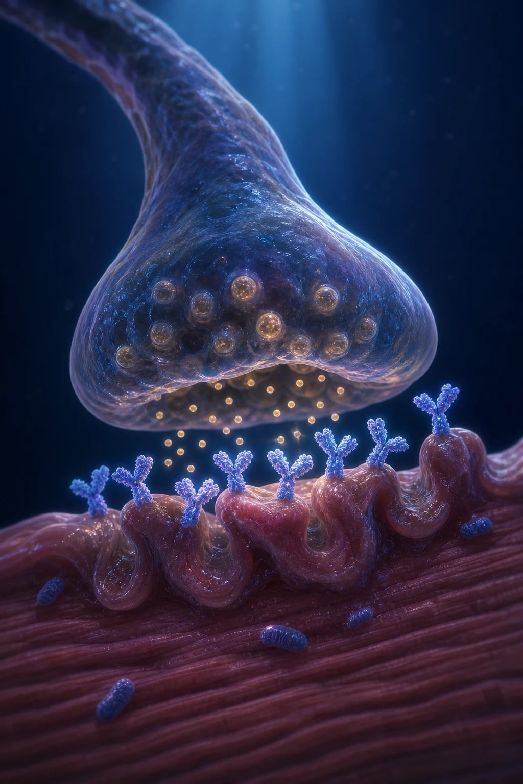
Normal neuromuscular transmission depends on the safety factor: each motor nerve action potential releases enough acetylcholine from the presynaptic terminal to depolarise many more receptors than the threshold needed to trigger a muscle action potential. The redundant receptor pool is the safety factor. In MG, autoantibodies reduce the receptor pool, the safety factor falls, and neuromuscular transmission fails intermittently — the failure shows up under load (sustained activity, repeated firing, fever, infection) when acetylcholine release physiologically drops and the depleted receptor pool is no longer sufficient.[1][2]
The antibody-mediated attack in anti-AChR MG is tripartite. First, the antibodies block acetylcholine binding to the receptor. Second, they cross-link receptors on the muscle membrane and trigger accelerated internalisation and degradation (antigenic modulation) — the muscle actively dismantles its own receptors. Third, the IgG1 and IgG3 subclasses fix complement, forming the membrane attack complex (C5b-9) on the junctional folds, which lyses the membrane, simplifies the folds, and reduces the surface area for receptor expression. The end result is a postsynaptic membrane with about one-third the normal receptor density and a markedly reduced safety factor.[1][9]
Anti-MuSK MG has a distinct mechanism. The antibodies are IgG4, which do not fix complement; they bind the extracellular domain of MuSK and disrupt the agrin-LRP4-MuSK signalling cascade that maintains AChR clustering at the junction. The junctional-fold architecture is relatively preserved, but the receptor cluster is dispersed; muscle involvement favours bulbar, facial, tongue and respiratory muscles (where MuSK is highly expressed) and spares the thymus (which is usually normal). Because the problem is not a deficit of acetylcholine availability, acetylcholinesterase inhibition with pyridostigmine is often ineffective or even counterproductive in MuSK MG.[7]
Anti-LRP4 MG is mediated by IgG1 antibodies that block the binding of the neuronal ligand agrin to LRP4, reducing MuSK activation and AChR clustering; complement contributes. The phenotype is often milder and overlaps AChR MG.[8]
The thymus is central to the disease in AChR-positive MG. Thymic germinal centres harbour autoreactive B cells and T-helper cells that present AChR-derived antigen. Thymic hyperplasia (germinal-centre formation) is found in early-onset AChR-positive MG; thymoma (often cortical-type with aberrant selection of immature T cells) drives a more severe form of AChR MG with anti-titin and anti-ryanodine antibodies. Removing the thymus removes a major source of autoreactive lymphocytes — the rationale for thymectomy in early-onset generalised AChR-positive MG.[3][4]
The modern targeted therapies target distinct steps in this cascade. Eculizumab blocks cleavage of C5, preventing formation of the membrane attack complex and sparing the postsynaptic membrane from complement-mediated lysis (hence its indication in refractory AChR-positive MG).[9] The FcRn inhibitors efgartigimod and rozanolixizumab block the neonatal Fc receptor that recycles IgG; this accelerates the degradation of pathogenic IgG and lowers autoantibody titres.[10][12] The B-cell depleters rituximab (anti-CD20) and inebilizumab (anti-CD19) deplete the B-cell precursors of antibody-producing plasma cells, with the greatest effect in anti-MuSK MG.[13]
Clinical Presentation
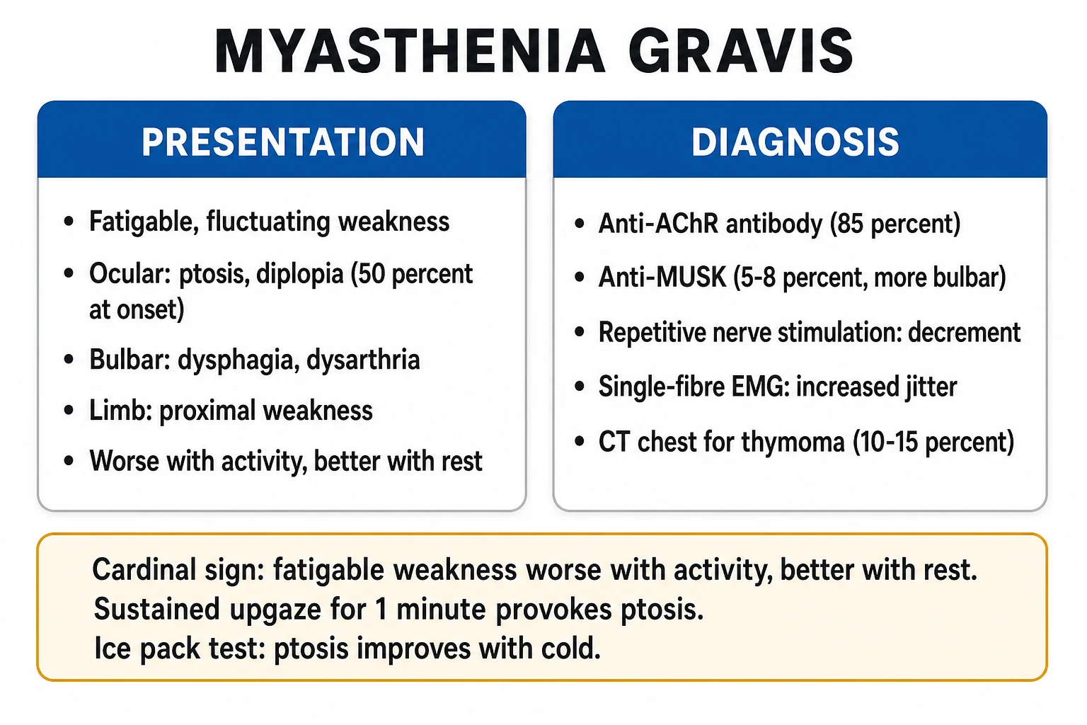
The cardinal feature of MG is fatigable, fluctuating weakness that worsens with sustained activity and improves with rest. There is characteristic diurnal variation — the patient is at their best in the morning and worst in the evening, after a meal, or after exercise. Sensation, tendon reflexes and bowel-bladder function are preserved; their loss should prompt a search for an alternative diagnosis.[2]
The pattern of muscle involvement is characteristic.[1][2]
Ocular
- 50 percent present with ptosis, diplopia, or both
- Ptosis often asymmetric; worsens with sustained upgaze (Simpson test)
- Diplopia from variable extraocular weakness — the affected eye may shift over hours
- Curtain sign (Cogan lid twitch) — ptotic lid jumps up on shifting from downgaze to primary position
Bulbar
- Dysphagia, nasal regurgitation, food sticking in the throat
- Dysarthria — nasal or fading voice; voice fades with counting to 100
- Weak cough, aspiration risk, facial weakness
- Jaw fatigue — the jaw droops while chewing; head droops from neck-extensor weakness
Limb
- Predominantly proximal; arms more than legs
- Difficulty lifting arms above the head, rising from a chair, climbing stairs
- Fatigable on repeated activity
- Wasting is a late feature — its early presence suggests MND or myopathy
Respiratory
- Diaphragmatic and intercostal weakness
- Breathlessness on exertion and then at rest
- Declining FVC, weak cough, poor secretion clearance
- Respiratory failure = myasthenic crisis
The bedside fatigability tests are quick, free, and diagnostic when positive. Sustained upgaze for one minute provokes ptosis (the Simpson test); sustained lateral gaze provokes diplopia; repeated squats or arm elevation reproduces limb weakness; counting aloud to 100 makes the voice fade. The two-minute ice pack test (improvement of ptosis by at least 2 millimetres after a crushed-ice pack over the closed eyelid) exploits cold-mediated reduction of acetylcholinesterase activity and is positive in about 90 percent of patients with ptosis.[2][6]
Atypical presentations are common and examiners test them deliberately. MuSK MG may present with respiratory failure or tongue wasting without ptosis or diplopia.[7] Late-onset MG in an older man may present as isolated dysphagia or neck-extensor weakness mislabelled as motor neuron disease. MG in pregnancy may worsen in the puerperium and present as respiratory failure after delivery. MG may be unmasked by an anaesthetic or by a precipitating drug (a course of gentamicin, a beta-blocker for hypertension, intravenous magnesium for pre-eclampsia). Transient neonatal MG presents in 10 to 20 percent of infants of MG mothers with hypotonia, weak suck and ptosis within 48 hours of birth, resolving over 2 to 4 weeks.[2]
Differential Diagnosis
The differential of fatigable or fluctuating weakness is wide; the examiner wants the candidate to distinguish each with a specific feature, not just list names.[1][2]
LEMS
- Presynaptic — IgG against voltage-gated calcium channel
- Weakness IMPROVES with repeated activity (facilitation)
- Reflexes reduced or absent; recover after brief exercise
- Autonomic features — dry mouth, constipation, impotence
- Small-cell lung cancer in 50 to 60 percent
- Anti-P/Q-type VGCC antibody positive
Botulism
- Presynaptic — toxin blocks SNARE-mediated acetylcholine release
- Descending paralysis (cranial nerves first then symmetric descending)
- Dilated pupils, parasympathetic failure, ileus
- Wound, food-borne, or infant exposure
- Treat with botulism antitoxin
CMS
- Inherited (not antibody-mediated)
- Onset in infancy or childhood; family history often positive
- No antibodies — AChR, MuSK, LRP4 all negative
- Do not respond to immunotherapy
- Subtype-specific therapy (e.g. Dok-7 → salbutamol/ephedrine; slow-channel → quinidine; some subtypes worsen with pyridostigmine)
OPMD
- Onset after 40 years; autosomal dominant
- Slowly progressive ptosis and dysphagia
- Family history positive; French-Canadian ancestry classic
- No fatigability; AChR antibody negative
- Normal RNS and SFEMG
CPEO
- Mitochondrial myopathy
- Slowly progressive symmetric ptosis and ophthalmoplegia
- No diplopia (extraocular muscles fail symmetrically)
- Associated with pigmentary retinopathy, deafness, diabetes, cardiomyopathy (Kearns-Sayre)
Graves
- Diplopia from extraocular muscle entrapment
- Proptosis, lid retraction (lid lag), conjunctival injection
- Abnormal thyroid function
- No fatigability; AChR antibody negative
MND
- UMN signs (brisk jaw jerk, pseudobulbar features)
- Tongue wasting and fasciculations
- Progressive course without fluctuation
- No antibody response
- EMG shows denervation, not a transmission defect
HypoKPP
- Recurrent episodes of painless paralysis
- Areflexic during the attack; low serum potassium
- No ocular involvement
- Resolves with potassium replacement
- Thyrotoxic periodic paralysis commoner in Asian men
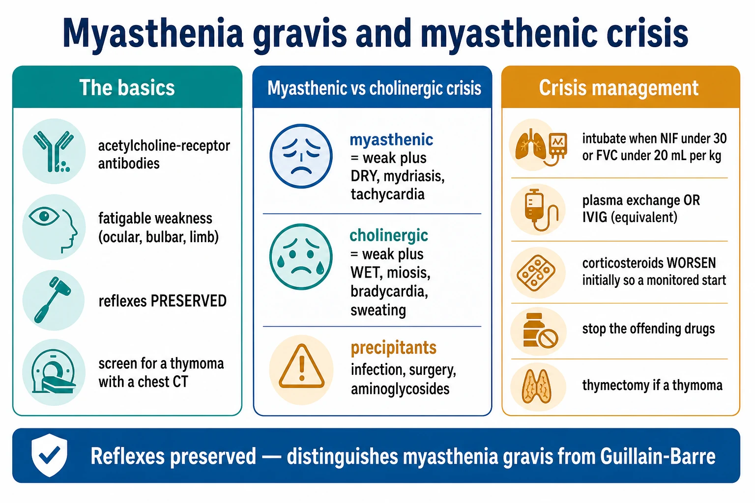
The clinical distinction between myasthenic crisis and cholinergic crisis is examined verbatim — both produce weakness, but the autonomic signatures are mirror images.[2][6]
Myasthenic
- Pupils — mydriasis (dilated)
- Skin — dry; mouth dry; no sweating
- Gut — constipation
- Heart — tachycardia
- Muscles — weak cough, weak swallow; no fasciculations
- Manage — IVIg or plasma exchange; support ventilation
Cholinergic
- Pupils — miosis (constricted)
- Skin — sweating, salivation, lacrimation
- Gut — abdominal cramps, diarrhoea
- Heart — bradycardia
- Muscles — fasciculations, cramps
- Manage — stop pyridostigmine, supportive; atropine for severe bradycardia
Clinical & Bedside Assessment
The focused bedside assessment for suspected MG has three aims: demonstrate fatigable weakness, assess bulbar and respiratory status (which determine disposition), and screen for thymoma (which is asymptomatic on examination).[2][6]
Ocular fatigability. Ask the patient to look up at the ceiling and hold upgaze for one minute. Ptosis on the affected side develops within 30 to 60 seconds (the Simpson test). The curtain sign (Cogan lid twitch) — a brief upward twitch of a ptotic lid when the patient shifts gaze from downgaze to primary position — reflects the momentary recovery of acetylcholine release at the start of contraction. The two-minute ice pack test improves ptosis by at least 2 millimetres in about 90 percent of patients with ptosis and is the most useful bedside confirmation when antibodies are pending.[2]
Bulbar fatigability. Ask the patient to count aloud from 1 to 100 in one breath; the voice fades at 30 to 50 in MG. Listen for nasal emission on /p/, /b/, /s/ sounds. Watch the patient swallow a sip of water — nasal regurgitation, cough, or a wet voice suggests aspiration. Test for jaw fatigue by asking the patient to chew; the jaw droops with repetition. Cough strength is a surrogate for respiratory reserve — a weak cough predicts crisis.[2][6]
Limb fatigability. Ask the patient to hold the arms outstretched at 90 degrees for one minute — drift, drop, or fatigable pronation develops. Ask the patient to rise from a chair repeatedly (5 to 10 times) — leg fatigue develops. Power on formal MRC grading may be normal at rest; the deficit is exposed only by sustained effort.[2][6]
Respiratory surveillance. In any patient with worsening MG, measure serial FVC (sitting and supine), negative inspiratory force (NIF), and peak expiratory flow (PEF) every 4 to 6 hours. The thresholds that mandate ICU admission are FVC under 20 mL/kg (under 1.5 L in an adult), NIF weaker than negative 30 cmH2O, PEF under 200 L/min, or a greater than 30 percent fall in FVC over 24 hours, or any bulbar weakness with weak cough.[6] A postural drop in FVC of greater than 25 percent from sitting to lying predicts diaphragmatic weakness.
Investigations
The diagnostic sequence in MG is serology first, electrophysiology second, imaging third.[1][2]
Anti-AChR antibody
- 85 percent positive in generalised MG, 50 percent in ocular-only MG
- Specificity over 99 percent — the primary serological test
- Three assays — binding (most sensitive), blocking, modulating
- Negative result does NOT exclude MG; test MuSK next
Anti-MuSK antibody
- 5 to 8 percent of all MG; 30 to 40 percent of AChR-seronegative generalised MG
- Order in every AChR-negative case
- Phenotype — bulbar, facial, tongue, respiratory; less ocular
- Predicts poor response to pyridostigmine; predicts benefit from rituximab
Anti-LRP4 antibody
- 2 to 5 percent overall; commoner in AChR and MuSK double-negative
- Order in seronegative cases
- Often milder phenotype
RNS
- Decrement of more than 10 percent in CMAP amplitude between 1st and 4th/5th stimulus
- Sensitivity about 75 percent in generalised MG, lower in ocular MG
- Facial, accessory, and proximal nerves most informative
- Augment by 1 minute of ischaemic or voluntary exercise
SFEMG
- Gold standard — sensitivity over 95 percent in specialist hands
- Increased jitter and impulse blocking in voluntary muscle
- Test an affected muscle (e.g. frontalis in ocular MG)
- Not specific to MG — any neuromuscular transmission disorder raises jitter
CT or MRI chest
- Every newly diagnosed MG patient — to identify thymoma (10 to 15 percent) and characterise thymic hyperplasia
- Contrast-enhanced CT preferred; MRI for soft-tissue characterisation
- Re-image if antibody profile suggests paraneoplastic disease
Bedside tests
- Two-minute ice pack test for ptosis — sensitivity 90 percent
- Sustained upgaze for one minute (Simpson test)
- Count to 100 in one breath — voice fades in bulbar MG
Routine bloods
- TFTs (TSH and free T4) — autoimmune thyroid disease in 5 to 10 percent
- ANA, anti-dsDNA — co-existing autoimmunity
- CK — to exclude myopathy; B12 — combined degeneration
- Anti-titin and anti-ryanodine — late-onset or thymoma-associated MG (not routine)
The edrophonium (Tensilon) test is no longer recommended in many centres for safety reasons (bronchospasm, bradycardia, syncope, asystole). When no alternative exists, it requires resuscitation equipment, atropine drawn up at the bedside, continuous ECG monitoring, and an experienced clinician. Most centres rely instead on antibodies, RNS, single-fibre EMG, and the bedside ice pack test.[2][6]
Management — Resuscitation
Myasthenic crisis is the neurological emergency of MG. The resuscitation bundle is airway, breathing, circulatory support, identification of the precipitant, and rapid disease-modifying therapy.[6]
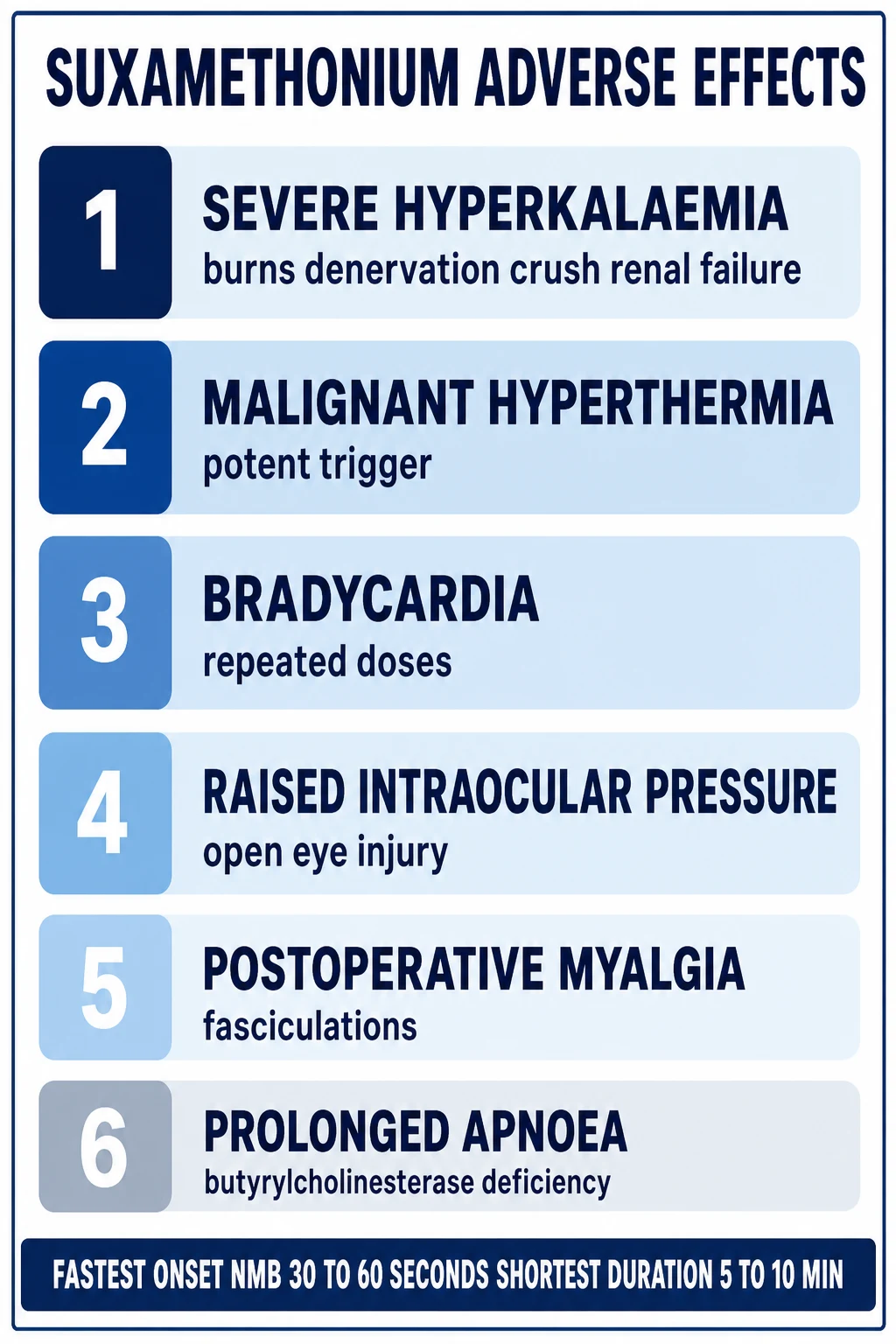
Avoid suxamethonium in MG — the response is unpredictable (relative resistance with prolonged blockade in a well-treated patient, or hyperkalaemic response with chronic denervation). Use a reduced dose of a non-depolarising agent (rocuronium 0.6 mg/kg or less, vecuronium, or cisatracurium), titrated to a peripheral-nerve stimulator, and anticipate the need for prolonged mechanical ventilation.[6]
Acute disease-modifying therapy. Once the airway is secure, give either intravenous immunoglobulin 0.4 g/kg per day for 5 days (total 2 g/kg) OR plasma exchange — 5 sessions of one plasma volume (about 50 mL/kg) on alternate days over 1 to 2 weeks. The two are equally effective and are not combined.[11] IVIg is simpler and has fewer access-related complications; plasma exchange works faster (effect within days) and is preferred when volume overload is a concern. The Cochrane meta-analysis (Gajdos 2012) confirmed equivalence of IVIg and plasma exchange and the lack of additional benefit from combining them.[11]
Supportive bundle. Mechanical ventilation (or non-invasive ventilation once weaning), nasogastric feeding, DVT prophylaxis (enoxaparin 40 mg subcutaneously daily), pressure-area care, careful secretion management (suction, mucolytics), and treatment of the precipitant — antibiotics for infection (avoiding aminoglycosides and fluoroquinolones), levothyroxine for hypothyroidism, withdrawal of offending drugs.[6]
Drugs to stop in MG crisis (the high-yield list examiners reward verbatim): aminoglycosides (gentamicin, neomycin, streptomycin), fluoroquinolones (ciprofloxacin, levofloxacin), macrolides (azithromycin, erythromycin, clarithromycin), beta-blockers (propranolol, timolol eye drops), calcium-channel blockers (verapamil), parenteral magnesium, iodinated contrast, neuromuscular blockers, telithromycin, D-penicillamine, and botulinum toxin injections.[2][6] Safe antibiotics in MG: penicillins, cephalosporins, carbapenems, vancomycin, doxycycline, cotrimoxazole, linezolid, aztreonam.[2]
Management — Definitive & Stepwise
The treatment ladder is layered, not sequential — most patients end up on several agents simultaneously.[1][6]
Step 1 — Symptomatic
- Pyridostigmine 30 to 60 mg orally four times daily; titrate to response over days to weeks
- Max single dose 120 mg; max total daily 1200 mg
- Onset 30 minutes; peak 2 hours; duration 4 to 6 hours
- Acetylcholinesterase inhibitor — raises synaptic acetylcholine, partially corrects safety factor
- Symptomatic only — not disease-modifying; usually insufficient alone for generalised MG
- Common side effects — diarrhoea, abdominal cramps, sweating, salivation (muscarinic); muscle fasciculations (nicotinic)
Step 2 — Corticosteroid
- Prednisolone 1 to 1.5 mg/kg on alternate days, titrated up from a low starting dose of 10 to 20 mg alternate days
- Increase by 5 mg every 2 to 3 days until minimal-manifestation status
- Watch for steroid-induced worsening in the first 1 to 2 weeks (give as inpatient if bulbar or respiratory involvement, or co-administer IVIg or PLEX)
- Once stable for 4 to 8 weeks, taper by 5 to 10 percent every 2 to 4 weeks
- Add bone protection (calcium/vitamin D; bisphosphonate), gastric protection, and PJP prophylaxis if on long-term over 20 mg per day with a second agent
Step 2b — Steroid-sparing
- Azathioprine 1 to 3 mg/kg per day (50 to 150 mg/day) orally — first-line; check TPMT before starting
- Mycophenolate mofetil 1 to 2 g per day orally in two divided doses — alternative; teratogenic, avoid in pregnancy
- Methotrexate 7.5 to 15 mg orally once weekly plus folic acid 5 mg weekly — cost-effective alternative
- Tacrolimus 3 to 5 mg orally at night — alternative; monitor trough levels and renal function
- Ciclosporin 2.5 to 5 mg/kg per day — older agent; nephrotoxic
- Take 6 to 12 months for full effect — bridge with corticosteroid
Step 3 — Thymectomy
- Always for thymoma (any MGFA class)
- AChR-antibody-positive generalised MG, MGFA Class II to IV, age 18 to 65 years, disease duration under 5 years (MGTX)
- Extended transsternal approach preferred
- Benefit accrues over 3 to 5 years; sustained at 5 years in the MGTX extension
- Not recommended in anti-MuSK MG; not first-line in purely ocular MG
Step 4 — Acute therapy
- IVIg 0.4 g/kg/day for 5 days (total 2 g/kg) — crisis, perioperative cover, refractory flares
- Plasma exchange — 5 sessions of one plasma volume (about 50 mL/kg) on alternate days over 1 to 2 weeks
- Equally effective — choose based on access, fluid status, contraindications
- Effect within 1 to 2 weeks; lasts weeks to a few months
Step 5 — Targeted
- Eculizumab — terminal complement (C5) inhibitor; refractory AChR-positive MG; 900 mg IV weekly x 4, then 1200 mg at week 5 and 1200 mg every 2 weeks; vaccinate against meningococcus at least 2 weeks before first dose
- Efgartigimod — FcRn blocker; AChR-positive generalised MG; 10 mg/kg IV weekly x 4 per cycle, repeat as needed at least 8 weeks apart
- Rozanolixizumab — subcutaneous anti-FcRn; AChR or MuSK MG; 7 to 10 mg/kg subcutaneously weekly x 6
- Rituximab — anti-CD20 B-cell depleter; 375 mg/m2 weekly x 4 or 1 g on days 1 and 15; especially effective in refractory MuSK MG
- Inebilizumab — anti-CD19 B-cell depleter; AChR or MuSK MG; 300 mg IV days 1 and 15 then week 26 (AChR-positive) or maintained (MuSK-positive)
In India and many other resource-limited settings, pyridostigmine, prednisolone, and azathioprine are the backbone of MG therapy; IVIg is scarce and costly so plasma exchange is often the practical crisis therapy; targeted biological therapies (eculizumab, efgartigimod, rozanolixizumab, inebilizumab) are restricted to affluent patients or trial centres. The clinician's job is to use the cheap effective agents (pyridostigmine, prednisolone, azathioprine) correctly and to use rituximab (which is more widely available and less expensive than the newer biologicals) for refractory MuSK MG.[1][6]
Tapering immunosuppression. Once minimal-manifestation status is stable for at least 6 to 12 months, taper the corticosteroid first (slowly — 5 to 10 percent every 2 to 4 weeks), then taper the steroid-sparing agent. Thymectomy is not a reason to stop immunosuppression abruptly. Patients should be told that immunosuppression may be lifelong.[6]
Pre-operative optimisation for thymectomy. Optimise strength with IVIg or plasma exchange in the weeks before surgery if there is bulbar or respiratory compromise; arrange anaesthetic review to plan the avoidance of neuromuscular blockers; plan post-operative monitoring in a high-dependency setting for 24 to 48 hours because 8 to 10 percent of patients have a transient post-thymectomy myasthenic crisis.[3][6]
Specific Subtypes & Scenarios
Ocular MG is ptosis and diplopia only, sustained for at least 2 years. Treat with pyridostigmine; add low-dose prednisolone if symptoms are disabling. CT chest for thymoma. Thymectomy is considered only if generalisation is anticipated or thymoma is present.[2]
Anti-MuSK MG has a distinctive phenotype — prominent bulbar, facial, tongue and respiratory weakness with less ocular disease — and a distinctive response to therapy. Pyridostigmine is often ineffective or causes cholinergic excess; thymectomy is not indicated (the thymus is normal); rituximab is highly effective in refractory disease. Treat with prednisolone plus an early steroid-sparing agent (mycophenolate or tacrolimus); reserve plasma exchange for crisis.[7]
Thymoma-associated MG is an absolute indication for thymectomy by extended transsternal approach; check anti-titin and anti-ryanodine antibodies; long-term immunosuppression for the MG; surveillance imaging for thymoma recurrence. The MG may be more severe and refractory.[1][5]
Late-onset MG (50 years or older) has male predominance, thymic atrophy, anti-titin and anti-ryanodine antibodies, less response to thymectomy, and more comorbidities. Managed with pyridostigmine plus immunosuppression. Consider paraneoplastic LEMS in smokers.[1]
MG in pregnancy may worsen in the first trimester and the puerperium. Pyridostigmine and prednisolone are safe. Azathioprine is the preferred steroid-sparing agent and is compatible with breastfeeding. Avoid mycophenolate and methotrexate (teratogenic). Rituximab may cause B-cell depletion in the neonate if given in pregnancy. Plan delivery at a centre with neonatal ICU for transient neonatal MG. Labour may require IVIg or plasma exchange. Epidural anaesthesia is preferred over general anaesthesia.[2][6]
Transient neonatal MG affects 10 to 20 percent of infants of MG mothers from transplacental transfer of maternal antibody; onset within 48 hours of birth with hypotonia, weak suck, weak cry, ptosis; resolves over 2 to 4 weeks as maternal antibody clears. The maternal antibody titre does not predict which infants will be affected. Treatment is supportive; pyridostigmine or exchange transfusion if severe.[2]
Juvenile MG presents from age 1 year with the same antibody pattern as adults. Ocular symptoms may dominate in prepubertal children and may remit spontaneously. Immunosuppression is cautious. Thymectomy is reserved for refractory cases in prepubertal children because the long-term risks and benefits are less clear than in adults.[2]
Refractory MG is failure to achieve minimal-manifestation status despite pyridostigmine plus at least one immunosuppressant at a tolerated dose for at least 12 months. Managed with a second immunosuppressant, rituximab (especially MuSK), or a complement or FcRn inhibitor.[6][9][10]
Complications & Pitfalls
The disease-related complications are myasthenic crisis (15 to 20 percent experience at least one), aspiration pneumonia from bulbar weakness, weight loss and malnutrition from dysphagia, respiratory failure, and the social isolation and depression of a chronic fluctuating illness.[2]
The treatment-related complications are the steroid-induced worsening in the first 1 to 2 weeks of high-dose prednisolone (start low, titrate up, or co-administer IVIg or plasma exchange), steroid side effects (osteoporosis, diabetes, hypertension, cataract, infection), azathioprine hepatotoxicity and myelosuppression (check FBC weekly for the first month then monthly), opportunistic infection (PJP) with prolonged immunosuppression, eculizumab-associated meningococcal infection (vaccinate at least 2 weeks before the first dose), IVIg-related thrombosis or haemolysis, and plasma-exchange-related hypotension and central-line complications.[6][9]
Prognosis & Disposition
With full modern treatment — pyridostigmine, immunosuppression, thymectomy where indicated, and targeted therapy where needed — most MG patients achieve minimal-manifestation status or pharmacological remission, and mortality from MG itself is now low (under 5 percent in developed countries).[1][2]
Worse prognosis is predicted by anti-MuSK positivity (more bulbar and respiratory crises, less response to pyridostigmine), thymoma-associated MG (more refractory, malignant-thymoma risk), late-onset MG with comorbidities, delayed diagnosis and treatment, recurrent crises, and non-adherence to immunosuppression.[1][7]
Better prognosis is predicted by early-onset AChR-positive MG with thymic hyperplasia (best response to thymectomy), female sex in early-onset, ocular-only MG for over 2 years without generalisation, adherence to immunosuppression, and absence of thymoma.[3][4]
The disposition of a patient in myasthenic crisis is ICU admission for ventilatory support and IVIg or plasma exchange, treatment of the precipitant, and weaning to a high-dependency or neurology ward once stable. The disposition of a stable outpatient is regular neurology follow-up, monitoring of respiratory and bulbar function, screening for steroid side effects, vaccination against influenza and pneumococcus (avoid live vaccines in the immunosuppressed), bone protection on long-term steroids (calcium and vitamin D, bisphosphonate if indicated), and patient education on precipitants and crisis warning signs.[6]
Every MG patient should carry an MG alert card listing the drugs to avoid and the warning signs of crisis (breathlessness, voice fading, weak cough, swallowing difficulty, progressive limb weakness).[2]
Special Populations
Pregnancy
- Pyridostigmine and prednisolone safe
- Azathioprine preferred steroid-sparing agent
- Avoid mycophenolate, methotrexate; ideally rituximab
- Plan delivery at centre with neonatal ICU
- Epidural preferred over general anaesthesia
- Risk of flare in first trimester and puerperium
Neonate
- Transient neonatal MG in 10 to 20 percent of infants of MG mothers
- Onset within 48 hours — hypotonia, weak suck, weak cry, ptosis
- Resolves over 2 to 4 weeks as maternal antibody clears
- Maternal titre does NOT predict which infants affected
- Supportive care; pyridostigmine if severe
Elderly
- Late-onset MG (50 years or older); male predominance
- Thymic atrophy; less response to thymectomy
- More comorbidities, higher surgical risk
- Minimise steroid burden (osteoporosis, diabetes, infection)
Children
- Juvenile MG from age 1 year
- Ocular symptoms may dominate and remit in prepubertal children
- Immunosuppression cautious
- Thymectomy reserved for refractory cases in prepubertal children
Immunocompromised
- PJP prophylaxis (cotrimoxazole 480 mg daily) on long-term prednisolone over 20 mg/day with a second agent
- Vaccinate against influenza and pneumococcus; avoid live vaccines
- Screen for CMV, candidiasis with prolonged immunosuppression
Perioperative
- Optimise strength with IVIg or PLEX pre-operatively if needed
- Plan anaesthesia to avoid neuromuscular blockers
- Monitor post-operatively in HDU 24 to 48 hours
- Continue pyridostigmine via NG tube if intubated
Breastfeeding
- Pyridostigmine, prednisolone, azathioprine compatible
- Mycophenolate and methotrexate contraindicated
- Counsel on transient neonatal MG risk
Evidence, Guidelines & Regional Differences
MGTX 2016
- Wolfe et al, NEJM 2016 (PMID 27509100)
- 126 patients, generalised non-thymomatous AChR-positive MG, MGFA Class II to IV, age 18 to 65 years, disease duration under 5 years
- Extended transsternal thymectomy plus alternate-day prednisone vs prednisone alone
- Thymectomy group had lower time-weighted QMG (6.15 vs 8.99, p under 0.001) and lower prednisone (44 vs 60 mg) at 3 years
- Landmark evidence supporting thymectomy in early-onset AChR-positive generalised MG
MGTX 2019
- Wolfe et al, Lancet Neurology 2019 (PMID 30692052)
- 5-year extension — sustained benefit (QMG 5.47 vs 9.34; prednisone 24 vs 48 mg)
- Confirms long-term benefit of thymectomy in early-onset AChR-positive generalised MG
REGAIN 2017
- Howard et al, Lancet Neurology 2017 (PMID 29066163)
- 125 patients, refractory AChR-positive generalised MG
- Eculizumab 900 mg weekly x 4, then 1200 mg at week 5 and every 2 weeks vs placebo
- Primary endpoint missed significance on worst-rank ANCOVA (p 0.0698) but secondary and sensitivity analyses favoured eculizumab
- Approved for refractory AChR-positive MG; vaccinate against meningococcus
ADAPT 2021
- Howard et al, Lancet Neurology 2021 (PMID 34146511)
- 167 patients, generalised MG (any antibody)
- Efgartigimod 10 mg/kg IV weekly x 4 per cycle vs placebo
- In AChR-positive patients, 68 percent of efgartigimod vs 30 percent of placebo were MG-ADL responders in cycle 1
- FcRn inhibition; first-in-class; well tolerated
MycarinG 2023
- Bril et al, Lancet Neurology 2023 (PMID 37059507)
- 200 patients, generalised AChR or MuSK MG
- Rozanolixizumab 7 mg/kg or 10 mg/kg subcutaneously weekly x 6 vs placebo
- Significant reduction in MG-ADL at day 43 for both doses
- Subcutaneous FcRn inhibition; useful for both AChR and MuSK MG
MINT 2025
- Nowak et al, NEJM 2025 (PMID 40202593)
- 238 patients, generalised AChR or MuSK MG
- Inebilizumab 300 mg IV days 1 and 15 (and day 183 for AChR-positive) vs placebo
- Significant reduction in MG-ADL and QMG at week 26
- B-cell depletion across both antibody subtypes
The MGFA international consensus guidance (Sanders 2016, MGFA/AAN RAND-UCLA appropriateness method) sets out the treatment statements that examiners expect to be quoted: pyridostigmine for symptoms; corticosteroid and a steroid-sparing agent for generalised MG; IVIg or plasma exchange for crisis and pre-operative optimisation; thymectomy for thymoma and selected non-thymomatous AChR-positive generalised MG; rituximab for refractory anti-MuSK MG; cautious immunosuppression in pregnancy and juvenile MG.[6]
The controversies that examiners probe: thymectomy in anti-MuSK MG (not recommended — the thymus is normal and MGTX did not include MuSK MG); thymectomy in purely ocular MG (reserved for anticipated generalisation or disabling symptoms); the long-term safety and access barriers of complement and FcRn inhibitors (meningococcal infection, infusion reactions, cost).[4][9]
Exam Pearls
MG-ONE-LINERS
Exam application bank (NEET-PG / INICET)
One-line answer
Myasthenia gravis (MG) is a B-cell-mediated autoimmune disorder of the postsynaptic neuromuscular junction in which autoantibodies damage or block the muscle acetylcholine receptor (or related proteins such as MuSK and LRP4), producing fluctuating, fatigable weakness that worsens with sustained activity and improves with rest. The hallmark is ocular involvement (ptosis, diplopia) in about half of patients, often spreading to bulbar (dysphagia, dysarthria, nasal voice), proximal limb, axial and respiratory muscles. Antibodies are positive in anti-AChR in 85 percent, anti-MuSK in 5 to 8 percent, anti-LRP4 in 2 to 5 percent, and 5 to 8 percent are seronegative. Diagnosis combines the clinical pattern with anti-AChR antibody, repetitive nerve stimulation (decremental response over 10 percent), single-fibre EMG (gold-standard sensitivity over 95 percent), and CT chest for thymoma (10 to 15 pe
Worked stems (answer without another resource)
Stem 1 — Classic presentation. Map symptoms to mechanism; name the first investigation and first treatment step with dose/route if drug therapy is standard. [1]
Stem 2 — Unstable / complicated. List red flags that force immediate resuscitation, theatre, ICU, antidote, or reperfusion — and what you do in the first 15 minutes. [1]
Stem 3 — Atypical group. Elderly, pregnancy, child, or immunocompromised: how presentation and thresholds change. [1]
Stem 4 — Differential trap. Name the three closest mimics and one discriminator for each. [1]
Stem 5 — Disposition. Who goes home with safety-netting, who is admitted, who needs HDU/ICU/theatre, and what follow-up is mandatory. [1]
Rapid viva checklist
- Definition + classification
- Pathophysiology chain
- Bedside signs / criteria
- Score with exact components (if any)
- Emergency bundle
- Definitive therapy with doses
- Complications of disease and of treatment
- Special populations
- Guideline/trial name if classic
- Three exam traps
Coverage self-check
If you cannot answer any stem above from this page alone, re-read the matching section — the page is intended to be self-sufficient for final-prof and NEET-PG/INICET questions on Myasthenia Gravis.
References
- [1]Gilhus NE, Verschuuren JJ. Myasthenia gravis: subgroup classification and therapeutic strategies Lancet Neurol, 2015.PMID 26376969
- [2]Ciafaloni E. Myasthenia Gravis and Congenital Myasthenic Syndromes Continuum (Minneap Minn), 2019.PMID 31794470
- [3]Wolfe GI, Kaminski HJ, Aban IB, et al. Randomized Trial of Thymectomy in Myasthenia Gravis N Engl J Med, 2016.PMID 27509100
- [4]Wolfe GI, Kaminski HJ, Aban IB, et al.; MGTX Study Group. Long-term effect of thymectomy plus prednisone versus prednisone alone in patients with non-thymomatous myasthenia gravis: 2-year extension of the MGTX randomised trial Lancet Neurol, 2019.PMID 30692052
- [5]Jaretzki A 3rd, Barohn RJ, Ernstoff RM, Kaminski HJ, Keesey JC, Penn AS, Sanders DB. Myasthenia gravis: recommendations for clinical research standards. Task Force of the Medical Scientific Advisory Board of the Myasthenia Gravis Foundation of America Ann Thorac Surg, 2000.PMID 10921745
- [6]Sanders DB, Wolfe GI, Benatar M, et al. International consensus guidance for management of myasthenia gravis: Executive summary Neurology, 2016.PMID 27358333
- [7]Hoch W, McConville J, Helms S, Newsom-Davis J, Melms A, Vincent A. Auto-antibodies to the receptor tyrosine kinase MuSK in patients with myasthenia gravis without acetylcholine receptor antibodies Nat Med, 2001.PMID 11231638
- [8]Higuchi O, Hamuro J, Motomura M, Yamanashi Y. Autoantibodies to low-density lipoprotein receptor-related protein 4 in myasthenia gravis Ann Neurol, 2011.PMID 21387385
- [9]Howard JF Jr, Utsugisawa K, Benatar M, et al.; REGAIN Study Group. Safety and efficacy of eculizumab in anti-acetylcholine receptor antibody-positive refractory generalised myasthenia gravis (REGAIN): a phase 3, randomised, double-blind, placebo-controlled, multicentre study Lancet Neurol, 2017.PMID 29066163
- [10]Howard JF Jr, Bril V, Vu T, et al.; ADAPT Investigator Study Group. Safety, efficacy, and tolerability of efgartigimod in patients with generalised myasthenia gravis (ADAPT): a multicentre, randomised, placebo-controlled, phase 3 trial Lancet Neurol, 2021.PMID 34146511
- [11]Gajdos P, Chevret S, Toyka KV. Intravenous immunoglobulin for myasthenia gravis Cochrane Database Syst Rev, 2012.PMID 23235588
- [12]Bril V, Druzdz A, Grosskreutz J, et al.; MG0003 study team. Safety and efficacy of rozanolixizumab in patients with generalised myasthenia gravis (MycarinG): a randomised, double-blind, placebo-controlled, adaptive phase 3 study Lancet Neurol, 2023.PMID 37059507
- [13]Nowak RJ, Benatar M, Ciafaloni E, et al.; MINT Investigators. A Phase 3 Trial of Inebilizumab in Generalized Myasthenia Gravis N Engl J Med, 2025.PMID 40202593