Neurology · Neurology
Parkinson Disease
Also known as Parkinson's disease · Idiopathic parkinsonism · Paralysis agitans · PD
Parkinson disease (PD) is a progressive neurodegenerative disorder characterised by loss of dopaminergic neurons of the substantia nigra pars compacta, with intracellular alpha-synuclein Lewy bodies. The cardinal motor features are TRAP: resting Tremor, Rigidity, Akinesia/bradykinesia (obligatory), and Postural instability. Diagnosis is clinical (MDS 2015 criteria); investigations exclude mimics. Treatment is symptomatic — levodopa combined with a peripheral decarboxylase inhibitor (carbidopa or benserazide) is the gold-standard and most effective therapy. Dopamine agonists, MAO-B inhibitors, COMT inhibitors and amantadine have defined roles; advanced disease uses apomorphine, levodopa-carbidopa intestinal gel, or deep brain stimulation. Long-term levodopa causes motor fluctuations and dyskinesia. Non-motor symptoms (anosmia, constipation, REM sleep behaviour disorder, depression, dementia, psychosis, dysautonomia) dominate late disease and drive disability. Dopamine-blocking drugs (metoclopramide, prochlorperazine, haloperidol) are contraindicated.
On this page & tools
Your progress
Saved locally on this device.
Exam tags
Red flags
Overview & Definition
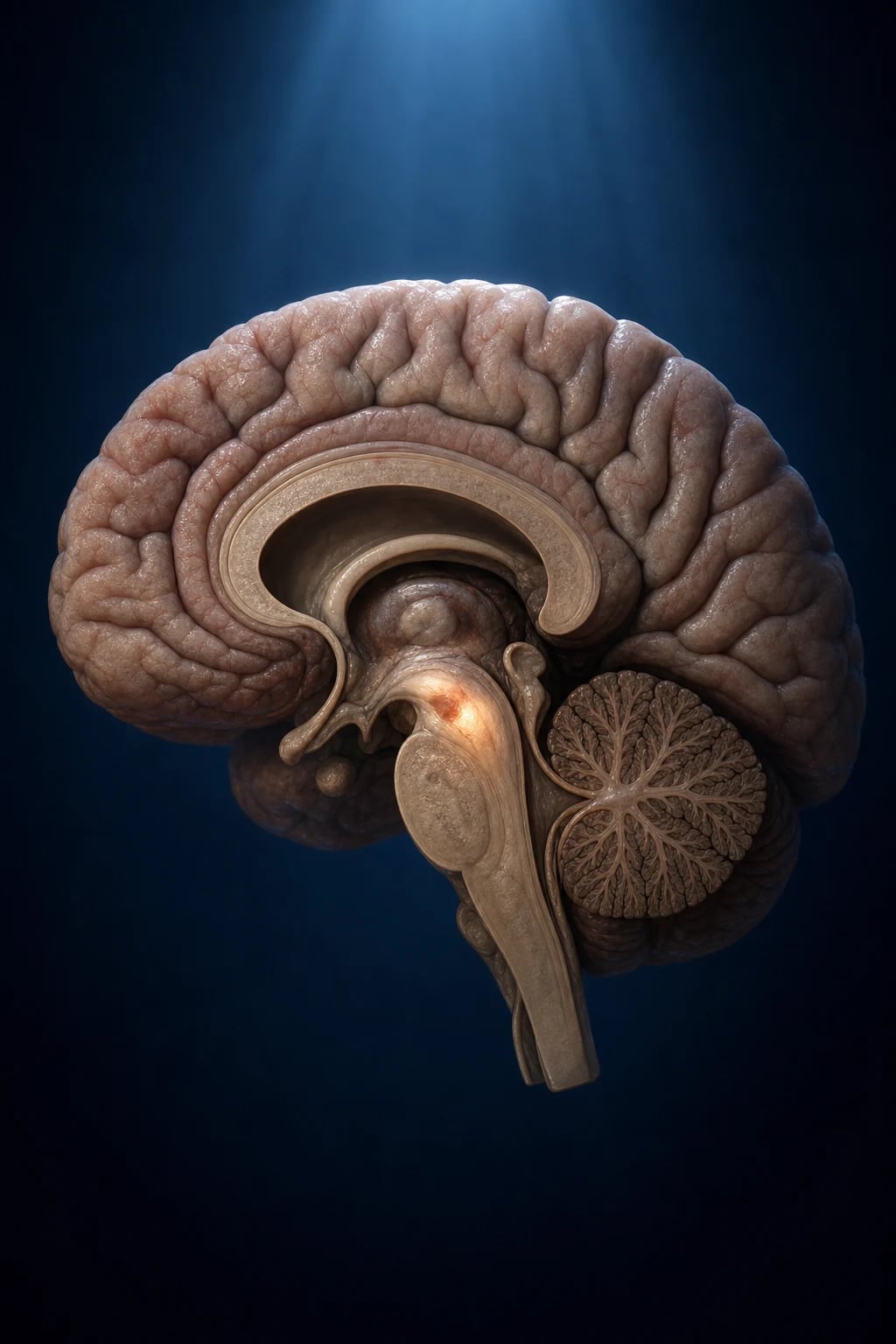
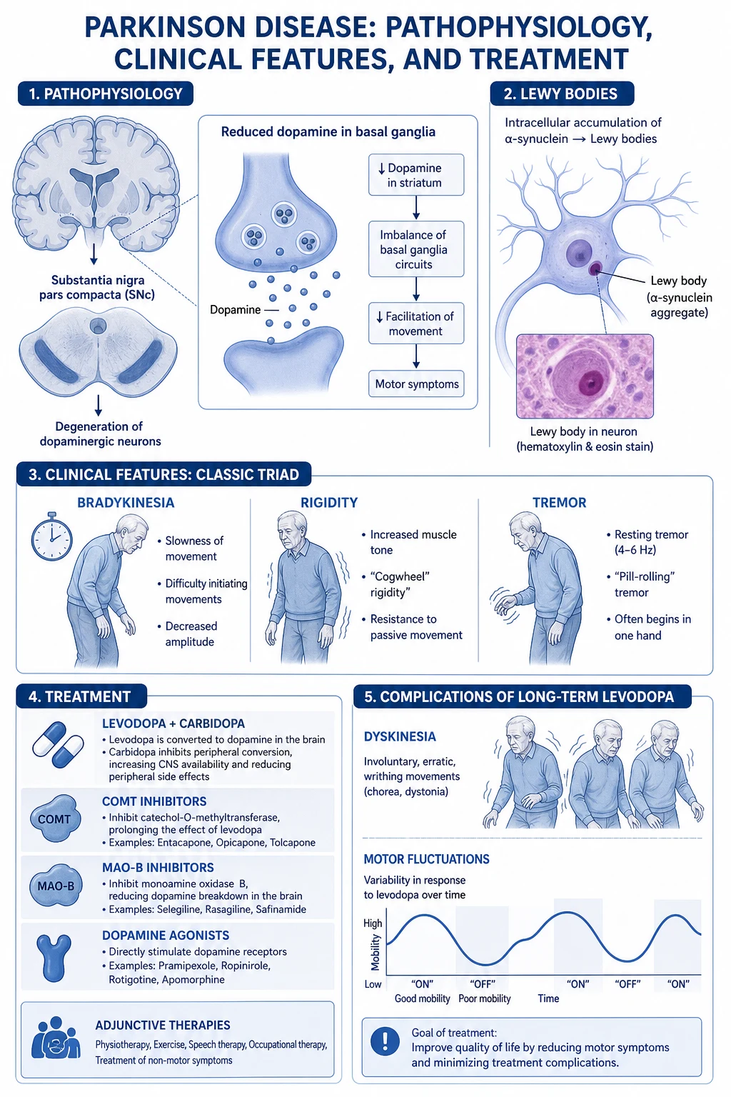
Parkinson disease (PD), originally described by James Parkinson in 1817 as the Shaking Palsy, is a chronic, progressive neurodegenerative disorder of the central nervous system characterised by loss of dopaminergic neurons in the substantia nigra pars compacta (SNpc) of the midbrain, with the presence of intracytoplasmic alpha-synuclein aggregates (Lewy bodies and Lewy neurites). The resulting dopamine depletion in the striatum (dorsolateral putamen) disinhibits the indirect basal ganglia pathway and disrupts the direct pathway, producing the cardinal motor triad of bradykinesia, rigidity, and resting tremor, with postural instability developing later.[1][4]
The clinical skill in PD is threefold. First, recognising parkinsonism (bradykinesia plus tremor or rigidity) and distinguishing idiopathic PD from the many mimics — drug-induced, vascular, and atypical parkinsonisms (PSP, MSA, DLB), essential tremor, and Wilson disease. Second, timing and selecting symptomatic therapy — when to start levodopa, when to use a dopamine agonist or MAO-B inhibitor, and how to manage the motor fluctuations and dyskinesia that complicate long-term levodopa. Third, never omitting dopaminergic medication in a hospitalised patient and never giving dopamine-blocking drugs (metoclopramide, prochlorperazine, haloperidol) that can precipitate catastrophic deterioration.[2]
There is no cure and no proven disease-modifying therapy; treatment is lifelong, symptomatic, and individualised, with the goal of maintaining function and quality of life for as long as possible.[1]
Classification
PD is classified in three useful ways — by aetiology, by motor phenotype, and by severity (Hoehn and Yahr). [1]
By aetiology — the key exam distinction:[2][3]
Idiopathic PD (sporadic)
- Most common (about 85 to 90%); cause unknown
- Insidious, asymmetric onset; clear, sustained levodopa response
- Alpha-synuclein Lewy body pathology in substantia nigra
- Diagnosis by MDS 2015 clinical criteria (no test confirms it in life)
Familial / genetic PD
- About 10 to 15%; autosomal dominant (LRRK2, SNCA) or recessive (Parkin/PARK2, PINK1, DJ-1/PARK7)
- LRRK2 (PARK8) is the commonest genetic cause worldwide; G2019S mutation
- Recessive forms cause young-onset PD (under 40 to 50), slower course, dystonia, less dementia
- GBA (glucocerebrosidase) variant is a strong risk factor, not a mendelian cause
Atypical parkinsonism (parkinson-plus)
- PSP, MSA, DLB, corticobasal degeneration (CBD)
- Poor or transient levodopa response; rapid progression; early falls/dementia/autonomic failure
- Different underlying pathology (tau, alpha-synuclein glial, TDP-43)
- Red-flag features in MDS criteria exclude a diagnosis of idiopathic PD
Secondary parkinsonism
- Drug-induced (neuroleptics, metoclopramide, prochlorperazine, cinnarizine, some CCBs)
- Vascular (multi-infarct, lower-body predominant, poor levodopa response)
- Toxic (MPTP, manganese, carbon monoxide, carbon disulfide)
- Structural (tumour, hydrocephalus, post-encephalitic, repetitive head trauma /拳击 parkinsonism pugilistica)
By motor phenotype (prognostically useful): [1]
- Tremor-dominant — prominent resting tremor, less rigidity/bradykinesia; slowest progression, best prognosis, least cognitive decline.
- Akinetic-rigid (postural-instability/gait difficulty, PIGD) — dominant bradykinesia, rigidity, gait freezing and falls; faster progression, more dementia and disability.
- Mixed / indeterminate — features of both.[1]
By severity — Hoehn and Yahr staging (1967):[4]
| Stage | Description |
|---|---|
| 1 | Unilateral involvement only (minimal functional impairment) |
| 1.5 | Unilateral plus axial involvement |
| 2 | Bilateral involvement, no impairment of balance/posture |
| 2.5 | Mild bilateral disease with recovery on pull test |
| 3 | Mild-to-moderate bilateral disease; some postural instability; physically independent |
| 4 | Severe disability; still able to walk or stand unaided |
| 5 | Wheelchair-bound or bedridden unless aided |
Young-onset PD (YOPD) is defined as onset under age 50 (some use under 40 or 45). It is more often genetic (Parkin, PINK1, DJ-1, LRRK2), progresses more slowly, has a higher risk of levodopa-induced dyskinesia, and is managed with a levodopa-sparing strategy (dopamine agonist or MAO-B inhibitor first).[4]
Epidemiology & Risk Factors
PD is the second commonest neurodegenerative disease after Alzheimer disease and the fastest-growing neurological disorder worldwide in terms of prevalence and disability.[1]
Prevalence and incidence: [1]
- Prevalence about 1% of the population over 60, rising to about 3% over 80; men affected about 1.5 times more than women.
- Incidence about 8 to 18 per 100,000 person-years overall; age-standardised incidence rises steeply with age.
- Lifetime risk approximately 2% in men, 1.3% in women.[1][4]
Parkinson disease — key numbers
Established risk factors:[1]
- Age — the strongest risk factor; rare before 40.
- Male sex — 1.4 to 2 times the risk of women.
- Genetics — family history; LRRK2, GBA, SNCA, Parkin/PINK1/DJ-1 mutations; first-degree relative risk about 2 to 3 times.
- Environmental exposures — pesticides and herbicides (paraquat, rotenone — both inhibit complex I); the MPTP discovery (a meperidine-analogue contaminant that caused acute parkinsonism in drug users) proved that a toxin can selectively destroy the SNpc; rural living, well water, farming; manganese, carbon monoxide, carbon disulfide (toxic).
- Head trauma — repeated (pugilistic parkinsonism); single severe TBI is a weaker association. [1]
Reported protective factors (examinable, controversial):[1][4]
- Caffeine / coffee — inverse association (adenosine A2A antagonism is the rationale for istradefylline).
- Cigarette smoking — strongest and most reproducible inverse association (dose-dependent); mechanism unclear (perhaps nicotine is neuroprotective, or smoking behaviour is itself reduced early in prodromal PD — reverse causation).
- Higher serum urate — antioxidant; associated with slower progression in men (the rationale behind inosine trials).
- Physical activity, NSAIDs, ibuprofen — weaker/inconsistent signals. [1]
These associations do not justify recommending smoking or treating with urate — the data are observational and confounded. [1]
Prodromal phase: motor symptoms are preceded by a prodromal phase of 10 to 20 years during which non-motor features accumulate — idiopathic REM sleep behaviour disorder (RBD), hyposmia/anosmia, constipation, depression, anxiety, orthostatic intolerance, and subtle motor signs. Probable RBD confers a very high (>80% over 14 years) risk of converting to an alpha-synucleinopathy (PD, DLB, MSA).[3]
Pathophysiology
PD is a synucleinopathy — the pathological hallmark is the misfolding and aggregation of alpha-synuclein into Lewy bodies (intracytoplasmic eosinophilic inclusions) and Lewy neurites (in neuronal processes).[1][4]
Site of degeneration. The vulnerable neurons are the melanin-pigmented dopaminergic neurons of the substantia nigra pars compacta (SNpc) — specifically the calbindin-negative neurons in the ventrolateral tier that project to the dorsolateral putamen (the nigrostriatal pathway). About 60 to 80% of these neurons (and a comparable fraction of striatal dopamine) must be lost before motor symptoms appear — a threshold that explains the long prodromal phase and the brain's capacity to compensate (upregulation of remaining neurons' firing and dopamine synthesis). Pigmented neurons in the locus coeruleus (noradrenergic), dorsal motor nucleus of the vagus, raphe (serotonergic), and nucleus basalis of Meynert (cholinergic) are also affected, accounting for non-motor features.[1]
Braak staging (2003). Braak proposed that Lewy pathology spreads caudal-to-rostral in a predictable six-stage sequence, beginning in the enteric nervous system and dorsal motor nucleus of the vagus (stages 1 to 2), ascending through the medulla and pons including the locus coeruleus (stage 2), the midbrain and substantia nigra (stage 3 — onset of motor symptoms), the basal forebrain and limbic cortex (stage 4), and finally the neocortex (stages 5 to 6 — dementia). This staging provides a unifying explanation for the prodromal non-motor symptoms preceding motor disease and for the late cognitive decline, and underpins the gut-brain hypothesis (alpha-synuclein propagating via the vagus) and the concept of prion-like cell-to-cell spread of misfolded alpha-synuclein.[1][4]
Basal ganglia circuitry — the mechanism of the motor signs.[4]
The basal ganglia normally facilitate wanted movement (direct pathway) and suppress unwanted movement (indirect pathway). Both originate from corticostriatal glutamatergic input to medium spiny neurons of the striatum and act on the thalamus, which then excites the motor cortex. [1]
- Direct pathway: cortex excites striatum then D1-dopamine-receptor MSNs which inhibit the internal globus pallidus (GPi) and substantia nigra pars reticulata (SNr), which themselves inhibit the thalamus — so disinhibiting the thalamus and facilitating movement. Dopamine stimulates D1, enhancing the direct pathway.
- Indirect pathway: cortex excites striatum then D2-MSNs which inhibit the external globus pallidus (GPe), which normally inhibits the subthalamic nucleus (STN), which excites the GPi/SNr — so the indirect pathway, when active, ultimately increases thalamic inhibition and suppresses movement. Dopamine stimulates D2, which inhibits the indirect pathway. [1]
In PD, loss of dopamine simultaneously weakens the direct pathway (less facilitation of movement) and removes inhibition of the indirect pathway (excess suppression of movement). The net effect is overactivity of the GPi/SNr (excessive thalamic inhibition) producing the hypokinetic features — bradykinesia and rigidity. This insight is the rationale for STN or GPi deep brain stimulation (reducing pathological output) and for levodopa/dopamine agonist therapy (restoring D1/D2 balance).[4]
Basal ganglia pathways — D1 Direct, D2 inDirect
DISC
D1 dopamine receptor drives the DIRECT pathway — facilitates movement. In PD, low dopamine weakens the direct pathway.
D2 dopamine receptor inhibits the INDIRECT pathway — when D2 is lost (PD), the indirect pathway runs unchecked, over-suppressing movement.
Loss of dopamine causes subthalamic nucleus and internal globus pallidus to become overactive — the target of deep brain stimulation.
The overactive GPi inhibits the thalamus, which fails to excite the motor cortex — bradykinesia and rigidity result.
Cell-death mechanisms — converging on mitochondrial dysfunction and oxidative stress.[1][4]
- Mitochondrial complex I dysfunction. SNpc neurons are exquisitely sensitive to complex I inhibition — both MPTP/MPP+ (which causes acute parkinsonism) and rotenone poison complex I. PD patients have a systemic complex I deficiency. This impairs the electron transport chain, reduces ATP, and leaks electrons to form superoxide.
- Oxidative stress. Dopamine metabolism by monoamine oxidase (MAO) generates hydrogen peroxide; iron-rich neuromelanin in the SNpc catalyses Fenton reactions producing hydroxyl radicals. Antioxidant defences (glutathione) are depleted early. Oxidative damage to lipids, proteins and DNA accelerates neuronal death.
- Alpha-synuclein misfolding and aggregation. Misfolded alpha-synuclein oligomerises and seeds further misfolding in a prion-like manner, spreading along connected neurons. Toxic gain-of-function injures membranes, mitochondria and proteostasis.
- Proteasomal and autophagy-lysosomal failure. Mutations in Parkin (PARK2), PINK1 (PARK6), DJ-1 (PARK7) and GBA impair ubiquitin-proteasome and mitophagy — defective clearance of damaged mitochondria and aggregates.
- Neuroinflammation. Activated microglia release cytokines (TNF-alpha, IL-1beta) that amplify neuronal injury. [1]
These mechanisms are mutually reinforcing (the vicious cycle of mitochondrial damage, oxidative stress, and protein aggregation) and are the targets of neuroprotective research — though no agent has yet shown convincing disease modification.[1]
Clinical Presentation
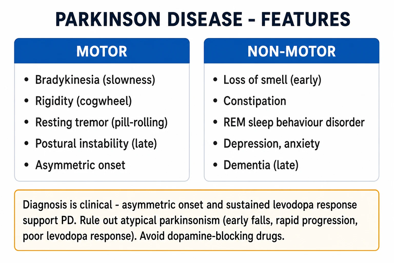
PD is insidious and asymmetric in onset; symptoms are grouped into motor and non-motor, the latter increasingly recognised as the dominant source of disability. [1]
Motor features — the cardinal tetrad (TRAP)
Resting Tremor
- 4 to 6 Hz, pill-rolling, at rest, abolished or reduced by action
- Asymmetric onset (often starts in one hand), worsened by stress
- Absent during sleep; present in about 70% at diagnosis
- Spared in akinetic-rigid subtype and in drug-induced parkinsonism (often)
Rigidity
- Velocity-independent increase in tone throughout range, both flexors and extensors
- Lead-pipe (uniform) or cogwheel (with superimposed tremor)
- Elicited on passive movement; enhanced by activation manoeuvre of contralateral limb (Froment sign)
Bradykinesia / Akinesia (OBLIGATORY)
- Slowness of movement (bradykinesia) and difficulty initiating/poverty of movement (akinesia)
- Decreasing amplitude and speed on repetitive tapping (fatiguing, hesitation, freezes)
- Manifests as hypomimia (mask face), micrographia, hypophonia, reduced blink, slowed chewing
Postural instability
- Late feature; loss of postural reflexes, retropulsion, propulsion, festination
- Assessed by the pull test (examiner pulls patient back at shoulders)
- Major cause of falls, fractures, loss of independence
Other motor signs: hypomimia (masked face, reduced facial expression), hypophonia (soft voice), hypersalivation/sialorrhoea (reduced swallow, not excess saliva), micrographia (small handwriting that gets smaller), reduced arm swing on walking, festination (accelerating, shuffling steps), freezing of gait (feet glued to floor, especially at turns, doorways, on starting — "off" freezing), camptocormia (stooped posture) and Pisa syndrome (lateral trunk flexion), blepharospasm, and dystonia (often a foot in young-onset disease).[1]
Non-motor features (often precede motor disease and dominate late)
Prodromal (pre-motor)
- Hyposmia/anosmia (universal early; UPSIT test abnormal)
- Constipation (years to decades before motor onset)
- REM sleep behaviour disorder (RBD) — act out dreams, shout, kick
- Depression, anxiety, orthostatic intolerance, subtle slowness
Established disease
- Depression (40 to 50%), anxiety, apathy, anhedonia
- Orthostatic hypotension, urinary urgency/incontinence, erectile dysfunction, constipation
- Sleep fragmentation, restless legs, RBD, daytime somnolence
- Pain, sensory disturbances (shoulder pain), seborrhoea, sweating
Late disease
- Dementia (PD dementia — develops after years of motor disease)
- Psychosis (visual hallucinations, delusions, paranoia)
- Severe dysphagia with aspiration risk, weight loss, immobility
- Marked autonomic failure; falls; pressure injury; infections
Atypical presentation in the elderly
In older patients PD may present atypically — with gait disturbance, falls, or cognitive decline rather than tremor, with more rapid progression, early dementia, prominent axial and postural instability, and marked autonomic failure. The tremor-dominant phenotype is rarer in the very old. A lower threshold to suspect atypical parkinsonism (especially PSP and DLB) is appropriate in this group.[2]
Differential Diagnosis
The differential of parkinsonism is broad. The discriminator questions are: Is the levodopa response clear and sustained? Is it asymmetric? Is there early dementia, early falls, gaze palsy, autonomic or cerebellar involvement? Is a drug responsible? Is the patient young (Wilson)? [1]
1. Essential tremor (the commonest mimic):[1]
- Postural/action tremor (not resting), 4 to 12 Hz, bilateral, hands and head (yes-yes), improves with alcohol, family history in over half, no bradykinesia or rigidity, normal gait. DAT-SPECT is normal (essential tremor) versus reduced (PD). Treat with propranolol or primidone. [1]
2. Drug-induced parkinsonism (always reversible, must be asked):[1]
- Caused by dopamine D2 antagonists — typical antipsychotics (haloperidol, chlorpromazine), atypicals (risperidone, olanzapine — less with quetiapine/clozapine), metoclopramide, prochlorperazine, antinauseants (promethazine), cinnarizine/flunarizine (calcium-channel blockers), some selective calcium-channel blockers, valproate (high dose), tetrabenazine, reserpine.
- Symmetric, less tremor-dominant, develops over weeks to months of the drug, and is reversible on stopping (may take weeks to months). DAT-SPECT is normal (no nigral degeneration). The trap: it coexists with idiopathic PD and unmasking a hidden parkinsonism. [1]
3. Vascular parkinsonism:[2]
- Multi-infarct state; lower-body predominant ("parkinsonism of the feet"), prominent gait disturbance (broad-based, marche a petits pas), pyramidal signs (hyperreflexia, Babinski), stepwise course, vascular risk factors, poor levodopa response, and white-matter/lacunar infarcts on MRI. [1]
4. Atypical parkinsonisms (parkinson-plus) — high-yield:[1][2]
Progressive supranuclear palsy (PSP)
- Tauopathy (4R tau); Richardson syndrome is the classic form
- Early falls (within first year, often backwards), axial (neck > limb) rigidity
- Supranuclear vertical gaze palsy (downgaze impaired first) — the cardinal sign
- Pseudobulbar, frontal dysfunction; poor levodopa response; MRI: hummingbird sign
Multiple system atrophy (MSA)
- Alpha-synuclein glial cytoplasmic inclusions; parkinsonism PLUS autonomic/cerebellar
- MSA-P (parkinsonian, poor levodopa response) or MSA-C (cerebellar ataxia)
- Prominent dysautonomia (orthostatic hypotension, urinary incontinence, erectile dysfunction)
- Stridor, pyramidal signs; MRI: putaminal atrophy, hot-cross-bun pontine sign
Dementia with Lewy bodies (DLB)
- Alpha-synuclein cortical Lewy bodies; fluctuating cognition, recurrent visual hallucinations
- Spontaneous parkinsonism; severe neuroleptic sensitivity; REM sleep behaviour disorder
- Dementia precedes or within 1 year of parkinsonism (the 1-year rule vs PD dementia)
- Reduced DAT-SPECT uptake; preserved medial temporal lobe (unlike Alzheimer)
Corticobasal degeneration (CBD)
- Tauopathy; strikingly asymmetric cortical and basal ganglia signs
- Apraxia, alien limb phenomenon, cortical sensory loss, myoclonus, dystonia
- Poor levodopa response; progressive asymmetry
5. Wilson disease (must exclude in anyone under 50 with new parkinsonism):[4]
- Autosomal recessive (ATP7B gene), copper accumulation; young age, hepatic dysfunction, Kayser-Fleischer rings (slit lamp), low serum ceruloplasmin (under 0.2 g/L), raised 24-hour urinary copper. Treat with penicillamine, trientine or zinc. [1]
6. Other mimics: normal pressure hydrocephalus (gait apraxia, dementia, urinary incontinence — triad), depression with psychomotor retardation (pseudo-parkinsonism), Alzheimer disease with extrapyramidal signs, Huntington disease (Westphal akinetic-rigid variant, especially juvenile), dopa-responsive dystonia (Segawa, dramatic levodopa response), spinal/stenosis gait disorders, and fragile X premutation tremor/ataxia syndrome (FXTAS).[1]
Clinical & Bedside Assessment
A focused neurological examination confirms parkinsonism and grades severity. [1]
Bradykinesia (obligatory):[2]
- Finger tapping (tap thumb and index repeatedly), hand pronation-supination, foot tapping, hand opening-closing. Look for decreasing amplitude and speed, hesitation/arrest, fatiguing over 10 to 15 seconds.
- Observe spontaneous movement: hypomimia, reduced blink rate (normal 12 to 20/min, reduced in PD), reduced gesture. [1]
Tone (rigidity): [1]
- Examine the wrist, elbow, shoulder, and neck by slow passive movement through full range. Lead-pipe rigidity is uniform; cogwheel is a ratchet-like feel superimposed on tremor.
- Activation manoeuvre (Froment manoeuvre): ask the patient to move the contralateral limb (open and close the opposite hand) — this augments rigidity on the tested side. [1]
Tremor: [1]
- Observe at rest in hands (pill-rolling), lips, jaw, lower limbs, and head. Re-examine during sustained posture (postural tremor in PD re-emerges after seconds — "re-emergent tremor") and action. PD rest tremor is suppressed by action; essential tremor is worst with action. [1]
Postural stability (pull test):[2]
- Stand behind the patient; give a quick backward pull on both shoulders and be ready to catch. Normal: recovery in one to two steps. Abnormal (retropulsion): takes more than two steps, or falls (no corrective step). Grade 0 to 4. [1]
Gait: observe arm swing (reduced, asymmetric), stride length (shortened, shuffling), turning (multiple small steps, "en bloc"), festination and freezing (especially at turns/doorways), posture (stooped, camptocormia). [1]
Glabellar tap (Myerson sign): tap repeatedly over the glabella. Sustained blinking (failure to habituate) is a frontal-release/parkinsonian sign; healthy subjects blink once or twice then stop. Supportive but not diagnostic. [1]
Other bedside signs: hypophonia, micrographia (write a sentence), sialorrhoea, seborrhoeic face, cogwheel at wrist, rest tremor in hands at rest, bradyphrenia (slow thinking).[1]
Standardised scales:[2]
- MDS-UPDRS (Movement Disorder Society — Unified Parkinson Disease Rating Scale): the gold-standard rating scale, four parts — I Non-motor aspects of daily living; II Motor aspects of daily living; III Motor examination (the objective part); IV Motor complications. Used to stage disease, monitor progression and treatment response.
- Hoehn and Yahr staging (1 to 5) — coarse functional staging (see Classification).
- Non-Motor Symptoms Scale (NMSS) — quantifies the burden of non-motor symptoms (gastrointestinal, urinary, sleep, mood, perceptual, sexual, cardiovascular, miscellaneous).
- Schwab and England Activities of Daily Living — percentage independence. [1]
Investigations
PD is a clinical diagnosis — there is no confirmatory test in life. Investigations are used to exclude mimics and, in selected cases, to support the diagnosis.[2][3]
When investigations are needed: [1]
- Atypical features (early falls, rapid progression, symmetric onset, early dementia, poor levodopa response, pyramidal/cerebellar/autonomic signs) → imaging and secondary-cause workup.
- Young onset (under 50) → exclude Wilson disease (ceruloplasmin, 24-hour urinary copper, slit-lamp for Kayser-Fleischer rings) and consider genetic testing.
- Diagnostic uncertainty (e.g., PD vs drug-induced parkinsonism or essential tremor) → DAT-SPECT. [1]
First-line tests (baseline and to exclude mimics):[2]
- MRI brain — usually normal in PD (or shows mild age-related atrophy). Its role is to exclude vascular disease, hydrocephalus, tumour, and to show pattern signs of atypical parkinsonism: hummingbird sign (midbrain atrophy in PSP), hot-cross-bun sign (pontine cruciform hyperintensity in MSA), putaminal atrophy (MSA-P), asymmetric frontoparietal atrophy (CBD).
- Bloods — FBC, U&E, LFTs, TFTs (exclude rare endocrine myxoedema mimics), vitamin B12, glucose. In young patients: serum ceruloplasmin and 24-hour urinary copper. [1]
Functional/imaging tests: [1]
- DAT-SPECT (DaTscan) — dopamine transporter SPECT: shows the density of presynaptic dopamine transporters in the striatum. Reduced, asymmetric putaminal uptake in PD, MSA, PSP, DLB (all have nigral degeneration). Normal in essential tremor and drug-induced parkinsonism (no structural degeneration). Use when the diagnosis is in doubt between PD and these two mimics. It does not distinguish PD from atypical parkinsonisms.[2]
- Olfactory testing (UPSIT / Sniffin' Sticks): hyposmia is present in over 90% of PD and helps distinguish PD (abnormal) from MSA, PSP, CBD and drug-induced parkinsonism (often normal smell). A simple, cheap discriminator.
- Cardiac MIBG scintigraphy: assesses postganglionic sympathetic cardiac denervation — reduced in PD and DLB but normal in MSA (the lesion is preganglionic). Useful in research; limited clinical availability.
- Acute levodopa challenge: a single dose of soluble levodopa/benserazide or dispersible levodopa/carbidopa (e.g., 100/25 mg) producing a clear, sustained motor improvement supports PD and predicts a good chronic response. Not routinely required but useful in selected cases.
- Alpha-synuclein seed amplification assay (SAA) in CSF and skin biopsy: the newest biomarker — detects misfolded alpha-synuclein with very high sensitivity and specificity for PD (and prodromal PD/DLB). Entering clinical practice; not yet widely available in India.[2]
MDS clinical diagnostic criteria (2015) — reproduced
The MDS 2015 criteria replaced the 1992 UK Parkinson's Disease Society Brain Bank criteria and are the current international standard.[3]
Step 1 — Diagnose parkinsonism: [1]
- Bradykinesia (obligatory) PLUS either rest tremor or rigidity. [1]
Step 2 — Apply absolute exclusions (any one excludes PD): [1]
- Clear cerebellar signs (gaze-evoked nystagmus, cerebellar gait, limb ataxia, dysarthria).
- Downward vertical supranuclear gaze palsy, or selective slowing of downward saccades.
- Diagnosis of frontotemporal dementia, primary progressive aphasia, or dementia with Lewy bodies (within 5 years, see 1-year rule).
- Parkinsonism restricted to the lower limbs for more than 3 years.
- Treatment with a dopamine blocker (neuroleptic) or dopamine-depleting drug at onset or onset within 5 years of starting.
- Absence of response to high-dose levodopa (despite at least 400 mg/day).
- Cortical sensory impairment, intact sphincters with severe dysautonomia (MSA-type).
- Normal dopamine transporter scan (DAT-SPECT).
- A proven alternative cause (Wilson disease, structural lesion). [1]
Step 3 — Supportive criteria (any one supports PD — 2 or more weighted): [1]
- Clear dramatic beneficial response to dopaminergic therapy.
- Marked levodopa-induced dyskinesia.
- Rest tremor of a limb documented.
- Olfactory loss, OR cardiac sympathetic denervation on MIBG. [1]
Step 4 — Red flags (warn against PD): [1]
- Rapid gait impairment (within 3 years).
- No progression of motor features.
- Early bulbar dysfunction (within 5 years).
- Early severe dysautonomia (within 5 years).
- Falls within 3 years of onset.
- Anterocollis or contractures within 10 years.
- Absence of common non-motor features (sleep, autonomic) despite 5 years of disease.
- Pyramidal signs bilaterally.
- Bilateral ptosis or vertical gaze restriction. [1]
Step 5 — Final categories: [1]
- Clinically established PD: absence of absolute exclusions + at least 2 supportive criteria + no red flags.
- Clinically probable PD: absence of absolute exclusions + at least 2 supportive criteria + fewer red flags than supportive criteria.[3]
Management — Resuscitation
There is no disease-modifying therapy; the goal is symptomatic control of motor and non-motor function and preservation of quality of life.[1]
The two inviolable rules in any hospitalised PD patient:[2][1]
- Never omit or delay dopaminergic medication. PD drugs are time-critical. Missing doses (especially levodopa, which has a short half-life of 60 to 90 minutes) precipitates wearing-off, akinesia, and — in severe withdrawal — parkinsonism-hyperpyrexia syndrome. In hospital, chart levodopa at the patient's home timing (often every 3 to 4 hours), not at standard drug-round times.
- Never give dopamine-blocking drugs. Metoclopramide, prochlorperazine, haloperidol, chlorpromazine, risperidone, olanzapine (and tetrabenazine) cross the blood-brain barrier, block D2 receptors, and worsen parkinsonism or precipitate a crisis. Safe antiemetics: domperidone (does not cross the BBB) and ondansetron (5-HT3). For acute agitation/psychosis use quinine-free approaches or low-dose quetiapine, clozapine, or pimavanserin. [1]
Nil-by-mouth (perioperative or acutely ill):[1]
- Convert levodopa to enteral (NG/NJ) administration if the gut is working; levodopa can be given via NG tube (crushed dispersible levodopa/carbidopa).
- If the gut is not working (e.g., postoperative ileus, severe vomiting), use transdermal rotigotine patch (continuous dopamine delivery, no gut absorption needed) as a bridge.
- Apomorphine (subcutaneous) is another parenteral option. [1]
Acute precipitants of sudden deterioration in a known PD patient:[2]
- Infection — urinary tract infection (commonest) and chest infection/aspiration pneumonia are the leading causes of acute deterioration ("sick parkinsonian"). Always dipstick urine and examine the chest.
- Drug omission — NBM without enteral/parenteral cover.
- Dehydration — worsens orthostatic hypotension and cognitive function.
- Constipation/faecal impaction — worsens absorption and motor function.
- Metabolic disturbance, new medication interaction, aspiration. [1]
Parkinsonism-hyperpyrexia syndrome (PHS)
A rare, life-threatening emergency analogous to neuroleptic malignant syndrome, triggered by abrupt withdrawal or rapid reduction of dopaminergic therapy (also by adding a dopamine blocker). Features: fever, marked rigidity, altered consciousness/stupor, autonomic instability (tachycardia, labile BP, diaphagoresis), elevated creatine kinase, leucocytosis. Rhabdomyolysis may cause acute kidney injury.[1]
Management: [1]
- Reintroduce dopaminergic therapy immediately (oral levodopa if possible; rotigotine patch or subcutaneous apomorphine if gut compromised).
- Supportive care — IV fluids, cooling, DVT prophylaxis; ICU monitoring for airway, autonomic instability and renal failure.
- Dantrolene (1 mg/kg IV, up to 10 mg/kg/day) for severe rigidity/hyperthermia (as in NMS).
- Treat precipitating infection. [1]
Management — Definitive & Stepwise
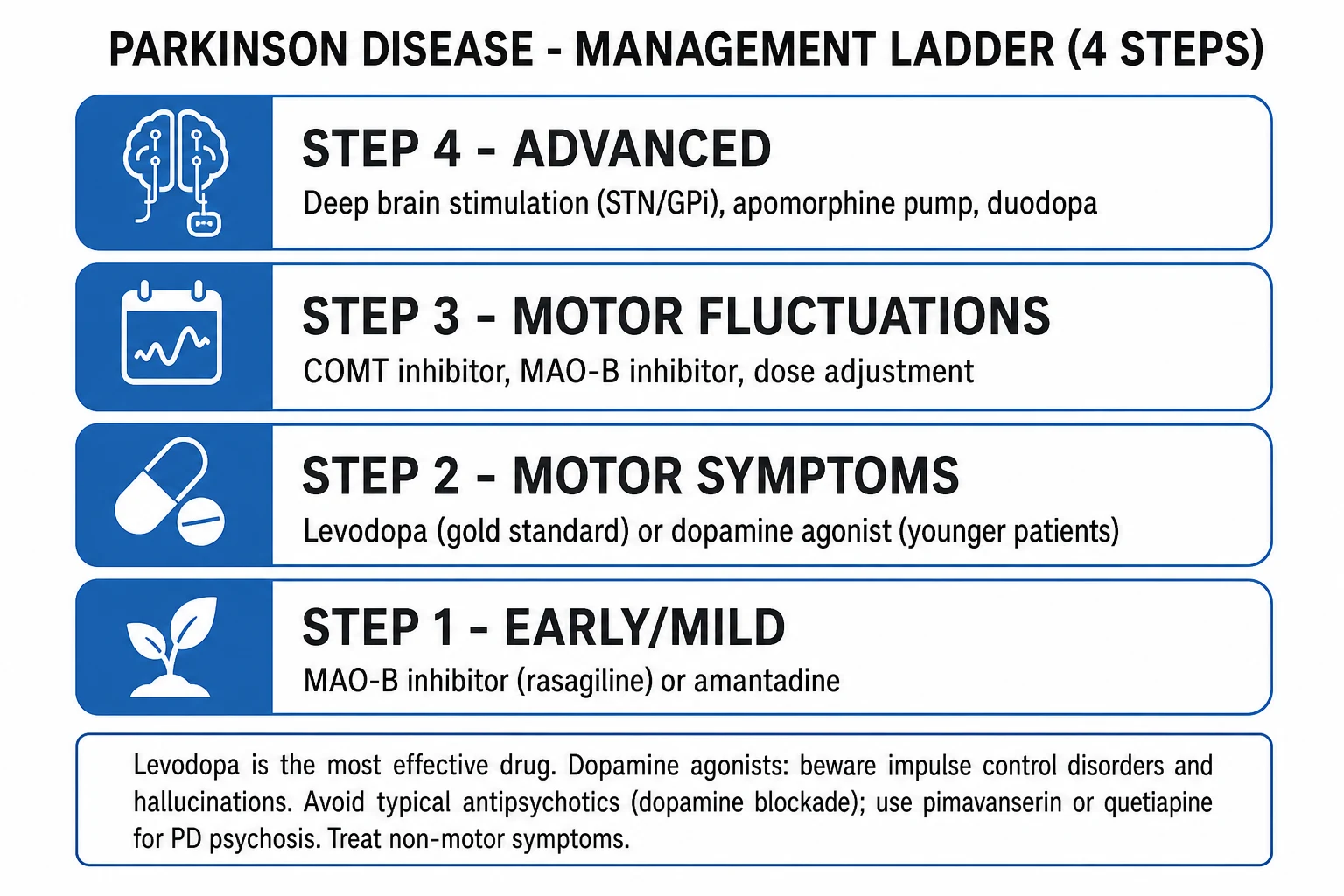
Therapy is symptomatic, individualised, and lifelong. The choice of initial agent depends on age, cognitive status, motor subtype, occupation, and patient preference.[1][2]
1. Levodopa — the gold standard
Levodopa combined with a peripheral dopa-decarboxylase inhibitor (carbidopa or benserazide) is the most effective symptomatic therapy for PD. The decarboxylase inhibitor prevents peripheral conversion of levodopa to dopamine (which causes nausea, hypotension, and is wasted peripherally), allowing more levodopa to reach the brain, where surviving nigral (and other) neurons convert it to dopamine via aromatic L-amino acid decarboxylase.[1]
- Starting dose: levodopa/carbidopa (Sinemet) or levodopa/benserazide (Madopar) — begin 50/12.5 mg or 100/25 mg three times daily, taken with meals initially to reduce nausea, then titrate over weeks to clinical effect. Controlled-release formulations are useful for overnight/early-morning off periods.
- Maintenance: typical total levodopa dose 300 to 1000 mg/day in 3 to 5 divided doses. The principle is lowest dose that controls function — high doses hasten motor complications.
- Side effects: nausea, postural hypotension, vivid dreams, hallucinations, daytime somnolence, and — with long-term use — motor fluctuations (wearing-off, on-off) and levodopa-induced dyskinesia. Supplemental peripheral decarboxylase inhibitor (extra carbidopa) or domperidone (10 to 20 mg, 30 minutes before levodopa) manages nausea.[2]
The outdated fear of "saving" levodopa for later (the levodopa-sparing doctrine) is no longer supported — early levodopa is safe, effective, and the PD-MED and ELLDOPA evidence shows no convincing increase in disease progression. Delaying levodopa deprives the patient of the most effective drug.[2][1]
2. Dopamine agonists (DAs)
Non-ergot agonists (preferred — no fibrotic/valvular risk) — ropinirole, pramipexole, rotigotine (transdermal) — act directly on D2/D3 receptors. Use first-line in younger patients (to delay levodopa and dyskinesia) or as adjuncts in advanced disease.[1]
- Pramipexole — start 0.088 mg three times daily (0.125 mg salt formulation), titrate to 0.54 to 1.1 mg three times daily; available as once-daily prolonged-release.
- Ropinirole — start 0.25 mg three times daily, titrate to up to 8 mg three times daily; prolonged-release once-daily available.
- Rotigotine patch — 2 to 16 mg/24h, once-daily continuous delivery; useful when oral route fails (perioperative, vomiting) and for overnight off periods. [1]
Ergot agonists (bromocriptine, pergolide, cabergoline) — largely abandoned because of cardiac valve fibrosis and pulmonary/retroperitoneal fibrosis; require echocardiographic monitoring if used.[1]
Key adverse effects of dopamine agonists (exam critical): [1]
- Impulse control disorders (ICDs) — pathological gambling, hypersexuality, compulsive shopping/eating, punding — particularly with D3-preferring agonists (pramipexole, ropinirole); ask proactively; reduce/stop the agonist.
- Visual hallucinations and psychosis — especially in older/cognitively impaired patients.
- Sleep attacks / sudden onset of sleep (warn about driving).
- Peripheral oedema, orthostatic hypotension, hallucinations, confusion. [1]
3. MAO-B inhibitors (selegiline, rasagiline)
Block monoamine oxidase B, reducing central dopamine breakdown; modest benefit as monotherapy in early mild disease and as adjuncts for wearing-off.[2]
- Selegiline (deprenyl) — 5 mg twice daily (morning and lunch; not evening — insomnia); amphetamine metabolite may cause insomnia and agitation.
- Rasagiline — 1 mg once daily; better tolerated; the ADAGIO trial suggested possible disease modification but not confirmed. [1]
Interactions: tyramine reaction (rare with selective MAO-B doses; avoid large tyramine meals at higher doses); serotonin syndrome with SSRIs/SNRIs/tramadol/pethidine — caution and review. Contraindicated with meperidine (pethidine).[1]
4. COMT inhibitors (entacapone, opicapone)
Inhibit catechol-O-methyltransferase, which otherwise breaks down levodopa in the periphery (and to a lesser extent centrally). Entacapone (200 mg with each levodopa dose; harmless orange-brown discolouration of urine) prolongs levodopa half-life; opicapone (50 mg once daily) is a longer-acting alternative. Use for wearing-off to extend "on" time.[2]
5. Amantadine
A weak NMDA antagonist (also antiviral, enhances dopamine release). Useful for levodopa-induced dyskinesia (the main modern indication) and tremor in early disease. Dose 100 mg once or twice daily (morning, lunch); reduce in renal impairment; causes livedo reticularis, ankle oedema, confusion/hallucinations, dry mouth.[1]
6. Anticholinergics (trihexyphenidyl/benzhexol)
Block central muscarinic receptors (restoring dopamine-acetylcholine balance). Use only in young patients with severe tremor unresponsive to other drugs. Dose trihexyphenidyl 1 to 2 mg twice daily, up to 6 to 15 mg/day. Limited by cognitive impairment, confusion (especially elderly), dry mouth, blurred vision, urinary retention, constipation — avoid in the elderly and cognitively impaired.[1]
7. Advanced therapies (for motor fluctuations refractory to oral optimisation)
Apomorphine
- Subcutaneous dopamine agonist; rescue pen (e.g., 2 to 10 mg as needed) for unpredictable off periods
- Continuous infusion (50 to 100 mg over 12 to 18 h) for severe fluctuations
- Pre-treat with domperidone for 2 days (anti-nausea); side effects — nausea, hypotension, nodules, somnolence
Levodopa-carbidopa intestinal gel (LCIG / Duodopa)
- Continuous intrajejunal infusion via PEG-J tube; smooth plasma levodopa levels
- Best for severe fluctuations and dyskinesia; very effective but invasive (PEG-J, pump)
- Highest cost; complications — tube displacement, infection, polyneuropathy (B12 deficiency)
Deep brain stimulation (DBS)
- Stereotactic implantation of electrodes in STN or GPi, connected to a pulse generator
- Best for: medically refractory motor fluctuations, dyskinesia, and tremor in cognitively intact patients with clear levodopa response
- Reduces motor fluctuations and dyskinesia, allows ~50% levodopa dose reduction
- Does NOT help (and may worsen) axial/non-motor: speech, freezing, gait, cognition
DBS selection criteria: clear levodopa-responsive idiopathic PD; motor fluctuations/dyskinesia despite optimised oral therapy; no dementia or major psychiatric illness; realistic expectations. Follett (2010, NEJM) showed STN and GPi stimulation were similarly effective on motor scores at 2 years; GPi may be preferable when dyskinesia control is paramount or cognitive risk exists.[7]
Non-motor symptom management
- Depression — SSRIs (citalopram, sertraline) or SNRIs; avoid tricyclics in older patients (anticholinergic, orthostatic). ECT for severe refractory depression. Note SSRIs can rarely trigger serotonin syndrome with MAO-B inhibitors — review.
- Psychosis — first treat reversible causes (infection, dehydration, drugs). Then reduce or stop anticholinergics, amantadine, dopamine agonists. For persistent hallucinations, use pimavanserin (a selective 5-HT2A inverse agonist; Cummings 2014 trial evidence; the only drug FDA-approved specifically for PD psychosis), or quetiapine (preferred atypical; sedating) or clozapine (very effective but needs weekly FBC monitoring for agranulocytosis). Never use haloperidol, risperidone, or olanzapine.[5]
- Dementia (PD dementia) — rivastigmine (a cholinesterase inhibitor; patch or oral 1.5 to 6 mg twice daily) is the only agent with trial evidence and FDA approval in PD dementia; donepezil also used.
- Orthostatic hypotension — non-pharmacological first (adequate hydration/salt, small frequent meals, compression stockings, raise head of bed, avoid hot baths, slow mobilisation); fludrocortisone (0.1 to 0.2 mg/day), midodrine (2.5 to 10 mg three times daily; supine hypertension is the limit), droxidopa (synthetic noradrenaline precursor).
- REM sleep behaviour disorder — clonazepam (0.25 to 0.5 mg at night) or melatonin (3 to 12 mg); protect the bed partner.
- Constipation — bulk-forming laxatives, macrogol, stimulants; review drugs.
- Urinary urgency — review for prostatic hypertrophy; cautious antimuscarinics (oxybutynin — but cognitive risk) or mirabegron (beta-3 agonist).
- Drooling/sialorrhoea — speech therapy; sublingual atropine drops or botulinum toxin into salivary glands.
- Dysphagia — speech and language therapy, texture modification, gastrostomy in late disease.[1][1]
Multidisciplinary care
PD demands a team: neurologist/movement-disorder specialist, specialist PD nurse (key coordinator), physiotherapy (gait, balance, cueing strategies, falls prevention), occupational therapy (activities of daily living, home safety, equipment), speech and language therapy (voice, swallow), dietetics (weight, protein-timing with levodopa, constipation), neuropsychology/psychiatry (mood, cognition), and social work. Driving assessment and advance care planning should be introduced proactively.[1]
Specific Subtypes & Scenarios
Young-onset PD (YOPD, onset under 50)
More often genetic (recessive: Parkin/PARK2, PINK1/PARK6, DJ-1/PARK7; dominant: LRRK2). Features slower progression, more dystonia (especially foot), less dementia, but markedly higher risk of levodopa-induced dyskinesia. Management prioritises a levodopa-sparing strategy — start with a dopamine agonist or MAO-B inhibitor, deferring levodopa until functionally necessary. Genetic counselling offered. Contraception and pregnancy planning discussed (levodopa crosses placenta but is generally continued; avoid amantadine and MAO-B inhibitors in pregnancy).[4]
Tremor-dominant PD
Prominent resting tremor, less bradykinesia and rigidity. Best prognosis, slowest progression, least dementia. Tremor can be refractory to dopaminergic therapy — anticholinergics, clozapine, DBS (especially thalamic/STN) help. Important to distinguish from essential tremor (use DAT-SPECT, olfaction, levodopa response).[1]
The atypical parkinsonisms — management implications
- PSP — poor levodopa response (try up to 1000 mg/day but usually disappointing); treat falls (physiotherapy, walking aids), blepharospasm (botulinum toxin), dysphagia. Median survival 6 to 9 years.
- MSA — poor levodopa response; aggressive treatment of autonomic failure (orthostatic hypotension, urinary); treat stridor (CPAP, ENT); median survival 6 to 10 years.
- DLB — levodopa-sensitive but with marked neuroleptic sensitivity (haloperidol can be fatal — never give typicals); cholinesterase inhibitors (rivastigmine, donepezil) for cognition and hallucinations; pimavanserin/quetiapine for psychosis.[2]
Drug-induced parkinsonism
Reversible on stopping the offending drug (over weeks to months). The common offenders are dopamine D2 antagonists: typical antipsychotics, metoclopramide, prochlorperazine, cinnarizine, some calcium-channel blockers. Management: withdraw the drug (or switch to quetiapine/clozapine if an antipsychotic is essential), consider anticholinergic cover (benztropine) if severe. Do not start chronic PD therapy unless symptoms persist after drug washout.[1]
PD psychosis vs PD dementia and the 1-year rule
- PD dementia (PDD): dementia develops in an established PD patient, at least 1 year after the onset of motor symptoms (the 1-year rule).[2]
- Dementia with Lewy bodies (DLB): dementia precedes or occurs within 1 year of parkinsonism. Both are alpha-synucleinopathies on a spectrum; the timing distinguishes them for clinical and research purposes.[2]
Freezing of gait (FOG)
Episodes of feeling "the feet are glued to the floor," classically at starts, turns, doorways, stress, or in crowds. Three types: off freezing (dopaminergic responsive), on freezing (dopaminergic non-responsive), and pseudo-on. Management: cues (laser pointer, striped floor, rhythmic counting, marching music), optimise dopaminergic therapy for off-freezing, apomorphine rescue, DBS (variable), physiotherapy. Often refractory and a major cause of falls.[1]
Complications & Pitfalls
Motor complications of long-term levodopa
After 5 years of levodopa, over 50% of patients develop motor complications; higher in young-onset disease. They fall into two groups.[1]
1. Motor fluctuations (wearing-off / off periods): [1]
- Wearing-off (end-of-dose deterioration): benefit of each levodopa dose shortens from 4 to under 2 to 3 hours; symptoms return before the next dose. Manage by more frequent dosing, controlled-release formulations, add a COMT inhibitor (entacapone/opicapone) or MAO-B inhibitor (rasagiline), or a dopamine agonist.
- Delayed-on / no-on / dose failure: the dose takes longer to work or fails (often due to protein competition for gut absorption — advise taking levodopa 30 to 60 minutes before high-protein meals).
- Unpredictable on-off fluctuations: sudden, unpredictable switches between on and off, unrelated to dose timing; advanced therapies (apomorphine, LCIG, DBS) indicated. [1]
2. Levodopa-induced dyskinesia (LID): [1]
Involuntary, often choreic or dystonic movements. Two patterns:[1]
- Peak-dose dyskinesia (commonest) — at the height of the levodopa effect; manage with lower individual doses (smaller, more frequent), amantadine (100 mg twice to three times daily — the mainstay), or advanced therapies.
- Biphasic (onset- and end-of-dose) dyskinesia — at the beginning and end of each dose; harder to treat; raise levodopa dose (counter-intuitively) or move to device-aided therapy.
- Off-period dystonia — early-morning painful foot dystonia; treat by extending "on" time overnight (controlled-release, rotigotine patch).[6]
Non-motor and long-term complications
- Falls and fractures — postural instability, osteoporosis, vitamin D deficiency; hip fractures are a major cause of morbidity.
- Dysphagia and aspiration pneumonia — the leading cause of death in PD; assess swallow, texture-modify, SLT.
- Dementia — develops in the majority after 10 to 15 years.
- Depression, anxiety, apathy, psychosis.
- Autonomic failure — orthostatic hypotension, urinary symptoms, constipation, erectile dysfunction.
- Sleep disorders — RBD, restless legs, sleep fragmentation, daytime somnolence.
- Weight loss, malnutrition, pressure injury, pressure sores, deep vein thrombosis, contractures. [1]
Classic pitfalls
- Giving dopamine-blocking drugs (metoclopramide, prochlorperazine, haloperidol) — the single most preventable error.
- Withholding levodopa for fear of dyskinesia — outdated; deprives the patient of the most effective therapy.
- Missing atypical parkinsonism (early falls, gaze palsy, symmetric, poor levodopa response, early dementia) and labelling it "PD that has stopped responding."
- Missing impulse control disorders on dopamine agonists — ask about gambling, sexuality, spending.
- Abrupt drug withdrawal precipitating parkinsonism-hyperpyrexia syndrome.
- Failing to treat non-motor symptoms (the main driver of quality-of-life loss).
- Forgetting Wilson disease in any patient under 50 with new parkinsonism.[2]
Prognosis & Disposition
PD is progressive but not directly fatal; typical course from diagnosis to death is 10 to 20 years (longer in tremor-dominant and young-onset disease, shorter in PIGD phenotype and with early cognitive or autonomic involvement).[1]
Leading causes of death are the complications of immobility and neurodegeneration: aspiration pneumonia (the single commonest cause), falls and fractures, sepsis (urinary/chest), and the dementing process.[1]
Functional trajectory correlates with Hoehn and Yahr stage: stages 1 to 2 independent; stage 3 mild disability, still independent; stage 4 needs assistance; stage 5 bed-/chair-bound. PIGD phenotype, early freezing, early postural instability, and early dementia predict a worse course.[4]
No proven disease-modifying therapy. Multiple candidates — selegiline (DATATOP), coenzyme Q10, creatine, vitamin E, inosine, isradipine, exenatide — have been tested and none has shown convincing neuroprotection in adequately powered trials. Research continues on alpha-synuclein immunotherapy and early-intervention prodromal trials.[1]
Multidisciplinary input improves outcome — physiotherapy (gait, balance, cueing, falls reduction), occupational therapy (independence, home modification, driving), speech and language therapy (voice, swallow), dietetics, and the specialist PD nurse (the coordinator who materially improves quality of life and reduces admissions).[1]
Disposition: outpatient management under a neurologist or movement-disorder specialist with shared care in the community; admit for acute deterioration (find and treat infection), aspiration, fractures, severe psychosis, or for device-aided therapy work-up. Advance care planning and palliative care involvement in late disease. [1]
Special Populations
Elderly
- Start low and go slow; avoid anticholinergics (cognitive impairment, urinary retention, constipation) and cautious with dopamine agonists (hallucinations, confusion, orthostatic hypotension, sleep attacks).
- Levodopa is first-line in the elderly and cognitively impaired.
- Beware falls, fractures, osteoporosis (calcium/vitamin D, bone protection), orthostatic hypotension, dementia, aspiration.
- Polypharmacy — review sedatives, antihypertensives, anticholinergic burden.[2]
Young-onset PD (under 50)
- Genetic counselling and testing (Parkin, PINK1, DJ-1, LRRK2, GBA).
- Dopamine agonist or MAO-B inhibitor first to delay levodopa (and dyskinesia); levodopa when function requires it.
- Contraception and pregnancy planning; occupational and driving impact.[4]
Pregnancy
Limited data. Levodopa is generally the preferred dopaminergic agent (longest safety record; crosses placenta but no clear teratogenicity); amantadine (teratogenic in animals — avoid) and MAO-B inhibitors (limited data — avoid). Dopamine agonists have very limited safety data. Multidisciplinary maternal-fetal medicine + neurology planning.[4]
Perioperative
- Continue dopaminergic medication on time up to and after surgery; chart at home schedule.
- If nil-by-mouth: enteral levodopa via NG, or rotigotine patch to cover; apomorphine if needed.
- Avoid dopamine blockers; treat postoperative nausea with ondansetron or domperidone.
- Watch for aspiration and airway compromise (delayed gastric emptying, impaired swallow).[1]
PD with coexisting psychiatric illness requiring dopamine-blocking drugs
When an antipsychotic is unavoidable (schizophrenia, severe agitation): use pimavanserin (FDA-approved for PD psychosis), quetiapine (best-tolerated; sedating), or clozapine (most effective; requires weekly FBC for agranulocytosis). Never haloperidol, risperidone, olanzapine, or chlorpromazine.[5]
Evidence, Guidelines & Regional Differences
The MDS 2015 clinical diagnostic criteria
Replaced the 1992 UK Parkinson's Disease Society Brain Bank criteria because the latter were derived from autopsy-confirmed cases (highly selected) and missed early/atypical presentations. The MDS criteria formalise the two-step (parkinsonism then exclusions/supportive/red-flags) framework and the categories of clinically established and clinically probable PD. See Investigations.[3]
NICE NG71 (2017) — United Kingdom
Recommends diagnosis by a specialist (neurologist or geriatrician with an interest), referral before treatment is started, physiotherapy, occupational therapy, speech and language therapy early, regular review, avoid dopamine blockers, advance care planning, and specialist nurse involvement. Treatment should not be delayed while waiting for a non-specialist appointment if function is impaired.[1]
Landmark evidence
- Levodopa efficacy (ELLDOPA, 2004; PD-MED, 2014): levodopa is more effective than dopamine agonists or MAO-B inhibitors for early PD; long-term levodopa does not appear to accelerate progression. PD-MED (large UK pragmatic trial) found marginally better mobility with levodopa than with levodopa-sparing strategies, at no cost in dyskinesia-related disability.[2]
- Dopamine agonist monotherapy (rasagiline — ADAGIO; pramipexole/ropinirole trials): effective in early disease; delayed levodopa and dyskinesia at the cost of more ICDs and side effects.[1]
- Pimavanserin (Cummings 2014, Lancet): a selective 5-HT2A inverse agonist; the only drug with positive phase-3 RCT evidence and FDA approval specifically for PD psychosis, with no worsening of motor function.[5]
- Deep brain stimulation (Follett 2010, NEJM): STN and GPi stimulation comparably improve motor function at 2 years in advanced PD; the choice is individualised.[7]
- Alpha-synuclein seed amplification assay (Siderowf 2023, Lancet Neurology): detects misfolded alpha-synuclein in CSF with ~88% sensitivity and 96% specificity for PD — a transformative biomarker entering practice.[2]
Regional practical differences — India (ICMR / national formulary)
Exam Pearls
- MPTP (a meperidine-analogue contaminant) selectively destroys the substantia nigra and causes acute parkinsonism — proof that a toxin can cause PD.
- Pesticides (paraquat, rotenone — complex I inhibitors) are risk factors.
- Bromocriptine, pergolide, cabergoline (ergot agonists) cause cardiac valve fibrosis — abandoned.
- Amantadine treats levodopa-induced dyskinesia; causes livedo reticularis.
- Anticholinergics (trihexyphenidyl) — only in young tremor-dominant; cognitive/anticholinergic side effects.
- Olfaction is lost in PD (and in DLB) but normal in MSA, PSP, CBD, and drug-induced parkinsonism — a cheap discriminator.
- Cardiac MIBG reduced in PD/DLB; normal in MSA (preganglionic lesion).
- Rivastigmine is the only cholinesterase inhibitor with trial evidence in PD dementia.
- Parkinsonism-hyperpyrexia syndrome — abrupt withdrawal of levodopa; treat by restarting dopaminergic therapy + supportive care + dantrolene.
- MDS-UPDRS parts I to IV — the standard rating scale; Hoehn and Yahr 1 to 5 — coarse staging.
- Always exclude Wilson disease (ceruloplasmin, 24-hour urinary copper, Kayser-Fleischer rings) in any parkinsonism under 50. [1]
Exam application bank (NEET-PG / INICET)
One-line answer
Parkinson disease (PD) is a progressive neurodegenerative disorder characterised by loss of dopaminergic neurons of the substantia nigra pars compacta, with intracellular alpha-synuclein Lewy bodies. The cardinal motor features are TRAP: resting Tremor, Rigidity, Akinesia/bradykinesia (obligatory), and Postural instability. Diagnosis is clinical (MDS 2015 criteria); investigations exclude mimics. Treatment is symptomatic — levodopa combined with a peripheral decarboxylase inhibitor (carbidopa or benserazide) is the gold-standard and most effective therapy. Dopamine agonists, MAO-B inhibitors, COMT inhibitors and amantadine have defined roles; advanced disease uses apomorphine, levodopa-carbidopa intestinal gel, or deep brain stimulation. Long-term levodopa causes motor fluctuations and dyskinesia. Non-motor symptoms (anosmia, constipation, REM sleep behaviour disorder, depression, dement
Worked stems (answer without another resource)
Stem 1 — Classic presentation. Map symptoms to mechanism; name the first investigation and first treatment step with dose/route if drug therapy is standard. [1]
Stem 2 — Unstable / complicated. List red flags that force immediate resuscitation, theatre, ICU, antidote, or reperfusion — and what you do in the first 15 minutes. [1]
Stem 3 — Atypical group. Elderly, pregnancy, child, or immunocompromised: how presentation and thresholds change. [1]
Stem 4 — Differential trap. Name the three closest mimics and one discriminator for each. [1]
Stem 5 — Disposition. Who goes home with safety-netting, who is admitted, who needs HDU/ICU/theatre, and what follow-up is mandatory. [1]
Rapid viva checklist
- Definition + classification
- Pathophysiology chain
- Bedside signs / criteria
- Score with exact components (if any)
- Emergency bundle
- Definitive therapy with doses
- Complications of disease and of treatment
- Special populations
- Guideline/trial name if classic
- Three exam traps
Coverage self-check
If you cannot answer any stem above from this page alone, re-read the matching section — the page is intended to be self-sufficient for final-prof and NEET-PG/INICET questions on Parkinson Disease.
References
- [1]Poewe W, Seppi K, Tanner CM, et al. Parkinson disease Nat Rev Dis Primers, 2017.PMID 28332488
- [2]Armstrong MJ, Okun MS. Diagnosis and Treatment of Parkinson Disease: A Review JAMA, 2020.PMID 32044947
- [3]Postuma RB, Berg D, Stern M, et al. MDS clinical diagnostic criteria for Parkinson's disease Mov Disord, 2015.PMID 26474316
- [4]Kalia LV, Lang AE. Parkinson's disease Lancet, 2015.PMID 25904081
- [5]Cummings J, Isaacson S, Mills R, et al. Pimavanserin for patients with Parkinson's disease psychosis: a randomised, placebo-controlled phase 3 trial Lancet, 2014.PMID 24183563
- [6]Fox SH, Katzenschlager R, Lim SY, et al. International Parkinson and movement disorder society evidence-based medicine review: Update on treatments for the motor symptoms of Parkinson's disease Mov Disord, 2018.PMID 29570866
- [7]Follett KA, Weaver FM, Stern M, et al. Pallidal versus subthalamic deep-brain stimulation for Parkinson's disease N Engl J Med, 2010.PMID 20519680