Neurology · General Medicine
Peripheral Neuropathy
Also known as Peripheral neuropathy · Polyneuropathy · Diabetic neuropathy · Sensorimotor neuropathy · Mononeuritis multiplex
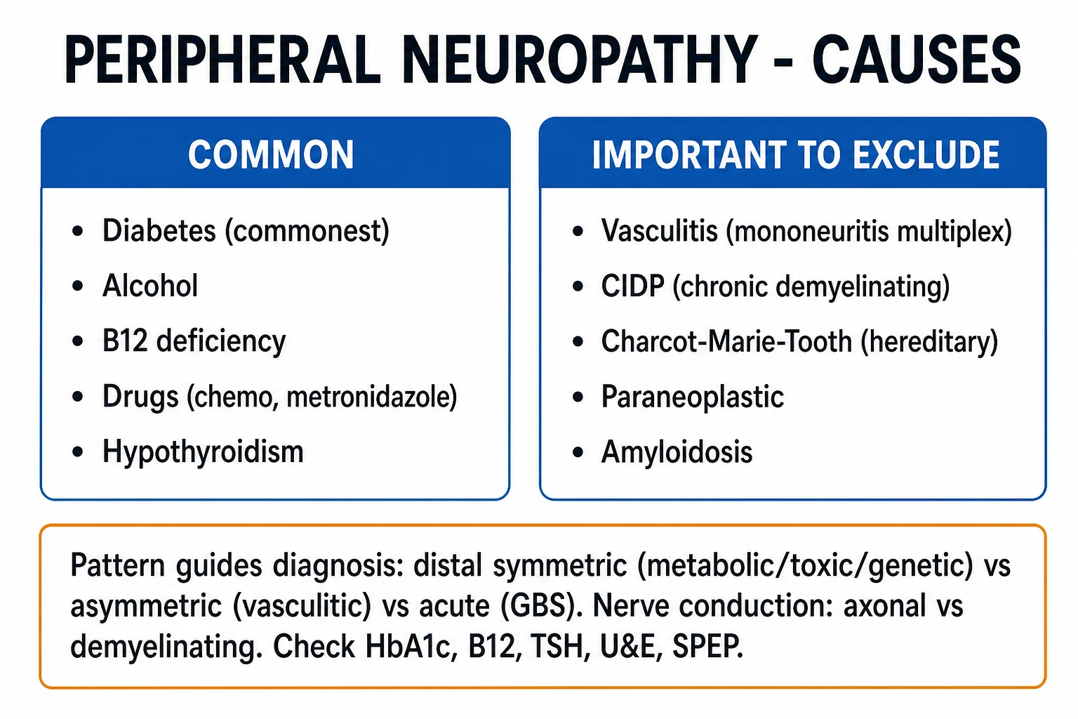
Peripheral neuropathy is any disorder of the peripheral nerves; clinically the term usually denotes polyneuropathy — a diffuse, usually symmetric process. The commonest pattern worldwide is a distal symmetric (length-dependent), sensorimotor, axonal polyneuropathy producing numbness, tingling and burning pain in a glove-and-stocking distribution, with loss of ankle reflexes. The commonest cause is diabetes mellitus (diabetic peripheral neuropathy, DPN); other leading causes are alcohol, vitamin B12 deficiency, drugs (chemotherapy, vincristine, isoniazid, metronidazole, amiodarone, phenytoin), uraemia, hereditary (Charcot-Marie-Tooth), vasculitic and CIDP. Diagnosis combines the clinical pattern, blood tests (HbA1c, B12, TSH, U&E, SPEP) and nerve conduction studies distinguishing axonal from demyelinating injury. Management is treat the underlying cause plus neuropathic pain relief (gabapentinoids, duloxetine, TCAs — not opioids) and meticulous foot care to prevent ulceration and amputation.
On this page & tools
Your progress
Saved locally on this device.
Practise this topic
Exam tags
Red flags
Overview & Definition
Peripheral neuropathy is among the commonest neurological disorders — its distal symmetric form is nearly universal in longstanding diabetes, and it underlies most non-traumatic lower-limb amputations worldwide. The discipline is to classify the pattern (because pattern predicts the cause), identify and treat the cause (diabetes, alcohol, B12, drugs, genetic, immune), protect the insensate foot from injury, and manage neuropathic pain rationally. The central red flag is acute, asymmetric, or motor-predominant neuropathy — which points away from the common metabolic causes toward Guillain-Barre syndrome (GBS), CIDP or vasculitic mononeuritis multiplex, all of which are neurological emergencies.[1][3]
The peripheral nerve carries three functional fibre populations, and which fibres are injured determines the symptoms. Large myelinated fibres subserve vibration, joint position (proprioception) and the deep tendon reflex arc; their loss produces numbness, sensory ataxia, a positive Romberg sign and areflexia. Small fibres (thinly myelinated A-delta and unmyelinated C) carry pain and temperature and innervate the autonomic end-organs; their loss produces burning pain, loss of protective sensation (painless injury and ulcers) and autonomic failure. Motor axons drive the muscles; their loss produces weakness, wasting and fasciculations. Most polyneuropathies are mixed, but recognising the predominant fibre type directs both the differential (a pure small-fibre neuropathy points to diabetes, amyloid or alcohol; a pure large-fibre neuropathy to B12, CIDP or CMT) and the investigation plan.[2]
Classification
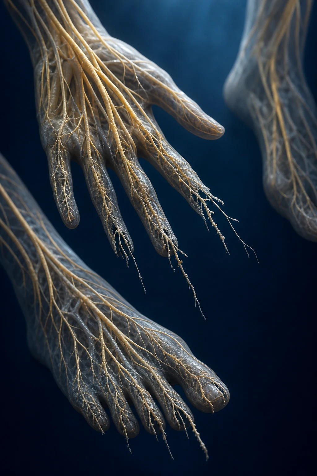
Peripheral neuropathy is classified along four axes — distribution, modality, pathology and course — and the combination directs the differential. The single most important split is between a mononeuropathy (a single nerve injured, usually by compression or trauma) and a polyneuropathy (a diffuse, symmetric process driven by a systemic or metabolic cause).[3]
By distribution
- Mononeuropathy — one nerve damaged, typically by compression, entrapment or trauma. The prototypes are carpal tunnel syndrome (median nerve at the wrist), ulnar neuropathy at the elbow, common peroneal (fibular) nerve palsy at the fibular head (foot drop), radial nerve palsy (Saturday-night palsy, wrist drop) and lateral femoral cutaneous nerve (meralgia paraesthetica). Bell's palsy is a mononeuropathy of the facial (VII) nerve — see below.
- Multiple mononeuropathy / mononeuritis multiplex — scattered, asynchronous individual nerve deficits that accumulate in a stepwise, painful fashion. The classic cause is vasculitis (polyarteritis nodosa, rheumatoid, ANCA-associated); diabetes and leprosy can also produce this pattern. Mononeuritis multiplex = vasculitis until proven otherwise.
- Polyneuropathy — diffuse, symmetric, usually distal and length-dependent. This is by far the commonest pattern and the default meaning of "peripheral neuropathy." It is further divided by modality and pathology (below). [1]
By modality (fibre type)
Predominantly sensory (large-fibre: vibration/proprioception loss, sensory ataxia — B12 deficiency, paraneoplastic; small-fibre: burning pain, autonomic features — diabetes, amyloid), predominantly motor (GBS, multifocal motor neuropathy, lead), autonomic prominent (diabetic autonomic neuropathy, amyloidosis), or mixed sensorimotor (the commonest — most metabolic/toxic neuropathies). The order of fibre involvement in typical axonal polyneuropathy is sensory then motor, with autonomic features late — a sequence worth memorising. [1]
By pathology (the electrophysiological split that drives the differential)
Axonal neuropathies (commonest — diabetes, alcohol, B12, toxic) reflect primary injury to the axon: nerve conduction shows reduced compound motor action potential (CMAP) and sensory nerve action potential (SNAP) amplitudes with near-normal conduction velocity. Demyelinating neuropathies (GBS, CIDP, CMT type 1, anti-MAG) reflect injury to myelin or the Schwann cell: conduction shows markedly slowed velocity (often below 35 m/s), prolonged distal latency, conduction block and temporal dispersion, with relatively preserved amplitude until secondary axonal loss supervenes. Mixed axonal-demyelinating patterns also occur (CMT type X, some toxic neuropathies). [1]
By course
Acute (days to 4 weeks — GBS, acute toxic such as vincristine or a paralytic tick), subacute (weeks to months — vasculitis, B12, paraneoplastic), and chronic (over years — diabetes, hereditary CMT, CIDP). CIDP is defined as demyelinating neuropathy progressing over more than 8 weeks, distinguishing it from GBS (under 4 weeks). [1]
Epidemiology & Risk Factors
Peripheral neuropathy affects roughly 2 to 8 percent of the general population, rising to around 50 percent of people with longstanding diabetes after two decades of disease.[1] Diabetes mellitus is the commonest cause worldwide; in primary-care neuropathy series the leading identifiable causes are, in order, diabetes, alcohol, vitamin B12 deficiency, drug toxicity (especially chemotherapy), chronic kidney disease and hereditary neuropathies. Charcot-Marie-Tooth disease, the commonest inherited neuromuscular disorder, affects about 1 in 2500 people. Even after a thorough work-up, 20 to 30 percent remain 'cryptogenic' (chronic idiopathic axonal polyneuropathy), a diagnosis of exclusion in older adults.
Risk factors for diabetic peripheral neuropathy include diabetes duration, poor glycaemic control (high HbA1c), older age, hypertension, dyslipidaemia, smoking and alcohol excess, and a low BMI in type 1.[2] In type 1 diabetes, intensive glycaemic control reduces the incidence of neuropathy by about 60 percent (DCCT), whereas in type 2 the effect of glucose lowering alone is modest, and multifactorial cardiovascular risk reduction (blood pressure, lipids, weight, smoking) is emphasised instead. Occupational exposure to neurotoxins (organic solvents, n-hexane, lead, arsenic, mercury) and specific chemotherapy regimens (platinum compounds — oxaliplatin; taxanes — paclitaxel; vinca alkaloids — vincristine; proteasome inhibitors — bortezomib; thalidomide) are important iatrogenic and toxic precipitants. Nutritional deficiency (B12 from vegan diet, pernicious anaemia, metformin or gastrectomy; thiamine in alcoholism and bariatric surgery) and HIV (both from the virus and from antiretrovirals, especially the d-drugs didanosine and stavudine) round out the global picture.[3]
Pathophysiology
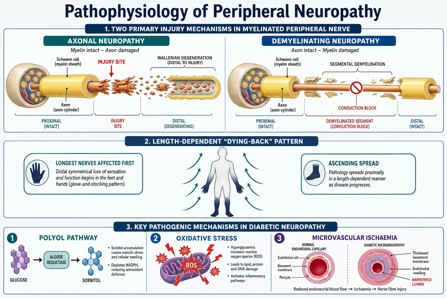
The mechanism of nerve injury divides into axonal and demyelinating pathology, each with a characteristic electrophysiology and tempo of recovery. In clinical practice, distinguishing these two on nerve conduction studies is the single most useful step after the history, because it partitions the differential into treatable immune/demyelinating disease versus metabolic/toxic/axonal disease.[2][3]
Axonal neuropathy is the commonest pathology (diabetes, alcohol, B12, toxic). The primary injury is to the axon itself. Because the neuronal cell body (in the dorsal root ganglion for sensory fibres, the anterior horn for motor fibres) can no longer sustain the long distal process, the axon undergoes distal 'dying-back' degeneration and, once transected, Wallerian degeneration of the segment distal to the lesion. The longest axons fail first — hence length dependence (the axon to the big toe may travel a metre or more, with the greatest metabolic demand and least trophic reserve). On nerve conduction this shows as low CMAP/SNAP amplitudes with preserved (near-normal) conduction velocity, and on needle EMG as fibrillations and positive sharp waves (denervation) in distal muscles with large, polyphasic motor units on reinnervation. Regrowth is slow, at about 1 mm per day (roughly 1 inch per month), and is only possible if the cause is removed and the cell body survives — so recovery spans many months to years.[3]
Demyelinating neuropathy (GBS, CIDP, CMT type 1, anti-MAG, diphtheria) targets myelin or the Schwann cell by immune (GBS, CIDP), dysimmune (anti-MAG) or genetic (CMT1 — PMP22 duplication) mechanisms. Stripped or thinned myelin causes conduction block, slowing and temporal dispersion, producing weakness and sensory loss out of proportion to axon loss. Nerve conduction shows markedly reduced conduction velocity (typically below 35 m/s, or below 70 to 80 percent of the lower limit of normal), prolonged distal and F-wave latencies, conduction block and temporal dispersion, with relatively preserved amplitudes. Recovery is possible because Schwann cells can remyelinate, and the immune forms respond dramatically to immunotherapy (plasma exchange, IVIg, steroids).[4]
The diabetic mechanism is multifactorial. Chronic hyperglycaemia drives the polyol (sorbitol) pathway: glucose is converted by aldose reductase to sorbitol and then fructose, depleting NADPH and myo-inositol, lowering Na+/K+-ATPase activity and impairing nerve conduction. Concurrently, hyperglycaemia generates mitochondrial oxidative stress (excess reactive oxygen species in the dorsal root ganglion neurons), forms advanced glycation end-products (AGEs) that crosslink nerve and vascular proteins, and causes microvascular ischaemia (thickened, hyalinised endoneurial capillary basement membranes that reduce nerve blood flow). Together these produce the length-dependent, predominantly axonal, mixed sensorimotor neuropathy of diabetes, with prominent small-fibre and autonomic involvement.[2]
Other mechanisms worth naming. Vitamin B12 deficiency impairs methylation in myelin synthesis and produces subacute combined degeneration of the dorsal columns and corticospinal tracts alongside the peripheral neuropathy. Alcohol injures nerve directly and indirectly through thiamine deficiency (which also produces Wernicke-Korsakoff). Vincristine and other vinca alkaloids inhibit microtubule formation and so block axonal transport, producing a sensorimotor axonal neuropathy; isoniazid competes with pyridoxine (B6) and causes a sensory neuropathy preventable with pyridoxine 25 to 50 mg daily; metronidazole (and the related nitroimidazoles) causes a predominantly sensory axonal neuropathy, classically after prolonged courses. Charcot-Marie-Tooth type 1A (the commonest CMT) is an autosomal dominant duplication of the PMP22 gene on chromosome 17p11.2 causing Schwann-cell dysfunction and uniformly slow conduction from childhood, often with palpable nerve hypertrophy. [1]
Clinical Presentation
The symptoms of a polyneuropathy follow the fibre type injured. A careful sensory, motor and autonomic history, with attention to tempo and distribution, usually predicts both the pathology and the cause before any test is done.[1][3]
Sensory — positive symptoms are tingling, pins-and-needles, burning, stabbing, electric-shock or 'tight-band' pain and allodynia (pain from a non-painful stimulus such as bedsheets), typically in a glove-and-stocking pattern and worse at night ('burning feet'). Small-fibre neuropathy in particular produces severe burning pain that may begin in the toes and ascend. Sensory — negative symptoms are numbness and loss of feeling; large-fibre loss (vibration, proprioception) causes sensory ataxia and a positive Romberg sign (loss of balance with eyes closed), while small-fibre loss (pain, temperature) allows painless injuries, burns and ulcers — the patient who cannot feel a pebble in the shoe. [1]
Motor symptoms and signs begin distally: weakness of ankle dorsiflexion (foot drop, causing tripping), intrinsic foot and hand muscles, and later weakness of grip; wasting of the intrinsic muscles, fasciculations, and clawing of the toes. Reflexes are lost early distally — ankle reflexes go first, then the knee jerks, then the upper-limb reflexes as the disease ascends. In hereditary CMT, chronic distal wasting gives the legs an inverted champagne-bottle ('stork leg') shape, and pes cavus (high arch) with hammer toes is typical from childhood. [1]
Autonomic symptoms are prominent in diabetic and amyloid neuropathy and include orthostatic hypotension (lightheadedness on standing, a fall in systolic blood pressure of at least 20 mmHg), resting tachycardia, gastroparesis (early satiety, nausea, vomiting, erratic glucose control), diabetic diarrhoea (often nocturnal), urinary retention, erectile dysfunction, and gustatory sweating (sweating of the face on eating cheese or chocolate). Severe autonomic neuropathy carries a poor prognosis and contributes to sudden death and anaesthesia-related haemodynamic instability. [1]
Foot complications are the most disabling sequelae. Painless neuropathic (plantar) ulcers typically occur under the metatarsal heads where repetitive unperceived pressure breaks down insensate skin. Charcot neuroarthropathy — a destructive arthropathy of an insensate joint — presents as a warm, swollen, erythematous, deformed foot with a rocker-bottom shape, easily mistaken for cellulitis; it demands immediate offloading. The combination of neuropathy, ischaemia and infection is the classic pathway to amputation.[2]
Atypical presentations the examiner tests deliberately: the elderly may present with falls from sensory ataxia or 'just numb feet'; diabetic small-fibre neuropathy may have normal reflexes and normal nerve conduction (small fibres are not tested by routine NCS — confirm with skin biopsy); and any acute asymmetry or motor predominance red-flags GBS, CIDP or vasculitis rather than ordinary metabolic disease. [1]
Differential Diagnosis
A structured differential uses the DANG THERAPIST frame and is the backbone of any neuropathy viva. The four conditions below are the high-yield discriminators that must be distinguished at the bedside.[3]
Diabetic distal symmetric polyneuropathy
- Commonest cause; slowly progressive, symmetric, distal sensorimotor with autonomic features
- Length-dependent (glove-and-stocking); absent ankle reflexes; loss of protective sensation on 10-g monofilament
- Nerve conduction: AXONAL (low amplitude, near-normal velocity)
- Management: glycaemic control, foot care, duloxetine or pregabalin for pain
Vitamin B12 deficiency
- Neuropathy PLUS dorsal-column signs (subacute combined degeneration): loss of vibration/proprioception, positive Romberg, spasticity
- Macrocytic anaemia (raised MCV); risk factors — vegan diet, pernicious anaemia, metformin, gastrectomy
- Partly REVERSIBLE with intramuscular hydroxocobalamin if treated early
- Check serum B12 (plus methylmalonic acid or homocysteine if borderline)
Vasculitic mononeuritis multiplex
- ASYMMETRIC, painful, STEPWISE accumulation of individual nerve deficits (e.g. foot drop then wrist drop)
- Systemic features — weight loss, purpura, renal; PAN, rheumatoid, ANCA-associated
- Nerve conduction: axonal, asymmetric; EMERGENCY — sural nerve biopsy confirms vasculitis
- Treat with high-dose corticosteroids plus cyclophosphamide or rituximab
Charcot-Marie-Tooth (CMT1)
- INHERITED; onset in childhood or young adulthood; autosomal dominant family history
- Pes cavus, hammer toes, inverted champagne-bottle legs; slowly progressive over decades
- Nerve conduction: UNIFORMLY slow velocity (PMP22 duplication on chromosome 17p11.2)
- No cure — supportive: physiotherapy, ankle-foot orthosis for foot drop
The full DANG THERAPIST frame is reproduced in the Exam Pearls below; in brief the additional causes are alcoholic (painful small-fibre, with thiamine deficiency), toxic/drug (vincristine, taxanes, platinum, bortezomib, metronidazole, amiodarone, phenytoin, isoniazid without B6, heavy metals), renal/uraemic, amyloid/paraprotein (anti-MAG, MGUS, familial amyloid polyneuropathy), pyridoxine (B6) excess (a pure sensory neuropathy — a favourite exam trap, as deficiency of B6 also causes neuropathy), infection (HIV, leprosy — the commonest cause worldwide in some tropical regions, mononeuritis multiplex with skin lesions and thickened nerves), systemic/sarcoid, and trauma/compression (mononeuropathies such as carpal tunnel). [1]
Clinical & Bedside Assessment
The focused examination defines the pattern and fibre type, which directs the diagnosis. The aim is to answer four questions: is the neuropathy distal symmetric, asymmetric/multifocal, acute motor, or pure sensory/autonomic?[3]
Large-fibre testing — 128-Hz tuning-fork vibration at the hallux (compare with a proximal bony point; a vibration perception threshold above 25 V on a neurothesiometer is abnormal), proprioception (toe up/down position), and the 10-g Semmes-Weinstein monofilament applied to six foot sites per foot (dorsum of the hallux and the 1st, 3rd and 5th metatarsal heads, plus the heel). Inability to feel the monofilament at any site indicates loss of protective sensation and predicts ulceration — this is the single most useful bedside test in diabetes. [1]
Small-fibre testing — pinprick (a neurological pin) and temperature (cold tuning fork or test tube) distally; small-fibre neuropathy may spare reflexes and vibration, so a normal large-fibre exam does not exclude it. [1]
Motor and reflex — test ankle then knee reflexes (ankle reflexes are lost earliest in length-dependent neuropathy), distal power (ankle dorsiflexion, toe extension, intrinsic hand muscles), and look for wasting, fasciculations and foot deformity (pes cavus, claw toes). Gait and Romberg — sensory ataxia gives a wide-based, high-stepping or stamping gait; a positive Romberg (balance lost with eyes closed) signals proprioceptive (large-fibre) loss. [1]
Orthostatic blood pressure — measure lying and standing for 3 minutes; a systolic drop of 20 mmHg or more (or diastolic 10 mmHg) within 3 minutes indicates orthostatic hypotension from autonomic failure. Inspect the neuropathic foot for callus, ulcers (especially under metatarsal heads and at the heel), deformity, Charcot changes (rocker-bottom, warmth, swelling), and palpate pulses (dorsalis pedis, posterior tibial) to screen for coexisting ischaemia. A warm swollen deformed foot must be treated as possible acute Charcot until proven otherwise. [1]
Investigations
Investigation is targeted by the clinical pattern — there is no 'screen everything' panel. The goals are to (a) confirm the neuropathy is peripheral, (b) distinguish axonal from demyelinating pathology, and (c) identify a treatable cause.[1][3]
First-line bloods — fasting glucose and HbA1c, vitamin B12 (plus methylmalonic acid or homocysteine if borderline, since these rise early and confirm tissue deficiency), full blood count (MCV for macrocytosis pointing to B12 or alcohol), U&E and creatinine (uraemia), LFTs, TSH, folate and thiamine (if alcohol or malnutrition), and lipid profile. [1]
Second-line tests (when the pattern is atypical or first-line unrevealing) — serum protein electrophoresis (SPEP) with immunofixation and anti-MAG antibody (paraproteinaemic neuropathy), ANA, ANCA, rheumatoid factor, complement and cryoglobulins (vasculitis), HIV serology, hepatitis B and C, serum copper and caeruloplasmin, serum lead level, ACE (sarcoid), anti-GQ1b and anti-GM1 antibodies (GBS/MFN), and genetic testing for CMT (PMP22 duplication/deletion first, then MPZ, GJB1, MFN2 by phenotype). CSF examination (when a demyelinating process such as CIDP is suspected) shows cytoalbuminologic dissociation — a high protein (often over 0.55 g/L) with a normal cell count — a hallmark of GBS and CIDP.[4]
Nerve conduction studies (NCS) with needle EMG are the key diagnostic test. They distinguish axonal from demyelinating injury and define distribution and severity. The interpretation is summarised below. [1]
| Parameter | Axonal neuropathy | Demyelinating neuropathy |
|---|---|---|
| CMAP/SNAP amplitude | Reduced (axons lost) | Relatively preserved (until late) |
| Conduction velocity | Near-normal (may be mildly reduced) | Markedly slowed — often below 35 m/s |
| Distal latency | Normal or mildly prolonged | Prolonged |
| F-wave latency | Mildly prolonged | Markedly prolonged or absent |
| Conduction block / temporal dispersion | Absent | Present (hallmark) |
| Needle EMG | Fibrillations, positive sharp waves (denervation); large polyphasic units on reinnervation | Denervation only if secondary axonal loss |
| Typical causes | Diabetes, alcohol, B12, toxic, uraemic | GBS, CIDP, CMT1, anti-MAG, diphtheria |
Small-fibre testing — when symptoms (burning pain, autonomic features) suggest small-fibre neuropathy but routine NCS are normal: punch skin biopsy measuring intra-epidermal nerve fibre density (reduced below age- and sex-adjusted fifth percentile confirms the diagnosis), quantitative sensory testing, sudomotor tests (QSUDOR, QSART) and corneal confocal microscopy. MRI of the spine or plexus is reserved for radicular, plexus or mass lesions. Sural nerve biopsy is rarely needed — reserve it for suspected vasculitis or amyloid, where histology changes management (a contralateral muscle biopsy combined with sural nerve increases the diagnostic yield).[3]
Management — Resuscitation

Most neuropathies are chronic and need no resuscitation, but several presentations are neurological emergencies that must be recognised at the bedside.[3]
- Acute ascending weakness with areflexia (suspected GBS) — admit, monitor forced vital capacity (FVC) and negative inspiratory force every 4 to 6 hours; impending respiratory failure (FVC under 20 mL/kg, or falling by more than 30 percent, or NIF below minus 40 cmH2O) needs ICU and early intubation before respiratory arrest; treat autonomic instability (arrhythmia, blood-pressure swings) and consider early plasma exchange or IVIg.
- Acute asymmetric painful neuropathy / mononeuritis multiplex (suspected vasculitis) — urgent sural nerve (± muscle) biopsy to confirm vasculitis, then high-dose corticosteroids (methylprednisolone 1 g IV daily for 3 to 5 days, then oral prednisolone 1 mg/kg/day) plus cyclophosphamide or rituximab for severe multi-organ disease; untreated vasculitic neuropathy progresses to irreversible axonal loss within days.
- Neuropathic foot ulcer or acute Charcot foot — offloading immediately (total-contact cast or non-removable knee-high device for Charcot), debridement of necrotic tissue, swab and deep tissue culture, empiric antibiotics for infection (as per local diabetic foot protocols), and urgent podiatry and vascular referral for any ischaemic or non-healing ulcer.
- Severe autonomic neuropathy (orthostatic hypotension) — withdraw aggravating drugs (diuretics, alpha-blockers, tricyclics), expand volume with salt and fluids, compression stockings, and arrange autonomic function testing; admit if syncope, injurious falls, or supine hypertension with severe postural drop. [1]
Management — Definitive & Stepwise
Definitive management has five strands: treat the cause, control neuropathic pain, protect the feet, manage autonomic symptoms, and provide multidisciplinary support.[1][2]
1. Treat the underlying cause — the only disease-modifying step for most. Tight glycaemic control slows diabetic neuropathy (especially in type 1, where the DCCT showed a 60 percent reduction in incident neuropathy; the effect is weaker in type 2). Intramuscular hydroxocobalamin (1 mg on alternate days for two weeks, then 1 mg every 2 to 3 months maintenance) can partly reverse early B12 neuropathy. Alcohol cessation and thiamine (100 mg daily) halt alcoholic neuropathy. Withdraw or reduce the offending drug (switch off vincristine, reduce metronidazole, replace isoniazid-related pyridoxine). Levothyroxine for hypothyroidism, renal optimisation and transplant for uraemic neuropathy. For immune neuropathies: CIDP responds to corticosteroids, IV immunoglobulin or plasma exchange; vasculitic neuropathy to high-dose steroids plus cyclophosphamide or rituximab; multifocal motor neuropathy to IVIg — and steroids can worsen it, so distinguishing MMN from CIDP is critical.[4]
2. Neuropathic pain — first-line by class, never opioids. Start one agent, titrate to effect or tolerability, and combine classes on partial response. Pregabalin 150 mg/day in two divided doses, titrate every 3 to 7 days to 300 to 600 mg/day (maximum 600 mg/day; reduce dose in renal impairment). Gabapentin 300 mg nocte day 1, 300 mg twice daily day 2, 300 mg three times daily day 3, then titrate to 900 to 3600 mg/day in three divided doses (renally excreted — adjust by creatinine clearance). Duloxetine 30 mg once daily for one week, then 60 mg once daily (the preferred agent for painful diabetic neuropathy per the ADA; up to 120 mg/day; avoid in severe hepatic impairment and uncontrolled hypertension). Amitriptyline (or nortriptyline) 10 to 75 mg nocte (start at 10 mg, titrate weekly; anticholinergic burden limits use in the elderly). Topical lidocaine 5% medicated plaster (one to three plasters for up to 12 hours per 24 hours) or capsaicin 8% patch (Qutenza, applied by clinician for 30 minutes on the feet every 3 months; or low-potency capsaicin 0.075% cream three to four times daily) for localised pain. Tramadol may be used short-term for breakthrough pain only. Avoid strong opioids (morphine, oxycodone) for chronic neuropathic pain — ineffective, addictive, and associated with harm; this stance is shared by NICE NG193 and the ADA.[2]
3. Foot care — prevents amputation. Daily foot inspection (use a mirror for the sole), well-fitting cushioned footwear, regular podiatry, callus debridement, moisturise dry skin (not between the toes), never walk barefoot, avoid hot water bottles and barefoot bathroom trips, cut nails straight across, and early treatment of any ulcer with offloading (total-contact cast or knee-high offloading boot), sharp debridement and infection control. Structured patient and family foot-care education is the cornerstone. [1]
4. Autonomic management. Orthostatic hypotension — withdraw aggravating drugs, increase salt (2 to 10 g/day) and fluid (2 to 3 L/day), compression stockings, then fludrocortisone (0.1 to 0.3 mg/day; watch for oedema and supine hypertension), midodrine (2.5 to 10 mg three times daily) or droxidopa (100 to 600 mg three times daily); raise the head of the bed. Gastroparesis — small frequent low-fat low-fibre meals, metoclopramide (5 to 10 mg before meals; beware tardive dyskinesia), domperidone or erythromycin 125 to 250 mg three times daily. Erectile dysfunction — a PDE5 inhibitor (sildenafil 50 mg, tadalafil 10 mg). Diabetic diarrhoea — loperamide or clonidine; hyperhidrosis — topical antiperspirant or glycopyrrolate. Urinary retention — intermittent self-catheterisation. [1]
5. Multidisciplinary support. Physiotherapy (strengthening, range-of-motion, gait re-education, an ankle-foot orthosis for foot drop), occupational therapy (aids and adaptations, falls prevention), dietetics, the pain team, orthotics, podiatry and structured diabetes education. [1]
Peripheral neuropathy — key numbers
Specific Subtypes & Scenarios
The major subtypes each carry high-yield exam facts that must be reproduced accurately.[1][3][6]
Diabetic peripheral neuropathy (DPN) — the commonest form worldwide; distal symmetric, mixed sensorimotor with autonomic features, predominantly axonal on NCS. A painful small-fibre variant (burning feet, normal reflexes and normal NCS) is increasingly recognised and may precede large-fibre loss. Diabetic amyotrophy (lumbosacral radiculoplexus neuropathy) is a distinct, asymmetric, painful proximal thigh weakness with weight loss, usually in type 2 diabetes, typically monophasic and improving over months; it may respond to steroids or IVIg. Screening: type 2 diabetes at diagnosis and type 1 from 5 years after diagnosis, then annually (10-g monofilament, vibration, ankle reflexes). Management is glycaemic control, foot care, and duloxetine or pregabalin for pain.[1]
Guillain-Barre syndrome (GBS) — covered in its own topic; in brief, an acute (under 4 weeks), ascending, predominantly motor demyelinating neuropathy with areflexia and cytoalbuminologic dissociation in the CSF, often post-Campylobacter. Treat with IVIg (0.4 g/kg/day for 5 days) or plasma exchange; monitor respiratory function and autonomic stability. Distinguish from CIDP by tempo.[4]
Chronic inflammatory demyelinating polyneuropathy (CIDP) — a chronic (over 8 weeks), usually symmetric, proximal and distal demyelinating motor-predominant neuropathy; cytoalbuminologic dissociation in CSF; conduction block and slowing on NCS. Unlike GBS it is steroid-responsive. First-line is IVIg (2 g/kg over 2 to 5 days, then maintenance 1 g/kg every 3 weeks), oral prednisolone (60 mg/day taper) or plasma exchange. The ICE, PREDICT and Immune trials established IVIg efficacy; IVIg is preferred for motor-predominant disease, and steroids are contraindicated in multifocal motor neuropathy (a CIDP mimic positive for anti-GM1).[4]
Vitamin B12 deficiency neuropathy — neuropathy combined with subacute combined degeneration of the cord (dorsal columns and lateral corticospinal tracts): loss of vibration and proprioception, positive Romberg, spastic paraparesis and extensor plantars, plus a macrocytic anaemia and pancytopenia. Risk factors: pernicious anaemia, vegan diet, metformin and proton-pump inhibitors (which deplete B12), ileal disease, gastrectomy, nitrous-oxide abuse. Hydroxocobalamin 1 mg IM on alternate days for 2 weeks, then every 2 to 3 months lifelong; partly reversible if treated early. [1]
Alcoholic neuropathy — a slowly progressive, often painful small-fibre sensorimotor axonal neuropathy, driven by direct alcohol neurotoxicity and thiamine (and other B-vitamin) deficiency. Management: abstinence, thiamine 100 mg daily (give before glucose to avoid precipitating Wernicke), a B-complex preparation, and nutritional rehabilitation. Painful symptoms respond to the standard neuropathic pain ladder.[3]
Chemotherapy-induced peripheral neuropathy (CIPN) — caused by platinum (oxaliplatin also causes an acute cold-induced syndrome of throat tightness and jaw pain within hours of infusion), taxanes, vincristine, bortezomib and thalidomide. A 'coasting' effect — worsening for weeks after the drug is stopped — is characteristic and traps the unwary. Prevention is dose reduction or discontinuation; duloxetine (30 to 60 mg daily) is the only agent with evidence for established CIPN pain. Counsel patients on cold avoidance for oxaliplatin and falls risk.[2]
Charcot-Marie-Tooth disease (CMT) — the commonest inherited neuropathy. CMT1 (most common) is demyelinating from PMP22 duplication on chromosome 17p11.2 (autosomal dominant), with uniformly slow conduction from childhood, palpable nerve hypertrophy, pes cavus, hammer toes, stork legs and a family history. CMT2 is axonal (normal or mildly slowed velocity); CMTX (GJB1/connexin-32) is X-linked and intermediate. Management is supportive only — physiotherapy, ankle-foot orthosis for foot drop, orthopaedic correction of deformity, and genetic counselling.[5]
Vasculitic neuropathy / mononeuritis multiplex — asymmetric, painful, stepwise accumulation of individual nerve deficits (a classic stem is 'foot drop then wrist drop then third-nerve palsy'); systemic features (weight loss, purpura, renal) point to polyarteritis nodosa, rheumatoid, ANCA-associated vasculitis, or cryoglobulinaemia (hepatitis C). Sural nerve (± contralateral muscle) biopsy confirms vasculitis; treat with high-dose corticosteroids (methylprednisolone 1 g IV daily for 3 to 5 days then oral prednisolone 1 mg/kg/day) plus cyclophosphamide or rituximab for severe disease. Untreated, it progresses to irreversible axonal loss within days — a true emergency.[3]
Bell's palsy — an acute, unilateral, lower-motor-neuron (LMN) facial (VII) nerve palsy of the peripheral segment, of presumed viral (HSV) aetiology. It differs from an upper-motor-neuron facial weakness because it involves the forehead (frontalis paralysed — the patient cannot wrinkle the brow on the affected side) and the orbicularis oculi (incomplete eye closure, Bell's phenomenon — the eye rolls upward on attempted closure). Associated features depend on the site of the lesion: hyperacusis (stapedius), loss of taste in the anterior two-thirds of the tongue (chorda tympani), and reduced lacrimation/salivation. Examination must include the ear (vesicles in the concha or ear canal with otalgia = Ramsay Hunt syndrome, herpes zoster oticus — worse prognosis, treat with antivirals) and a search for alternative causes (otitis media, parotid tumour, Lyme disease, stroke). Management: oral prednisolone 60 mg daily for 5 days, then taper by 10 mg daily (60 mg days 1 to 5, 50 mg day 6, down to 10 mg day 10) — best started within 72 hours of onset; antivirals (valaciclovir or aciclovir) added for severe palsy or Ramsay Hunt. Eye protection is mandatory — lubricating drops by day and ointment plus eyelid taping (or a chamber patch) at night to prevent exposure keratitis. Most recover fully within 6 months; poor prognostic markers are complete palsy at onset, age over 60, Ramsay Hunt, and absent motor unit potentials on facial EMG at 2 weeks.[6]
Carpal tunnel syndrome (CTS) — a compression mononeuropathy of the median nerve beneath the transverse carpal ligament at the wrist. The commonest entrapment neuropathy; risk factors are female sex, pregnancy, diabetes, hypothyroidism, rheumatoid arthritis, acromegaly, amyloidosis, and occupational repetitive wrist use. Symptoms: nocturnal pain, tingling and numbness in the thumb, index, middle and radial half of the ring finger, often relieved by 'shaking out' the hand (the flick sign). Signs: weakness of thumb abduction (abductor pollicis brevis), thenar wasting in advanced disease, and positive Tinel's sign (tapping the wrist reproduces symptoms) and Phalen's test (wrist flexion for 60 seconds reproduces symptoms). Diagnosis is clinical and confirmed by nerve conduction (prolonged median motor and sensory distal latencies across the wrist). Management: nocturnal wrist splint in neutral, local corticosteroid injection (methylprednisolone into the carpal tunnel) for moderate symptoms, and carpal tunnel decompression (surgical division of the flexor retinaculum) for severe, persistent or recurrent disease, or when thenar wasting develops.[3]
Complications & Pitfalls
- Foot ulceration and lower-limb amputation — the most feared complication; the neuropathy-ischaemia-infection pathway is the route to amputation, and the great majority of diabetes-related amputations are preceded by a (preventable) ulcer. Loss of protective sensation on the 10-g monofilament is the single best predictor.
- Charcot neuroarthropathy — painless fracture, dislocation and joint destruction producing a rocker-bottom deformity; a warm swollen foot must be offloaded immediately in a total-contact cast. The classic pitfall is mistaking acute Charcot for cellulitis or gout and treating with antibiotics or NSAIDs instead of offloading.[2]
- Falls, fractures and burns — from sensory ataxia, proprioceptive loss and foot drop; pressure injuries in insensate limbs; thermal burns from hot water bottles the patient cannot feel.
- Respiratory failure — in GBS and severe CIDP, diaphragmatic weakness causes type 2 respiratory failure; monitor FVC and intubate early.
- Treatment-related harm — opioid dependence (avoid), sedation, dizziness and ataxia (gabapentinoids, TCAs — increase falls), hyponatraemia (TCAs and SSRIs — check sodium in the elderly), hepatotoxicity (monitor LFTs with duloxetine), postural hypotension from polypharmacy, and worsening of multifocal motor neuropathy by steroids.
- Diagnostic pitfalls — treating painful feet as 'small-fibre neuropathy' while missing a treatable cause (B12, hypothyroidism, paraprotein, vasculitis, CIDP); forgetting that a normal nerve conduction study does NOT exclude small-fibre neuropathy (confirm with skin biopsy); and missing GBS in a patient presenting with 'just numbness' before weakness develops.[3]
Prognosis & Disposition
Prognosis depends entirely on the cause and pathology. Reversible or partly reversible: B12 deficiency (if treated early), hypothyroidism, some toxic neuropathies, and CIDP with immunotherapy. Arrestable (progression halts once the cause is removed but deficit persists): diabetes, alcohol, drug withdrawal. Progressive despite treatment: hereditary CMT and amyloid (without disease-modifying therapy such as patisiran/tafamidis). Axonal neuropathies recover slowly — nerve regrows at about 1 mm per day — so recovery spans months to years if the cause is removed and the cell body is intact. Demyelinating neuropathies (GBS, CIDP) can recover quickly through remyelination and respond to immunotherapy. In GBS, mortality is 3 to 7 percent and about 20 percent are left with residual disability. Tight glycaemic control slows but does not reverse established DPN, and foot protection is the single most effective intervention to prevent amputation and preserve mobility. Bell's palsy recovers fully in about 70 to 85 percent with early steroids; carpal tunnel decompression cures the majority of surgical candidates.[1][2][6]
Prevention
Prevention is cause-specific but centres on diabetic foot care and glycaemic control: intensive glycaemic management in type 1 (DCCT), multifactorial cardiovascular risk reduction in type 2, annual foot screening with the 10-g monofilament, structured foot-care education (never walk barefoot, daily inspection, well-fitting shoes), and smoking cessation. Avoid alcohol excess; supplement thiamine in alcoholism. Check B12 before and during metformin, and give pyridoxine (B6) 25 to 50 mg daily with isoniazid to prevent sensory neuropathy. In chemotherapy patients, baseline nerve conduction for high-risk regimens and proactive dose reduction or discontinuation at the first sign of CIPN. Ergonomic measures and wrist splints reduce occupational CTS. Vaccination against influenza, COVID-19 and, where relevant, Zika reduces the infective triggers of GBS. [1]
Special Populations
- Elderly — high prevalence; may present with falls or imbalance rather than sensory complaint; multi-morbidity and polypharmacy increase both neuropathy risk and treatment harms, so dose-reduce gabapentinoids and TCAs (falls, delirium, hyponatraemia), avoid opioids, and screen for reversible causes (B12, hypothyroidism).
- Diabetes — annual foot screening (10-g monofilament, vibration, ankle reflexes); glycaemic and multifactorial risk control; structured patient foot-care education; early podiatry for any callus or deformity.
- Pregnancy — gabapentinoids are generally avoided in pregnancy (teratogenic concern); prefer non-pharmacological measures and topical agents for neuropathic pain; gestational diabetes drives DPN risk; carpal tunnel syndrome of pregnancy usually resolves postpartum.
- Renal impairment — uraemic neuropathy is common and partly reversible with transplantation; dose-reduce renally cleared drugs (gabapentin, pregabalin, duloxetine) and supplement for dialysis losses.
- Chemotherapy patients — baseline nerve conduction for high-risk regimens, dose-reduction or discontinuation decisions with oncology, and cold avoidance for oxaliplatin's acute cold-induced syndrome.
- Immunocompromised / HIV — antiretroviral toxic neuropathy (the d-drugs) and HIV-distal sensory polyneuropathy are common; distinguish from CMV polyradiculopathy (acute, cauda equina pattern).
- Anticoagulated or ischaemic limbs — a neuropathic foot ulcer in an ischaemic limb needs urgent vascular assessment alongside neuropathy care; compression contraindicated in arterial insufficiency. [1]
Evidence, Guidelines & Regional Differences
- Screening — the ADA recommends screening all patients with type 2 diabetes at diagnosis and type 1 from 5 years after diagnosis, then annually, using a 10-g monofilament with or without vibration or a tuning fork; NICE (NG19) offers a similar structured annual foot risk review stratifying risk into low, moderate, high and active foot disease.[1]
- Neuropathic pain — NICE (NG193) offers amitriptyline, duloxetine, gabapentin or pregabalin as first-line (chosen by comorbidity and cost), combining classes on partial response and avoiding opioids except for short-term rescue; the ADA specifically recommends duloxetine or pregabalin for DPN pain. The AAN guideline lists pregabalin, duloxetine, amitriptyline, gabapentin and venlafaxine as having varying levels of support.[2]
- Glycaemic control — tight control reduces DPN in type 1 (DCCT, with a 60 percent reduction in incident neuropathy) but the effect is weaker in type 2, where multifactorial risk reduction (BP, lipids, weight, smoking) is emphasised (Steno-2, ACCORD).
- CIDP — corticosteroids, IVIg and plasma exchange are all effective (evidence from the ICE, PREDICT and Immune trials); IVIg is often first-line for motor-predominant disease, and steroids are contraindicated in multifocal motor neuropathy.[4]
- Bell's palsy — the Cochrane review confirms that early oral corticosteroids increase the proportion recovering to a good outcome (number needed to treat about 11); antivirals alone confer no benefit but may be added for severe palsy or Ramsay Hunt.[6]
The Australian Therapeutic Guidelines and the Royal Australasian College of General Practitioners align with NICE/ADA: gabapentinoids, duloxetine and TCAs as first-line for neuropathic pain with opioids avoided. Gabapentinoid prescribing has been tightened (initiation restrictions, dose review, and controls on misuse/diversion). For DPN pain, duloxetine or pregabalin are preferred first-line; the National Association of Diabetes Centres supports annual foot screening with the 10-g monofilament.
Exam Pearls
Causes of peripheral neuropathy — DANG THERAPIST
DANG THERAPIST
commonest cause worldwide; distal symmetric sensorimotor; painful small-fibre variant
painful small-fibre; ± thiamine deficiency; give thiamine before glucose
B12 (subacute combined degeneration), thiamine, folate; post-bariatric
Charcot-Marie-Tooth — pes cavus, stork legs, uniform slowing, PMP22 duplication
chemotherapy (platinum, taxanes, vincristine, bortezomib), metronidazole, amiodarone, phenytoin, isoniazid, heavy metals
hypothyroidism; acromegaly
uraemic neuropathy — partly reversible with transplant
anti-MAG, MGUS, familial amyloid polyneuropathy
compression mononeuropathies — CTS, ulnar, peroneal; also Pyridoxine (B6) EXCESS
leprosy (commonest worldwide in tropics — thickened nerves), HIV, Lyme, diphtheria
mononeuritis multiplex — asymmetric and stepwise; sarcoid
mononeuropathies such as CTS, peroneal palsy, radial (Saturday-night) palsy
- Axonal vs demyelinating on NCS. Axonal (diabetes, alcohol, B12, toxic) = low amplitude, near-normal velocity. Demyelinating (CIDP, GBS, CMT1, anti-MAG) = slow velocity (below 35 m/s), prolonged latency, conduction block, temporal dispersion.
- Glove-and-stocking, longest nerves first = length-dependent axonal neuropathy; classic for diabetes. The axon to the big toe is the longest in the body, so it fails first.
- Small-fibre neuropathy = burning pain with autonomic features, normal reflexes and normal NCS — confirm with skin biopsy (intra-epidermal nerve fibre density).
- Mononeuritis multiplex = asymmetric, painful, stepwise = vasculitis until proven otherwise — biopsy and immunosuppress.
- CIDP vs GBS — both demyelinating with cytoalbuminologic dissociation; CIDP over 8 weeks with proximal-and-distal weakness, GBS under 4 weeks ascending.
- Neuropathic pain — pregabalin (150 to 600 mg) or gabapentin (900 to 3600 mg), duloxetine (30 to 60 mg), amitriptyline (10 to 75 mg nocte) — NOT opioids; duloxetine preferred for diabetic pain.
- Foot care prevents amputation; loss of protective sensation on a 10-g monofilament predicts ulceration; never walk barefoot.
- B6 EXCESS (not deficiency) causes a sensory neuropathy — a favourite exam trap; deficiency of B6 (from isoniazid) also causes neuropathy, prevented by pyridoxine 25 to 50 mg daily.
- Bell's palsy is an LMN VII lesion — forehead involved, give prednisolone 60 mg for 5 days within 72 hours; vesicles in the ear = Ramsay Hunt (add antivirals, worse prognosis).
- Subacute combined degeneration of the cord (B12) — dorsal columns + lateral corticospinal tracts; combine neuropathy with proprioception loss, positive Romberg and a macrocytic anaemia.
- Charcot foot = warm, swollen, deformed foot in a neuropathic patient — offload, do not confuse with cellulitis. [1]
Exam application bank (NEET-PG / INICET)
One-line answer
Peripheral neuropathy is any disorder of the peripheral nerves; clinically the term usually denotes polyneuropathy — a diffuse, usually symmetric process. The commonest pattern worldwide is a distal symmetric (length-dependent), sensorimotor, axonal polyneuropathy producing numbness, tingling and burning pain in a glove-and-stocking distribution, with loss of ankle reflexes. The commonest cause is diabetes mellitus (diabetic peripheral neuropathy, DPN); other leading causes are alcohol, vitamin B12 deficiency, drugs (chemotherapy, vincristine, isoniazid, metronidazole, amiodarone, phenytoin), uraemia, hereditary (Charcot-Marie-Tooth), vasculitic and CIDP. Diagnosis combines the clinical pattern, blood tests (HbA1c, B12, TSH, U&E, SPEP) and nerve conduction studies distinguishing axonal from demyelinating injury. Management is treat the underlying cause plus neuropathic pain relief (gabap
Worked stems (answer without another resource)
Stem 1 — Classic presentation. Map symptoms to mechanism; name the first investigation and first treatment step with dose/route if drug therapy is standard. [1]
Stem 2 — Unstable / complicated. List red flags that force immediate resuscitation, theatre, ICU, antidote, or reperfusion — and what you do in the first 15 minutes. [1]
Stem 3 — Atypical group. Elderly, pregnancy, child, or immunocompromised: how presentation and thresholds change. [1]
Stem 4 — Differential trap. Name the three closest mimics and one discriminator for each. [1]
Stem 5 — Disposition. Who goes home with safety-netting, who is admitted, who needs HDU/ICU/theatre, and what follow-up is mandatory. [1]
Rapid viva checklist
- Definition + classification
- Pathophysiology chain
- Bedside signs / criteria
- Score with exact components (if any)
- Emergency bundle
- Definitive therapy with doses
- Complications of disease and of treatment
- Special populations
- Guideline/trial name if classic
- Three exam traps
Coverage self-check
If you cannot answer any stem above from this page alone, re-read the matching section — the page is intended to be self-sufficient for final-prof and NEET-PG/INICET questions on Peripheral Neuropathy.
References
- [1]Selvarajah D, Kar D, Khunti K, et al. Diabetic peripheral neuropathy: advances in diagnosis and strategies for screening and early intervention Lancet Diabetes Endocrinol, 2019.PMID 31624024
- [2]Sloan G, Selvarajah D, Tesfaye S. Pathogenesis, diagnosis and clinical management of diabetic sensorimotor peripheral neuropathy Nat Rev Endocrinol, 2021.PMID 34050323
- [3]Watson JC, Dyck PJB. Peripheral Neuropathy: A Practical Approach to Diagnosis and Symptom Management Mayo Clin Proc, 2015.PMID 26141332
- [4]Lehmann HC, Burke D, Kuwabara S, et al. Chronic inflammatory demyelinating polyneuropathy: update on diagnosis, immunopathogenesis and treatment J Neurol Neurosurg Psychiatry, 2019.PMID 30992333
- [5]Berciano J, Garcia A, Gallardo E, et al. Intermediate Charcot-Marie-Tooth disease: an electrophysiological reappraisal and systematic review J Neurol, 2017.PMID 28364294
- [6]Madhok VB, Gagyor I, Daly F, et al. Corticosteroids for Bell's palsy (idiopathic facial paralysis) Cochrane Database Syst Rev, 2016.PMID 27428352