Neurology · General Medicine
Spinal Cord Compression
Also known as Spinal cord compression · Malignant spinal cord compression · MSCC · Metastatic epidural spinal cord compression · MESCC · Cauda equina syndrome · Cord compression
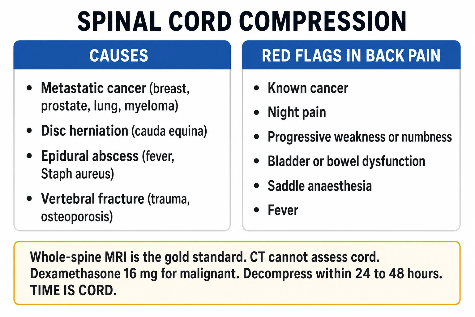
Spinal cord compression is a neurological emergency in which the spinal cord, conus medullaris or cauda equina is compressed by metastatic cancer (commonest: breast, prostate, lung, myeloma, kidney), intervertebral disc herniation, epidural abscess, epidural haematoma or vertebral collapse. Presentation: progressive, often nocturnal back pain, a sensory level, weakness, and — late and ominous — bladder and bowel dysfunction. Cauda equina syndrome adds saddle anaesthesia, bilateral sciatica, urinary retention and erectile dysfunction. Diagnosis is urgent whole-spine MRI. Malignant spinal cord compression: dexamethasone 16 mg immediately, then radiotherapy or surgical decompression within 24 to 48 hours. Cauda equina from disc: emergency surgical decompression within 24 to 48 hours. Time is cord — delay causes permanent paralysis.
On this page & tools
Your progress
Saved locally on this device.
Exam tags
Red flags
Overview & Definition
Spinal cord compression is one of the few true neurological emergencies in which hours, not days, decide whether a patient walks or is paralysed for life. The clinical discipline is to recognise the pattern — progressive back pain plus a neurological deficit — to image urgently with whole-spine magnetic resonance imaging rather than computed tomography, to give dexamethasone immediately when the cause is malignant, and to decompress within 24 to 48 hours by surgery or radiotherapy. The most important single clinical sign is bladder dysfunction: once painless urinary retention appears, the window for neurological recovery is closing rapidly, and the chance of recovering a useful bladder falls steeply.[1][2]
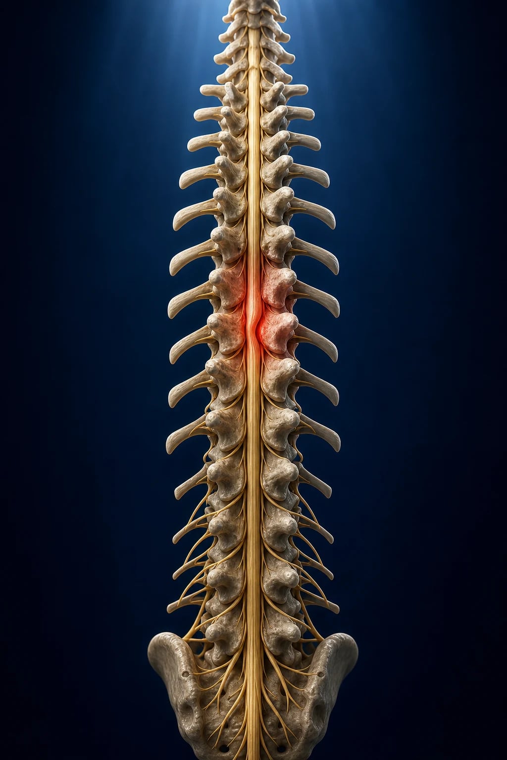
Spinal cord compression is defined as any pathological process that compromises the neural tissue of the spinal cord, the conus medullaris, or the cauda equina within the spinal canal, producing progressive neurological deficit below the level of the lesion. By the time the syndrome is clinically recognisable, the compressing mass — tumour, disc, pus, blood or bone — has already begun the cascade of venous congestion, oedema and ischaemia that, left untreated, terminates in irreversible cord infarction. The condition is classified as a neurological emergency precisely because the early, oedematous phase is pharmacologically and surgically reversible while the late, infarcted phase is not. The biological clock is the reason every guideline, every pathway and every examiner returns to the same mantra: time is cord.[1]
Definition and distinguishing terms
Several overlapping terms appear in the literature and in exam stems, and a candidate must use them precisely. Spinal cord compression is the umbrella clinical syndrome. Malignant spinal cord compression (MSCC) — also called metastatic epidural spinal cord compression (MESCC) when the compressing tumour lies in the epidural space — refers specifically to compression by metastatic cancer, the commonest cause overall and the entity to which the dexamethasone-and-radiotherapy pathway applies.[1][6] Cauda equina syndrome (CES) is compression of the lumbosacral nerve roots below the conus medullaris (the cord ends at the level of the L1 to L2 vertebral body), producing a lower motor neurone pattern with saddle anaesthesia and severe early sphincter disturbance rather than the upper motor neurone pattern of true cord compression.[8] Conus medullaris syndrome sits at the junction between the two, mixing upper and lower motor neurone signs with characteristically early and severe bladder and bowel dysfunction. Distinguishing these three syndromes at the bedside — cord, conus, cauda — is one of the highest-yield clinical judgements an examiner tests, because each carries a different anatomy, a different urgency, and a different operation.[2][8]
Classification
Spinal cord compression can be classified along three axes that together determine the urgency, the operation, and the prognosis. By anatomical level it is divided into compression above the conus (true cord compression, giving upper motor neurone signs and a sensory level), at the conus medullaris (mixed upper and lower motor neurone signs with early sphincter involvement), and below the conus (cauda equina syndrome, lower motor neurone signs with saddle anaesthesia). By tempo it is classified as acute (hours to days — typically trauma, epidural haematoma, or abscess), subacute (days to weeks — typical of malignant compression), or chronic (months — degenerative canal stenosis, slow-growing intradural tumours). By aetiology it falls into four groups: neoplastic (metastatic and, rarely, primary), degenerative and mechanical (disc herniation, canal stenosis), infective (epidural abscess, tuberculous spondylodiscitis), and vascular and traumatic (epidural haematoma, vertebral fracture, penetrating injury).[1][2]
For malignant epidural disease, radiologists and spine surgeons use the Bilsky epidural spinal cord compression (ESCC) grading scale, a six-point scale that grades the degree of epidural tumour and cord deformation on axial MRI and guides the choice between radiotherapy and surgery. Grade 0 denotes bone-only disease; grade 1 describes epidural tumour that abuts but does not deform the cord (subdivided 1a, 1b, 1c by circumferential extent); grade 2 denotes cord displacement or compression without cerebrospinal fluid (CSF) block visible; and grade 3 — high-grade compression — is cord compression with a complete CSF block and cord deformation. High-grade (2 and 3) compression, an unstable spine, or a radio-resistant tumour favour surgical decompression; low-grade compression of a radiosensitive tumour favours radiotherapy.[1][6]
Epidemiology & Risk Factors
Malignant spinal cord compression is overwhelmingly a disease of advanced cancer and is one of the most devastating complications of malignancy. Approximately 5 to 10 per cent of all cancer patients develop clinically evident MSCC at some point in their illness, rising to as much as 10 per cent in myeloma and prostate cancer. In the United Kingdom, MSCC is the second commonest neurological complication of cancer after brain metastasis, with an estimated annual incidence of around 40 to 80 cases per million population. The mean age at presentation is the seventh decade, reflecting the age distribution of the common primary tumours.[1][5]
The five commonest primary tumours responsible for MSCC, in rough order, are breast, prostate, lung, myeloma or lymphoma, and kidney (renal cell) carcinoma — together accounting for over three-quarters of cases. A useful clinical mnemonic that also captures the relative frequencies is "BLPT-RK": Breast, Lung, Prostate, Thyroid/myeloma, Renal, Kidney — the same cancers that make up the classic "metastatic spine" group. The thoracic spine is the commonest site of metastatic cord compression (around 60 to 70 per cent of cases), because it has the greatest number of vertebrae and the narrowest canal; the lumbosacral region follows, then the cervical. Prostate cancer has a particular tendency to seed the lumbar and sacral spine and pelvis via the Batson venous plexus — a valveless vertebral venous network that allows tumour emboli to bypass the lungs and lodge in the axial skeleton.[1][6]
The risk factors for non-malignant cord compression differ by cause. For spinal epidural abscess, the recognised risk factors are intravenous drug use, diabetes mellitus, immunosuppression (including HIV and chronic steroids), bacteraemia or endocarditis, recent spinal procedure or epidural injection, indwelling vascular catheters, and alcohol misuse; Staphylococcus aureus is the causative organism in over half of cases.[7] For cauda equina syndrome from disc herniation, the principal risk factors are a large central or paracentral disc protrusion, older age, heavy occupational loading, and acute trauma superimposed on pre-existing degenerative disc disease. For epidural haematoma, the dominant risk factor is iatrogenic or therapeutic anticoagulation, especially in the setting of an epidural or spinal anaesthetic. In the Indian subcontinent and other high-tuberculosis-burden regions, Pott's spine (tuberculous spondylodiscitis) remains a leading cause of cord compression, and Mycobacterium tuberculosis must be considered alongside malignancy in any patient with destructive vertebral disease.[1]
Pathophysiology
The cord is destroyed in a cascade, and the entire clinical urgency of the condition flows from a single biological fact: the early stages of compression are reversible, while the late stages are not. This is why time is cord. The cascade proceeds in four overlapping stages. Stage one is mechanical deformation: the tumour, disc, abscess, haematoma or bone fragment physically distorts the cord or nerve roots and raises the pressure within the spinal canal. Stage two is venous congestion and vasogenic oedema: obstruction of the epidural venous plexus and the small intramedullary venules produces venous hypertension, capillary leakage and vasogenic oedema — visible on MRI as T2 hyperintensity within the cord substance. This stage is potentially reversible if the compression is relieved. Stage three is arterial ischaemia: continuing compression compromises the small arterial feeders and the pia-arachnoid vasculature, producing cytotoxic oedema and demyelination; the deficit is now deteriorating but the cord is still salvageable. Stage four is infarction and necrosis: axonal death, gliosis and permanent cavitation. Once the cord has infarcted, neurological recovery is impossible no matter how promptly the mass is removed. Early decompression rescues the oedematous, ischaemic-but-not-yet-infarcted cord; after infarction, the deficit is permanent.[1][2]
Why specific tracts fail in a specific order
The arterial supply of the spinal cord explains both the watershed vulnerability and the tract-selective pattern of compression. The cord is supplied by a single anterior spinal artery (derived from the vertebrals at the foramen magnum and reinforced by segmental medullary arteries, most importantly the artery of Adamkiewicz, which usually enters on the left between T9 and L2) and the paired posterior spinal arteries. The anterior spinal artery perfuses the anterior two-thirds of the cord — the corticospinal (motor) and spinothalamic (pain and temperature) tracts and the anterior horns — while the posterior spinal arteries perfuse the posterior third, including the dorsal columns (proprioception and vibration). Because compression preferentially impairs the more tenuous anterior arterial supply, motor and pain-and-temperature loss typically precede loss of proprioception and vibration. The watershed zone between T4 and T8, where collateral supply between adjacent segmental arteries is poorest, is the most vulnerable segment of the cord to ischaemic injury — a fact the examiner rewards when asking why a suddenly hypotensive patient develops a T6 sensory level.[1][2]
How compression appears on MRI
Vasogenic oedema within the cord appears as T2 hyperintensity (a bright signal) with corresponding T1 hypointensity, often with cord expansion. The compressing mass itself is well demonstrated: metastatic tumour usually arises in the vertebral body and produces an anterior epidural mass displacing the cord posteriorly; a disc is a discrete posteriorly-protruding fragment continuous with the parent disc; an abscess shows rim enhancement with central low signal and surrounding soft-tissue phlegmon; a haematoma shows signal characteristics that vary with age (acute isointense on T1, hyperintense on T2; subacute becomes T1-hyperintense). MRI therefore simultaneously confirms the diagnosis, identifies the level, characterises the cause, and demonstrates the cord signal change that determines prognosis.[1][6]
Aetiology
The four broad aetiologic groups are neoplastic, degenerative and mechanical, infective, and vascular or traumatic. Metastatic cancer is the single commonest cause of cord compression overall, with the five primaries already listed (breast, prostate, lung, myeloma or lymphoma, kidney) dominating. Metastatic tumour reaches the epidural space most often by haematogenous spread to the vertebral body, then contiguous extension through the posterior vertebral cortex into the anterior epidural space; this is why the typical compressing mass is anterior and why decompressive surgery often involves a lateral or posterior approach with vertebral-body resection (corpectomy) and stabilisation. Far less commonly, tumour reaches the epidural space by direct extension from a paravertebral mass through the intervertebral foramen (characteristic of lymphoma and of paediatric neuroblastoma). Primary intradural tumours — meningioma, schwannoma and ependymoma — are a smaller, slower-growing group that presents with chronic progressive myelopathy rather than acute catastrophe; their MRI signature is an intradural extramedullary or intramedullary mass, not an epidural mass arising from bone.[1][2]
The commonest cause of acute cauda equina syndrome is a large central or paracentral lumbar disc herniation, typically at L4 to L5 or L5 to S1, that compresses the cauda equina roots against the posterior canal. Degenerative lumbar canal stenosis — hypertrophy of the ligamentum flavum and facet joints producing gradual narrowing of the spinal canal — produces neurogenic claudication (leg pain, heaviness and paraesthesia brought on by walking and relieved by sitting or spinal flexion) rather than true cord compression; the absence of saddle anaesthesia, retention, or a motor level distinguishes claudication from CES at the bedside. Spinal epidural abscess is the prototypical infective cause: a collection of pus in the epidural space, most often seeded haematogenously from a distant focus (skin, endocarditis, intravenous drug use) or by direct extension from vertebral osteomyelitis, that compresses the cord both by direct mass effect and by septic thrombosis of the epidural venous plexus.[7]
Pott's spine (tuberculous spondylodiscitis) merits separate attention because of its importance in the Indian context. Mycobacterium tuberculosis typically seeds the lower thoracic and upper lumbar spine, producing a subacute destructive spondylodiscitis with anterior vertebral-body collapse, kyphotic deformity (the classic Pott's gibbus) and an anterior epidural abscess or granulation tissue that compresses the cord. Unlike pyogenic infection, Pott's spine often presents subacutely with constitutional symptoms (low-grade fever, night sweats, weight loss), and paraplegia may develop slowly over weeks; management combines prolonged anti-tubercular therapy with surgical decompression and stabilisation when there is neurological deficit, severe kyphosis, or instability. Spinal epidural haematoma classically presents as sudden, severe back pain ("aphonic" pain) followed within hours by a progressive neurological deficit, often in an anticoagulated patient or after a recent spinal/epidural anaesthetic procedure; urgent surgical evacuation with reversal of anticoagulation is the treatment. Traumatic cord compression from vertebral fracture with retropulsion of bone fragments, or from penetrating injury, is discussed in the spinal-cord-injury topic but shares the same decompressive principle.[1]
Clinical Presentation
The clinical presentation is dominated by three cardinal features — pain, neurological deficit, and autonomic dysfunction — whose tempo and pattern localise the lesion and time the emergency. Back pain is the first and most constant symptom, present in approximately 95 per cent of patients with MSCC at presentation and often preceding neurological deficit by days to weeks. The pain of malignant compression is characteristically progressive, worse at night, and unrelieved by rest — the inverse of mechanical back pain, which is typically worse with activity and relieved by lying down. Three pain mechanisms contribute: local biological pain from periosteal stretch and tumour infiltration of the vertebral body (deep, aching, often nocturnal); radicular pain from nerve-root compression (sharp, shooting, in a dermatomal band, often bilateral in thoracic compression); and mechanical pain from vertebral-body collapse and instability (worse on movement, sitting up, or loading the spine, and dramatically relieved by lying flat). A patient who cannot find a comfortable position in bed, who reports a worsening pain pattern over days, or who has nocturnal pain disproportionate to activity has cord compression until imaging proves otherwise.[1][2]
Motor, sensory and autonomic features
Below the level of a true cord compression, the motor examination reveals progressive upper motor neurone weakness — initially a sense of heaviness, stiffness, and difficulty climbing stairs or rising from a chair, evolving to overt paraparesis or tetraparesis. The weakness is typically symmetric for cord lesions (in contrast to the asymmetric, radicular weakness of cauda equina). Tone is increased, deep tendon reflexes are brisk, and the plantar responses are extensor (positive Babinski sign); clonus may be present at the ankles. Sensory examination reveals a sensory level — a horizontal line on the trunk below which pinprick and temperature (spinothalamic) sensation is lost — which is the single most useful clinical sign for localising the lesion. The sensory level on the trunk is typically one to two segments below the anatomical cord level, because fibres ascend several segments in the dorsal root entry zone before decussating; a T10 sensory level therefore usually corresponds to a T8 to T9 cord lesion. Posterior-column modalities (vibration and joint position sense) are relatively spared early in compression, because the posterior spinal arterial supply is less affected.[1][2]
Autonomic dysfunction is a late and ominous feature. The earliest autonomic sign is urinary retention with overflow incontinence — the bladder becomes palpable and percussible, the patient may describe passing small volumes or dribbling, and a post-void residual measured by bladder ultrasound is markedly raised. Faecal incontinence from anal sphincter denervation, erectile dysfunction, and reduced anal sphincter tone on rectal examination follow. The appearance of sphincter disturbance signals that the damage has reached the conus or the sacral roots and that the window for neurological recovery is closing; once complete retention is established, the prognosis for bladder recovery is poor even after decompression. Cervical cord compression may additionally produce autonomic instability (blood-pressure fluctuation, cardiac arrhythmia) and, in high cervical lesions, respiratory failure from diaphragmatic involvement (C3 to C5 roots).[1]
The reflex pattern and the epidural abscess triad
The reflex pattern is a high-yield discriminator at the bedside. In true cord compression above the conus, reflexes below the lesion are brisk (hyperreflexia), with clonus and extensor plantars, while reflexes at the level may be depressed (due to anterior horn cell or root involvement at the compressed segment). In cauda equina syndrome, by contrast, reflexes below the lesion are depressed or absent — specifically, the ankle jerks are lost (S1 to S2 roots) and often the knee jerks (L3 to L4) — and the plantars are flexor; the weakness is flaccid. The spinal epidural abscess classically presents with a triad of severe back pain, fever, and progressive neurological deficit — although only a minority of patients have all three at first presentation, the combination of fever with raised inflammatory markers (C-reactive protein, erythrocyte sedimentation rate) and back pain should trigger urgent MRI even before neurological signs appear.[7]
Cauda Equina Syndrome
Cauda equina syndrome deserves dedicated attention because it is the commonest acute spinal emergency a junior doctor will encounter on call, because its recognition is time-critical, and because its medicolegal consequences when missed are severe. The cauda equina ("horse's tail") is the leash of lumbosacral nerve roots below the conus medullaris, which marks the caudal end of the spinal cord at approximately the L1 to L2 vertebral level in adults (lower in infants). Compression of these roots — most often by a large central lumbar disc herniation at L4 to L5 or L5 to S1 — produces a characteristic clinical picture that every final-professional candidate must be able to recite from memory.[8]
The four cardinal features of cauda equina syndrome are: saddle anaesthesia (loss of sensation over the perineum, buttocks, posterior thighs and genitalia — the dermatomes of S2 to S4, which contact the saddle in a rider); bilateral sciatica or bilateral radicular leg pain; bladder dysfunction (urinary retention, incontinence, or loss of urethral sensation); and bowel and sexual dysfunction (faecal incontinence, reduced anal tone, erectile dysfunction). To these are added the lower motor neurone signs of flaccid weakness, hypotonia, and areflexia in the affected root distributions, and a markedly raised post-void residual volume. The rectal examination is the key bedside investigation: reduced anal sphincter tone, absent anal wink, and saddle sensory loss are the findings that, in combination with new urinary symptoms, mandate emergency MRI and surgical referral.[8]
CES-R versus CES-I — the timing distinction
A clinically important subclassification, increasingly used in guidelines and emphasised in the Lavy consensus, distinguishes cauda equina syndrome with retention (CES-R) — the complete syndrome with painless urinary retention and overflow incontinence, typically with a palpable bladder and a post-void residual well above 200 mL — from cauda equina syndrome incomplete (CES-I), in which the patient has bilateral neurological symptoms and signs (saddle anaesthesia, bilateral sciatica, motor or sensory deficit) but voluntary bladder control is preserved and retention is not yet established. The distinction matters because CES-I is the urgent window: a patient with CES-I can still recover full bladder and motor function if decompressed within 24 to 48 hours, whereas once retention is established (CES-R) the prospect of recovering a useful bladder is markedly reduced even with prompt surgery. A patient with CES-R is a true surgical emergency to be decompressed as soon as possible, but the realistic goal is now bladder preservation rather than bladder recovery. Suspected CES-I must therefore be imaged and referred with the same urgency as CES-R, because the opportunity to prevent progression to the complete syndrome is the one that changes outcome.[8]
The post-void residual (PVR) volume is quantified by bladder ultrasound or catheterisation: a residual above 100 to 200 mL in the setting of suspected cauda equina is abnormal and requires urgent imaging; residual above 500 mL with a palpable bladder is consistent with established retention and full CES-R. New bilateral sciatica, new urinary symptoms (hesitancy, poor stream, dribbling, loss of urethral sensation), or any saddle sensory disturbance in a patient with back pain must trigger urgent MRI of the whole spine (lumbar emphasis) within hours, not days.[8]
Cord, Conus and Cauda Compared
The three compressive syndromes of the thoracolumbar spine must be distinguished at the bedside, because each carries a different urgency, a different operation, and a different prognosis. The distinction rests on the level of the lesion (above the conus, at the conus, or in the cauda equina) and on the resulting upper versus lower motor neurone pattern, the symmetry and tempo of the deficit, and the timing and severity of sphincter involvement.[2][8]
Cord compression (above conus)
- Upper motor neurone signs below the level — hyperreflexia, Babinski, clonus, spasticity
- Symmetric weakness with a clear sensory level on the trunk
- Pain often a band around the chest or abdomen; radicular component possible
- Bladder dysfunction is late; dexamethasone for malignant disease
- Decompress (surgery or radiotherapy) within 24 to 48 hours
- Typical cause: metastatic tumour in the thoracic vertebral body
Conus medullaris (T12 to L1)
- Mixed upper and lower motor neurone signs — UMN in legs but LMN at the sacral segments
- Early and severe bladder, bowel and sexual dysfunction
- Saddle anaesthesia often symmetric; back pain less prominent than in cauda equina
- Bulbocavernosus and anal reflexes absent
- Causes: conus tumours (ependymoma, metastasis), L1 fracture
- Surgical decompression; outcome for bladder often poor
Cauda equina (below conus)
- Lower motor neurone signs — areflexia, hypotonia, flaccid weakness
- Asymmetric, severe radicular pain; bilateral sciatica
- Saddle anaesthesia; reduced anal tone; sexual dysfunction
- Urinary retention (overflow) — early and severe
- No routine steroids; emergency microdiscectomy or decompression within 24 to 48 hours
- Typical cause: large central lumbar disc herniation
Clinical & Bedside Assessment
A focused but complete neurological examination is required whenever cord compression is suspected — both to confirm the syndrome and to localise the level, which directs the MRI and the surgical approach. The examination has four components: motor, sensory, reflex, and autonomic (sphincter). Motor examination documents tone, power (Medical Research Council grade 0 to 5 in each myotome), and the distribution of weakness (symmetric versus asymmetric, proximal versus distal, upper versus lower motor neurone). Sensory examination maps pinprick and light touch on each dermatome to identify a sensory level on the trunk — the line below which sensation is lost, which is the single most reliable bedside localising sign. Vibration sense is tested at the toes and the pubic ramus (the most sensitive posterior-column modality for early cord dysfunction). Reflex examination records the biceps, triceps, supinator, knee and ankle reflexes and the plantar responses; a pattern of brisk reflexes in the legs with extensor plantars localises above the conus, while depressed ankle jerks with flexor plantars localises to the cauda. Autonomic examination requires a digital rectal examination for anal sphincter tone and the anal wink and bulbocavernosus reflexes, plus a bladder scan for post-void residual volume.[1][8]
The named signs and manoeuvres examined in this context include the Babinski sign (extension of the great toe on plantar stimulation, indicating corticospinal tract damage), ankle clonus (sustained rhythmic contraction on sudden dorsiflexion, indicating upper motor neurone pathology), the Lhermitte sign (electric-shock sensation down the spine on neck flexion, suggesting cervical cord or posterior-column irritation — classically in multiple sclerosis but also in cervical cord compression), and the anal wink (contraction of the external anal sphincter on pricking the perianal skin, reflecting S2 to S4 integrity; its absence indicates sacral root or conus involvement). The bulbocavernosus reflex — contraction of the bulbocavernosus muscle on squeezing the glans or tugging the Foley catheter — tests the integrity of the S2 to S4 reflex arc and is used in spinal-cord-injury assessment to determine whether spinal shock has resolved; its return marks the end of spinal shock and the emergence of reliable upper motor neurone signs.[1]
A general examination completes the assessment and often reveals the underlying cause: cachexia, a mastectomy scar, a prostate or breast mass, lymphadenopathy, stigmata of intravenous drug use, fever and a heart murmur suggesting endocarditis, or the bruising of anticoagulation. In the long-case setting, the candidate who presents the examination by system, then level, then likely cause, then urgency — and explicitly states the bladder and sphincter findings — will cover every domain an examiner marks.[1]
Differential Diagnosis
A compressive lesion must be excluded first: every patient with a possible sensory level or progressive leg weakness gets an urgent whole-spine MRI before any other diagnosis is entertained, because the cost of missing a decompressible cord lesion is permanent paraplegia. Once imaging has excluded compression, the differential of a subacute myelopathy or progressive leg weakness opens up. The high-yield mimics each have a recognisable MRI signature or bedside test.[1][2]
[1] [2]A separate question is the conus versus cauda distinction at the bedside. A conus medullaris lesion (at the T12 to L1 vertebral body, where the cord terminates) characteristically produces early and severe sphincter dysfunction, a symmetric saddle anaesthesia, mixed upper and lower motor neurone signs in the legs (because the lesion spans the junction of cord and roots), and relatively little radicular pain. A cauda equina lesion below the conus produces severe asymmetric radicular pain, prominent lower motor neurone signs, and bladder dysfunction that is late relative to the radicular pain. The distinction is not always clean at the bedside — large lesions can involve both — but it directs the surgical approach and the prognostic conversation.[2][8]
Investigations
The investigation of suspected cord compression has one imperative test, several adjunctive blood tests for aetiology, and one bedside test for cauda equina. The imperative test is urgent whole-spine magnetic resonance imaging (MRI), which is the gold standard and the only imaging modality that reliably demonstrates the spinal cord, the cauda equina roots, the compressing mass, and the cord signal change that predicts prognosis. A protocol of sagittal and axial T1 and T2 sequences, with gadolinium if an abscess, a primary tumour, or leptomeningeal disease is suspected, will demonstrate the level, the cause, and the cord signal change in a single study. Whole-spine rather than limited imaging is required because metastatic disease is often multilevel, the clinical sensory level is only approximate, and a second asymptomatic compressive lesion that changes the surgical plan is found in a meaningful minority of patients.[1][5]
Plain computed tomography (CT) of the spine shows bone beautifully but cannot assess the cord, the roots, or the soft-tissue mass, and is therefore insufficient to exclude cord compression — a fact the examiner tests repeatedly. CT is valuable for assessing vertebral-body destruction, fracture, and spinal instability, and is used in surgical planning. CT myelography — CT after intrathecal injection of contrast via lumbar puncture — is the alternative when MRI is contraindicated (a non-MRI-compatible pacemaker, certain intracranial aneurysm clips, cochlear implants, or severe claustrophobia). Plain radiographs are insensitive and have no role in excluding cord compression; they may show vertebral-body collapse or a destructive lesion but cannot assess the cord.[1]
Blood tests and the bedside bladder scan
Blood tests serve three purposes: to identify the cause, to assess fitness for intervention, and to monitor for complications. The panel for suspected malignant compression includes a full blood count (anaemia of chronic disease, leucoerythroblastic picture in marrow infiltration), erythrocyte sedimentation rate and C-reactive protein (raised in infection, malignancy, and inflammation — and a near-obligate finding in epidural abscess), renal function and electrolytes (dehydration, hypercalcaemia), liver function and albumin, coagulation screen, serum calcium and albumin (hypercalcaemia of malignancy, myeloma), prostate-specific antigen (PSA) in men if prostate cancer is possible, and a myeloma screen — serum protein electrophoresis, serum free light chains, and urine Bence Jones protein — when multiple myeloma is suspected. In suspected infection, blood cultures are taken before antibiotics whenever possible. A coagulation screen is essential in any patient on anticoagulation or with a suspected epidural haematoma, and an international normalised ratio above the therapeutic range in a patient with new back pain and neurological deficit raises epidural haematoma to the top of the differential.[1][7]
The bedside bladder scan — a portable ultrasound measurement of bladder volume after attempted micturition — is the practical test for cauda equina. A post-void residual above 100 mL raises concern, above 200 mL is significantly abnormal, and above 500 mL with a palpable bladder and overflow incontinence is consistent with established retention (CES-R). The bladder scan is rapid, non-invasive, and reproducible, and its result is one of the data points that triggers emergency MRI. Cerebrospinal fluid analysis (lumbar puncture) has essentially no role in suspected acute cord compression: a lumbar puncture below a complete spinal block can precipitate neurological deterioration, it adds nothing acutely, and it is contraindicated until imaging has excluded a compressive lesion. CSF may be examined later if, after a normal MRI, a diagnosis such as Guillain-Barre syndrome or transverse myelitis is being pursued.[1][8]
Spinal cord compression — the numbers that matter
Management — Resuscitation
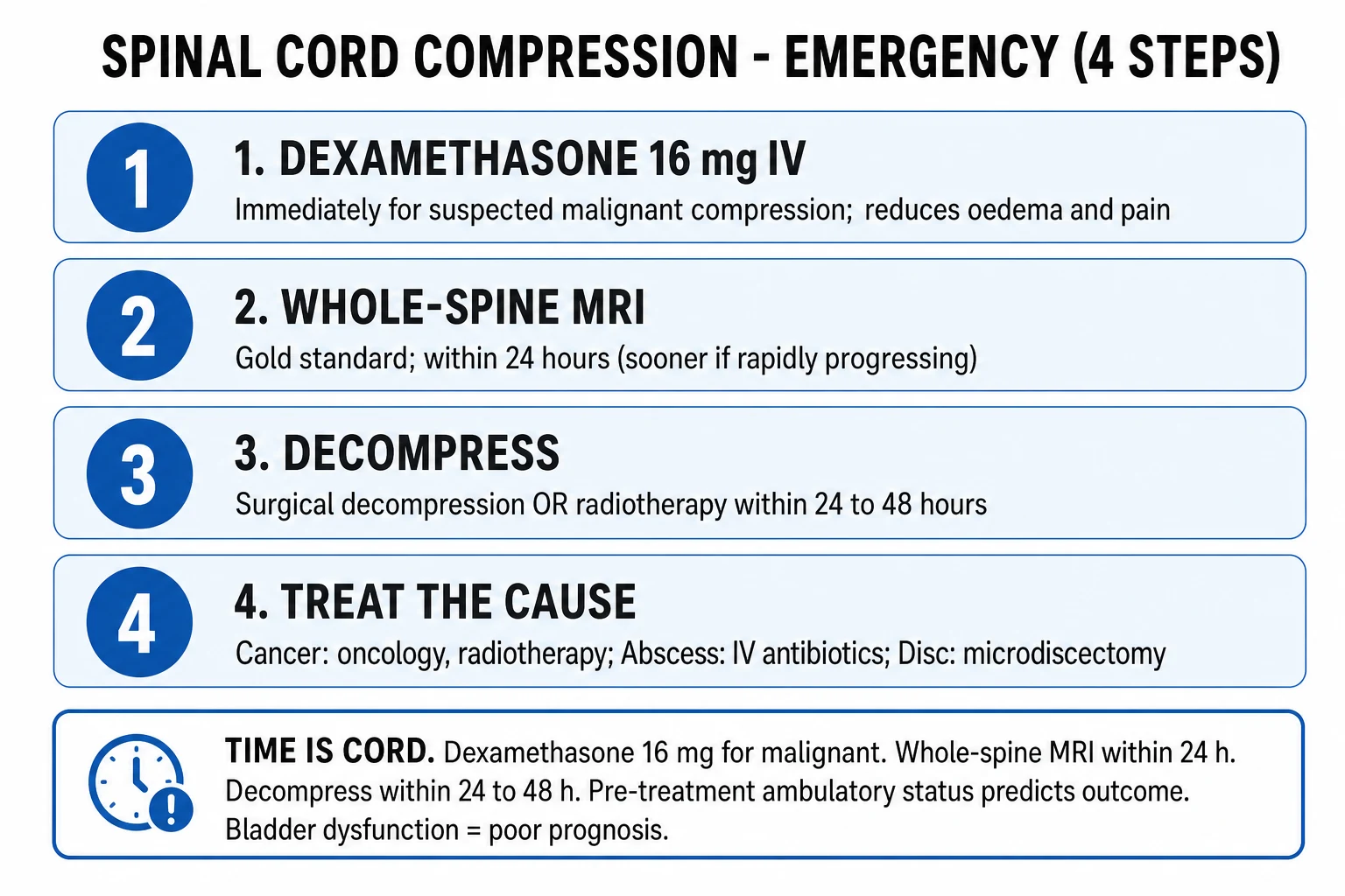
The immediate management of suspected cord compression is a time-critical bundle that should begin in the emergency department or the oncology clinic the moment the syndrome is recognised, in parallel with — not after — arranging the MRI. The resuscitation bundle has six components: recognise and protect, image, steroid, analgesia, nursing and bladder care, and refer.[1][6]
Recognise and protect. The diagnosis is made clinically on the history and examination; it should not await imaging. The patient is nursed flat with log-rolling for all movements to avoid displacing an unstable spine (especially in traumatic or destructive malignant vertebral-body collapse), and strict pressure-area care is begun immediately — paralysed or weak patients develop pressure ulcers within hours. Vital signs, cardiac monitoring, and intravenous access are established; cervical cord compression and high thoracic lesions can cause autonomic instability and respiratory compromise, so airway and breathing are reassessed frequently.[1]
Image. Arrange urgent whole-spine MRI within 24 hours of suspicion for MSCC (NICE NG12 standard), and within hours for cauda equina syndrome, rapidly progressive deficit, or suspected epidural abscess or haematoma. While the MRI is being arranged, take the bloods listed above and a bladder-scan reading. Steroid. For malignant spinal cord compression, give dexamethasone 16 mg as a loading dose intravenously (or orally if the patient can take tablets) immediately, followed by a maintenance regimen of 16 mg per day (commonly 8 mg twice daily or, in some protocols, 8 mg four times daily, then taper) — the goal is to reduce vasogenic cord oedema, relieve pain, and protect the cord from ischaemic injury during the wait for definitive decompression. Steroids are not routinely given for cauda equina syndrome from disc herniation (no proven benefit and may worsen outcome), for epidural abscess (may impair infection control), or for epidural haematoma. The evidence base for the 16 mg dexamethasone dose is largely observational, derived from the systematic reviews of Loblaw and colleagues; the randomised trials of higher-dose corticosteroids were stopped because of unacceptable side effects and did not establish a benefit.[5][6]
Analgesia, nursing and bladder care, and refer. Give analgesia following the World Health Organization ladder, escalating from paracetamol and a non-steroidal anti-inflammatory (if not contraindicated) to a weak then a strong opioid; for neuropathic or radicular pain add gabapentin or pregabalin and consider a short course of dexamethasone if not already given. Insert a urinary catheter for established retention (to relieve pain, prevent bladder damage, and provide accurate output monitoring), and institute venous thromboembolism prophylaxis (mechanical and, once any surgical bleeding risk is addressed, pharmacological) because the immobile cancer patient is at very high risk of deep-vein thrombosis and pulmonary embolism. Finally, refer urgently to the spinal surgery or neurosurgical team, the clinical oncology team, and the radiology team — ideally through a single coordinated spinal metastasis or cord-compression pathway — so that the decision between surgery, radiotherapy, and best-supportive care is made by the multidisciplinary team and not in isolation.[1]
Management — Definitive: Surgery versus Radiotherapy
The definitive treatment of malignant spinal cord compression is a choice between direct decompressive surgery followed by radiotherapy and radiotherapy alone, made by the multidisciplinary team on the basis of the patient's performance status, the number of compressive levels, the tumour's radiosensitivity, the presence of spinal instability, the prognosis, and the patient's wishes. The framework the modern decision rests on is the landmark randomised trial of Patchell and colleagues (2005).[3][1]
The Patchell trial and what it changed
Patchell and colleagues randomised 101 patients with a single level of metastatic cord compression to either direct decompressive surgical resection followed by radiotherapy (30 Gy in 10 fractions) within 14 days or radiotherapy alone. The trial was closed early at interim analysis because of a clear benefit of surgery: significantly more patients in the surgery arm remained ambulatory or regained ambulation (84 per cent versus 57 per cent), more regained the ability to walk after losing it (62 per cent versus 19 per cent), surgery patients retained the ability to walk for longer, and they had a significantly longer median survival (126 days versus 100 days). The trial established that, for the single-level, fit patient with metastatic cord compression, direct decompressive surgery plus radiotherapy is superior to radiotherapy alone and is the standard of care. The caveat — repeated in every guideline since — is that the result applies to a single compressive level in a patient with a reasonable performance status and prognosis, and does not extend to patients with multiple levels, very poor performance status, or a life expectancy measured in days.[3][1]
Indications for surgery and for radiotherapy
The indications for surgical decompression (with or without instrumented stabilisation) in MSCC are: a single level of compression in a fit patient; spinal instability or a retropulsed bone fragment; an unknown primary tumour for which a histological diagnosis is needed; a radio-resistant tumour (renal cell carcinoma, melanoma, sarcoma, some non-small-cell lung cancers); recurrence after prior radiotherapy to the same site; or neurological deterioration during or after radiotherapy. The indications for radiotherapy alone are: multiple compressive levels; a radiosensitive tumour (breast, prostate, myeloma, lymphoma, small-cell lung cancer); a patient unfit for surgery; or a very poor projected survival in whom the burden of surgery is not justified. Whatever the modality, the treatment must be delivered within 24 to 48 hours of diagnosis to maximise the chance of neurological recovery.[1][6]
The radiotherapy fractionation schedules in common use are 8 Gy in a single fraction (the commonest palliative regimen, providing rapid pain relief and equivalent ambulation to longer courses in patients with short prognosis), 20 Gy in 5 fractions, or 30 Gy in 10 fractions (used for better-prognosis patients and for radiosensitive tumours). All three are effective for pain; the longer courses may provide more durable local control at the cost of more treatment days. The choice is individualised to prognosis, performance status, and tumour type.[5]
Cauda equina, abscess and haematoma — the definitive treatments
For cauda equina syndrome from a large central lumbar disc herniation, the definitive treatment is emergency surgical decompression — microdiscectomy, hemilaminectomy or laminectomy — within 24 to 48 hours of the onset of complete retention; the goal is to relieve root compression before irreversible root infarction. Steroids are not routinely given for cauda equina of disc origin (no proven benefit and possible harm); the patient is taken to theatre once imaging and consent are complete. The timing evidence consistently shows that decompression within 48 hours is associated with markedly better motor, bladder, and sensory recovery than decompression after 48 hours of complete paralysis, although the exact cutoff and the role of surgery in CES-R remain debated.[8]
For spinal epidural abscess, the definitive treatment combines urgent surgical decompression with prolonged intravenous antibiotics directed at the cultured organism for 4 to 6 weeks (often followed by a period of oral therapy). Empiric intravenous antibiotic therapy before culture results must cover Staphylococcus aureus (including methicillin-resistant strains) and Gram-negative organisms; a typical regimen is intravenous vancomycin plus a third-generation cephalosporin such as ceftriaxone (or vancomycin plus piperacillin-tazobactam if Gram-negatives including Pseudomonas are likely, as in many post-procedural cases). Selected patients with small abscesses, no neurological deficit, and a surgically-correctable source may be managed with antibiotics alone under close observation, but any neurological deficit, sepsis, or worsening on antibiotics is an indication for surgical decompression. The timing literature reviewed by Tuchman and colleagues supports early surgery for patients with neurological deficit; antibiotic-only management is reserved for selected stable patients and carries a risk of deterioration that requires low threshold to abandon for surgery.[7]
For epidural haematoma, the definitive treatment is urgent surgical evacuation with reversal of anticoagulation (vitamin K and prothrombin complex concentrate for warfarin; specific reversal agents such as andexanet alfa or prothrombin complex concentrate for the direct oral anticoagulants, depending on the agent and the time since last dose). The window for neurological recovery after evacuation is similar to that of the disc and the abscess — within 24 to 48 hours for the best outcome. For Pott's spine with neurological deficit, severe kyphosis, or instability, the treatment combines prolonged anti-tubercular therapy (standard four-drug regimen: isoniazid, rifampicin, pyrazinamide, ethambutol for two months, then isoniazid and rifampicin for a further four to seven months or longer) with surgical decompression and stabilisation. For traumatic cord compression, the initial management is advanced trauma life support with spinal immobilisation, followed by urgent imaging and, where indicated, surgical decompression and stabilisation; the role of high-dose methylprednisolone is discussed under controversies.[1][4]
Specific Subtypes & Scenarios
Traumatic cord injury and the NASCIS controversy
In traumatic spinal cord injury with persistent cord compression by bone fragment or haematoma, urgent surgical decompression and stabilisation is performed once the patient is resuscitated. The National Acute Spinal Cord Injury Studies (NASCIS-2 and NASCIS-3) examined high-dose intravenous methylprednisolone given within 8 hours of injury — the NASCIS-3 regimen being a 30 mg/kg intravenous bolus over 15 minutes, followed after a 45-minute pause by a 5.4 mg/kg/hour infusion for 23 hours (or for 48 hours in selected patients, per NASCIS-3). The trials reported a modest neurological benefit in subgroup analyses, but the magnitude and clinical meaningfulness of that benefit, and the risks (increased infection, gastrointestinal bleeding, and the need for prolonged intensive care), have made the use of high-dose methylprednisolone in traumatic cord injury controversial; current AANS/CNS guidelines treat it as an option rather than a standard, and many centres have abandoned it. A candidate should know the regimen, the time window (within 8 hours of injury), and that the evidence is contested.[4]
Cervical cord compression — why it is more dangerous
Cervical cord compression is anatomically more dangerous than thoracic or lumbar compression for three reasons: the cervical cord carries the pathways to all four limbs, so a cervical lesion produces quadriparesis or quadriplegia rather than paraparesis; high cervical lesions (above C5) compromise the phrenic nerve roots (C3 to C5) and the diaphragm, producing respiratory failure; and the cervical sympathetic outflow traverses this segment, so high lesions can produce Horner syndrome, autonomic instability, neurogenic shock (hypotension with bradycardia from loss of sympathetic tone), and life-threatening cardiac arrhythmia. The sensory level is high — at the clavicles (C4) or below — and weakness involves the arms (cervical myotomes) as well as the legs. The management is the same in principle (urgent MRI, decompression within 24 to 48 hours) but the airway, breathing, and circulatory consequences demand intensive-care-level monitoring.[1]
Pregnancy, age, and the immunocompromised
In pregnancy, the same urgency applies but the MRI can be performed safely (without gadolinium) in any trimester when clinically indicated; the patient is positioned in the left lateral decubitus position to avoid aortocaval compression, and a multidisciplinary plan involving obstetrics, anaesthetics, spinal surgery, and neonatology addresses the timing of delivery relative to decompression. In the elderly, degenerative cervical myelopathy — chronic cord compression from cervical spondylosis and ligamentum-flavum hypertrophy — is the commonest cause of non-compressive-on-imaging-but-truly-compressive myelopathy in older adults, presenting with hand clumsiness, gait disturbance, upper motor neurone signs in the legs, and often neck pain; it requires decompressive surgery (anterior cervical discectomy and fusion, or posterior laminoplasty) for established myelopathy. The immunocompromised patient (HIV, transplant, chemotherapy) has a broadened differential — epidural abscess with atypical organisms, toxoplasmosis, lymphoma, fungal infection — and a lower threshold for empirical infection cover and for biopsy. The anticoagulated patient with sudden severe back pain has epidural haematoma until imaging proves otherwise, and the international normalised ratio must be checked and reversed urgently.[1][7]
Complications & Rehabilitation
The complications of cord compression divide into those caused by delayed decompression and those caused by the resulting immobility and neurological deficit. The principal disease complication is permanent paraplegia or quadriplegia — the direct consequence of cord infarction from delayed decompression — with permanent bladder and bowel dysfunction from sacral cord or cauda equina damage, and chronic neuropathic pain below the compression level (a burning, shooting, or electric sensation often refractory to simple analgesics and requiring gabapentinoids, tricyclic antidepressants, or serotonin-noradrenaline reuptake inhibitors). Spasticity develops below the lesion in the weeks after injury, producing painful flexor or extensor spasms that are managed with baclofen, tizanidine, botulinum toxin, or intrathecal baclofen pumps. Autonomic dysreflexia — a life-threatening paroxysmal sympathetic surge producing dangerous hypertension, bradycardia, and headache — occurs in patients with cord lesions above T6, triggered by noxious stimuli below the lesion (a distended bladder, a faecally-loaded rectum, a pressure sore); management is to identify and remove the trigger, sit the patient upright, and treat the blood pressure with a short-acting antihypertensive such as nifedipine or glyceryl trinitrate.[1]
The complications of immobility are the same as in any paralysed patient and must be prevented aggressively from the day of admission: pressure ulcers (turn every two hours, pressure-relieving mattress, daily skin inspection); deep-vein thrombosis and pulmonary embolism (mechanical and pharmacological prophylaxis — the latter initiated once surgical bleeding risk is acceptable); urinary tract infection from catheterisation (use intermittent catheterisation where possible, change catheters aseptically); contractures (passive range-of-movement exercises, splinting); heterotopic ossification; pneumonia (deep-breathing exercises, incentive spirometry, early mobilisation); and constipation and faecal impaction (structured bowel care with stool softeners and scheduled toileting). The complications of high-dose dexamethasone — hyperglycaemia (monitor blood glucose), increased infection risk, Candida oral thrush, peptic ulceration and gastrointestinal bleeding (consider a proton-pump inhibitor), psychiatric disturbance (mood elevation, insomnia, steroid psychosis in the elderly), proximal myopathy, fluid retention, and avascular necrosis of the femoral head — must be anticipated and the steroid tapered as soon as the definitive treatment is delivered and the oedema controlled.[1][6]
Rehabilitation begins from the day of admission, not after discharge: early physiotherapy for chest, joint range, and strength; occupational therapy for activities of daily living and home modification; bladder and bowel retraining (intermittent self-catheterisation for the neurogenic bladder, structured bowel programme for the neurogenic bowel); psychological support for the patient adjusting to a sudden change in independence; and a coordinated discharge plan involving the community spinal-cord-injury or rehabilitation team. The complexity of these needs is the reason patients with significant residual deficit are best managed through a regional spinal injuries or rehabilitation centre.[1]
Prognosis & Disposition
The single strongest predictor of post-treatment ambulatory outcome in malignant spinal cord compression is the pre-treatment ambulatory status — a finding replicated across every cohort and trial. Patients who are walking before treatment overwhelmingly remain ambulatory after treatment (around 80 to 90 per cent); patients who are non-ambulatory but have some motor function may regain ambulation in roughly 30 to 60 per cent of cases; patients who are completely paraplegic before treatment rarely regain ambulation (under 10 per cent in most series). The duration and severity of the neurological deficit before treatment also matter: a deficit of less than 24 to 48 hours carries a far better outlook than one of a week or more. Bladder dysfunction at presentation is an adverse prognostic marker for both motor and bladder recovery.[1][3]
The median survival after a diagnosis of malignant spinal cord compression is approximately 3 to 6 months, varying markedly with the primary tumour (prostate and breast cancer patients often survive a year or more; lung cancer patients a few months; myeloma patients variable, often many months to years with modern therapy) and with the patient's performance status and burden of visceral metastases. This median survival is the reason the treatment decision must weigh the burden of surgery against the projected benefit in remaining life: a major decompression with instrumented fusion is justified in a fit prostate-cancer patient with a single level and a projected survival of a year, but may be disproportionate in a bedbound lung-cancer patient with visceral metastases and a projected survival of weeks, for whom single-fraction radiotherapy and best supportive care may be the kinder and more appropriate choice. The conversation about prognosis, goals of care, and the trade-off between treatment intensity and quality of remaining life is a central part of the multidisciplinary decision and must be had honestly with the patient and family.[1][5]
For cauda equina syndrome from disc herniation, decompression within 48 hours is associated with markedly better motor, sensory, and bladder recovery than decompression after 48 hours of complete paralysis. The recovery of bladder function in particular is strongly time-dependent: patients decompressed within 24 to 48 hours often regain a continent bladder, while those decompressed late frequently have a permanent neurogenic bladder requiring lifelong intermittent catheterisation. The risk of permanent bladder and bowel dysfunction once established retention has occurred is substantial even with prompt surgery, which is why the prevention of progression from CES-I to CES-R is the single most impactful clinical decision in the pathway. The disposition of any patient treated for cord compression — malignant or mechanical — is to a monitored bed or high-dependency unit initially, with planned transfer to a rehabilitation facility for those with residual deficit, and clear safety-net advice to return urgently if new or worsening symptoms develop.[8]
Prevention, Screening & Oncology Follow-up
Because the outcome of MSCC is so dependent on the pre-treatment neurological status, early recognition before neurological deficit develops is the single most effective intervention. Two prevention strategies are central. The first is patient and primary-care education: every patient with a cancer that has a high propensity for spinal metastasis (breast, prostate, lung, myeloma, kidney) should be told, in writing and verbally, the red-flag features of incipient cord compression — new or progressive back pain, night pain, radicular pain, limb weakness, numbness, or difficulty walking — and instructed to present urgently to the oncology team or the emergency department. The second is a low threshold for proactive spinal MRI in patients with high-risk primaries who report new back pain, before neurological signs appear, because the outcome of treatment for early asymptomatic or pain-only MSCC is dramatically better than for MSCC with established deficit.[1][5]
Bone-modifying agents have a specific preventive role. Bisphosphonates (zoledronic acid, pamidronate) and the RANKL inhibitor denosumab reduce the rate of skeletal-related events — pathologic fracture, spinal cord compression, the need for radiotherapy or surgery to bone, and malignant hypercalcaemia — in patients with bone metastases from breast, prostate, myeloma, and other solid tumours. Their routine use in patients with radiographic bone metastasis is a guideline-supported component of metastatic disease management and reduces (though does not abolish) the incidence of MSCC. After MSCC treatment, follow-up imaging is guided by symptoms and the primary tumour, and the multidisciplinary team monitors for local recurrence, new compressive lesions at other levels, and spinal instability.[1][5]
The medicolegal importance of acting on red-flag back pain cannot be overstated. Missed or delayed cauda equina syndrome is one of the commonest and most costly sources of medical negligence litigation in many jurisdictions; the legal duty is to document the presence or absence of red flags explicitly, to perform and record the neurological and rectal examination, to arrange urgent imaging and refer to the spinal service within the recommended timeframe, and to communicate clearly with the patient about the warning signs and when to return. A defensible record demonstrates that the red flags were sought, the imaging pathway was followed, and the patient was given written safety-net advice. The same duty applies in reverse: the absence of red flags and a documented normal neurological examination support a non-imaging decision and protect the clinician.[8]
Evidence, Guidelines & Regional Differences
Spinal cord compression sits at the intersection of neurology, oncology, spinal surgery, infectious diseases, and emergency medicine, and the regional guidelines reflect these perspectives. The United Kingdom NICE NG12 guideline ("Suspected cancer: recognition and referral") and the Spinal Cord Impairment pathway standardise the cancer-back-pain referral and the 24-hour MRI standard for suspected MSCC, with a clear pathway from primary care through to the spinal surgery and oncology teams. The British Association of Surgical Oncology and the Royal College of Radiologists have published detailed MSCC pathways. In the United States, the American Association of Neurological Surgeons and Congress of Neurological Surgeons (AANS/CNS) guidelines on the treatment of metastatic epidural spinal cord compression endorse the Patchell framework — direct decompressive surgery plus radiotherapy for the single-level, fit patient — and set out the indications and contraindications for surgery. The NCCN (National Comprehensive Cancer Network) guidelines incorporate the MSCC pathway into the disease-specific guidelines for the relevant primary tumours.[1][3]
The dexamethasone evidence remains the most contentious element. The standard 16 mg loading dose derives from observational series and from the systematic review by Loblaw and colleagues (2005); no large modern randomised trial has established the optimal corticosteroid regimen, and a Dutch trial (the CHEMOTOP and related studies) is examining whether lower doses or no steroids are non-inferior, given the substantial side-effect burden of high-dose dexamethasone. Some centres have moved to a lower routine dose (for example 8 to 10 mg loading) on the grounds that the higher-dose evidence is observational and the side-effect burden is dose-dependent; the 16 mg dose remains the widely cited standard and the dose a candidate should state in an exam.[5][6]
The radiotherapy fractionation debate has been largely settled by randomised trials showing that, for pain control and ambulation, 8 Gy in a single fraction is equivalent to longer fractionation in patients with short prognosis, although retreatment is more often needed after single-fraction treatment and longer courses may be preferred for patients with better prognosis and for locally aggressive tumours. The timing of surgery in cauda equina syndrome remains a live area — the classic dogma of decompression within 48 hours is supported by observational data but not by a randomised trial (which would be ethically difficult), and there is debate about whether the cutoff is 24, 36, or 48 hours; what is not debated is that decompression should occur as soon as possible after diagnosis, ideally within 24 hours. The NASCIS high-dose methylprednisolone regimen for traumatic cord injury, described above, is the other major controversy in the field — the trials' subgroup analyses are contested, the side-effect burden is real, and practice varies between centres and between countries.[4][8]
[1] [1] [3]Indian context and high-tuberculosis-burden regions — Pott's spine (tuberculous spondylodiscitis) is a leading cause of cord compression and must be considered alongside malignancy in any destructive vertebral lesion, especially in younger patients and those with constitutional symptoms. Cost-limited access to urgent whole-spine MRI is a real constraint; in resource-limited settings, CT of the spine (showing vertebral-body destruction, paravertebral abscess, or canal compromise) is a pragmatic first step, with referral for MRI where the diagnosis is not established. Empiric anti-tubercular therapy may be started on strong clinical and radiological suspicion while awaiting microbiological confirmation in high-prevalence settings.
Exam Pearls & High-Yield Minutiae
The examiner rewards the candidate who can move fluently between the syndrome (recognise the pattern), the level (cord, conus, or cauda), the cause (the five primaries plus disc, abscess, haematoma, fracture), the test (urgent whole-spine MRI), the drug (dexamethasone 16 mg for malignant), and the operation (decompress within 24 to 48 hours). The most common single mark-losing errors are: confusing cauda equina (lower motor neurone, areflexia, saddle anaesthesia, early severe sphincter) with cord compression (upper motor neurone, hyperreflexia, sensory level, late sphincter); giving steroids for cauda equina of disc origin (no proven benefit); ordering CT rather than MRI to exclude cord compression (CT shows bone, not cord); or forgetting to check the bladder and the perianal sensation in a patient with back pain (the post-void residual and the rectal examination are the two most-missed and most-decisive bedside tests).[1][8]
The five primaries — BLPT-RK (and Batson)
BLPT-RK
commonest in many series; thoracic predilection; radiosensitive
poor prognosis; often presents with established deficit
lumbar and sacral via Batson venous plexus; osteoblastic metastases; radiosensitive
myeloma is highly radiosensitive; thyroid is radio-resistant
radio-resistant; surgery preferred
radio-resistant; surgery preferred for single-level disease
Why time is CORD — the compression cascade
CORD
mechanical deformation by tumour, disc, abscess or haematoma — stage 1
vasogenic oedema, T2 hyperintensity on MRI — stage 2, still reversible
ischaemia, demyelination — stage 3, deteriorating but salvageable
axonal infarction, necrosis, gliosis — stage 4, irreversible
The five red flags in back pain — NAVES
NAVES
weakness, sensory level, saddle anaesthesia, sphincter disturbance
onset after age 50, or known cancer
weight loss, fever, night sweats — infection or malignancy
urinary retention, incontinence, erectile dysfunction — cauda equina
progressive, unremitting, nocturnal — not mechanical
Exam application bank (NEET-PG / INICET)
One-line answer
Spinal cord compression is a neurological emergency in which the spinal cord, conus medullaris or cauda equina is compressed by metastatic cancer (commonest: breast, prostate, lung, myeloma, kidney), intervertebral disc herniation, epidural abscess, epidural haematoma or vertebral collapse. Presentation: progressive, often nocturnal back pain, a sensory level, weakness, and — late and ominous — bladder and bowel dysfunction. Cauda equina syndrome adds saddle anaesthesia, bilateral sciatica, urinary retention and erectile dysfunction. Diagnosis is urgent whole-spine MRI. Malignant spinal cord compression: dexamethasone 16 mg immediately, then radiotherapy or surgical decompression within 24 to 48 hours. Cauda equina from disc: emergency surgical decompression within 24 to 48 hours. Time is cord — delay causes permanent paralysis. [1]
Worked stems (answer without another resource)
Stem 1 — Classic presentation. Map symptoms to mechanism; name the first investigation and first treatment step with dose/route if drug therapy is standard. [1]
Stem 2 — Unstable / complicated. List red flags that force immediate resuscitation, theatre, ICU, antidote, or reperfusion — and what you do in the first 15 minutes. [1]
Stem 3 — Atypical group. Elderly, pregnancy, child, or immunocompromised: how presentation and thresholds change. [1]
Stem 4 — Differential trap. Name the three closest mimics and one discriminator for each. [1]
Stem 5 — Disposition. Who goes home with safety-netting, who is admitted, who needs HDU/ICU/theatre, and what follow-up is mandatory. [1]
Rapid viva checklist
- Definition + classification
- Pathophysiology chain
- Bedside signs / criteria
- Score with exact components (if any)
- Emergency bundle
- Definitive therapy with doses
- Complications of disease and of treatment
- Special populations
- Guideline/trial name if classic
- Three exam traps
Coverage self-check
If you cannot answer any stem above from this page alone, re-read the matching section — the page is intended to be self-sufficient for final-prof and NEET-PG/INICET questions on Spinal Cord Compression.
[8]References
- [1]Lawton AJ, Lee KA, Cheville AL, et al. Assessment and Management of Patients With Metastatic Spinal Cord Compression: A Multidisciplinary Review J Clin Oncol, 2019.PMID 30395488
- [2]Wu J, Ranjan S. Neoplastic Myelopathies Continuum (Minneap Minn), 2018.PMID 29613896
- [3]Patchell RA, Tibbs PA, Regine WF, et al. Direct decompressive surgical resection in the treatment of spinal cord compression caused by metastatic cancer: a randomised trial Lancet, 2005.PMID 16112300
- [4]Bracken MB, Shepard MJ, Holford TR, et al. Administration of methylprednisolone for 24 or 48 hours or tirilazad mesylate for 48 hours in the treatment of acute spinal cord injury. Results of the Third National Acute Spinal Cord Injury Randomized Controlled Trial. National Acute Spinal Cord Injury Study JAMA, 1997.PMID 9168289
- [5]Loblaw DA, Perry J, Chambers A, Laperriere NJ. Systematic review of the diagnosis and management of malignant extradural spinal cord compression: the Cancer Care Ontario Practice Guidelines Initiative's Neuro-Oncology Disease Site Group J Clin Oncol, 2005.PMID 15774794
- [6]Byrne TN. Metastatic epidural spinal cord compression: update on management Semin Oncol, 2006.PMID 16769419
- [7]Tuchman A, Pham M, Hsieh PC. The indications and timing for operative management of spinal epidural abscess: literature review and treatment algorithm Neurosurg Focus, 2014.PMID 25081968
- [8]Lavy C, Marks P, Dangas K, et al. Cauda equina syndrome-a practical guide to definition and classification Int Orthop, 2022.PMID 34862914