Neurology · Neurology
Stroke
Also known as Cerebrovascular accident · CVA · Ischaemic stroke · Haemorrhagic stroke · Brain attack
Stroke is sudden focal neurological deficit from vascular injury to the brain: ischaemic (85%) from arterial occlusion, or haemorrhagic (15%) from vessel rupture. FAST (Face, Arm, Speech, Time) triggers recognition; a non-contrast CT brain is the first, most urgent investigation to distinguish the two — since the acute treatments are opposite. Ischaemic stroke: IV alteplase within 4.5 hours, thrombectomy up to 24 hours for large-vessel occlusion with favourable imaging. Haemorrhagic stroke: blood-pressure control, reversal of anticoagulation, and neurosurgical evacuation where indicated.
On this page & tools
Your progress
Saved locally on this device.
Exam tags
Red flags
Overview & Definition
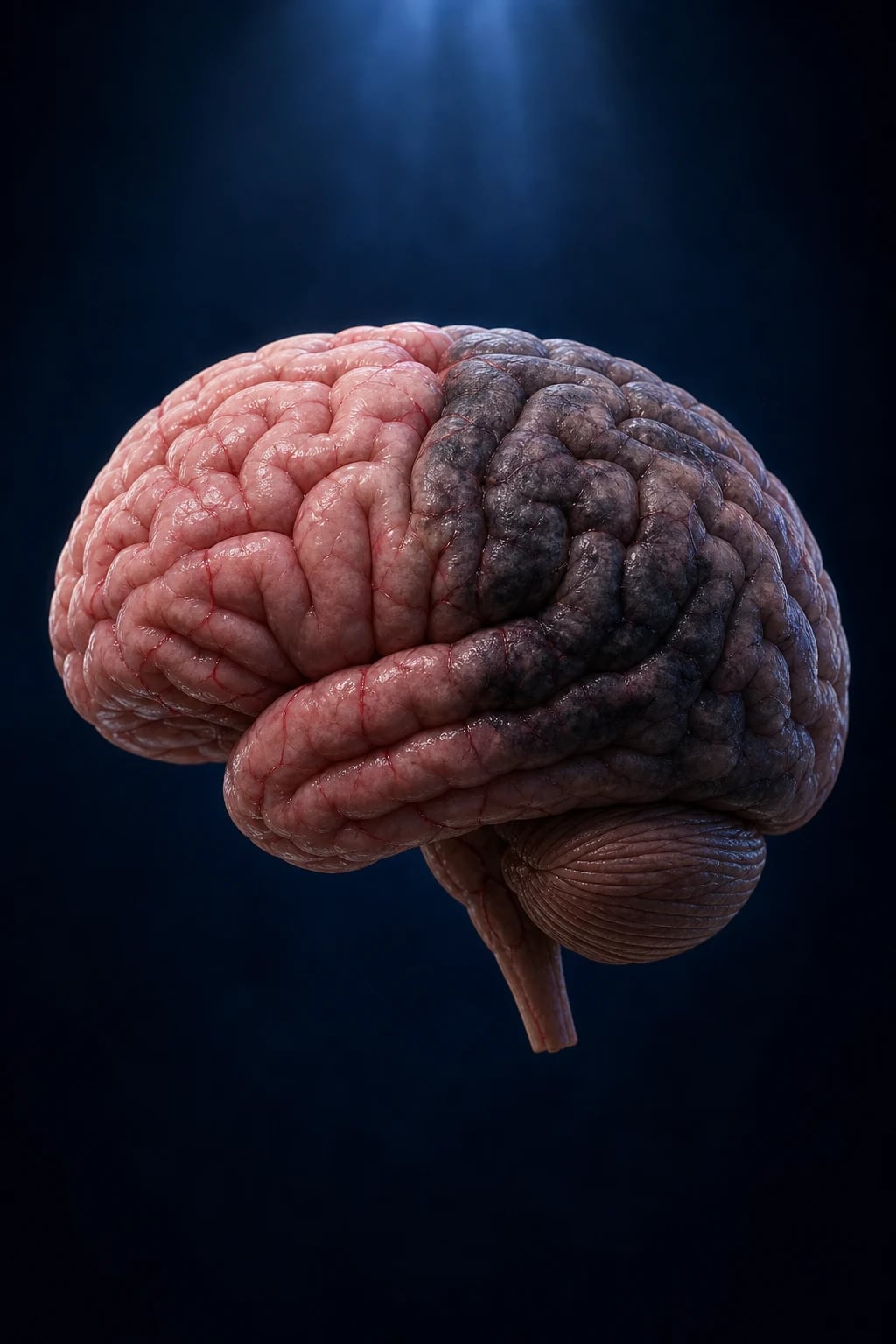
The WHO definition of stroke, still the bedrock of undergraduate and postgraduate examinations, is "rapidly developing clinical signs of focal (or global) disturbance of cerebral function, lasting more than 24 hours or leading to death, with no apparent cause other than that of vascular origin." Three elements are essential and examiners probe each: (1) a rapid, sudden onset — stroke is a * vascular event*, not a progressive degenerative one; (2) focal neurological deficit referable to a vascular territory (global disturbance alone suggests subarachnoid haemorrhage or severe bilateral ischaemia rather than a discrete territorial stroke); and (3) a vascular cause, with no better explanation such as tumour, trauma, seizure, or metabolic derangement.[11]
The boundary with transient ischaemic attack (TIA) has been deliberately redrawn. The old, time-based rule defined TIA as a focal deficit resolving within 24 hours; stroke was anything lasting longer. That 24-hour threshold was arbitrary and misleading, because diffusion-weighted MRI shows that a substantial minority of deficits resolving clinically within 24 hours have nonetheless already infarcted. The modern, tissue-based definition therefore defines TIA as a transient episode of neurological dysfunction caused by focal brain, spinal cord, or retinal ischaemia without acute infarction on imaging — and stroke as the same process with infarction. The practical consequence is that every "TIA" now warrants urgent brain MRI, because a clinically brief event may already be a completed stroke on tissue. [1]
Stroke is the second leading cause of death worldwide and the leading cause of acquired adult disability. It is broadly divisible into two mechanistically opposite categories that share only their clinical presentation — ischaemic stroke (about 85%) from arterial occlusion, and haemorrhagic stroke (about 15%) from a ruptured vessel. Haemorrhagic stroke further divides into intracerebral haemorrhage (ICH, about 10% of all strokes), bleeding into brain parenchyma, and subarachnoid haemorrhage (SAH, about 5%), bleeding into the subarachnoid space, most often from a ruptured berry aneurysm. This split matters more than almost any other classification in medicine, because the two halves are managed in directly opposite ways — antithrombotic therapy that rescues ischaemic brain will kill a patient with an undiagnosed haemorrhage. The single most important acute investigation, the non-contrast CT brain, exists precisely to make that distinction. [1]
Classification
Ischaemic stroke
- ~85% of all strokes
- Large-artery atherosclerosis, cardioembolism (AF), or small-vessel (lacunar) disease — TOAST classification
- Treated with thrombolysis/thrombectomy then antiplatelet or anticoagulant secondary prevention
Intracerebral haemorrhage
- ~10% of all strokes
- Hypertensive lipohyalinosis (basal ganglia, thalamus, pons, cerebellum) or cerebral amyloid angiopathy (lobar, elderly)
- Treated with BP control, reversal of anticoagulation, selective neurosurgical evacuation
Subarachnoid haemorrhage
- ~5% of all strokes
- Ruptured berry (saccular) aneurysm in most cases; smoking and hypertension are risk factors
- Presents as sudden 'thunderclap' headache — needs CT ± lumbar puncture, then neurosurgical/interventional securing of the aneurysm
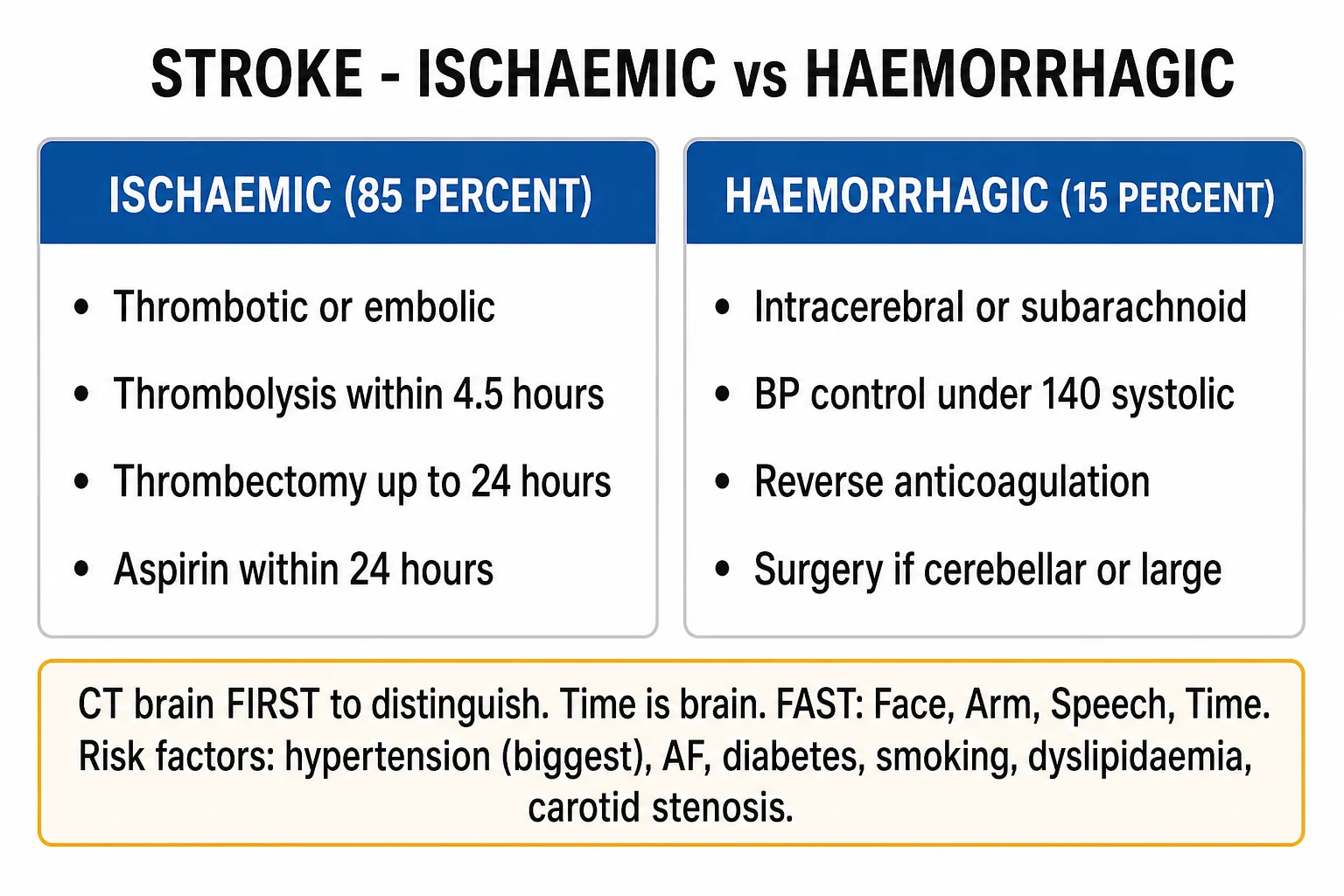
Within ischaemic stroke, the TOAST classification (Trial of ORG 10172 in Acute Stroke Treatment) is the universally examined scheme and divides ischaemic stroke into five mechanistic groups. Large-artery atherosclerosis accounts for roughly 20% and arises from plaque rupture or in-situ thrombosis in a major intracranial or extracranial artery, with artery-to-artery embolism of plaque debris distally. Cardioembolism accounts for about 20% and originates from clot formed in the heart — classically in the left atrial appendage in atrial fibrillation, but also from prosthetic or diseased valves, a mural thrombus following myocardial infarction, or endocarditis. Small-vessel (lacunar) occlusion accounts for about 25% and results from lipohyalinosis of small penetrating arteries, producing small, deep infarcts. Other determined cause (dissection, vasculitis, thrombophilia, moyamoya, sickle cell, drug use) is uncommon. The remainder are undetermined (cryptogenic) — roughly a third — where no cause is found despite a full work-up, often prompting prolonged cardiac monitoring for paroxysmal atrial fibrillation. Cardioembolic and large-artery strokes tend to involve the cortex and produce cortical signs (aphasia, neglect, visual field loss); lacunar strokes spare the cortex and produce pure motor, pure sensory, or ataxic-hemiparetic syndromes with a comparatively better prognosis. [1]
A further practical distinction examiners expect is thrombotic versus embolic ischaemic stroke. Thrombotic strokes (large-artery atherosclerosis or small-vessel lipohyalinosis) tend to have a stuttering, stepwise or progressive onset, often waking with the deficit, and occur in the territory of the diseased vessel. Embolic (cardioembolic or artery-to-artery) strokes characteristically produce the maximal deficit at onset, often during activity, and may produce multiple infarcts in different vascular territories if the source is the heart. A stuttering versus a sudden-maximal tempo at the bedside is a high-yield discriminator that points to mechanism and to the aetiological work-up that follows. [1]
Epidemiology & Risk Factors
Stroke — the headline epidemiology
Risk factors divide cleanly into modifiable and non-modifiable groups, and examiners expect both lists. Among the modifiable factors, hypertension is the single largest contributor to stroke overall — it is the dominant driver of both small-vessel (lacunar) ischaemic infarction and of hypertensive intracerebral haemorrhage, and the risk gradient is continuous from "normal" upwards, with no threshold below which harm accrues. Atrial fibrillation is the single most important treatable cause of cardioembolic stroke: untreated non-valvular AF carries an annual stroke risk of roughly 4 to 5% (higher with additional CHA₂DS₂-VASc risk factors), which oral anticoagulation reduces by about two-thirds. Diabetes mellitus roughly doubles stroke risk and worsens outcome after stroke. Dyslipidaemia, smoking (which doubles ischaemic stroke risk and is the dominant modifiable risk factor for subarachnoid haemorrhage), obesity and physical inactivity, carotid artery stenosis, obstructive sleep apnoea, excess alcohol, and a prior TIA or stroke complete the modifiable list. Sickle cell disease is a specific, important cause in its affected populations. [1]
The non-modifiable risk factors are age (risk approximately doubles each decade after 55), male sex (women still have a higher lifetime risk because they live longer), family history and genetic factors, and ethnicity (Black and South Asian populations have higher stroke incidence and a higher proportion of haemorrhagic stroke than White populations, partly driven by hypertension prevalence). Different risk-factor profiles preferentially produce different stroke types, and this mapping is heavily examined: hypertension, anticoagulation, and cerebral amyloid angiopathy predispose to intracerebral haemorrhage; berry aneurysm, smoking, and hypertension predispose to subarachnoid haemorrhage; large-vessel atherosclerosis, carotid stenosis, and AF predispose to large cortical ischaemic stroke; and chronic hypertension and diabetes predispose to lacunar (small-vessel) ischaemic stroke. [1]
In younger patients (under 45 years), the standard atherosclerotic framework often does not apply, and the differential broadens to include cervical or intracranial arterial dissection, patent foramen ovale with paradoxical embolism, thrombophilias (antiphospholipid syndrome, factor V Leiden, protein C/S deficiency), substance use (cocaine, amphetamines), moyamoya disease, sickle cell disease, and oral contraceptive use combined with smoking. A young stroke warrants a dedicated, different work-up — failure to consider dissection or thrombophilia in a 30-year-old is a classic pitfall. [1]
Pathophysiology
The ischaemic cascade and the penumbra
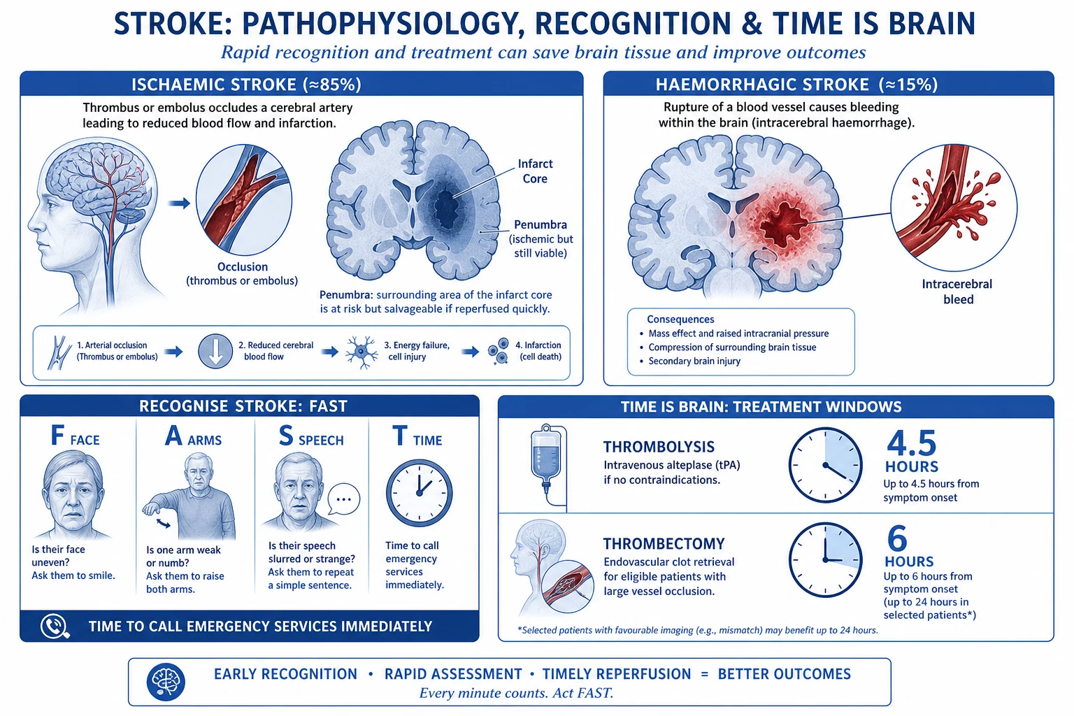
In ischaemic stroke, arterial occlusion cuts off oxygen and glucose delivery, triggering the ischaemic cascade. Within seconds, energy (ATP) failure disables Na⁺/K⁺-ATPase ion pumps, neurons can no longer maintain their resting membrane potential, and they depolarise. Depolarisation triggers massive glutamate release (excitotoxicity); glutamate opens NMDA and AMPA receptors, calcium floods into the neuron, and calcium-activated proteases, lipases, and endonucleases dismantle the cell from within. Cell death is fastest and most complete in the ischaemic core, where cerebral blood flow has fallen below about 10 mL/100 g/min and tissue is irreversibly infarcted within minutes. [1]
The genius of acute stroke therapy rests on the ischaemic penumbra — a rim of tissue surrounding the core where blood flow is reduced (typically 10 to 20 mL/100 g/min) but not yet below the threshold for irreversible injury. Penumbra is hypoperfused and electrically silent (it produces the deficit) but metabolically viable because collateral circulation keeps it just above the infarction threshold. The penumbra is unstable: it progressively converts to core the longer the artery stays occluded. This is the entire physiological basis of time-critical reperfusion — restore flow, and the penumbra recovers; delay, and it dies. This is the meaning of "time is brain" — modelled estimates suggest approximately 1.9 million neurons and roughly 12 km of myelinated fibres are lost per minute of untreated large-vessel ischaemic stroke, with the brain ageing the equivalent of about 3.1 weeks per minute without flow. [1]
Mechanisms of ischaemic stroke
The mechanism of occlusion differs by TOAST subtype. In large-artery atherosclerosis, an atherosclerotic plaque in the carotid bifurcation, the carotid siphon, or a major intracranial artery either ruptures to expose its lipid core (triggering in-situ thrombosis) or sheds plaque debris downstream as artery-to-artery emboli that lodge in a distal branch — both produce territorial infarcts, often with cortical features. In cardioembolism, thrombus that has formed in a static or dyskinetic portion of the heart (the left atrial appendage in atrial fibrillation, the ventricular wall after anterior MI, on a diseased or prosthetic valve, or on vegetations in endocarditis) dislodges and embolises to the brain, where it characteristically lodges in a major cerebral artery (often the middle cerebral artery) and produces a sudden-maximal, often large, territorial deficit. In small-vessel (lacunar) disease, chronic hypertension and diabetes cause lipohyalinosis — a destructive thickening and fibrinoid degeneration of small penetrating arteries (the lenticulostriate, pontine, and thalamoperforator branches) — that culminates in tiny (under 1.5 cm) deep infarcts in the basal ganglia, thalamus, internal capsule, or pons. Watershed (border-zone) infarction occurs at the junctions between two arterial territories during profound hypoperfusion — profound hypotension, cardiac arrest, or a tight proximal carotid stenosis — producing characteristic "string of beads" superficial infarcts or linear deep white-matter lesions. [1]
Mechanisms of haemorrhagic stroke
Haemorrhagic stroke follows a different mechanism entirely. Hypertensive intracerebral haemorrhage results from chronic hypertension causing lipohyalinosis and Charcot-Bouchard microaneurysms in small penetrating arteries; these microaneurysms (classically at the bifurcation of the lenticulostriate, thalamoperforator, pontine, and cerebellar perforating arteries) rupture, producing deep haematomas in the basal ganglia (putamen is the single commonest site), thalamus, pons, and cerebellum. Cerebral amyloid angiopathy — deposition of β-amyloid in the walls of small cortical and leptomeningeal vessels — weakens them and produces lobar (peripheral, cortical) haemorrhages in older patients, often without any hypertensive history; lobar ICH in the elderly, sometimes recurrent and sometimes with associated leptomeningeal "sulcal" siderosis, is the signature. Arteriovenous malformations (AVMs), cavernous haemangiomas, intracranial aneurysm rupture into parenchyma, anticoagulant and thrombolytic therapy, bleeding diatheses, tumours (especially metastatic melanoma, renal cell, choriocarcinoma), and sympathomimetic drug use (cocaine, amphetamines) are further causes. [1]
Subarachnoid haemorrhage is most often caused by rupture of a saccular (berry) aneurysm at a branch point of the circle of Willis — the posterior communicating artery, anterior communicating artery, and middle cerebral artery bifurcation being the classic sites. Perimesencephalic (non-aneurysmal) SAH is a milder, distinct pattern. In both ischaemic and haemorrhagic stroke, cerebral autoregulation is disrupted locally — which is why blood-pressure management differs by stroke type and is handled cautiously in both directions: permissive (untreated) in most acute ischaemic stroke to preserve penumbral perfusion, aggressively lowered in intracerebral haemorrhage to limit haematoma expansion. [1]
Clinical Presentation
The cardinal presentation is a sudden-onset focal neurological deficit. The public-recognition mnemonic FAST (Face drooping, Arm weakness, Speech difficulty, Time to call for help) captures the commonest pattern; the extended BE-FAST adds Balance and Eyes, capturing posterior-circulation deficits that FAST alone misses. The clinical syndrome localises to the vascular territory involved, and recognising a territory is the single most examined localisation skill in stroke medicine. [1]
[1]The distinction between the dominant (usually left) hemisphere and the non-dominant (usually right) hemisphere is heavily tested. A left MCA stroke classically produces a right hemiparesis with aphasia — expressive (Broca) if the superior division is involved, receptive (Wernicke) if the inferior division, or global if the whole territory. A right MCA stroke produces a left hemiparesis with hemispatial neglect (the patient ignores the left side of space), anosognosia (denial of the deficit), and sometimes aprosodia (flattened emotional tone of speech). The rule of thumb: dominant-hemisphere strokes are recognised by language disturbance; non-dominant strokes are missed because neglect masquerades as the patient "just not caring." [1]
Atypical presentations are deliberately tested. In the elderly, diabetics, and the immunocompromised, stroke can present as confusion, an unwitnessed fall, a subtle gait disturbance, a gradual cognitive decline, or even "just not their usual self" — a cortical sign may be absent and a focal deficit subtle. Silent infarcts (imaging evidence of infarction without a history of any deficit) are common in the elderly and in diabetics. A high index of suspicion is essential: any sudden neurological change in an at-risk patient is stroke until proven otherwise. [1]
Differential Diagnosis
Several conditions mimic stroke closely enough that they must be actively excluded, because treating a mimic as a stroke (or vice versa) causes harm. [1]
Hypoglycaemia
- Produces an IDENTICAL focal hemiparesis or aphasia
- Bedside glucose is the first test in EVERY suspected stroke, before imaging
- Correct with IV dextrose — the deficit resolves
Postictal (Todd) paresis
- Transient focal weakness after a seizure, resolving over hours
- Look for a witnessed seizure or epilepsy history; the deficit can be indistinguishable from stroke
Hemiplegic migraine
- Focal deficit with a spreading aura and preceding headache
- Younger patient with a migraine history; a diagnosis of exclusion after stroke excluded
Subdural haematoma
- Gradual onset; trivial or forgotten head injury, especially elderly/anticoagulated
- CT shows a crescentic extra-axial collection crossing suture lines
Brain tumour
- Subacute progressive deficit; can present acutely with haemorrhage into tumour or seizure
- CT/MRI shows a mass with oedema; often a tumour 'stroke' is the first presentation of metastasis
Functional / neurological deficit
- Inconsistent, non-anatomical examination findings; Hoover sign positive
- A diagnosis only after organic disease is excluded — never the default
Sepsis / delirium
- Fluctuating confusion unmasking an old deficit or mimicking a new one
- Excludes a stroke mimic; sepsis work-up needed
Two specific high-yield distinctions deserve emphasis. Hypoglycaemia is the most important reversible mimic — a bedside glucose must be checked and corrected in every suspected stroke before any other step, because untreated hypoglycaemia causes permanent neuronal injury and a "stroke" due to hypoglycaemia resolves with dextrose. Hyponatraemia can similarly produce focal or generalised neurological dysfunction. Distinguishing vestibular vertigo from a posterior-circulation stroke is a specific, frequently tested skill: the bedside HINTS examination (Head Impulse, Nystagmus, Test of Skew) identifies "dangerous" central patterns that favour stroke over benign peripheral vertigo — a normal head-impulse test, direction-changing or vertical nystagmus, or skew deviation on alternate-cover testing in a patient with acute vestibular syndrome is a central ("bad") HINTS and points to posterior-circulation stroke even when early CT is normal. [1]
Clinical & Bedside Assessment
The first 10 minutes — the stroke pathway at the bedside
Glucose check first — exclude and correct hypoglycaemia before attributing any deficit to stroke
FAST/BE-FAST recognition; activate the stroke team; document the EXACT last-known-well time
Assess ABC; oxygen only if hypoxic; two IV lines; baseline bloods including coagulation
Brief targeted exam: NIHSS, GCS, finger-prick glucose, blood pressure both arms, cardiac rhythm, and a check for posterior-circulation signs (nystagmus, limb ataxia, crossed deficits)
Immediate non-contrast CT brain — the test that decides ischaemic versus haemorrhagic
If large-vessel occlusion suspected (severe NIHSS, gaze deviation, dense hemiplegia) — CT angiography at the same sitting
The ROSIER score (Recognition of Stroke in the Emergency Room) is the validated ED tool: it excludes hypoglycaemia as a first step, then scores features that favour stroke (asymmetric face, arm, or leg weakness; speech disturbance; visual field defect: each +1) against features that favour a mimic (loss of consciousness or syncope, seizure activity: each −1). A positive total (above zero) supports a diagnosis of stroke and triggers the imaging and thrombolysis pathway. [1]
The NIH Stroke Scale (NIHSS) is the standard severity assessment used throughout stroke care — every candidate must know it exists, what it measures, its range, and how it is interpreted. It quantifies neurological deficit across 11 domains: level of consciousness (responsiveness, questions, commands), best gaze, visual fields, facial palsy, motor arm (right and left), motor leg (right and left), limb ataxia, sensory, best language, dysarthria, and extinction/inattention (neglect). Each domain is scored 0 (normal) up to a maximum, and the totals sum to a range from 0 (no deficit) to 42 (comatose with quadriplegia). Severity bands: 1 to 4 mild, 5 to 15 moderate, 16 to 20 moderate-to-severe, 21 to 42 severe. The NIHSS directly informs the thrombolysis/thrombectomy decision (a low NIHSS in a minor stroke may sway the thrombolysis risk-benefit; a high NIHSS with cortical signs points to a large-vessel occlusion and triggers CTA for thrombectomy selection) and is the single strongest predictor of outcome. [1]
NIHSS severity bands and what they mean
Moderate — thrombolysis candidate; CTA if cortical signs
Recording the exact "last known well" time is essential at first contact — it is the anchor for both the 4.5-hour thrombolysis window and the (up to) 24-hour thrombectomy window. For wake-up strokes, "last known well" is the time the patient was last seen normal, not the time they woke with the deficit. Its accurate documentation is frequently the single detail that determines whether a patient is eligible for time-critical therapy. [1]
Investigations
Stroke — the four numbers every candidate must know
Non-contrast CT brain is the first and most urgent investigation. Its purpose in the hyperacute phase is not primarily to diagnose the ischaemic stroke (which may be CT-silent in the first hours) but to exclude haemorrhage, since that single distinction determines the entire treatment pathway. Acute haemorrhage appears within minutes as high-density (white) extra-axial or intra-axial blood. Early ischaemic changes on CT are subtle and easily missed: the hyperdense MCA sign (a clot in the M1 segment appearing as a bright dot or line), loss of the insular ribbon, effacement of cortical sulci, loss of grey-white matter differentiation, and hypodensity of the basal ganglia. A large area of early hypodensity (over one-third of the MCA territory) is a relative contraindication to thrombolysis, because it represents an already-established large infarct at high risk of haemorrhagic transformation. [1]
CT angiography (CTA) is performed when a large-vessel occlusion is suspected — to select thrombectomy candidates — and is ideally acquired at the same sitting as the non-contrast CT. CT perfusion (or MR perfusion) maps the core-versus-penumbra mismatch that extends the thrombectomy window beyond 6 hours up to 24 hours, by identifying a small irreversibly-infarcted core surrounded by a large volume of salvageable penumbra. MRI — specifically diffusion-weighted imaging (DWI) — is the most sensitive test for early (within minutes) ischaemia, restricted diffusion appearing bright on DWI with corresponding low signal on the apparent diffusion coefficient (ADC) map. MRI DWI-FLAIR mismatch (a visible lesion on DWI but not on FLAIR) is used to select wake-up stroke patients for thrombolysis, on the principle that the infarct is recent. [1]
Beyond acute imaging, the aetiological work-up seeks the underlying cause and drives secondary prevention: an ECG (looking for atrial fibrillation); prolonged cardiac monitoring (24-hour Holter, an inpatient telemetry monitor, or an ambulatory implantable loop recorder) when paroxysmal AF is suspected despite a normal presenting ECG — cryptogenic stroke is the classic indication for prolonged monitoring; transthoracic echocardiography (with a bubble study to look for a patent foramen ovale, and transoesophageal echo where a cardiac source, left atrial thrombus, or endocarditis is suspected); carotid Doppler ultrasound or CTA/MRA to quantify carotid stenosis; and blood tests — glucose, HbA1c, full blood count, lipid profile, electrolytes including sodium, renal and liver function, coagulation (PT/INR, APTT), and a thrombophilia screen in younger patients or those with an unexplained cryptogenic stroke. [1]
Named scores reproduced
Two risk-stratification scores are heavily examined and must be reproduced verbatim. [1]
ABCD2 score risk-stratifies a suspected TIA for the early risk of recurrent stroke and the urgency of specialist assessment: Age 60 or older = 1; Blood pressure 140/90 mmHg or higher = 1; Clinical features unilateral weakness = 2, speech impairment without weakness = 1, other = 0; Duration 60 minutes or longer = 2, 10 to 59 minutes = 1, under 10 minutes = 0; Diabetes present = 1. The total ranges 0 to 7: low risk 0 to 3, moderate 4 to 5, high 6 to 7. A high score warrants same-day specialist assessment, because the early (48-hour to 7-day) stroke risk after TIA is substantial. [1]
CHA₂DS₂-VASc estimates annual stroke risk in atrial fibrillation and guides the decision to anticoagulate: Congestive heart failure = 1; Hypertension = 1; Age 75 or older = 2; Diabetes = 1; Stroke/TIA/thromboembolism = 2; Vascular disease (MI, PAD, aortic plaque) = 1; Age 65 to 74 = 1; Sex category (female) = 1. Modern guidance anticoagulates men with a score of 2 or more and women with a score of 3 or more (a score of 0 in men, or 1 in women from sex alone, does not require anticoagulation). The HAS-BLED score estimates bleeding risk on anticoagulation — Hypertension, Abnormal renal/liver function (1 each), Stroke, Bleeding history or predisposition, Labile INR, Elderly (over 65), Drugs/alcohol concomitantly — a high score (3 or more) does not withhold anticoagulation but flags the need for caution and correctable risk-factor management. [1]
Management — Resuscitation
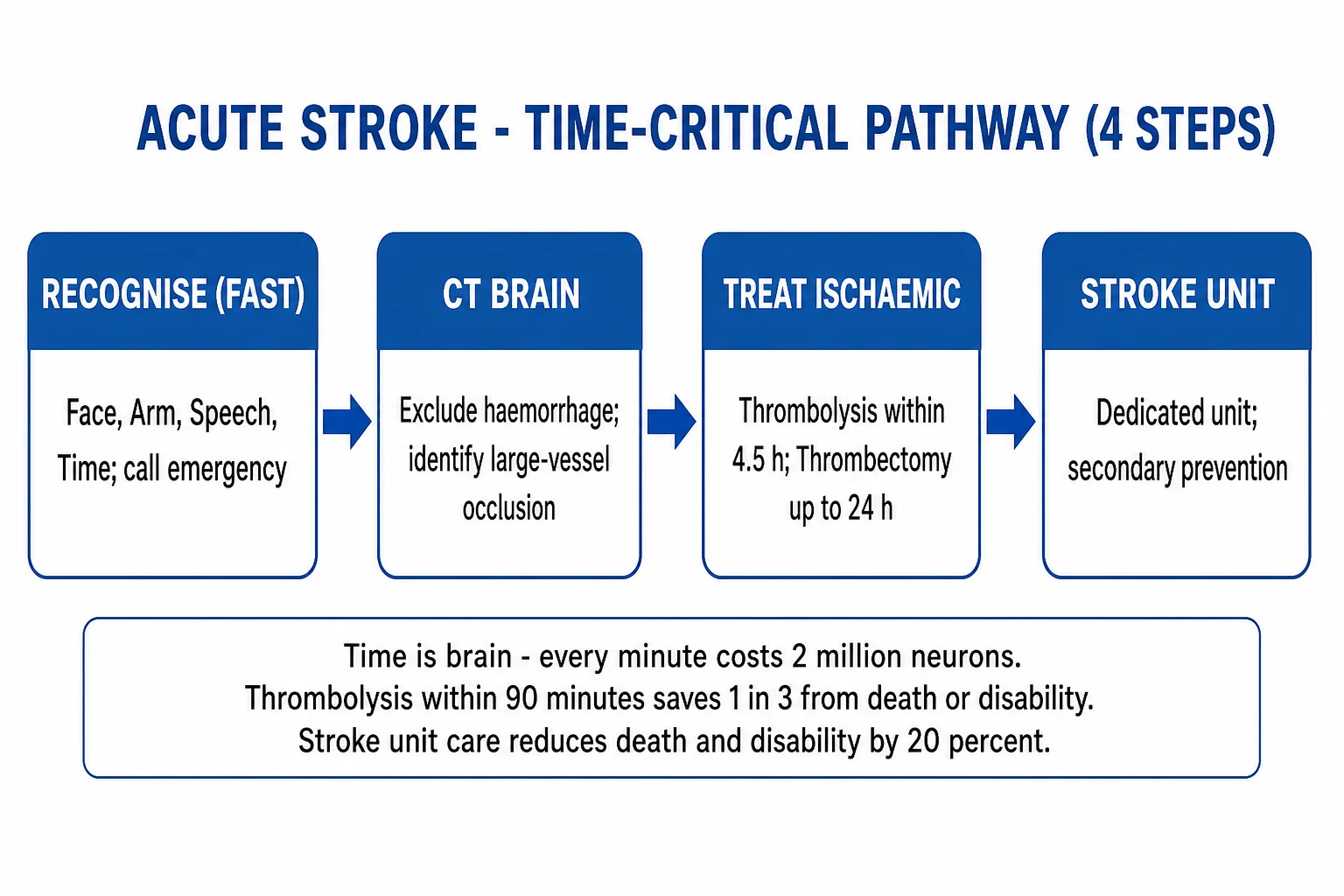
Stroke is a time-critical ABCDE emergency that runs in parallel with neuroimaging and reperfusion decisions — do not let monitoring delay the CT. [1]
First 15 minutes
- Airway — recovery position if unconscious vomiting; early intubation if GCS low or airway unprotected.
- Breathing — oxygen only if SpO2 <94% (avoid routine hyperoxia).
- Circulation — IV access; treat shock; continuous monitoring.
- Disability — glucose immediately (hypoglycaemia mimics stroke — treat if low); record NIHSS; note exact last known well time.
- Exposure — fever control, seizure management, trauma signs, neck stiffness (SAH/meningitis differentials).
Blood pressure before thrombolysis / generally
- Do not aggressively lower BP in ischaemic stroke unless BP is extreme or required to meet thrombolysis thresholds, aortic dissection, ACS, or other compelling indication.
- For candidates for IV alteplase, typical requirement: BP <185/110 mmHg before and <180/105 mmHg for 24 h after — use labetalol IV, nicardipine, or local agent per protocol.
- In ICH, early BP lowering toward systolic ~140 mmHg is often targeted if presenting 150–220 (INTERACT2 / guideline-dependent nuance) — avoid precipitous drops.
NPO, glucose, temperature, DVT prophylaxis
- Keep nil by mouth until swallow screen passed.
- Treat hypo; avoid aggressive intensive glucose lowering.
- Treat fever; investigate aspiration/infection.
- Early mechanical DVT prophylaxis; start anticoagulation for DVT prevention only after haemorrhagic risk assessed (timing depends on infarct size/haemorrhagic transformation risk).
Activate pathways
Door-to-CT and door-to-needle clocks start at registration. Parallel bloods (glucose, FBC, coags, renal) — do not delay CT waiting for full labs unless on anticoagulants with unknown INR and that result will change therapy.
Management — Definitive & Stepwise
Ischaemic stroke — IV thrombolysis (alteplase)
IV alteplase (recombinant tissue plasminogen activator, rt-PA) is the only medical therapy proven to improve outcome in acute ischaemic stroke. It is dosed at 0.9 mg/kg (maximum 90 mg), given as a 10% bolus over 1 minute followed by the remaining 90% infused over 1 hour, within 4.5 hours of symptom onset or last known well, once haemorrhage on CT and the contraindications below have been excluded. Its efficacy and the accompanying real but net-favourable bleeding risk were established by the landmark NINDS rt-PA trial (1995, benefit within 3 hours)[1] and extended to the 3 to 4.5-hour window by ECASS III[2]; the Emberson individual-patient-data meta-analysis (2014) confirmed that benefit is strongly time-dependent — earlier treatment within the window yields substantially better outcomes, and although benefit shrinks with time it persists out to 4.5 hours.[3]
Alteplase 0.9 mg/kg (max 90 mg)
Key exclusion criteria (the list every candidate must know in outline) include: haemorrhage on CT; time over 4.5 hours (or unknown onset without imaging mismatch); severe uncontrolled hypertension (over 185/110 mmHg despite treatment); recent intracranial or spinal surgery, serious head trauma, or intracranial haemorrhage ever; active internal bleeding; recent GI or urinary tract haemorrhage (under 21 days); recent major surgery or trauma (under 14 days); arterial puncture at a non-compressible site (under 7 days); current anticoagulation with an elevated INR (over 1.7) or a DOAC taken within 24 to 48 hours; platelet count under 100,000; glucose under 2.7 mmol/L (correct before deciding); large early infarct (over one-third of MCA territory); recent MI; seizure at onset (unless residual deficit is clearly postictal-imaging-proven stroke); and rapidly improving or minor, non-disabling deficit. Pregnancy is a relative, not absolute, contraindication. Age alone is not a contraindication. [1]
Ischaemic stroke — mechanical thrombectomy
Mechanical thrombectomy is the only procedure proven to improve outcome in large-vessel ischaemic stroke and is now standard of care. It is offered for anterior-circulation large-vessel occlusion — the intracranial internal carotid artery, the M1 or M2 segments of the middle cerebral artery — and increasingly for selected basilar artery occlusions. The two time windows are heavily examined: [1]
Thrombectomy time windows — what the evidence supports
The 2015 five-trial package (MR CLEAN, ESCAPE, EXTEND-IA, SWIFT-PRIME, REVASCAT) established thrombectomy for large-vessel occlusion within 6 hours.[4] DAWN[5] and DEFUSE-3[6] extended the window to 24 hours using imaging-based selection (a small core infarct with a large volume of salvageable penumbra on CT perfusion or MR perfusion-diffusion mismatch). Recognising a thrombectomy candidate clinically — a severe NIHSS (typically 6 or above), gaze deviation, dense hemiplegia — should trigger urgent CTA rather than waiting for a full standard work-up; a missed large-vessel occlusion is a missed reperfusion opportunity.
After reperfusion — the immediate 24 hours
After successful thrombolysis, the blood-pressure target is under 180/105 mmHg for 24 hours to reduce the risk of symptomatic haemorrhagic transformation, with frequent (every 15 minutes for the first 2 hours, then every 30 minutes for 6 hours, then hourly) neuro-observation and blood-pressure checks. A repeat CT brain is performed at 24 hours (or sooner if the patient deteriorates) to exclude haemorrhage before starting antiplatelet therapy. No antiplatelet or anticoagulant is given for 24 hours after thrombolysis. Swallow assessment is mandatory before any oral intake (including medications) — nil by mouth until a formal swallow screen is passed. [1]
Antithrombotic and disease-modifying therapy
Antithrombotic secondary prevention depends on the stroke mechanism, started once haemorrhage has been excluded on CT and, where thrombolysis was given, after the 24-hour delay: [1]
Non-cardioembolic ischaemic stroke
- Aspirin 300 mg loading then 75 mg once daily, OR clopidogrel 75 mg once daily as monotherapy
- For minor stroke (NIHSS ≤3) or high-risk TIA: DUAL antiplatelet therapy — aspirin plus clopidogrel — for 21 DAYS, then step down to clopidogrel monotherapy (CHANCE, POINT)
- Long-term monotherapy thereafter
Cardioembolic (AF) stroke
- ANTICOAGULATION, not antiplatelet — antiplatelets are ineffective in AF
- Direct oral anticoagulant (DOAC — apixaban, rivaroxaban, dabigatran, edoxaban) preferred over warfarin for non-valvular AF
- Timing balances haemorrhagic transformation vs early recurrence — typically 2 to 14 days post-stroke (earlier for small infarcts/TIA, later for large infarcts)
The evidence for short-course dual antiplatelet therapy comes from CHANCE (2013)[7] and POINT (2018)[8], which together established that aspirin plus clopidogrel for 21 days (then monotherapy) reduces recurrent stroke in minor ischaemic stroke and high-risk TIA, with the benefit concentrated in the first 10 to 21 days and a rising haemorrhage risk thereafter — hence the 21-day cap. The DOAC-over-warfarin preference in non-valvular AF rests on the Ruff meta-analysis (2014), showing roughly a 50% reduction in intracranial haemorrhage and similar or superior stroke prevention with DOACs.[10]
For every patient regardless of mechanism, the secondary-prevention package is completed by a high-intensity statin (e.g. atorvastatin 80 mg daily, or the maximally tolerated dose — targeting an LDL reduction of 50% or more and LDL under 1.8 mmol/L), blood-pressure control (typically an ACE inhibitor or angiotensin-receptor blocker ± a thiazide-like diuretic, target under 130/80 mmHg), glycaemic control (HbA1c target individualised, around 53 mmol/mol / 7%), smoking cessation, lifestyle modification (Mediterranean diet, regular exercise, weight reduction, alcohol moderation), and — for symptomatic carotid stenosis of 70 to 99% in a surgically-fit patient — carotid endarterectomy ideally within two weeks of the qualifying event (the benefit is strongly time-dependent and largely lost by 12 weeks). [1]
Management — Haemorrhagic Stroke
The management of intracerebral haemorrhage is the opposite of ischaemic stroke in almost every respect. The three pillars are blood-pressure control, reversal of anticoagulation, and selective neurosurgical evacuation. [1]
Blood pressure — the largest modifiable determinant of haematoma expansion, which occurs mostly in the first few hours and drives early deterioration. The INTERACT2 trial established that rapid lowering of systolic blood pressure to under 140 mmHg (typically with IV labetalol, nicardipine, or urapidil) is safe and improves functional outcome compared with a more conservative target (under 180 mmHg), without a clear mortality benefit.[9]
Reversal of anticoagulation is mandatory in any anticoagulant-associated ICH and must be immediate. Warfarin is reversed with intravenous vitamin K (10 mg) plus prothrombin complex concentrate (PCC), which works within minutes (fresh-frozen plasma is an inferior, slower alternative). Dabigatran is reversed with the specific monoclonal antibody fragment idarucizumab (5 g IV); rivaroxaban and apixaban with the recombinant factor Xa decoy andexanet alfa (or, if unavailable, PCC). Heparin is reversed with protamine sulfate. Thrombocytopaenia and platelet dysfunction are addressed where relevant. A patient on an anticoagulant who has an ischaemic stroke usually cannot receive thrombolysis until the anticoagulant effect is confirmed absent or reversed; most proceed directly to thrombectomy if eligible, bypassing thrombolysis. [1]
Neurosurgical evacuation is selectively life-saving. Cerebellar haemorrhage is a neurosurgical emergency in its own right: a haematoma over about 3 cm (or one causing brainstem compression, fourth-ventricle effacement, hydrocephalus, or deterioration) warrants urgent surgical evacuation, because posterior-fossa swelling rapidly compresses the brainstem and obstructs CSF outflow — a small haematoma in a tight posterior fossa can kill. Lobar supratentorial haemorrhage may be considered for evacuation if superficial, large, and deteriorating, especially in younger patients. Deep (basal-ganglia/thalamic) haemorrhages are not usually evacuated surgically. Decompressive hemicraniectomy is the life-saving treatment for malignant MCA syndrome — massive cerebral oedema causing transtentorial herniation after a large hemispheric infarct — performed ideally within 48 hours in patients under 60 with a large (usually NIHSS above 15) MCA infarct, where it reduces mortality substantially (from about 80% to about 20%) at the cost of some degree of residual disability. [1]
Specific Subtypes & Scenarios
Large-vessel occlusion (LVO) — internal carotid artery, M1/M2, or basilar — is a thrombectomy candidate by definition. Recognising it clinically (severe NIHSS, gaze deviation, dense hemiplegia) should trigger urgent CT angiography at the same sitting as the non-contrast CT, rather than waiting for a full standard work-up. Time is the overriding determinant of outcome. [1]
Cerebellar haemorrhage is the surgical emergency of haemorrhagic stroke — evacuation is life-saving because of the risk of brainstem compression and obstructive hydrocephalus, even when the haematoma itself is modest in volume. A deterioration in consciousness is the trigger. [1]
Wake-up or unknown-onset stroke — found on waking, or last-known-well unclear — can still be eligible for thrombolysis or thrombectomy if MRI DWI-FLAIR mismatch (lesion visible on DWI but not on FLAIR, implying recency) or CT-perfusion mismatch suggests the infarct is recent and there is salvageable penumbra. [1]
Posterior-circulation and basilar occlusion produces the devastating locked-in syndrome if untreated; basilar thrombectomy is increasingly used and can extend the treatment window, because the consequence of a missed basilar occlusion is catastrophic. A sudden-onset vertigo with crossed signs, diplopia, ataxia, or a decreased level of consciousness is posterior-circulation stroke until proven otherwise — use the HINTS examination to separate central from peripheral vertigo. [1]
Stroke in an anticoagulated patient (warfarin or DOAC) usually cannot be thrombolysed until the anticoagulant effect is reversed or confirmed absent, given the high intracranial haemorrhage risk; most such patients proceed directly to thrombectomy if a large-vessel occlusion is present and they are otherwise eligible. [1]
Complications & Pitfalls
Ischaemic stroke complications
- Haemorrhagic transformation of the infarct
- Cerebral oedema and raised ICP — malignant MCA syndrome
- Seizures (early or late post-stroke epilepsy)
- Aspiration pneumonia — swallow assessment before any oral intake
- Venous thromboembolism (DVT/PE) — prophylactic LMWH once haemorrhage excluded
- Pressure ulcers, contractures, shoulder pain, falls
- Post-stroke depression (screen all patients)
IV-thrombolysis complications
- Symptomatic intracranial haemorrhage (~2–6% — the feared one)
- Orolingual angio-oedema (especially with ACE inhibitor use)
- Systemic bleeding from puncture sites or viscera
Classic pitfalls
- Missing a mimic (especially hypoglycaemia) and giving thrombolysis inappropriately
- Missing the time window through delayed recognition or imprecise last-known-well
- Failing to lower BP below the 185/110 threshold before thrombolysis
- Giving antiplatelet/anticoagulant before CT excludes haemorrhage
- Delayed thrombectomy referral for a large-vessel occlusion
- Withholding thrombolysis on age alone — age is NOT a contraindication
Malignant MCA syndrome — massive cerebral oedema after a complete MCA-territory infarct, causing midline shift and transtentorial herniation, peaking at 48 to 72 hours — is the most feared medical complication of ischaemic stroke. It is treated with decompressive hemicraniectomy within 48 hours in appropriately selected (typically under 60) patients, which reduces mortality from roughly 80% to roughly 20% at the cost of residual disability. Medical measures (mannitol, hypertonic saline, head-of-bed elevation) are temporising bridges to surgery. [1]
Prognosis & Disposition
Stroke-unit care reduces death and dependency by about one-fifth independent of reperfusion — admit to a dedicated unit whenever possible. Prognosis depends on age, NIHSS, infarct/haemorrhage volume, location (dominant hemisphere, brainstem), reperfusion success, glucose, fever, and complications (aspiration, VTE, raised ICP). [1]
NIHSS (concept for exams)
NIHSS quantifies deficit (0–42) across LOC, gaze, visual fields, facial palsy, motor arms/legs, ataxia, sensory, language, dysarthria, extinction. Higher scores → worse outcome and often stronger thrombectomy consideration when LVO present. Mild disabling deficits can still deserve thrombolysis even if NIHSS is "low."
Disposition
- Stroke unit for almost all acute strokes
- ICU/NSICU for large ICH, basilar occlusion, malignant MCA syndrome risk, post-thrombectomy instability, airway compromise
- Decompressive hemicraniectomy window for malignant MCA infarction in selected patients ≤60 years (and carefully selected older adults) before herniation peaks (~48 h)
- Early physio/OT/SALT, secondary prevention started in hospital, mood and cognition screening
Secondary prevention core (ischaemic)
- Antiplatelet: aspirin 300 mg loading then 75 mg (or dual short-course in selected minor stroke/high-risk TIA per POINT/CHANCE-style protocols — typically 21 days then monotherapy)
- Anticoagulation for AF (delay post large infarct to reduce haemorrhagic transformation — often 3–14 days stratified by severity)
- High-intensity statin; BP control after acute phase; diabetes management; carotid revascularisation for symptomatic severe stenosis in fit candidates; lifestyle [1]
Special Populations
Pregnancy
Thrombolysis is relative, not absolute, contraindication for disabling ischaemic stroke. Exclude pre-eclampsia/eclampsia and cerebral venous thrombosis (CVT) — CVT needs anticoagulation even with some haemorrhagic components under specialist care. Multidisciplinary obstetric-neurology decisions.
Elderly
Age alone is not a contraindication to alteplase or thrombectomy. Higher ICH risk and frailty alter goals of care conversations but must not automatically withhold time-critical therapy.
Young stroke work-up
Add dissection imaging, thrombophilia/autoimmune panel when indicated, bubble echo for shunt, drug history (cocaine), pregnancy test, HIV/syphilis where relevant, vasculitis clues.
On anticoagulation
VKA with high INR → reverse if ICH (PCC + vitamin K). DOAC-related ICH → specific reversal when available (idarucizumab for dabigatran; andexanet for factor Xa inhibitors in selected protocols) or PCC per local guidance. For ischaemic stroke on DOAC, thrombolysis timing depends on last dose, renal function, and level assays where available — often not given within 48 h of DOAC unless levels low.
Cerebral venous thrombosis
Headache, seizures, focal signs, often young women (OCP, puerperium). Diagnose with CT/MR venography. Treat with anticoagulation; endovascular options if deterioration.
Prevention
Stroke prevention operates at three levels. Primordial prevention addresses the development of risk factors in the first place — population-level blood-pressure control, diet, exercise, and tobacco control. Primary prevention targets individuals with risk factors but no prior stroke — control of hypertension (the highest-yield single intervention), oral anticoagulation for atrial fibrillation at sufficient CHA₂DS₂-VASc score, statin therapy for high-risk vascular disease, smoking cessation, diabetes and lipid management, and carotid revascularisation for asymptomatic high-grade stenosis in selected patients. Secondary prevention — applied to every stroke and TIA survivor — combines the disease-modifying package above (antiplatelet or anticoagulant, high-intensity statin, blood-pressure control, glycaemic control, lifestyle change) with timely carotid endarterectomy for symptomatic significant stenosis and prolonged cardiac monitoring for occult paroxysmal AF in cryptogenic stroke. The combination of these measures can reduce recurrent stroke risk by more than 80%. [1]
Evidence, Guidelines & Regional Differences
The landmark stroke trials — one-line takeaways
The overall framework — FAST recognition, CT-first imaging, time-windowed reperfusion, dedicated stroke-unit care — is globally consistent. AHA/ASA 2019 (US)[11] is the comprehensive current US framework: thrombolysis to 4.5 h, thrombectomy 0 to 6 h without and 6 to 24 h with imaging selection, dual antiplatelet for 21 days in minor stroke. NICE NG128 (UK) aligns on the core time windows and emphasises stroke-unit admission and swallow assessment; the UK historically leaned on clopidogrel monotherapy after the 2-week dual-antiplatelet window. AHA/ASA and the ESO/EAN (Europe) agree on thrombectomy to 24 h with imaging selection. Indian Stroke Association / ICMR guidance follows the same windows adapted to local resource availability — thrombolysis services are more widely available than thrombectomy, and warfarin remains in use alongside DOACs. Local variation is chiefly in thrombectomy service coverage and in DOAC versus warfarin uptake for atrial fibrillation.
The principal controversies are the lower boundary of thrombolysis in minor non-disabling stroke (where the bleeding risk may exceed benefit), the role of thrombolysis in patients already on a DOAC, the extended-window thrombectomy criteria (how mismatch is measured and the upper core-volume limit), and whether tenecteplase (a genetically modified variant with longer half-life and bolus administration) will replace alteplase as the default thrombolytic — current evidence supports non-inferiority and a trend toward better outcome. [1]
Thrombolysis Eligibility Exam Table & Worked Stems
IV alteplase core (typical adult protocol teaching)
| Item | Detail |
|---|---|
| Window | 0–4.5 h from last known well (0–3 h strongest; 3–4.5 h with extra exclusions in some older labels) |
| Dose | Alteplase 0.9 mg/kg (max 90 mg): 10% bolus, remainder over 60 min |
| Imaging | Non-contrast CT (or MRI) excludes haemorrhage; ASPECTS/early infarct signs considered |
| BP | <185/110 before; maintain <180/105 after |
| Key absolute CI (examples) | Acute ICH; prior ICH (relative/absolute depending); active internal bleeding; recent intracranial surgery/trauma; coagulopathy thresholds (platelets <100k, INR >1.7, etc.); infective endocarditis with septic embolus concerns in some protocols |
| Thrombectomy | Anterior circulation LVO typically within 6 h; 6–24 h if DAWN/DEFUSE-3 style imaging mismatch criteria met |
ABCD2 for TIA risk stratification (reproduce)
Age ≥60 (1); BP ≥140/90 (1); Clinical: unilateral weakness (2) or speech impairment without weakness (1); Duration ≥60 min (2) or 10–59 min (1); Diabetes (1). Score 0–7. Higher scores → higher short-term stroke risk — but do not use ABCD2 alone to send high-risk phenotypes home; dual antiplatelet strategies and same-day imaging pathways matter more than the number alone.
Worked stem — wake-up stroke
Patient wakes with aphasia and right hemiparesis; last seen normal 22:00; now 08:00. Not automatically out of options — if MRI DWI-FLAIR mismatch or CT perfusion mismatch criteria met, thrombolysis/thrombectomy may still apply in extended windows under protocol.
Worked stem — tPA vs bleed
Post-alteplase sudden headache, BP spike, neuro deterioration → stop infusion, urgent CT, reverse (cryoprecipitate/fibrinogen replacement protocols, TXA in some pathways), ICU neurosurgical consult.
Worked NEET-PG Stems — Stroke
- Last known well 2 h ago, NIHSS 12, CT no blood → alteplase 0.9 mg/kg if eligible; BP under 185/110.
- ICA/M1 occlusion at 4 h → thrombolysis if eligible + thrombectomy.
- Wake-up stroke → advanced imaging mismatch may still allow reperfusion.
- AF with large infarct day 0 → delay anticoagulation (haemorrhagic transformation risk); start antiplatelet interim per protocol.
- TIA with ABCD2 high + ipsilateral 80% carotid stenosis → early carotid revascularisation work-up + antithrombotic + statin.
- Thunderclap headache, neck stiffness → SAH pathway (not ischaemic tPA). [1]
Exam Pearls
Exam application bank (NEET-PG / INICET)
One-line answer
Stroke is sudden focal neurological deficit from vascular injury to the brain: ischaemic (85%) from arterial occlusion, or haemorrhagic (15%) from vessel rupture. FAST (Face, Arm, Speech, Time) triggers recognition; a non-contrast CT brain is the first, most urgent investigation to distinguish the two — since the acute treatments are opposite. Ischaemic stroke: IV alteplase within 4.5 hours, thrombectomy up to 24 hours for large-vessel occlusion with favourable imaging. Haemorrhagic stroke: blood-pressure control, reversal of anticoagulation, and neurosurgical evacuation where indicated.
Worked stems (answer without another resource)
Stem 1 — Classic presentation. Map symptoms to mechanism; name the first investigation and first treatment step with dose/route if drug therapy is standard. [1]
Stem 2 — Unstable / complicated. List red flags that force immediate resuscitation, theatre, ICU, antidote, or reperfusion — and what you do in the first 15 minutes. [1]
Stem 3 — Atypical group. Elderly, pregnancy, child, or immunocompromised: how presentation and thresholds change. [1]
Stem 4 — Differential trap. Name the three closest mimics and one discriminator for each. [1]
Stem 5 — Disposition. Who goes home with safety-netting, who is admitted, who needs HDU/ICU/theatre, and what follow-up is mandatory. [1]
Rapid viva checklist
- Definition + classification
- Pathophysiology chain
- Bedside signs / criteria
- Score with exact components (if any)
- Emergency bundle
- Definitive therapy with doses
- Complications of disease and of treatment
- Special populations
- Guideline/trial name if classic
- Three exam traps
Coverage self-check
If you cannot answer any stem above from this page alone, re-read the matching section — the page is intended to be self-sufficient for final-prof and NEET-PG/INICET questions on Stroke.
BE-FAST
NIHSS domains
Self-test: a 72-year-old man presents at 90 minutes with right face and arm weakness and expressive aphasia. CT shows no haemorrhage. BP 168/92. Glucose 6.4. INR 1.0. What is the next step?
He is within the 4.5-h window, has a disabling dominant-hemisphere ischaemic stroke, BP is under 185/110, glucose is normal, and there is no anticoagulation or other exclusion. The next step is IV alteplase 0.9 mg/kg (max 90 mg), 10% bolus over 1 min then 90% over 1 h, followed by an urgent CT angiogram to assess for a large-vessel occlusion that would make him a thrombectomy candidate, and admission to a stroke unit with BP kept under 180/105 for 24 h and aspirin 300 mg started after the 24-h repeat CT.
Self-test: a 68-year-old woman on dabigatran for AF presents with acute right hemiparesis and CT shows a left basal-ganglia haemorrhage. BP 180/100. What do you do?
This is an anticoagulant-associated intracerebral haemorrhage. Reverse the dabigatran immediately with idarucizumab 5 g IV; lower the BP rapidly to SBP under 140 mmHg (IV labetalol or nicardipine); admit to a stroke unit or neurocritical care; arrange urgent neurosurgical review (basal-ganglia haematomas are usually managed non-operatively, but cerebellar or large lobar haematomas need surgical consideration); and do NOT give thrombolysis. Restart anticoagulation only after a structured interval (typically 7 to 14 days, balancing re-embolism against re-bleed) once the bleeding risk has settled.
References
- [1]The National Institute of Neurological Disorders and Stroke rt-PA Stroke Study Group. Tissue plasminogen activator for acute ischemic stroke N Engl J Med, 1995.PMID 7477192
- [2]Hacke W, Kaste M, Bluhmki E, et al. Thrombolysis with alteplase 3 to 4.5 hours after acute ischemic stroke N Engl J Med, 2008.PMID 18815396
- [3]Emberson J, Lees KR, Lyden P, et al. Effect of treatment delay, age, and stroke severity on the effects of intravenous thrombolysis with alteplase for acute ischaemic stroke: a meta-analysis of individual patient data from randomised trials Lancet, 2014.PMID 25106063
- [4]Berkhemer OA, Fransen PSS, Beumer D, et al. Intraarterial treatment for acute ischemic stroke N Engl J Med, 2015.PMID 25785978
- [5]Nogueira RG, Jovin TG, Haussen DC, et al. Thrombectomy 6 to 24 Hours after Stroke with a Mismatch between Deficit and Infarct N Engl J Med, 2018.PMID 29129157
- [6]Albers GW, Marks MP, Kemp S, et al. Thrombectomy for Stroke at 6 to 16 Hours with Selection by Perfusion Imaging N Engl J Med, 2018.PMID 29364767
- [7]Wang Y, Wang Y, Zhao X, et al. Clopidogrel with aspirin in acute minor stroke or transient ischemic attack N Engl J Med, 2013.PMID 23803136
- [8]Johnston SC, Easton JD, Farrant M, et al. Clopidogrel and Aspirin in Acute Ischemic Stroke and High-Risk TIA N Engl J Med, 2018.PMID 29766750
- [9]Anderson CS, Heeley E, Huang Y, et al. Rapid blood-pressure lowering in patients with acute intracerebral hemorrhage N Engl J Med, 2013.PMID 23713578
- [10]Ruff CT, Giugliano RP, Braunwald E, et al. Comparison of the efficacy and safety of new oral anticoagulants with warfarin in patients with atrial fibrillation: a meta-analysis of randomised trials Lancet, 2014.PMID 24315724
- [11]Powers WJ, Rabinstein AA, Ackerson T, et al. Guidelines for the Early Management of Patients With Acute Ischemic Stroke: 2019 Update to the 2018 Guidelines for the Early Management of Acute Ischemic Stroke: A Guideline for Healthcare Professionals From the American Heart Association/American Stroke Association Stroke, 2019.PMID 31662037