Neurology · General Medicine
Subarachnoid Haemorrhage
Also known as Subarachnoid haemorrhage · SAH · Ruptured aneurysm · Thunderclap headache
Subarachnoid haemorrhage (SAH) is bleeding into the subarachnoid space, usually from a ruptured intracranial aneurysm (85 percent), presenting with a sudden, severe 'thunderclap' headache (maximum intensity within 1 minute) — often described as 'the worst headache of my life' — with neck stiffness, photophobia, nausea, vomiting, altered consciousness and sometimes seizures. Non-contrast CT brain (sensitivity about 100 percent within 6 hours on a modern scanner) is the first investigation; if the CT is negative or performed later than 6 hours, lumbar puncture for xanthochromia (yellow CSF supernatant from bilirubin) confirms the diagnosis. Management includes securing the aneurysm (endovascular coiling preferred over surgical clipping per ISAT), nimodipine 60 mg orally every 4 hours for 21 days (prevents delayed cerebral ischaemia from vasospasm), blood pressure control, and management of complications (rebleeding, vasospasm, hydrocephalus, hyponatraemia). SAH carries a 30-day mortality of around 30 percent; vasospasm is the leading cause of preventable death and disability.
On this page & tools
Your progress
Saved locally on this device.
Exam tags
Red flags
Overview & Definition
Subarachnoid haemorrhage (SAH) is bleeding into the subarachnoid space — the compartment between the arachnoid and pia mater through which cerebrospinal fluid (CSF) normally circulates. It is a neurological emergency and the third most common subtype of stroke after ischaemic stroke and intracerebral haemorrhage, accounting for roughly 5 percent of all strokes but a disproportionate share of stroke-related death and disability because it strikes younger people (mean age 50–55 years) and robs many productive years.[1][2]
The defining clinical event is the thunderclap headache: a headache that reaches maximum intensity within one minute of onset. This single symptom is the cornerstone of the disease — a thunderclap headache is SAH until proven otherwise, and the discipline of clinical practice is to take every sudden severe headache seriously, scan immediately, and perform a lumbar puncture if the CT is negative. The penalty for missing the diagnosis is high: an unsecured aneurysm re-ruptures in roughly a third of patients within two weeks, and rebleeding carries a mortality of about 70 percent.[1][11]
Once the diagnosis is made, management has two parallel time-critical objectives. The first is to prevent rebleeding by obliterating the aneurysm as soon as possible, ideally within 24 hours. The second is to prevent delayed cerebral ischaemia (DCI), the syndrome of vasospasm-driven brain injury that appears 4–14 days after the bleed and is the leading cause of preventable death and disability in those who survive the initial ictus. Every other element of management — blood pressure control, nimodipine, ventricular drainage, sodium correction — exists to serve these two objectives.[2][7]
Classification
SAH is classified by cause, by aneurysm site, and by clinical and radiological severity. These three axes together determine prognosis and treatment. [1]
By cause, aneurysmal rupture dominates — about 85 percent of spontaneous SAH. Perimesencephalic non-aneurysmal SAH accounts for roughly 10 percent and has a strikingly benign course, while extension of an intracerebral haemorrhage (hypertensive or arteriovenous malformation) into the ventricles and subarachnoid space accounts for the remainder.[1][2]
Aneurysmal SAH
~85 percent
- Rupture of a saccular ('berry') aneurysm at a Circle of Willis bifurcation
- Full SAH syndrome; rebleeding and vasospasm drive outcome
- Diagnosed by CT ± LP; aneurysm localised on CTA/DSA
- Requires urgent aneurysm securing (coiling or clipping) and nimodipine
Perimesencephalic non-aneurysmal SAH
~10 percent (PNSAH)
- Blood centred around the midbrain in the prepontine / perimesencephalic cisterns
- No aneurysm on digital subtraction angiography
- Benign course — vasospasm rare, prognosis excellent
- Nimodipine often still given; outcome good regardless
ICH extension / AVM
~5 percent
- Hypertensive ICH dissecting into the ventricles/subarachnoid space
- AVM rupture in younger patients, sometimes with prior seizures
- Managed as ICH (BP, ICP) ± AVM-specific treatment (surgery, embolisation, radiosurgery)
By aneurysm site, saccular aneurysms arise at the bifurcations of the Circle of Willis where the muscularis media and internal elastic lamina are naturally deficient. The anterior communicating artery (ACom) is the commonest site at roughly 30 percent, followed by the internal carotid / posterior communicating artery (PCom) at 25 percent, the middle cerebral artery (MCA) bifurcation at 20 percent, and the basilar tip / vertebrobasilar circulation at 7–10 percent. The site predicts the focal deficit at presentation: a third-nerve palsy points to a PCom aneurysm, paraparesis and abulia to an ACom aneurysm, and a hemiparesis/aphasia to an MCA aneurysm.[1][6]
By clinical severity, two scales are in routine use. The Hunt and Hess grade (1968) is symptom-based, while the World Federation of Neurosurgical Societies (WFNS) grade (1988) is based on the Glasgow Coma Scale (GCS) plus the presence of a motor deficit. Both predict outcome and are recorded at first assessment and on any change. A third scale, the Fisher scale, grades the amount of blood on the CT and predicts the risk of vasospasm. Each is reproduced verbatim in the Investigations section because examiners expect the components to be stated exactly.[6][10]
Epidemiology & Risk Factors
SAH has an annual incidence of roughly 6–10 per 100,000 people, with higher rates reported in Finland and Japan (around 20 per 100,000). The incidence has been declining over recent decades, attributed to smoking cessation, better blood pressure control, and improved detection of unruptured aneurysms. About 10–15 percent of patients die before reaching hospital, and the overall 30-day mortality is around 30 percent, with a further 10–20 percent of survivors left functionally dependent.[1][2]
The modifiable risk factors, in order of impact, are hypertension (the strongest), smoking (dose-dependent and synergistic with hypertension), excess alcohol, and sympathomimetic drugs such as amphetamines and cocaine (which produce acute blood-pressure surges). The non-modifiable risks include age (peaking at 40–60 years), female sex (about 1.6-fold increased risk after age 55), and a family history of aneurysmal SAH — two or more affected first-degree relatives confer a roughly 2–4-fold increased risk and are an indication for screening.[1][6]
Modifiable risks
- Hypertension — strongest modifiable risk factor
- Smoking — dose-dependent, synergistic with hypertension
- Excess alcohol; sympathomimetic drugs (cocaine, amphetamines) — acute BP surge
- Correcting these lowers population SAH incidence
Non-modifiable / genetic
- Age 40–60; female sex (1.6-fold after 55)
- Family history — 2 first-degree relatives → 2–4-fold risk (screening indicated)
- Autosomal dominant polycystic kidney disease (ADPKD) — screen all
- Ehlers-Danlos (vascular type), Marfan, neurofibromatosis type 1, fibromuscular dysplasia
Several heritable connective-tissue and vascular disorders predispose to intracranial aneurysms and are deliberately tested in exams. Autosomal dominant polycystic kidney disease (ADPKD) is the most important: 5–10 percent of ADPKD patients harbour an intracranial aneurysm, and all ADPKD patients should be screened with magnetic resonance angiography (MRA). The same is true of patients with two or more first-degree relatives who have had an aneurysmal SAH. The vascular type of Ehlers-Danlos syndrome (type IV, due to type III collagen deficiency) and Marfan syndrome (fibrillin-1) both weaken the arterial wall; neurofibromatosis type 1 and fibromuscular dysplasia are also associated.[1][7]
For an unruptured aneurysm found incidentally, the rupture risk depends on size, site, and patient factors. Small (under 7 mm) anterior-circulation aneurysms in patients without prior SAH carry a low annual rupture risk, whereas larger aneurysms, posterior-circulation aneurysms (especially PCom and basilar tip), symptomatic aneurysms, and aneurysms with a daughter lobe or documented growth carry higher risks that warrant repair.[6][7]
Pathophysiology
A saccular ('berry') aneurysm forms at an arterial bifurcation of the Circle of Willis. At these apices the muscularis media and the internal elastic lamina are physiologically deficient, so the intima bears the full haemodynamic load. Over years, sustained wall shear stress injures the endothelium, fragments the internal elastic lamina, depletes smooth muscle, and the intima bulges outward into a saccular pouch, often with a narrow neck and an asymmetric 'daughter sac' that is the usual point of rupture.[1]
Rupture typically occurs during a transient blood-pressure surge — exertion, straining, coitus, or emotional stress. Blood exits the aneurysm under systemic arterial pressure into the subarachnoid space. Three immediate consequences follow. First, the intracranial pressure (ICP) rises abruptly, often approaching diastolic blood pressure; cerebral perfusion pressure (CPP equals MAP minus ICP) collapses, producing transient global cerebral ischaemia and the characteristic brief loss of consciousness at the moment of rupture. Second, blood in the CSF irritates the meninges, generating neck stiffness, photophobia, and a positive Kernig sign over the following hours. Third, the massive sympathetic (catecholamine) surge damages the myocardium — the syndrome of neurogenic stunned myocardium, a reversible Takotsubo-like cardiomyopathy with troponin rise and ECG changes.[2][11]
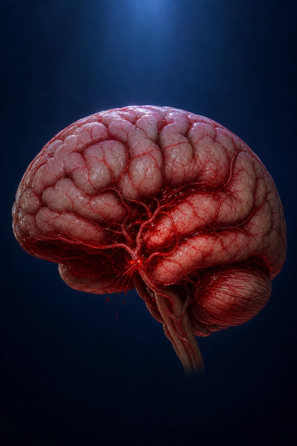
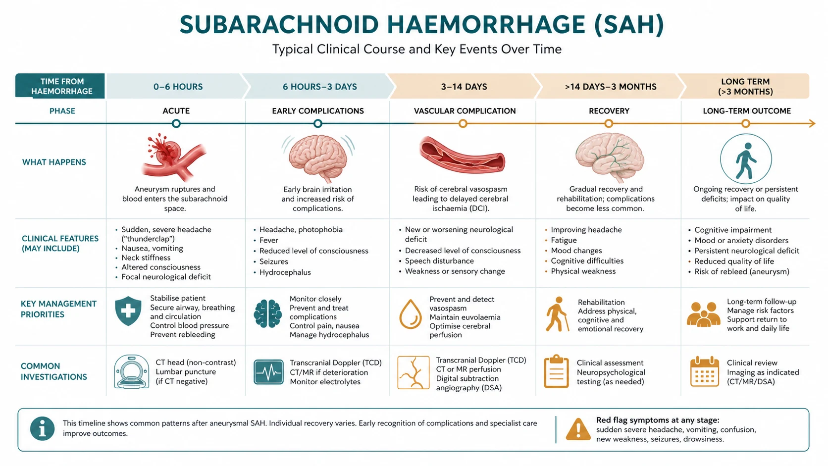
The most consequential secondary event is delayed cerebral ischaemia (DCI), which appears 4–14 days after the bleed (peak at days 7–8) and is the leading cause of preventable death and disability. Its mechanism is multifactorial. Breakdown of subarachnoid blood releases oxyhaemoglobin, which depletes the vasodilator nitric oxide, stimulates release of the potent vasoconstrictor endothelin-1, generates free radicals, and activates protein kinase C in vascular smooth muscle — together producing sustained large-vessel vasospasm (visible on angiography in 30–70 percent of patients, peaking at days 7–8). Vasospasm alone does not fully explain DCI: microvascular dysfunction, cortical spreading depolarisations, microthrombosis, and a marked inflammatory response all contribute, which is why nimodipine (which barely reverses large-vessel spasm) nonetheless improves outcome.[2][11]
Two further complications flow directly from the bleed. Hydrocephalus arises because blood obstructs CSF reabsorption at the arachnoid granulations (a communicating hydrocephalus, acute or chronic) or because intraventricular clot blocks the aqueduct or fourth-ventricle outlet (an obstructive hydrocephalus, acute). Hyponatraemia in roughly 30–40 percent of patients is most often cerebral salt wasting — the release of natriuretic peptides produces volume depletion with high urine sodium and osmolarity — rather than SIADH, and the distinction is one of the most heavily examined points in the disease (see Complications).[2][11]
Clinical Presentation
The cardinal symptom is the thunderclap headache: sudden onset, with maximum intensity reached within one minute. Patients reach for the same descriptors — "the worst headache of my life", "like being kicked in the back of the head", "an explosion". It is classically occipital or suboccipital but may be diffuse, and it persists for hours to days. It is reported by 75–95 percent of patients and is the single most important symptom in clinical medicine to take seriously: no thunderclap headache may be dismissed as migraine without excluding SAH.[1][4]
Accompanying features develop over hours. Meningism — neck stiffness, photophobia, and positive Kernig and Brudzinski signs — appears 3–12 hours after the bleed as blood irritates the meninges, and may be absent in the first hour, which is a dangerous pitfall. Nausea and vomiting (about 75 percent, sometimes projectile at onset) and a brief loss of consciousness at the moment of rupture (in about half, from the ICP surge) are common. Seizures occur at onset in 10–20 percent from acute cortical irritation. Focal deficits localise the aneurysm: a third-nerve palsy (ptosis, a dilated pupil, and the eye deviated 'down and out') indicates a PCom aneurysm compressing the oculomotor nerve; lower-limb-dominant weakness and abulia indicate an ACom aneurysm; a hemiparesis or aphasia indicates an MCA aneurysm.[1][2]
Classic presentation
- Thunderclap headache (max within 1 min) — 'worst ever'
- Neck stiffness, photophobia, positive Kernig/Brudzinski (after 3–12 h)
- Nausea/vomiting; brief loss of consciousness at onset (~50%)
- Focal deficit localises the aneurysm (III palsy = PCom; leg weakness = ACom)
Atypical / easily missed
- Elderly — confusion or a fall instead of classic headache
- Sentinel (warning) bleed days–weeks before — a resolving 'different' headache
- Pregnancy/puerperium — SAH is a leading non-obstetric cause of maternal death
- Isolated thunderclap headache with a normal exam — still SAH until excluded
- Terson syndrome — vitreous/preretinal haemorrhage on fundoscopy
Two atypical presentations deserve emphasis because examiners test them deliberately. The sentinel (warning) bleed is a minor leak days to weeks before the catastrophic rupture, presenting as a sudden but resolving headache; it is missed in about half of cases and dismissed as migraine, and recognising it allows intervention before the major rupture. Terson syndrome — vitreous or preretinal haemorrhage seen on fundoscopy from ICP transmitted along the optic nerve sheath — occurs in 15–20 percent and signals a larger bleed and a worse prognosis; always examine the fundi in any patient with a thunderclap headache.[1][11]
In the elderly, the headache may be muted or absent, and the patient may present with confusion, a fall, or coma after a collapse — the trap being to attribute the collapse to a simple fall rather than to an underlying SAH. In pregnancy and the puerperium, SAH is a leading non-obstetric cause of maternal death, and any thunderclap headache in pregnancy is SAH (alongside pre-eclampsia, HELLP, and cerebral venous sinus thrombosis) until proven otherwise.[2]
Differential Diagnosis
The differential of a sudden severe headache is wide, and the clinical task is not to name the mimics but to exclude SAH first. Any headache that is sudden, maximal within a minute, exertional, "worst ever", associated with neck stiffness, vomiting, loss of consciousness or a focal deficit — in any patient over 40, pregnant, or anticoagulated — mandates CT ± LP.[1][5]
Migraine
- Builds over minutes–hours; pulsating; ± aura; photophobia/phonophobia
- Recurrent similar episodes; family history
- NEVER assume a 'first or worst' migraine without excluding SAH
Bacterial / viral meningitis
- Headache + fever + neck stiffness evolving over hours–days (not thunderclap)
- Rash (meningococcal); CT normal, LP diagnostic (low glucose, high neutrophils, organisms)
Cerebral venous sinus thrombosis
- Subacute headache ± seizures, focal deficits, papilloedema
- Risk factors: OCP, pregnancy, dehydration, prothrombotic state; CT/MR venogram diagnostic
Cervical artery dissection
- Sudden unilateral neck/face pain ± Horner syndrome ± ischaemic stroke
- Younger patient; can coexist with or mimic SAH
Primary thunderclap headache / RCVS
- Diagnosis of EXCLUSION only — identical to SAH at onset; CT ± LP must be normal
- RCVS: recurrent thunderclaps over days–weeks, multifocal vasoconstriction on angiography
Pituitary apoplexy
- Sudden severe headache + ophthalmoplegia + visual field defect ± collapse
- In a patient with a pituitary adenoma; haemorrhage on CT/MRI
Which single feature most reliably separates SAH from its mimics?
The tempo of onset. SAH reaches maximum intensity within one minute; migraine builds over minutes to hours; meningitis evolves over hours; thunderclap headache of any cause (SAH included) shares the sudden onset — which is why primary thunderclap headache is a diagnosis of exclusion only after CT ± LP have excluded SAH.[1][4]
Clinical & Bedside Assessment
The focused assessment combines a precise history of onset, vital signs, the GCS, meningeal signs, a cranial-nerve and focal exam, and fundoscopy. The onset-to-peak time is the single most discriminating historical detail; a thunderclap headache (maximal within one minute) is SAH until excluded regardless of the rest of the exam.[1][5]
The Glasgow Coma Scale (GCS) is recorded at first contact and repeated frequently; it is the cornerstone of WFNS grading and ongoing monitoring, and a drop of even one point mandates urgent reassessment and CT. Vital signs typically show acute hypertension from the sympathetic surge (which does not by itself indicate chronic hypertension). Meningeal signs — neck stiffness on passive flexion, a positive Kernig sign (resistance or pain on knee extension with the hip flexed), and a positive Brudzinski sign (involuntary hip flexion on passive neck flexion) — appear from 3–12 hours and may be absent early. Fundoscopy checks for Terson syndrome (subhyaloid or vitreous haemorrhage) and papilloedema of raised ICP.[1]
A clinical decision rule, the Ottawa SAH Rule, guides imaging in alert, neurologically intact patients whose non-traumatic headache peaked within one hour. The rule is highly sensitive and used to rule out — that is, to decide who does and does not need a CT.[5]
The Ottawa SAH Rule is a Canadian-derived decision aid adopted in emergency departments internationally. The AHA/ASA 2023 guideline (Hoh) recommends urgent non-contrast CT in any patient with a sudden severe headache, with LP for xanthochromia if the CT is non-diagnostic — a workflow consistent with the Ottawa rule. Regional practice (UK/NICE, ANZ, India) follows the same CT-first principle, with local variation in access to neurosurgical services.[5][7]
Severity is graded at the bedside using the Hunt and Hess and WFNS scales (reproduced in Investigations), and the general exam looks for complications as they arise: hydrocephalus (depressed consciousness with a rising blood pressure and falling heart rate — the Cushing response), rebleeding (sudden deterioration), and vasospasm (a new focal deficit or confusion at days 4–14).[6][10]
Investigations
First test — non-contrast CT brain
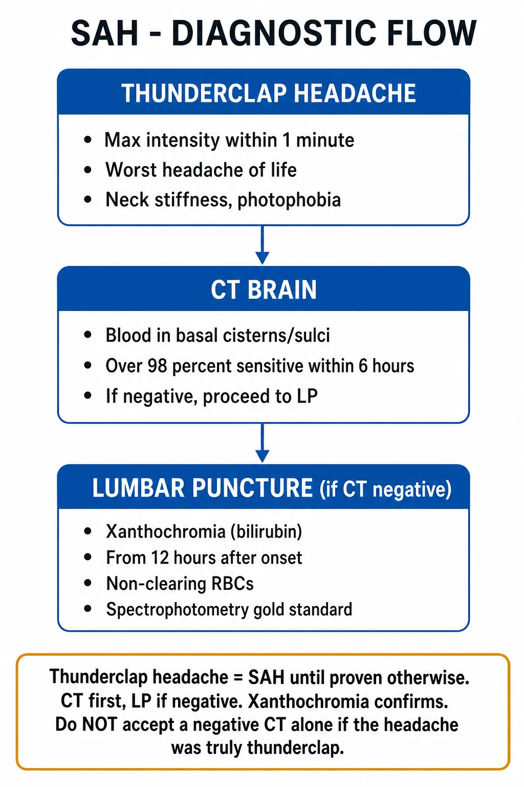
The non-contrast CT brain is the first and most important investigation. Performed within 6 hours of onset on a modern third-generation scanner, it has a sensitivity of about 100 percent (95 percent confidence interval 97 to 100) for SAH — supporting a CT-first approach without mandatory LP when the scan is performed that early.[4]
The blood of SAH lies in the basal cisterns, the Sylvian fissure, the interhemispheric fissure, and the superficial cortical sulci — appearing as hyperdense material. The CT also shows intracerebral or intraventricular extension, hydrocephalus, mass effect, and often suggests the aneurysm's location from the pattern of blood. Because CT sensitivity falls steeply with time (about 85 percent at 24 hours, 50 percent at one week, 30 percent at two weeks as blood is reabsorbed), a negative CT performed more than 6 hours after onset does not exclude SAH — the next step is a lumbar puncture.[1][4]
Lumbar puncture — xanthochromia
The lumbar puncture (LP) is performed if the CT is negative but suspicion persists, and ideally at least 6–12 hours after onset (preferably 12 hours) so that red-cell lysis has had time to generate bilirubin. The opening pressure is raised. The decisive finding is xanthochromia — a yellow tinge to the CSF supernatant produced by bilirubin, a breakdown product of haemoglobin released by red-cell lysis and not present in a traumatic tap (where the supernatant remains colourless). Xanthochromia appears from about 12 hours, peaks at 48 hours, and persists for about two weeks. Bilirubin is best confirmed by spectrophotometry. Comparing the red-cell count in the first and third tubes helps distinguish a true SAH (similar counts) from a traumatic tap (a falling count).[1][4]
Why does xanthochromia take hours to appear — and why is a 'blood-stained' CSF not enough?
Because bilirubin is generated by red-cell lysis in vivo, which takes several hours after the bleed. A blood-stained CSF sampled immediately could be a traumatic tap (the needle hitting a vessel). The yellow supernatant (bilirubin) — confirmed by spectrophotometry, and stable from 12 hours to about 2 weeks — is the signature of true SAH.[1]
Vessel imaging — CTA and DSA
Once SAH is confirmed, CT angiography (CTA) is the next step: it identifies the aneurysm, its size, neck, and location, and guides the choice between coiling and clipping, with a sensitivity of 95–98 percent for aneurysms over 3 mm. Digital subtraction angiography (DSA) remains the gold standard (about 99 percent sensitivity) and is reserved for cases where the CTA is negative or inconclusive but the blood pattern suggests an aneurysm, and for planning and performing endovascular treatment. A perimesencephalic non-aneurysmal SAH is confirmed by a negative DSA in the characteristic CT pattern (blood around the midbrain).[1][6]
Severity scales reproduced verbatim
The named scales are reproduced exactly as examiners expect them. The Hunt and Hess grade (1968) and the WFNS grade (1988) grade clinical severity and predict outcome; the Fisher scale grades the CT blood burden and predicts vasospasm.[6][10]
Hunt and Hess grade (I–V): [1]
| Grade | Description |
|---|---|
| I | Asymptomatic, or mild headache, slight nuchal rigidity |
| II | Moderate to severe headache, nuchal rigidity, no neurological deficit other than cranial-nerve palsy |
| III | Drowsy or confused, mild focal deficit |
| IV | Stupor, moderate to severe hemiparesis, early decerebrate rigidity, vegetative disturbance |
| V | Deep coma, decerebrate rigidity, moribund |
WFNS grade (based on GCS and motor deficit): [1]
| Grade | GCS | Motor deficit |
|---|---|---|
| I | 15 | Absent |
| II | 13–14 | Absent |
| III | 13–14 | Present |
| IV | 7–12 | Present or absent |
| V | 3–6 | Present or absent |
Fisher scale (CT blood burden): [1]
| Grade | CT finding |
|---|---|
| I | No blood detected |
| II | Diffuse or thin (under 1 mm) layer of subarachnoid blood; no clots |
| III | Localised clot and/or thick (over 1 mm) vertical layer of blood |
| IV | Intracerebral or intraventricular clot with diffuse or no SAH |
Bloods and surveillance
Blood tests include a full blood count, urea and electrolytes (watch for hyponatraemia), coagulation, glucose, liver function, and group-and-save or crossmatch. Troponin and an ECG detect neurogenic stunned myocardium (a troponin rise with QT prolongation and the 'cerebral T waves' of diffuse T-wave inversion). Transcranial Doppler (TCD) surveillance begins around day 3: a rising middle cerebral artery velocity (over 120 cm/s, or a Lindegaard ratio above 3 to distinguish vasospasm from hyperaemia) heralds vasospasm. EEG is reserved for suspected seizures or unexplained depressed consciousness.[2][11]
Management — Resuscitation
Resuscitation follows ABCDE, with two parallel time-critical objectives from the moment SAH is suspected: prevent rebleeding (the killer in the first 24 hours) and prevent delayed cerebral ischaemia (the killer at days 4–14). Protect the airway in comatose patients — intubate and ventilate if the GCS is 8 or lower — give oxygen to keep the saturation at or above 94 percent, and avoid both hypotension and hypoxia, which worsen secondary brain injury.[6][11]
Resuscitation bundle in the first hour
ABCDE + airway
Protect airway; intubate if GCS 8 or lower. Oxygen to SpO2 at least 94%. Avoid hypotension and hypoxia.
Control blood pressure
Before the aneurysm is secured, keep systolic BP below 160 mmHg with a titratable IV agent (labetalol 10–20 mg IV boluses or nicardipine infusion 5–15 mg/h). Avoid hypotension — it worsens cerebral perfusion in a brain at risk of vasospasm.
Analgesia, antiemesis, calm
Paracetamol ± small opioid doses; ondansetron. Keep the patient calm — coughing and straining spike BP and rebleed risk. Avoid over-sedation that obscures the neuro exam.
Seizures
IV levetiracetam 60 mg/kg (max 4.5 g) load then 1 g twice daily, or phenytoin 20 mg/kg load. Prophylaxis is selective (large cortical blood, ICH extension), not routine.
Reverse anticoagulation
Warfarin — vitamin K + prothrombin complex concentrate; dabigatran — idarucizumab; anti-Xa DOACs — andexanet alfa; antiplatelets — discuss platelet transfusion with neurosurgery.
Raised ICP / hydrocephalus
Head of bed 30 degrees, mild sedation, normocapnia; mannitol 0.5 g/kg or hypertonic saline; external ventricular drain (EVD) for acute obstructive hydrocephalus or deteriorating consciousness with ventriculomegaly.
Transfer to a neurosciences centre
Refer early to a high-volume centre — centralisation is associated with better outcomes.
Blood pressure control before the aneurysm is secured aims to lower the rebleed risk without sacrificing cerebral perfusion. A common target is a systolic blood pressure below 160 mmHg (or a mean arterial pressure below 110), achieved with a titratable agent; after the aneurysm is secured, the blood-pressure target is liberalised to maintain perfusion, and may be raised deliberately if vasospasm develops.[6][7]
Management — Definitive & Stepwise
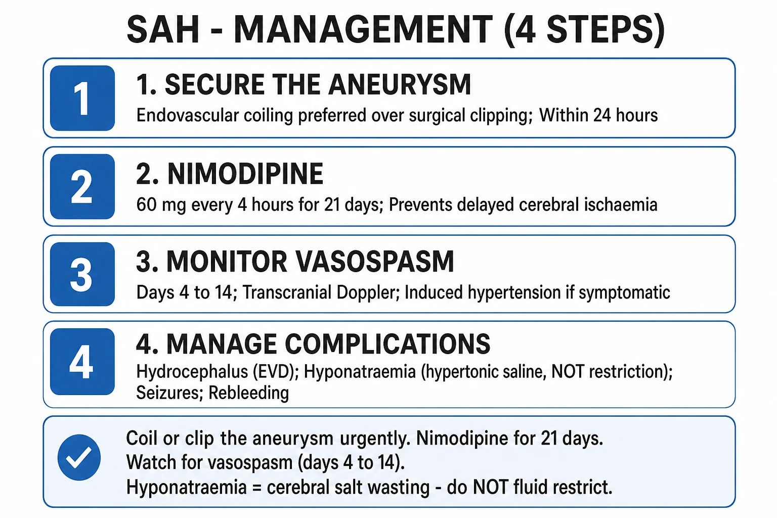
Definitive management has four pillars: secure the aneurysm (prevent rebleeding); prevent delayed cerebral ischaemia with nimodipine; manage hydrocephalus and raised ICP; and prevent and treat systemic complications.[6][7]
Pillar 1 — secure the aneurysm
The aneurysm is obliterated ideally within 24 hours by either endovascular coiling (detachable platinum coils delivered by catheter angiography, packing the aneurysm from the circulation) or surgical clipping (a craniotomy with a clip placed across the aneurysm neck). The landmark International Subarachnoid Aneurysm Trial (ISAT) established coiling as the preferred treatment for aneurysms suitable for either approach.[3][6]
ISAT — International Subarachnoid Aneurysm Trial
Lancet 2002 (Molyneux et al.)
Multicentre RCT of 2143 patients with a ruptured intracranial aneurysm suitable for both procedures, randomised to endovascular coiling or neurosurgical clipping.
Key finding
Death or dependency (modified Rankin 3–6) at 1 year: 23.7 percent with coiling versus 30.6 percent with clipping (relative risk reduction 22.6 percent, p equals 0.0019).
Practice change
Coiling became the preferred treatment for ruptured aneurysms suitable for either technique; long-term follow-up confirmed durable benefit, though late rebleed risk is slightly higher with coiling.
The choice between coiling and clipping depends on aneurysm anatomy, the patient, and local expertise. MCA bifurcation and wide-necked aneurysms often favour clipping; posterior circulation and elderly patients favour coiling. Flow-diverting stents are an option for complex aneurysms. Until the aneurysm is secured, the patient remains at high risk of rebleeding, which carries about 70 percent mortality — hence the urgency.[3][11]
Endovascular coiling
- Preferred when the aneurysm is amenable (ISAT: lower death/dependency at 1 year)
- Avoids craniotomy; shorter hospital stay; favoured in elderly and posterior circulation
- Slightly higher late rebleed risk — needs surveillance angiography
- Risk: thromboembolic stroke, intra-procedural rupture, coil migration
Surgical clipping
- Preferred for MCA bifurcation, wide-neck, and complex/giant aneurysms
- Allows evacuation of a haematoma and bony decompression if needed
- Lower late rebleed risk than coiling; durable obliteration
- Risk: cerebral ischaemia, infection, seizures, longer recovery
Pillar 2 — nimodipine
Nimodipine, a dihydropyridine calcium-channel blocker, is the only drug proven to improve outcome after SAH. It is given to all patients with aneurysmal SAH regardless of clinical grade, 60 mg orally (or via nasogastric tube) every 4 hours for 21 days, started within 4 days of the bleed.[8][9]
Nimodipine
Dihydropyridine calcium-channel blocker — prevents delayed cerebral ischaemia after SAH
Dose
60 mg PO/NG every 4 hours for 21 days
Across AHA/ASA (2023), NICE, ESO, and Indian neurology guidelines, oral nimodipine 60 mg every 4 hours for 21 days is the universal standard for aneurysmal SAH. The Dorhout Mees Cochrane review (2007) showed oral nimodipine reduced poor outcome with a relative risk of 0.67 and a number-needed-to-treat of about 19. Intravenous nimodipine (1 mg/h, titrating to 2 mg/h) is an alternative where the oral route is unavailable but carries a higher risk of hypotension and requires a central line.[9][7]
Pillar 3 — hydrocephalus and raised ICP
An external ventricular drain (EVD) is inserted for acute obstructive hydrocephalus, for intraventricular blood obstructing CSF flow, or for deteriorating consciousness with ventriculomegaly. A lumbar drain or serial LPs may be used for communicating hydrocephalus, and about 10–20 percent of patients develop chronic symptomatic hydrocephalus requiring a ventriculoperitoneal (VP) shunt. General ICP measures — head elevation to 30 degrees, normocapnia, sedation, and osmotic therapy (mannitol or hypertonic saline) — run in parallel.[6][11]
Pillar 4 — systemic complications
General measures keep the brain out of trouble while it recovers. Deep-vein thrombosis prophylaxis begins with compression stockings, with low-molecular-weight heparin added after the aneurysm is secured (typically more than 24 hours after). Normoglycaemia (avoid hyperglycaemia with an insulin sliding scale if needed), normothermia (paracetamol and cooling for fever), and treatment of anaemia (transfusion threshold generally a haemoglobin of 80–90 g/L) all matter — hyperglycaemia, fever, and anaemia are independently associated with poor outcome. Early enteral nutrition and stress-ulcer prophylaxis complete the bundle.[2][11]
Treating vasospasm when it occurs
When vasospasm or delayed cerebral ischaemia develops at days 4–14, the goal is to restore cerebral perfusion. Modern euvolaemic hypertensive therapy maintains euvolaemia and induces hypertension (raising the systolic blood pressure to 160–200 mmHg with vasopressors such as noradrenaline), but only after the aneurysm is secured — raising the blood pressure against an unsecured aneurysm invites rebleeding. This approach has replaced the older 'triple-H' therapy (hypervolaemia, haemodilution, hypertension), which caused pulmonary oedema, hyponatraemia, and haemorrhage. If refractory, intra-arterial vasodilators (milrinone, verapamil) or balloon angioplasty of the spastic vessel are options.[2][7]
Hunt and Hess severity — mortality climbs with grade
Drowsy / mild deficit
Drowsy or confused, mild focal deficit
Specific Subtypes & Scenarios
Aneurysmal SAH
- 85 percent of spontaneous SAH — full management pathway applies
- Urgent securing (coiling/clipping) + nimodipine + vasospasm surveillance
Perimesencephalic non-aneurysmal SAH
- ~10 percent; blood around the midbrain, no aneurysm on DSA
- Benign course — vasospasm rare, prognosis excellent; nimodipine often still given
AVM rupture
- Younger patients; sometimes prior seizures or a bruit
- Managed by surgical resection, embolisation, or stereotactic radiosurgery (Spetzler–Martin grade)
Traumatic SAH
- Cortical contusional blood over convexity sulci (not basal cisterns)
- Managed as traumatic brain injury — no nimodipine unless a co-existing aneurysm
Mycotic aneurysm
- Septic embolisation (endocarditis) — distal MCA, often multiple
- Prolonged antibiotics ± endovascular or surgical intervention depending on rupture
High-grade SAH (WFNS IV–V)
- Historically considered unsalvageable — but aggressive early care yields functional independence in a substantial minority of survivors
- Warrants aggressive initial management before any prognostic decision
The perimesencephalic non-aneurysmal SAH (PNSAH) is the subtype examiners love because it is the exception to the rule that SAH is dangerous. The blood is confined to the perimesencephalic and prepontine cisterns around the midbrain, no aneurysm is found on DSA, the patient is usually clinically well, and the outcome is excellent with vasospasm rare. Nimodipine is often still given for caution. At the other extreme, high-grade SAH (WFNS IV–V) was once treated palliatively, but modern series show that aggressive early management secures functional independence in a meaningful proportion of survivors — a finding that justifies maximal initial therapy before any limitation-of-treatment decision.[2][10]
Complications & Pitfalls
The complications of SAH follow a predictable timeline, and recognising them is the core of neurocritical care. [1]
Complications timeline after SAH
Rebleeding is the dominant early killer — about 3–4 percent of patients re-rupture in the first 24 hours, and rebleeding carries roughly 70 percent mortality. Prevented by early aneurysm securing and blood-pressure control.
Raised ICP, acute hydrocephalus (EVD), global cerebral oedema, and the catecholamine-driven neurogenic stunned myocardium (troponin rise, QT prolongation, 'cerebral T waves', reversible Takotsubo-like cardiomyopathy).
The leading cause of preventable death and disability — symptomatic in about 30 percent. New focal deficit, confusion, or a fall in conscious level. Prevented by nimodipine; treated by euvolaemia and induced hypertension (after the aneurysm is secured).
Hyponatraemia in 30–40 percent, usually cerebral salt wasting (hypovolaemic) — high urine sodium and output, volume depletion. Occasionally SIADH. Treat CSW with hypertonic saline ± fludrocortisone; do not fluid restrict.
Seizures (10–20 percent) from cortical blood; ventriculitis from the EVD; aspiration and ventilator-associated pneumonia; DVT/PE in the immobilised patient.
Communicating hydrocephalus (sometimes a normal-pressure-hydrocephalus picture) needing a VP shunt; cognitive impairment (attention, executive, memory), fatigue, and depression/anxiety in up to 30–40 percent of survivors.
The hyponatraemia question is the classic exam trap. After SAH, low sodium is most often cerebral salt wasting — a volume-depleted state driven by natriuretic peptide release — rather than SIADH, which is volume-replete or expanded. The two are distinguished by volume status, and the treatment diverges accordingly: fluid restriction (the correct treatment for SIADH) is dangerous in cerebral salt wasting because it worsens hypovolaemia, provokes vasospasm, and threatens cerebral ischaemia.[2][11]
Cerebral salt wasting
commonest after SAH
- Hypovolaemic hyponatraemia — high urine sodium and output, volume depletion
- Driven by brain natriuretic peptide release
- Treat with HYPERTONIC SALINE (2–3%) ± fludrocortisone 0.1–0.2 mg PO + volume replacement
- NEVER fluid restrict — worsens vasospasm and cerebral ischaemia
SIADH
less common after SAH
- Euvolaemic or hypervolaemic hyponatraemia — concentrated urine, low serum osmolarity
- Inappropriate ADH; volume-replete
- Treat with FLUID RESTRICTION, salt tablets ± vasopressin antagonists (tolvaptan)
- Restriction is the trap if you mislabel CSW as SIADH
The AHA/ASA 2023 guideline (Hoh) emphasises avoiding hypovolaemia at all stages of SAH and treating cerebral salt-wasting with sodium repletion rather than fluid restriction, consistent with NICE, ESO, and ANZ neurocritical-care practice.[7]
The classic errors that cost lives are: dismissing a thunderclap headache as migraine; not doing an LP after a negative CT performed more than 6 hours after onset; fluid restricting hyponatraemia in cerebral salt wasting; failing to give nimodipine for the full 21 days; and attributing early deterioration to 'expected' decline rather than to rebleeding, hydrocephalus, or vasospasm — each of which mandates an urgent CT.[1][11]
Prognosis & Disposition
About 10–15 percent of patients die before reaching hospital, and the overall 30-day mortality is around 30 percent; a further 10–20 percent of survivors remain functionally dependent. Outcomes have improved over decades with better diagnosis, early aneurysm repair, nimodipine, and centralised neurocritical care.[1][2]
Predictors of poor outcome are a high clinical grade (Hunt and Hess or WFNS IV–V), older age, a large aneurysm (over 10 mm), a posterior-circulation location, rebleeding, intraventricular blood or hydrocephalus, a thick subarachnoid clot (high Fisher or modified Fisher), symptomatic vasospasm, hyperglycaemia, fever, anaemia, and hypotension. Predictors of good outcome are a low WFNS grade (I–II), a small aneurysm, an anterior-circulation location, early aneurysm securing, no vasospasm, younger age, and management in a high-volume centre.[6][10]
Perimesencephalic non-aneurysmal SAH is the exception — vasospasm is rare and the prognosis is excellent. For aneurysmal SAH, vasospasm/delayed cerebral ischaemia is the leading cause of preventable death and disability, which is why nimodipine prophylaxis and vigilant surveillance through days 4–14 are non-negotiable. Even patients judged to have a 'good outcome' often have subtle cognitive deficits (attention, executive function, processing speed), fatigue, and depression — only about 30–50 percent return to their previous level of work without restriction.[2]
Disposition is a neurosciences intensive-care unit throughout days 0–14 (the vasospasm window), then a step-down ward and rehabilitation, with outpatient follow-up for cognition and mood and surveillance angiography for coiled aneurysms (which carry a slightly higher late rebleed risk and may recur).[3][7]
Special Populations
Pregnancy and the puerperium — SAH is a leading non-obstetric cause of maternal death. A thunderclap headache in pregnancy is SAH (alongside pre-eclampsia, HELLP, and cerebral venous sinus thrombosis) until excluded. Management is multidisciplinary (obstetrics, neurosurgery, anaesthesia); coiling is preferred where feasible (avoids craniotomy), nimodipine is given (the maternal risk of vasospasm outweighs theoretical teratogenicity concerns), and excessive blood-pressure drops are avoided.[2]
Elderly patients present atypically (confusion, a fall, less prominent headache) and have higher complication rates; coiling is preferred when feasible and outcomes are worse, but age alone does not justify withholding aggressive therapy. [1]
Anticoagulated patients have a higher rebleeding risk and worse outcome; anticoagulation is reversed urgently as part of resuscitation (prothrombin complex concentrate plus vitamin K for warfarin; idarucizumab for dabigatran; andexanet alfa for anti-Xa DOACs). [1]
ADPKD patients — 5–10 percent harbour an intracranial aneurysm; all are screened with MRA, and a family history of two or more first-degree relatives with SAH is also a screening indication. Marfan syndrome, the vascular type of Ehlers-Danlos (type IV), neurofibromatosis type 1, and fibromuscular dysplasia are further associations.[1][7]
Incidental unruptured aneurysms are managed by size, site, and patient factors: small (under 7 mm) anterior-circulation aneurysms in patients without prior SAH have a low annual rupture risk and may be observed, whereas larger, posterior-circulation, symptomatic, or growing aneurysms warrant repair.[6]
Evidence, Guidelines & Regional Differences
The evidence base for SAH is anchored by three landmark contributions. The ISAT trial (Molyneux 2002) made coiling the preferred treatment for aneurysms suitable for both techniques. The nimodipine evidence begins with Allen (1983) and is consolidated by the Dorhout Mees Cochrane review (2007), which confirmed that oral nimodipine reduces poor outcome with a relative risk of 0.67 and a number-needed-to-treat of about 19 — making nimodipine the only drug proven to improve outcome after SAH.[3][8][9]
Nimodipine for SAH — Allen 1983 (NEJM)
N Engl J Med 1983 (Allen et al.)
Multicentre prospective double-blind RCT of 125 neurologically normal patients within 96 h of SAH, randomised to nimodipine or placebo for 21 days.
Key finding
Severe deficit from cerebral arterial spasm: 1 of 56 nimodipine vs 8 of 60 placebo (p equals 0.03).
Practice change
Established nimodipine 60 mg every 4 h for 21 days as standard prophylaxis against delayed cerebral ischaemia — confirmed by the Dorhout Mees Cochrane review (2007).
The diagnostic evidence comes from Perry (2011, BMJ), which showed that modern CT performed within 6 hours of headache onset has about 100 percent sensitivity for SAH, and from Perry (2017, CMAJ), which validated the Ottawa SAH Rule (sensitivity about 100 percent) for ruling out SAH in alert, neurologically intact patients.[4][5]
The guidelines are the AHA/ASA Connolly 2012 guideline, now superseded by the AHA/ASA Hoh 2023 guideline — the current comprehensive framework endorsing early aneurysm repair, oral nimodipine, euvolaemic hypertensive therapy for delayed cerebral ischaemia, and centralisation to high-volume centres.[6][7]
AHA/ASA (2023, Hoh), NICE (UK), ESO (Europe), and Indian Academy of Neurology guidance converge on the same pillars: CT-first diagnosis with LP for xanthochromia if CT is non-diagnostic; coiling preferred over clipping where feasible (ISAT); oral nimodipine 60 mg every 4 h for 21 days; euvolaemic hypertensive therapy for DCI; and avoidance of fluid restriction in cerebral salt wasting. Regional variation is mainly in access to neurosurgical and endovascular services.[7]
Negative trials are equally important because examiners test them. MASH-II (Dorhout Mees 2012) showed that magnesium sulphate did not improve outcome after SAH — magnesium is therefore not recommended. STASH (Kirkman 2014) showed that simvastatin did not improve outcome. Both are reminders that, despite decades of trials, nimodipine remains the only pharmacotherapy proven to help.[9]
Exam Pearls
SAH pearls — the facts that decide an answer
SAH
Thunderclap headache — max within 1 minute — is SAH until proven otherwise; CT first, LP if negative
Ruptured saccular aneurysm in 85 percent; ACom commonest site (30 percent)
Usually cerebral salt wasting, NOT SIADH — do NOT fluid restrict; use hypertonic saline ± fludrocortisone
Diagnosis
- CT brain first — about 100 percent sensitive within 6 h (Perry 2011)
- If CT negative or over 6 h old — LP for xanthochromia (bilirubin, from 12 h)
- CTA localises the aneurysm; DSA is the gold standard
- Ottawa SAH Rule (sensitivity ~100%) — rule-OUT decision aid
Management
- Secure the aneurysm within 24 h — coiling preferred over clipping (ISAT)
- Nimodipine 60 mg every 4 h for 21 days — only drug proven to improve outcome
- Vasospasm (days 4–14): euvolaemia + induced hypertension after aneurysm secured
- Rebleeding (first 24 h): BP control (SBP under 160) before securing
Exam application bank (NEET-PG / INICET)
One-line answer
Subarachnoid haemorrhage (SAH) is bleeding into the subarachnoid space, usually from a ruptured intracranial aneurysm (85 percent), presenting with a sudden, severe 'thunderclap' headache (maximum intensity within 1 minute) — often described as 'the worst headache of my life' — with neck stiffness, photophobia, nausea, vomiting, altered consciousness and sometimes seizures. Non-contrast CT brain (sensitivity about 100 percent within 6 hours on a modern scanner) is the first investigation; if the CT is negative or performed later than 6 hours, lumbar puncture for xanthochromia (yellow CSF supernatant from bilirubin) confirms the diagnosis. Management includes securing the aneurysm (endovascular coiling preferred over surgical clipping per ISAT), nimodipine 60 mg orally every 4 hours for 21 days (prevents delayed cerebral ischaemia from vasospasm), blood pressure control, and management of [1]
Worked stems (answer without another resource)
Stem 1 — Classic presentation. Map symptoms to mechanism; name the first investigation and first treatment step with dose/route if drug therapy is standard. [1]
Stem 2 — Unstable / complicated. List red flags that force immediate resuscitation, theatre, ICU, antidote, or reperfusion — and what you do in the first 15 minutes. [1]
Stem 3 — Atypical group. Elderly, pregnancy, child, or immunocompromised: how presentation and thresholds change. [1]
Stem 4 — Differential trap. Name the three closest mimics and one discriminator for each. [1]
Stem 5 — Disposition. Who goes home with safety-netting, who is admitted, who needs HDU/ICU/theatre, and what follow-up is mandatory. [1]
Rapid viva checklist
- Definition + classification
- Pathophysiology chain
- Bedside signs / criteria
- Score with exact components (if any)
- Emergency bundle
- Definitive therapy with doses
- Complications of disease and of treatment
- Special populations
- Guideline/trial name if classic
- Three exam traps
Coverage self-check
If you cannot answer any stem above from this page alone, re-read the matching section — the page is intended to be self-sufficient for final-prof and NEET-PG/INICET questions on Subarachnoid Haemorrhage.
References
- [1]Macdonald RL, Schweizer TA. Spontaneous subarachnoid haemorrhage Lancet, 2017.PMID 27637674
- [2]Claassen J, Park S. Spontaneous subarachnoid haemorrhage Lancet, 2022.PMID 35985353
- [3]Molyneux A, Kerr R, Stratton I, et al. (ISAT Collaborative Group). International Subarachnoid Aneurysm Trial (ISAT) of neurosurgical clipping versus endovascular coiling in 2143 patients with ruptured intracranial aneurysms: a randomised trial Lancet, 2002.PMID 12414200
- [4]Perry JJ, Stiell IG, Sivilotti ML, et al. Sensitivity of computed tomography performed within six hours of onset of headache for diagnosis of subarachnoid haemorrhage: prospective cohort study BMJ, 2011.PMID 21768192
- [5]Perry JJ, Sivilotti MLA, Sutherland J, et al. Validation of the Ottawa Subarachnoid Hemorrhage Rule in patients with acute headache CMAJ, 2017.PMID 29133539
- [6]Connolly ES Jr, Rabinstein AA, Carhuapoma JR, et al. Guidelines for the management of aneurysmal subarachnoid hemorrhage: a guideline for healthcare professionals from the American Heart Association/american Stroke Association Stroke, 2012.PMID 22556195
- [7]Hoh BL, Ko NU, Amin-Hanjani S, et al. 2023 Guideline for the Management of Patients With Aneurysmal Subarachnoid Hemorrhage: A Guideline From the American Heart Association/American Stroke Association Stroke, 2023.PMID 37212182
- [8]Allen GS, Ahn HS, Preziosi TJ, et al. Cerebral arterial spasm--a controlled trial of nimodipine in patients with subarachnoid hemorrhage N Engl J Med, 1983.PMID 6338383
- [9]Dorhout Mees SM, Rinkel GJ, Feigin VL, et al. Calcium antagonists for aneurysmal subarachnoid haemorrhage Cochrane Database Syst Rev, 2007.PMID 17636626
- [10]Ironside N, Buell TJ, Chen CJ, et al. High-Grade Aneurysmal Subarachnoid Hemorrhage: Predictors of Functional Outcome World Neurosurg, 2019.PMID 30735864
- [11]Busl KM, Bogossian EG, Claassen J, et al. Beyond the bleed: complications after aneurysmal subarachnoid hemorrhage. Pathophysiology, clinical implications, and management strategies: a review Crit Care, 2025.PMID 41029753