Neurology · General Medicine
Transient Ischaemic Attack (TIA)
Also known as Transient ischaemic attack · TIA · Mini-stroke
Transient ischaemic attack (TIA) is a transient episode of neurological dysfunction caused by focal brain, spinal cord or retinal ischaemia without acute infarction (tissue-based definition). It is a medical emergency, not a benign event — the risk of completed stroke is 5 to 10 percent within 48 hours and 10 to 20 percent within 90 days without treatment. The ABCD2 score (Age, Blood pressure, Clinical features, Duration, Diabetes) stratifies early stroke risk and drives triage urgency. Presentation is a sudden focal neurological deficit that resolves within 24 hours (usually under 1 hour) — unilateral weakness, aphasia, or monocular visual loss (amaurosis fugax). Urgent workup includes CT then MRI-DWI brain, carotid Doppler or CT angiography, ECG (atrial fibrillation) and echocardiography. Management is aggressive secondary prevention: dual antiplatelet therapy (aspirin plus clopidogrel for 21 days, then monotherapy), high-intensity statin, blood-pressure control, anticoagulation for atrial fibrillation, and carotid endarterectomy for symptomatic stenosis over 50 percent (especially over 70 percent) within 2 weeks — together preventing up to 80 percent of predicted strokes.
On this page & tools
Your progress
Saved locally on this device.
Practise this topic
Exam tags
Red flags
Overview & Definition
A TIA is best understood as a near-miss stroke — a transient episode of focal brain, spinal-cord or retinal ischaemia that resolves before tissue infarction completes, but which predicts an imminent completed stroke. The single most important concept an examiner tests is that TIA is a medical emergency: the risk of stroke is highest in the first 24 to 48 hours, which is why modern guidelines mandate urgent (within 24 hours) specialist assessment and imaging rather than routine outpatient referral.[1][2]
The modern tissue-based definition (endorsed by the American Heart Association/American Stroke Association in 2009) is: "a transient episode of neurological dysfunction caused by focal brain, spinal cord, or retinal ischaemia without acute infarction." This deliberately replaces the older time-based definition (symptoms resolving within 24 hours) because symptom duration is a poor discriminator of tissue fate — up to 30 percent of clinically diagnosed TIAs demonstrate infarction on diffusion-weighted MRI and are therefore reclassified as ischaemic stroke.[1] The practical consequence is that the boundary between TIA and minor stroke is set by imaging, not the clock: a five-minute deficit with infarction is a stroke, whereas a six-hour deficit without infarction is a TIA.
The clinical discipline in managing a TIA is to treat every suspected event as the warning it is: establish the onset time, exclude mimics and haemorrhage, identify the mechanism (large-vessel atherosclerosis, cardioembolism, small-vessel lacunar disease, or another determined cause), and implement mechanism-directed secondary prevention before the stroke arrives. The phrase "time is brain" applies as much to TIA as to acute stroke, because the highest-risk period for recurrence is the first 48 hours.[1]
Definition & Classification
Time-based versus tissue-based definitions
Old (time-based)
pre-2009 convention
- Sudden focal neurological deficit resolving **completely within 24 hours**
- Arbitrary time cut-off — purely clinical, no imaging
- Risked dismissing patients with MRI infarction as 'benign'
- **Superseded** by the tissue-based definition
Modern (tissue-based)
current standard (AHA/ASA 2009)
- Transient focal dysfunction from brain, spinal-cord or retinal ischaemia **without acute infarction**
- Discriminated by **MRI diffusion-weighted imaging (DWI)**, not the clock
- Typical duration **under 1 hour** (most true TIAs)
- About 30 percent of clinically diagnosed TIAs show DWI infarction — reclassified as **stroke**
The shift matters because the time-based definition underestimated risk. A patient whose symptoms lasted 20 minutes could still harbour a small infarct and carry the recurrence risk of a stroke, not the supposedly benign prognosis of a TIA. The tissue definition forces a structural answer (is there infarction?) and ensures such patients are not falsely reassured.[1]
Classification by vascular mechanism (TOAST)
The TOAST classification (Trial of Org 10172 in Acute Stroke Treatment) categorises cerebral ischaemia by aetiology, and the same framework drives TIA workup because the mechanism determines secondary prevention. The five categories are reproduced below. [1]
| TOAST category | Typical source in TIA | Hallmark and management |
|---|---|---|
| Large-artery atherosclerosis | Carotid bifurcation plaque (artery-to-artery embolism) | Carotid endarterectomy if symptomatic stenosis over 50 percent (especially over 70 percent); aggressive risk-factor control |
| Cardioembolism | Atrial fibrillation, prosthetic valve, mural thrombus, recent MI | Anticoagulation (DOAC preferred), NOT antiplatelets |
| Small-vessel occlusion (lacunar) | Lipohyalinosis of penetrating arteries (hypertension, diabetes) | Pure motor / pure sensory / ataxic hemiparesis syndrome; risk-factor control |
| Other determined cause | Arterial dissection, vasculitis, hypercoagulable state, substance use | Commonest in the young; targeted workup and specific therapy |
| Undetermined (cryptogenic) | No identified source despite workup, or two competing causes | Prolonged cardiac monitoring (paroxysmal AF); empiric best medical therapy |
The commonest mechanisms overall are large-artery atherosclerosis and cardioembolism; the mechanisms carrying the highest early recurrence are severe carotid stenosis and atrial fibrillation — precisely the targets the workup is designed to capture.[1]
Epidemiology & Risk Factors
TIA is common and frequently under-recognised. Population-based studies (notably the Oxford Vascular Study, OXVASC) report a community incidence of roughly 50 per 100,000 per year, rising steeply with age. The cardinal epidemiological fact — and the reason TIA is treated as a vascular emergency — is the short-term stroke risk:[2]
TIA — the numbers an examiner wants
Modifiable risk factors are the standard atherosclerotic and thromboembolic set, and they are also the targets of secondary prevention: [1]
- Hypertension — the single biggest modifiable risk factor and the most important to control long term.
- Atrial fibrillation — the most important treatable cause; untreated AF carries an annual stroke risk of roughly 4 to 5 percent, rising steeply with additional CHA2DS2-VASc factors.
- Diabetes mellitus — both a risk factor and, via hypoglycaemia, a dangerous mimic.
- Dyslipidaemia, smoking, obesity, sedentary lifestyle, excessive alcohol.
- Age, male sex, family history, prior TIA or stroke (non-modifiable).
- Carotid stenosis and symptomatic peripheral arterial disease — markers of systemic atherosclerosis. [1]
The factors that predict the highest early recurrence after TIA are atrial fibrillation, severe symptomatic carotid stenosis, crescendo (multiple) events, and diabetes — the same features the ABCD2 score and the aetiological workup are designed to surface.[1]
In the young (under 45 years), the risk profile differs: consider carotid or vertebral artery dissection, patent foramen ovale, thrombophilia, substance use (cocaine, amphetamines), autoimmune vasculitis, and sickle-cell disease. The ABCD2 score underperforms in this group because young patients score low on age yet can still harbour a high-risk mechanism.[2]
Pathophysiology
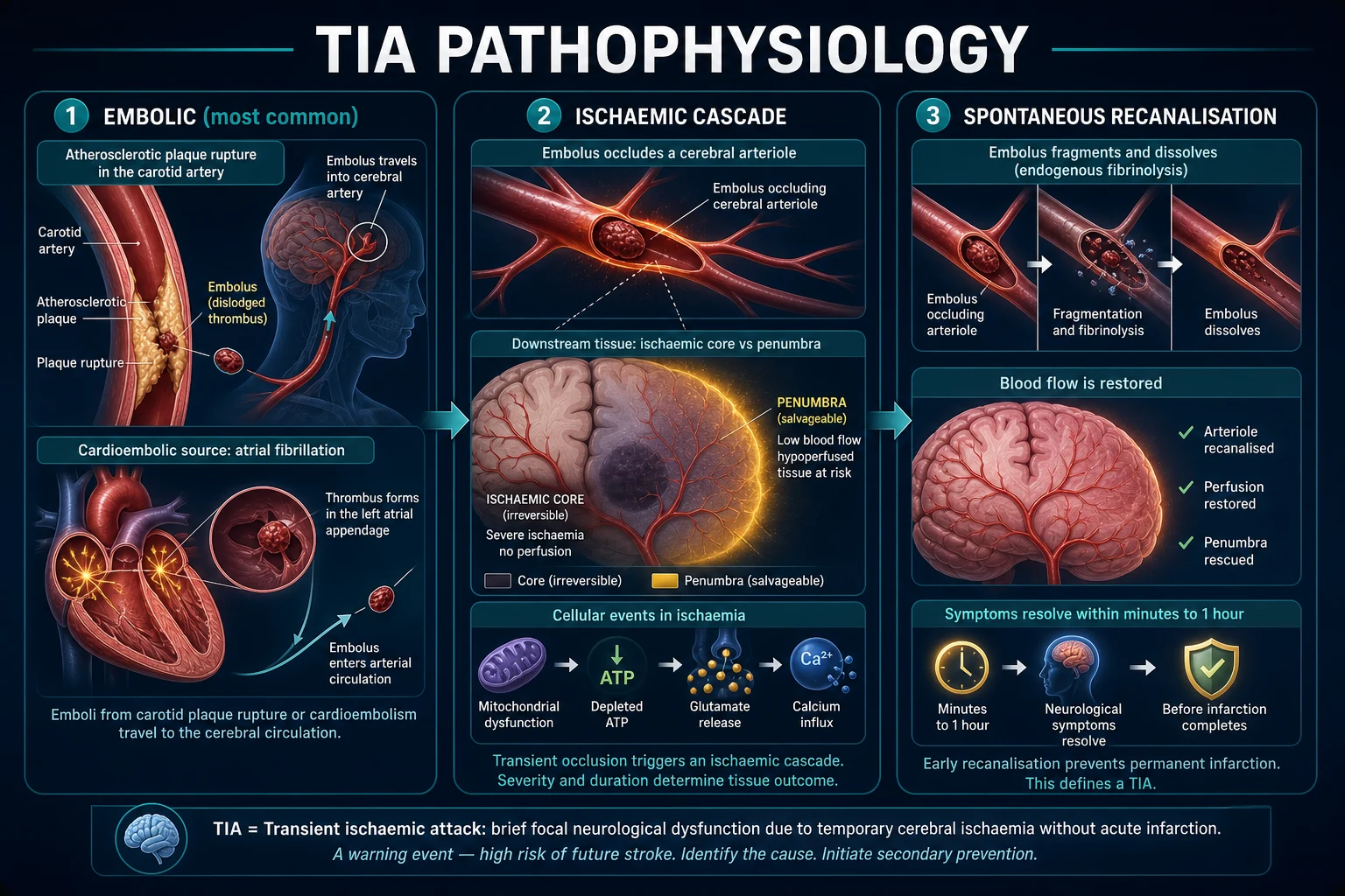
A TIA shares the same ischaemic cascade as a completed stroke. What differs is that cerebral blood flow is restored before the penumbra progresses to irreversible infarction.[1]
The ischaemic cascade
- An embolus (from a ruptured carotid plaque, a left atrial thrombus in atrial fibrillation, or an aortic arch lesion) lodges in a cerebral or retinal artery, or a small penetrating artery transiently occludes (lacunar mechanism).
- Downstream tissue becomes ischaemic: adenosine triphosphate (ATP) depletion causes failure of the sodium-potassium pump and cytotoxic oedema; glutamate release activates NMDA receptors, driving calcium influx and excitotoxic neuronal injury.
- A central core of the most severely ischaemic tissue is surrounded by the penumbra — hypoperfused but still salvageable tissue that is the target of all acute stroke therapy.
- In TIA, the embolus fragments, lyses (endogenous fibrinolysis), or passes distally, and the collateral circulation restores perfusion — symptoms resolve before the penumbra infarcts. [1]
If recanalisation is delayed or incomplete, the penumbra is recruited into the core and the event becomes a completed stroke. Why symptoms resolve is therefore the crux: spontaneous recanalisation plus collateral perfusion rescue the penumbra within minutes to an hour.[1] The physiology is governed by cerebral blood flow (CBF) thresholds. Normal cortical CBF is about 50 mL per 100 g per minute. As flow falls, tissue passes through discrete functional states: at 20 to 30 mL per 100 g per minute neurons become electrically silent but are still viable (the penumbra), and below 10 mL per 100 g per minute irreversible infarction begins within minutes. The duration of ischaemia matters as much as its depth — tissue that would survive an hour of moderate hypoperfusion will infarct within minutes if flow is profoundly reduced. In a TIA, flow is restored while the tissue is still in the penumbra range, which is why the duration of most true TIAs is under one hour. The collateral circulation — chiefly the Circle of Willis, leptomeningeal anastomoses, and ophthalmic collaterals — buys time: a patient with robust collaterals can tolerate a transient carotid occlusion that would infarct immediately in someone with an incomplete circle. This is why two patients with an identical embolus can have very different outcomes.
The three mechanisms in detail
Artery-to-artery embolism
large-vessel
- Atherosclerotic plaque at the **carotid bifurcation** ruptures
- Platelet-fibrin or cholesterol (**Hollenhorst**) emboli travel distally
- Produces anterior-circulation TIA or **amaurosis fugax**
- Managed with **CEA** for severe symptomatic stenosis plus antiplatelet
Cardioembolism
cardiac source
- Thrombus forms in the **left atrial appendage** in atrial fibrillation
- Larger clots → more severe deficits, higher recurrence
- Often produces **cortical** syndromes (aphasia, neglect)
- Managed with **anticoagulation (DOAC)**, not antiplatelets
Small-vessel (lacunar)
penetrating artery
- **Lipohyalinosis** of deep penetrating arteries from hypertension/diabetes
- Produces **lacunar syndromes** (pure motor, pure sensory)
- Good short-term prognosis but high lifetime vascular risk
- Managed with aggressive risk-factor control and antiplatelet
Amaurosis fugax — the retinal equivalent
Amaurosis fugax is the retinal counterpart of a cerebral TIA: a cholesterol (Hollenhorst) plaque or platelet-fibrin embolus from the carotid bifurcation lodges transiently in the central retinal artery, producing sudden, painless monocular visual loss that clears as the embolus moves distally. Fundoscopy may reveal a bright, refractile cholesterol plaque at an arterial bifurcation — a pathognomonic finding pointing to ipsilateral carotid disease.[1]
Clinical Presentation
The cardinal feature of a TIA is a sudden-onset focal neurological deficit that is identical to stroke at onset, reaches its maximum immediately (negative deficit, maximal at onset), and then resolves completely — usually within minutes, almost always within one hour.[2]
Anterior (carotid) circulation
- Unilateral weakness or clumsiness of face, arm and/or leg (middle cerebral artery territory typically affects face and arm more than leg; anterior cerebral artery affects the leg preferentially).
- Speech disturbance — dysphasia or aphasia (dominant hemisphere, usually left), or dysarthria.
- Neglect, visuospatial disturbance (non-dominant, right hemisphere).
- Homonymous hemianopia (optic radiation or occipital involvement).
- Amaurosis fugax — transient, painless monocular visual loss ("a curtain descending"), lasting seconds to minutes (ophthalmic/retinal territory — the only TIA that produces monocular symptoms). [1]
Posterior (vertebrobasilar) circulation
- Diplopia, vertigo, dysarthria, dysphagia (brainstem and cranial nerve involvement).
- Bilateral or alternating limb weakness, crossed sensory loss, and ataxia.
- Bilateral or isolated homonymous hemianopia (occipital cortex).
- Drop attacks (sudden loss of tone without loss of consciousness) and, rarely, transient locked-in states.
- Crossed signs (e.g. ipsilateral cranial nerve palsy with contralateral motor or sensory deficit) are the hallmark of a brainstem lesion. [1]
Lacunar syndromes
Lacunar TIAs produce one of the classic lacunar syndromes from small deep penetrator disease: pure motor hemiparesis (face, arm, leg of equal severity), pure sensory stroke, ataxic hemiparesis, and the dysarthria–clumsy-hand syndrome. They have a good short-term prognosis but a high lifetime vascular risk, because they signal systemic small-vessel disease. [1]
[1]Atypical presentations
Examiners deliberately test atypical scenarios. Older patients may present with confusion or reduced consciousness rather than a clean focal deficit. Diabetic patients may have hypoglycaemia masquerading as a focal deficit — always check finger-prick glucose first. Isolated vertigo can be a posterior-circulation TIA; the HINTS examination (Head Impulse, Nystagmus, Test of Skew) is the bedside discriminator — a central, "bad" pattern (normal head-impulse test, direction-changing nystagmus, skew deviation) points to stroke, not benign vestibular neuritis.[2]
Differential Diagnosis
TIA is a diagnosis of inclusion (a sudden focal ischaemic deficit) that requires exclusion of mimics. The single most important mimic, and the cheapest to reverse, is hypoglycaemia — check a finger-prick glucose in every patient before labelling the event a TIA.[2]
| Mimic | Key distinguishing feature |
|---|---|
| Hypoglycaemia | Low glucose; may produce a focal deficit; resolves with dextrose — check first |
| Migraine aura | Positive, spreading sensory or visual symptoms that march over several minutes; scintillating scotoma; often followed by headache |
| Postictal (Todd) paresis | Preceded by a seizure; slower resolution; often an accompanying confusional state |
| Syncope | Loss of tone or posture, pallor, brief; no persistent focal deficit |
| Functional neurological disorder (FND) | Internally inconsistent, non-anatomical pattern; context of psychological stress; give-away weakness |
| Subdural haematoma | Fluctuating deficit, headache, history of (often minor) head injury; seen on CT |
| Brain tumour | Progressive rather than transient; may produce "stroke-like" episodes; mass on imaging |
| Vestibular vertigo | Isolated vertigo; HINTS — a central pattern (normal head-impulse, direction-changing nystagmus, skew deviation) points to stroke |
The tempo is the chief discriminator: a TIA is sudden, negative, and maximal at onset, whereas a migraine aura is positive and marching, a postictal paresis follows a seizure and resolves more slowly, and a tumour is progressive. Hypoglycaemia and a subdural haematoma can fool the unwary — glucose and CT close both traps.[2]
Clinical & Bedside Assessment
The ABCD2 score
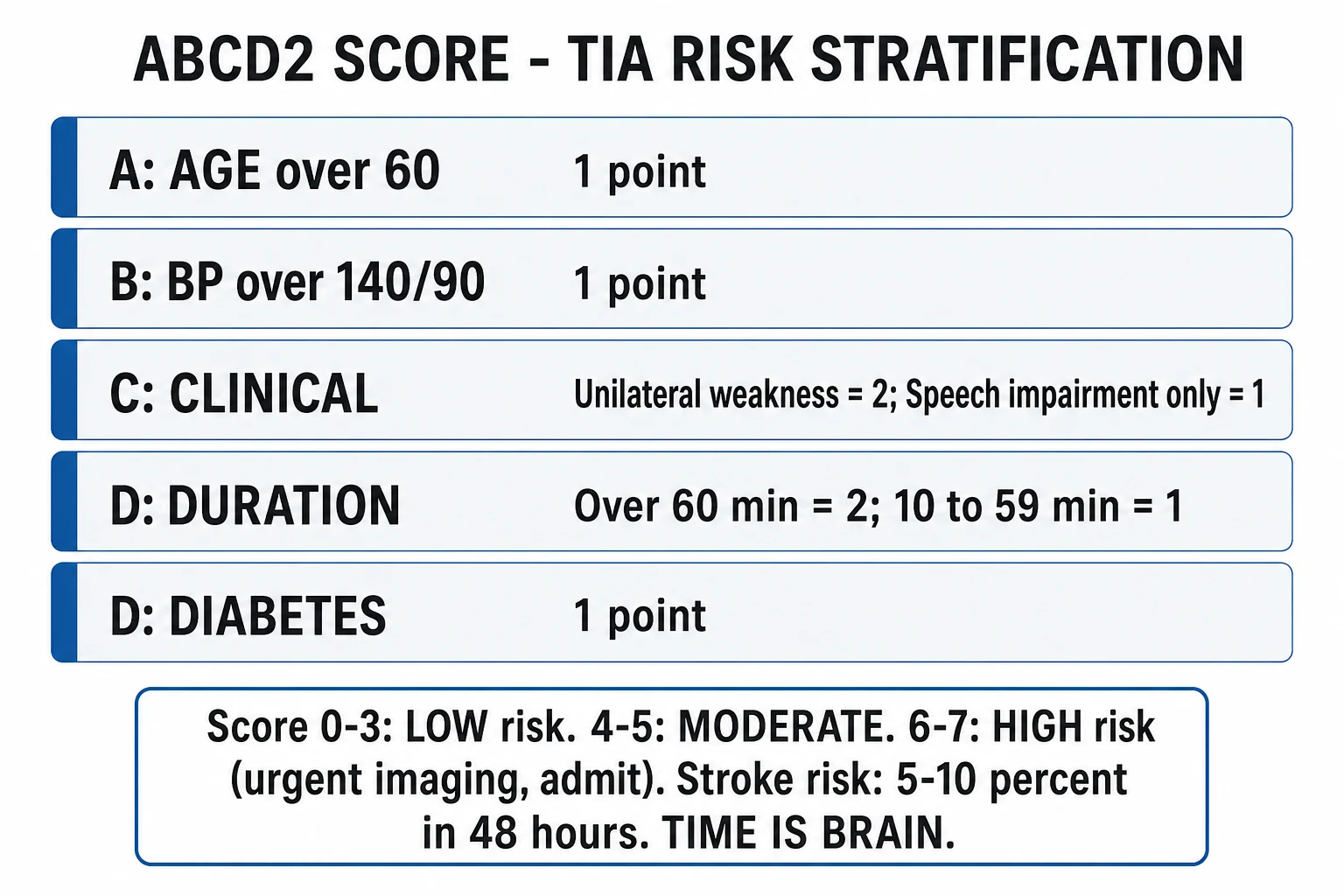
The ABCD2 score stratifies the two-day stroke risk after a suspected TIA and drives triage urgency. It does not diagnose TIA and it does not exclude mimics — it is purely a risk-prediction tool derived and validated by Johnston and colleagues in the Lancet (2007) across four independent cohorts.[3]
| Component | Criterion | Points |
|---|---|---|
| Age | 60 years or older | 1 |
| Blood pressure | 140 mmHg systolic or higher, or 90 mmHg diastolic or higher | 1 |
| Clinical features | unilateral weakness | 2 |
| speech impairment without weakness | 1 | |
| Duration | 60 minutes or longer | 2 |
| 10 to 59 minutes | 1 | |
| Diabetes | present | 1 |
The total ranges from 0 to 7. The original validation reported the following two-day stroke risks: high risk (6 to 7) — 8.1 percent; moderate risk (4 to 5) — 4.1 percent; low risk (0 to 3) — 1.0 percent.[3]
ABCD2 risk bands — 2-day stroke risk
High risk
Highest priority — admit or same-day TIA clinic, urgent imaging, expedite carotid and cardiac workup, and start dual antiplatelet therapy once haemorrhage is excluded.
Limitations of ABCD2
The ABCD2 score has important limits an examiner probes deliberately. It underestimates risk in young patients (who score low on age yet can harbour dissection or thrombophilia), it ignores carotid stenosis and atrial fibrillation (the two highest-risk mechanisms), and it does not capture recurrent events. Refinements such as ABCD3 (adding a point for dual TIA events) and ABCD3-I (adding carotid stenosis and DWI positivity) improve discrimination but are less widely used. The lesson: a low ABCD2 score never reassures when the clinical story or aetiological workup suggests high risk.[1]
Bedside assessment
At the bedside, record the exact onset and last-known-well time (it drives urgency and thrombolysis decisions if the event declares itself as stroke) and the symptom duration in minutes. Perform a full neurological examination (including NIHSS scoring while symptomatic) and a cardiovascular examination: auscultate for a carotid bruit (ipsilateral to anterior-circulation symptoms suggests stenosis), check the pulse for atrial fibrillation, measure blood pressure, and examine the heart for murmurs or failure. Posterior-circulation or large-vessel signs — crossed deficits, decreased consciousness, isolated vertigo — mandate urgent CT angiography.[1]
ABCD2 — early stroke risk after TIA
ABCD2
60 years or older — 1 point
140/90 mmHg or higher — 1 point
unilateral weakness 2; speech only 1
60 min or more 2; 10 to 59 min 1
present — 1 point (total 0 to 7)
Investigations
The workup serves two aims: exclude mimics and haemorrhage, and identify the mechanism that will direct secondary prevention. The minimum dataset is: brain imaging, carotid imaging, ECG and cardiac monitoring, and baseline bloods.[1]
- Brain imaging — CT brain is performed first to exclude haemorrhage and mass lesions; MRI with diffusion-weighted imaging (DWI) is the most sensitive modality and confirms or excludes infarction (the tissue-based discriminator). Infarction on MRI means the event is a stroke, not a TIA — which upstages both prognosis and management.
- Carotid imaging within 24 hours (for any anterior-circulation event) — duplex Doppler ultrasound is first-line to detect and grade stenosis; CT or MR angiography confirms the degree of stenosis (graded by the NASCET method) and maps the arch anatomy to plan surgery.
- ECG — to detect atrial fibrillation, the commonest cardioembolic source. If the initial ECG is normal, consider prolonged monitoring (24-hour Holter, external loop recorder, or an implantable loop recorder) to detect paroxysmal atrial fibrillation, which is the commonest finding in cryptogenic TIA.
- Echocardiography — transthoracic echo if a cardioembolic source is suspected; transoesophageal echo (TOE) for suspected valvular disease, left atrial thrombus, or a patent foramen ovale (especially in the young).
- Bloods — glucose (and HbA1c), fasting lipids, full blood count, urea and electrolytes, and coagulation. In the young, add a thrombophilia screen, toxicology, autoimmune/vasculitic markers, and dedicated dissection imaging (fat-suppressed MRI of the neck vessels or CTA). [1]
The sensitivity and specificity of the modalities matter: MRI-DWI is markedly more sensitive than CT for small posterior-circulation and lacunar infarcts; carotid Doppler is operator-dependent but excellent for bifurcation disease; CTA is rapid and ideal for acute decision-making. Each test is chosen for the question it answers.[1]
Management — Urgency and Resuscitation
A suspected TIA is a medical emergency. The standard of care is specialist (neurology or stroke physician) assessment and brain imaging within 24 hours via a dedicated TIA service or the emergency department.[2]
Who to admit
Admit patients with any of the following: an ABCD2 score of 4 or higher, crescendo or recurrent events, diagnostic uncertainty, those unable to comply with outpatient care or living alone without support, and any patient with posterior-circulation symptoms or a positive DWI (which means stroke, not TIA).[2]
The immediate bundle
Immediate management of suspected TIA
Check glucose
Treat hypoglycaemia before labelling the event — it is a cheap, dangerous mimic
Brain imaging
CT first to exclude haemorrhage; MRI-DWI to confirm or exclude infarction
Start antiplatelet therapy
After imaging excludes haemorrhage: aspirin 300 mg loading then 75 mg, plus clopidogrel 300 mg then 75 mg daily for 21 days (non-cardioembolic)
Do NOT aggressively lower BP
A stenotic territory may be perfusion-dependent — allow permissive hypertension until stable, then titrate to target as an outpatient
Carotid imaging within 24 h
For anterior-circulation events — Doppler or CTA to identify operable stenosis
The public-facing message that drives every downstream benefit is FAST — Face (facial droop), Arm (limb weakness), Speech (slurred or absent), Time (emergency, call for help immediately). Immediate presentation after symptom onset is the rate-limiting step for all effective therapy.[2]
Management — Definitive and Secondary Prevention
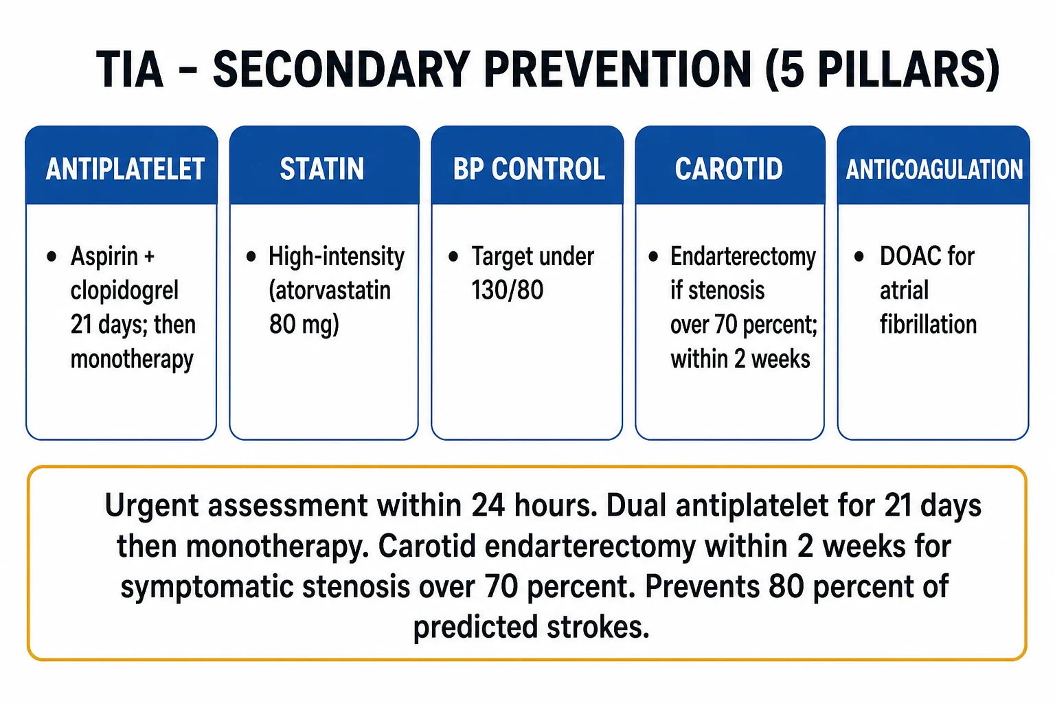
The aim of secondary prevention is to prevent the completed stroke that the TIA predicts. Aggressive, mechanism-directed therapy can prevent up to 80 percent of predicted 90-day strokes.[6]
1. Antiplatelet therapy for non-cardioembolic TIA
For a non-cardioembolic TIA (no atrial fibrillation), give dual antiplatelet therapy — aspirin plus clopidogrel — for 21 days, then step down to monotherapy (clopidogrel 75 mg once daily, or aspirin with modified-release dipyridamole). The loading regimen is aspirin 300 mg then 75 mg once daily, plus clopidogrel 300 mg then 75 mg once daily.[1]
The evidence for the 21-day dual window comes from two landmark trials. [1]
CHANCE (2013)
N Engl J Med
Randomised double-blind placebo-controlled trial in 5170 Chinese patients with minor stroke or high-risk TIA within 24 h. Clopidogrel 300 mg load then 75 mg daily for 90 days (plus aspirin 75 mg for 21 days) versus aspirin alone.
Key finding
90-day stroke: 8.2 percent (dual) versus 11.7 percent (aspirin alone); hazard ratio 0.68 (p under 0.001), with no increase in haemorrhage.
Practice change
Established short-term dual antiplatelet therapy as standard after high-risk TIA and minor stroke.
POINT (2018)
N Engl J Med
International randomised double-blind trial in 4881 patients with minor stroke or high-risk TIA. Clopidogrel 600 mg load then 75 mg daily plus aspirin versus aspirin alone, started within 12 h.
Key finding
90-day major ischaemic event: 5.0 percent (dual) versus 6.5 percent (aspirin); hazard ratio 0.75 (p 0.02). Major haemorrhage: 0.9 percent versus 0.4 percent (hazard ratio 2.32, p 0.02).
Practice change
Confirmed the ischaemic benefit but showed the bleeding signal accrues earliest — underpinning the 21-day cap on dual therapy.
Taken together, CHANCE showed the benefit and POINT showed that the bleeding risk appears early and continues, which is why dual therapy is capped at 21 days before stepping down to monotherapy.[4][5]
2. Cardioembolic TIA (atrial fibrillation)
If the mechanism is atrial fibrillation, give anticoagulation — not antiplatelets. A direct oral anticoagulant (DOAC) (apixaban, rivaroxaban, dabigatran, or edoxaban) is preferred over warfarin for non-valvular AF on the basis of the Ruff 2014 meta-analysis, which pooled the four pivotal DOAC trials (RE-LY, ROCKET AF, ARISTOTLE, ENGAGE AF-TIMI 48, over 71,000 patients) and found DOACs reduced stroke or systemic embolism by 19 percent and intracranial haemorrhage by about half versus warfarin, with similar major bleeding.[8]
Ruff DOAC meta-analysis (2014)
Lancet
Prespecified random-effects meta-analysis of 71,683 patients with atrial fibrillation across RE-LY, ROCKET AF, ARISTOTLE, ENGAGE AF-TIMI 48.
Key finding
DOACs reduced stroke or systemic embolism by 19 percent (RR 0.81) and intracranial haemorrhage (RR 0.48) versus warfarin, with reduced all-cause mortality and similar major bleeding, at the cost of more gastrointestinal bleeding.
Practice change
Established DOACs as first-line over warfarin for stroke prevention in non-valvular atrial fibrillation.
Because a TIA by definition produces no infarct, anticoagulation can be started immediately — there is no haemorrhagic-transformation delay as there is after a completed cardioembolic stroke.[1]
3. Carotid revascularisation
For symptomatic carotid stenosis (i.e. producing the TIA), offer carotid endarterectomy (CEA) for stenosis of 50 percent or more, with the greatest benefit and lowest NNT for stenosis over 70 percent (NNT about 3). Timing is critical: the operation should be performed within 2 weeks of the event, because the benefit collapses rapidly with delay — the NNT rises from about 3 within 2 weeks to over 100 beyond 12 weeks. Carotid artery stenting is an alternative in selected patients (younger, high surgical risk, hostile neck) but carries a higher periprocedural stroke risk in older patients.[7]
4. Vascular risk-factor targets for all patients
- Blood pressure: target under 130/80 mmHg (an ACE inhibitor or angiotensin receptor blocker plus a thiazide-like diuretic is a common regimen).
- Lipids: a high-intensity statin (atorvastatin 80 mg daily) aiming for LDL reduction of 50 percent or more.
- Glycaemic control: optimise diabetes (HbA1c target individualised).
- Smoking cessation — the single most impactful modifiable behaviour.
- Lifestyle: Mediterranean-style diet, weight reduction, regular exercise, alcohol moderation. [1]
5. The cumulative benefit
The full bundle — antiplatelet or anticoagulant, statin, blood-pressure control, smoking cessation, and carotid intervention where indicated — can prevent up to 80 percent of predicted strokes after TIA. This figure derives from the EXPRESS study, which showed that simply accelerating assessment and treatment reduced the 90-day stroke risk from 10.3 percent to 2.1 percent (an 80 percent relative reduction, hazard ratio 0.20).[6]
Specific Subtypes and Scenarios
- Crescendo TIA (two or more events within 24 hours): very high risk of imminent completed stroke — admit, perform urgent imaging and a large-vessel workup, and expedite definitive therapy (carotid endarterectomy or anticoagulation) once the mechanism is identified.
- Severe symptomatic carotid stenosis (over 70 percent): CEA within 2 weeks plus best medical therapy — the highest-NNT intervention in stroke medicine.
- Cardioembolic TIA (atrial fibrillation): anticoagulation with a DOAC replaces antiplatelets; start immediately as there is no infarct to transform.
- Posterior-circulation TIA: obtain urgent CT angiography of head and neck; if symptoms progress to a basilar artery occlusion, this is a mechanical thrombectomy emergency.
- Young patient (under 45): a different aetiological workup — carotid or vertebral dissection (fat-suppressed neck MRI or CTA), patent foramen ovale (bubble echo and TOE), thrombophilia (protein C, protein S, antithrombin, antiphospholipid antibodies, factor V Leiden, prothrombin gene mutation), substance use (cocaine, amphetamines), and autoimmune vasculitis. The ABCD2 score underperforms here.[2]
Complications and Pitfalls
Complications
- Completed stroke — the principal complication: 5 to 10 percent within 48 hours and 10 to 20 percent within 90 days without treatment.
- Cognitive impairment and vascular dementia — even without a completed stroke, recurrent TIAs contribute to multi-infarct cognitive decline.
- Myocardial infarction — TIA patients share systemic atherosclerosis; comprehensive vascular risk reduction is mandatory, not optional.
- Depression, anxiety, and falls — common after a TIA and frequently overlooked. [1]
Classic pitfalls
- Labelling a mimic (hypoglycaemia, migraine aura, postictal paresis) as a TIA — or, conversely, dismissing a true TIA as trivial.
- Failing to exclude haemorrhage on imaging before starting antiplatelets — a microbleed or early intracerebral haemorrhage can present with a transient deficit.
- Over-relying on ABCD2 — it underestimates risk in the young and ignores carotid stenosis and atrial fibrillation.
- Delaying carotid endarterectomy beyond 2 weeks — the benefit collapses with delay.
- Giving antiplatelets instead of anticoagulants in atrial fibrillation.[1]
Prognosis and Disposition
With urgent assessment and aggressive secondary prevention, the 90-day stroke risk can be reduced by up to 80 percent (EXPRESS: 10.3 percent to 2.1 percent).[6] The highest-risk period is the first 48 hours, which is exactly why urgent rather than routine assessment is mandated. The features that predict the highest early stroke risk are an ABCD2 score of 6 or higher, severe carotid stenosis, atrial fibrillation, crescendo events, and a positive DWI on MRI.[1]
Long term, TIA patients have a reduced life expectancy, mostly from cardiovascular disease, which is why lifelong vascular risk reduction is essential rather than a finite course of treatment. Beyond stroke prevention, secondary prevention also reduces the risk of cognitive decline and vascular dementia.[2]
The 90-day post-TIA trajectory
A patient is safe for discharge from the TIA service once the workup is complete, secondary prevention has been started, carotid imaging has been arranged (or performed), and follow-up is booked. Clear safety-net advice must be given: return immediately if symptoms recur. [1]
Driving advice
In the United Kingdom (DVLA), a patient must not drive for at least one month after a single TIA; after multiple TIAs within a short period they must not drive until symptoms have been controlled for three months, and the DVLA must be notified. Regional rules differ — the patient should be advised to check their local licensing authority's guidance.[2]
[1]Special Populations
- Atrial fibrillation — anticoagulation (DOAC), not antiplatelets; start immediately as there is no infarct.[1]
- Symptomatic carotid stenosis — endarterectomy within 2 weeks for stenosis over 50 percent; NNT about 3 for over 70 percent; benefit falls rapidly after 2 weeks.[7]
- Crescendo TIA — very high stroke risk; admit, image, and consider urgent intervention.
- Pregnancy — a hypercoagulable state raising TIA and stroke risk; aspirin is safe, DOACs are avoided, and low-molecular-weight heparin is used if anticoagulation is required.
- Elderly — higher comorbidity and atypical presentation (confusion, reduced consciousness), but the urgency of assessment is unchanged.
- Diabetic — higher risk and worse outcome; tight glucose and blood-pressure control; remember that hypoglycaemia is a mimic.
- Already on anticoagulants presenting with TIA — assess adherence and the INR or DOAC level; exclude intracranial bleed on imaging before continuing or escalating therapy.[2]
Evidence, Guidelines and Regional Differences
The landmark evidence underpinning modern TIA management is summarised below. [1]
EXPRESS (2007)
Lancet
Prospective population-based before-and-after sequential comparison (OXVASC) of urgent versus standard outpatient assessment and treatment of TIA and minor stroke.
Key finding
90-day recurrent stroke fell from 10.3 percent to 2.1 percent (adjusted hazard ratio 0.20, an 80 percent reduction) with urgent, same-day specialist assessment and immediate treatment.
Practice change
Established that urgent TIA assessment and early treatment reduce early recurrent stroke by 80 percent — the foundation of the 24-hour specialist-assessment standard worldwide.
AHA/ASA 2019 Acute Ischaemic Stroke Guideline
Stroke
Comprehensive update of the 2018 guidelines covering prehospital care, urgent evaluation, and early secondary prevention of acute ischaemic stroke and high-risk TIA.
Key finding
Recommends dual antiplatelet therapy (aspirin plus clopidogrel) for 21 days after high-risk TIA or minor stroke (Class 1), early carotid revascularisation for symptomatic stenosis, and anticoagulation for cardioembolic sources.
Practice change
Codified the 21-day dual-antiplatelet window and the urgency of carotid intervention into US practice.
Regional guideline comparison
[1]The European Stroke Organisation (ESO) guidelines align closely with the AHA/ASA and NICE on the urgency of assessment, the 21-day dual-antiplatelet window, and carotid intervention timing. In India, where access to carotid endarterectomy and rapid imaging is uneven, best medical therapy (antiplatelet, high-intensity statin, blood-pressure and diabetes control, smoking cessation) is the most widely applicable intervention and must be optimised for every patient.
Exam Pearls
- TIA = transient focal neurological deficit from ischaemia WITHOUT infarction; resolves within 24 hours, usually under 1 hour.
- NOT benign — 5 to 10 percent stroke in 48 hours, 10 to 20 percent in 90 days without treatment.
- The five numbers: under 24 h (old definition), under 1 h (typical duration), 5 to 10 percent stroke in 48 h, 10 to 20 percent in 90 d, CEA within 2 weeks.
- ABCD2: Age, BP, Clinical, Duration, Diabetes. Score of 4 or more = high risk; assess within 24 hours.
- Non-cardioembolic: dual antiplatelet (aspirin + clopidogrel) for 21 days then monotherapy; statin; BP under 130/80.
- AF: anticoagulation (DOAC), not antiplatelets.
- Symptomatic carotid stenosis over 70 percent: CEA within 2 weeks (NNT about 3).
- Always exclude hypoglycaemia first; exclude haemorrhage on CT before antiplatelets.
- Amaurosis fugax = ipsilateral carotid stenosis until proven otherwise.
- Crescendo TIA = admit.
- EXPRESS showed urgent care prevents 80 percent of predicted strokes; CHANCE and POINT underpin the 21-day dual window. [1]
Quick self-test: A 72-year-old diabetic with a 30-min episode of right face and arm weakness and dysphasia, now resolved. BP 168/96, pulse irregular. What is the ABCD2 score and the immediate management?
ABCD2: Age (1) + BP (1) + unilateral weakness (2) + duration 30 min (1) + diabetes (1) = 6 (high risk). Plus the irregular pulse flags atrial fibrillation. Immediate management: admit or urgent TIA clinic within 24 hours, CT to exclude haemorrhage, ECG and cardiac monitoring for AF, carotid Doppler, and — once AF is confirmed — anticoagulation with a DOAC (not antiplatelets); if AF is excluded, dual antiplatelet for 21 days then monotherapy.
Exam application bank (NEET-PG / INICET)
One-line answer
Transient ischaemic attack (TIA) is a transient episode of neurological dysfunction caused by focal brain, spinal cord or retinal ischaemia without acute infarction (tissue-based definition). It is a medical emergency, not a benign event — the risk of completed stroke is 5 to 10 percent within 48 hours and 10 to 20 percent within 90 days without treatment. The ABCD2 score (Age, Blood pressure, Clinical features, Duration, Diabetes) stratifies early stroke risk and drives triage urgency. Presentation is a sudden focal neurological deficit that resolves within 24 hours (usually under 1 hour) — unilateral weakness, aphasia, or monocular visual loss (amaurosis fugax). Urgent workup includes CT then MRI-DWI brain, carotid Doppler or CT angiography, ECG (atrial fibrillation) and echocardiography. Management is aggressive secondary prevention: dual antiplatelet therapy (aspirin plus clopidogrel
Worked stems (answer without another resource)
Stem 1 — Classic presentation. Map symptoms to mechanism; name the first investigation and first treatment step with dose/route if drug therapy is standard. [1]
Stem 2 — Unstable / complicated. List red flags that force immediate resuscitation, theatre, ICU, antidote, or reperfusion — and what you do in the first 15 minutes. [1]
Stem 3 — Atypical group. Elderly, pregnancy, child, or immunocompromised: how presentation and thresholds change. [1]
Stem 4 — Differential trap. Name the three closest mimics and one discriminator for each. [1]
Stem 5 — Disposition. Who goes home with safety-netting, who is admitted, who needs HDU/ICU/theatre, and what follow-up is mandatory. [1]
Rapid viva checklist
- Definition + classification
- Pathophysiology chain
- Bedside signs / criteria
- Score with exact components (if any)
- Emergency bundle
- Definitive therapy with doses
- Complications of disease and of treatment
- Special populations
- Guideline/trial name if classic
- Three exam traps
Coverage self-check
If you cannot answer any stem above from this page alone, re-read the matching section — the page is intended to be self-sufficient for final-prof and NEET-PG/INICET questions on Transient Ischaemic Attack (TIA).
References
- [1]Mendelson SJ, Prabhakaran S. Diagnosis and Management of Transient Ischemic Attack and Acute Ischemic Stroke: A Review JAMA, 2021.PMID 33724327
- [2]Perry JJ, Yadav K, Syed S, Shamy M. Transient ischemic attack and minor stroke: diagnosis, risk stratification and management CMAJ, 2022.PMID 36220167
- [3]Johnston SC, Rothwell PM, Nguyen-Huynh MN, Giles MF, Elkins JS, Bernstein AL, Sidney S. Validation and refinement of scores to predict very early stroke risk after transient ischaemic attack Lancet, 2007.PMID 17258668
- [4]Wang Y, Wang Y, Zhao X, Liu L, Wang D, Wang C, Wang C, Li H, Meng X, Cui L, Jia J, Dong Q, Xu A, Zeng J, Li Y, Wang Z, Xia H, Johnston SC; CHANCE Investigators. Clopidogrel with aspirin in acute minor stroke or transient ischemic attack N Engl J Med, 2013.PMID 23803136
- [5]Johnston SC, Easton JD, Farrant M, Barsan W, Conwit RA, Elm JJ, Kim AS, Lindblad AS, Palesch YY; POINT Investigators. Clopidogrel and Aspirin in Acute Ischemic Stroke and High-Risk TIA N Engl J Med, 2018.PMID 29766750
- [6]Rothwell PM, Giles MF, Chandratheva A, Marquardt L, Geraghty O, Redgrave JN, Lovelock CE, Binney LE, Bull LM, Cuthbertson FC, Welch SJ, Bosch S, Alexander FC, Silver LE, Gutnikov SA, Mehta Z; EXPRESS study. Effect of urgent treatment of transient ischaemic attack and minor stroke on early recurrent stroke (EXPRESS study): a prospective population-based sequential comparison Lancet, 2007.PMID 17928046
- [7]Powers WJ, Rabinstein AA, Ackerson T, Adeoye OM, Bambakidis NC, Becker K, Biller J, Brown M, Demaerschalk BM, Hoh B, Jauch EC, Kidwell CS, Leslie-Mazwi TM, Ovbiagele B, Scott PA, Sheth KN, Southerland AM, Summers DV, Tirschwell DL. Guidelines for the Early Management of Patients With Acute Ischemic Stroke: 2019 Update to the 2018 Guidelines for the Early Management of Acute Ischemic Stroke: A Guideline for Healthcare Professionals From the American Heart Association/American Stroke Association Stroke, 2019.PMID 31662037
- [8]Ruff CT, Giugliano RP, Braunwald E, Hoffman EB, Deenadayalu N, Ezekowitz MD, Camm AJ, Weitz JI, Lewis BS, Parkhomenko A, Yamashita T, Antman EM. Comparison of the efficacy and safety of new oral anticoagulants with warfarin in patients with atrial fibrillation: a meta-analysis of randomised trials Lancet, 2014.PMID 24315724