Neurology · General Medicine
Trigeminal Neuralgia & Bell Palsy
Also known as Trigeminal neuralgia · Tic douloureux · Bell palsy · Facial nerve palsy · Idiopathic facial paralysis
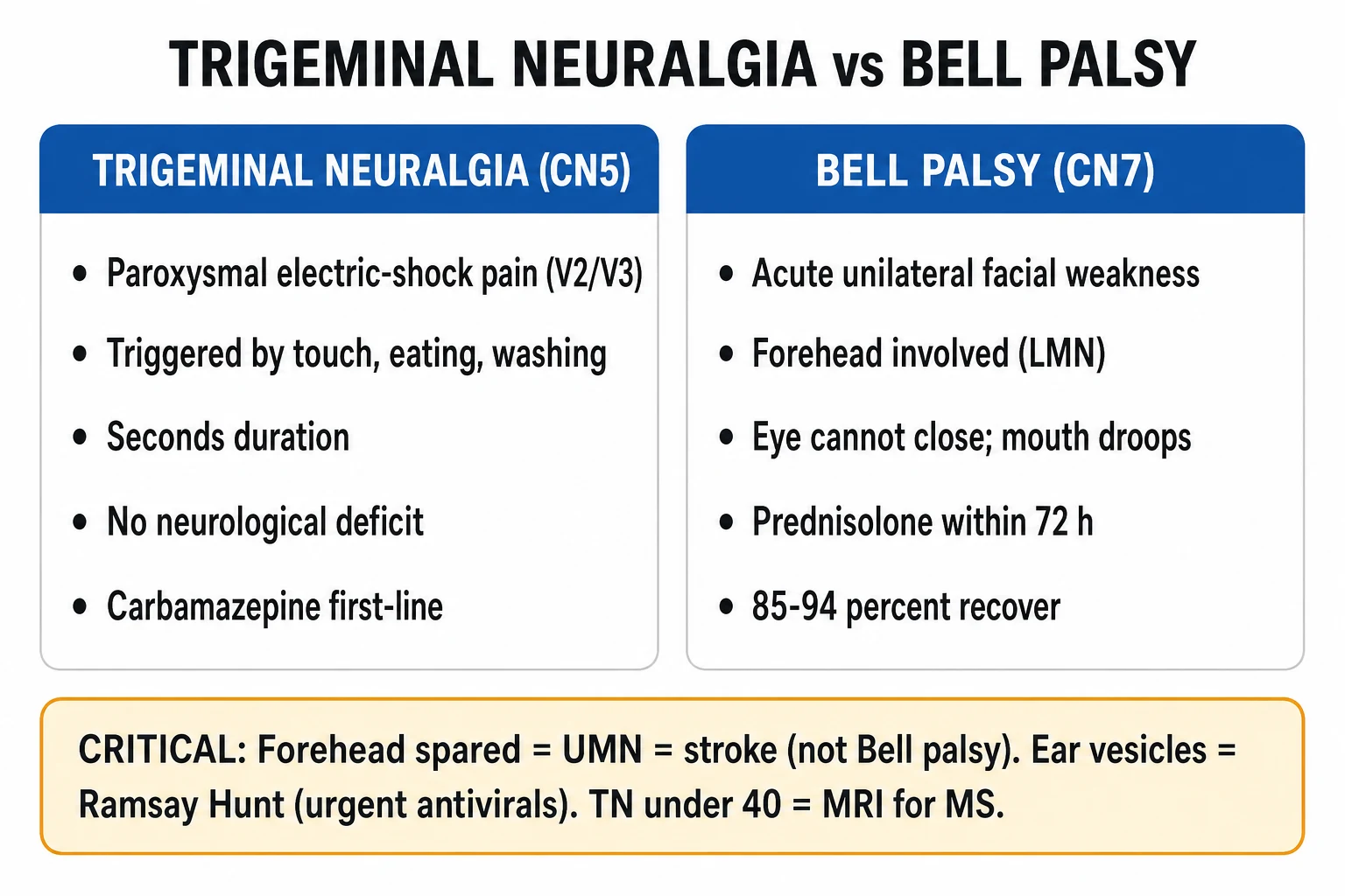
Trigeminal neuralgia (TN, tic douloureux) is severe, paroxysmal, electric-shock-like facial pain in the distribution of the trigeminal nerve (usually V2/V3), lasting seconds and provoked by light touch (washing, eating, cold air). It is usually caused by neurovascular compression of the trigeminal root at the root-entry zone, producing focal demyelination and ephaptic transmission. Carbamazepine is first-line; refractory cases need microvascular decompression (Jannetta), gamma knife or percutaneous procedures. Bell palsy is an acute, unilateral lower motor neurone (LMN) facial nerve (CN VII) palsy of idiopathic (likely viral) cause producing facial asymmetry with forehead involvement, inability to close the eye, drooping mouth and altered taste. Treatment is oral prednisolone 60 mg daily for 5 days then taper, started within 72 hours, plus mandatory eye protection; antivirals add limited benefit. Most recover within 3 to 6 months. The single highest-yield discriminating fact: forehead spared = upper motor neurone (stroke); forehead involved = LMN (Bell palsy).
On this page & tools
Your progress
Saved locally on this device.
Exam tags
Red flags
Overview & Definition
These are two of the commonest cranial nerve syndromes in clinical practice and a frequent subject of short cases and vivas. They are paired not because they share a mechanism — they do not — but because both present acutely to the same clinics, both are clinical diagnoses made at the bedside, and both hinge on a single decisive discriminator that an examiner will probe relentlessly. Confusing the two, or worse, labelling a stroke as Bell palsy, is a classic and serious error. [1]
Trigeminal neuralgia (TN, tic douloureux) is defined by its characteristic pain quality rather than by any imaging finding. The International Classification of Headache Disorders (ICHD-3, as reflected in the European Academy of Neurology 2019 guideline) defines it as recurrent, unilateral, paroxysmal facial pain in the distribution of one or more divisions of the trigeminal nerve, lasting from a fraction of a second to two minutes, of severe intensity, with an electric-shock, stabbing or shooting quality, and precipitated by innocuous stimuli — washing, shaving, eating, brushing the teeth, a cold wind, or even light touch to a trigger zone. Critically, there is no persistent neurological deficit between attacks in classical TN; the patient is pain-free and neurologically intact. Recognition of this phenotype alone usually makes the diagnosis.[1]
Bell palsy is an acute, unilateral, peripheral (lower motor neurone) facial-nerve palsy of unknown (idiopathic) cause that develops over less than 72 hours and is not attributable to any other identifiable cause (stroke, infection, tumour, trauma). The pivotal clinical skill is distinguishing an LMN lesion (Bell palsy — forehead and eye involved) from a UMN lesion (cortical stroke — forehead spared), because the management and urgency are entirely different. The diagnosis of Bell palsy is one of exclusion: secondary causes of an acute LMN facial palsy must be considered and ruled out at the bedside before the label is applied.[2][3]
Classification
Trigeminal neuralgia is classified by aetiology (ICHD-3 / EAN 2019) into three mutually exclusive categories. Classical TN is attributed to a neurovascular conflict — an artery or vein compressing the trigeminal root at the root-entry zone — with demonstrable morphological change in the nerve (atrophy or flattening) on MRI; the offending vessel is most often the superior cerebellar artery. Secondary TN is attributed to a demyelinating plaque of multiple sclerosis or a tumour (meningioma, vestibular or trigeminal schwannoma, epidermoid cyst) at the nerve root or Gasserian ganglion. Idiopathic TN is diagnosed when no cause — neither a neurovascular conflict with morphological change nor an identifiable structural lesion — is found.[1]
Clinically, TN is further split by pain phenotype into purely paroxysmal TN (only the electric-shock attacks) and TN with concomitant continuous pain (TN type 2 — a constant, dull background ache beneath the paroxysms). This distinction matters at the bedside: TN type 2 is harder to control medically, responds less well to surgery, and carries a poorer prognosis, and its presence should prompt the examiner to question whether the diagnosis is truly classical TN or an atypical facial pain syndrome. [1]
Facial weakness is classified by the level of the lesion within the motor pathway, which is the single most important diagnostic fork. A lower motor neurone (LMN) palsy — the Bell palsy pattern — affects the whole ipsilateral hemiface: forehead, eye and mouth all fail, because the facial nerve nucleus and all its peripheral branches are damaged. An upper motor neurone (UMN) palsy affects only the contralateral lower face with forehead sparing, because the upper face receives bilateral cortical innervation from both motor cortices, so a unilateral cortical lesion (stroke) still leaves one intact pathway to the forehead. Severity of any facial palsy is then graded objectively by the House-Brackmann system, reproduced in the Investigations section below. [1]
Trigeminal neuralgia
- Paroxysmal electric-shock pain, V2/V3, seconds, triggerable by touch
- No neurological deficit between attacks (classic)
- Neurovascular compression at the root-entry zone
- Carbamazepine first-line; surgery if refractory
- Atypical features (sensory loss, young, bilateral) → MRI for MS/tumour
Bell palsy (LMN)
- Acute unilateral facial weakness over under 72 hours
- Forehead AND eye AND mouth involved on one side
- Idiopathic, likely viral (HSV-1) inflammation of CN VII
- Prednisolone within 72 hours plus mandatory eye protection
- About 85 percent recover untreated; 94 percent with early steroids
UMN facial palsy (stroke)
- Forehead SPARED — only contralateral lower face weak
- Often with arm/leg weakness or other stroke signs
- Cortical/internal capsule lesion (UMN pathway)
- Treat as acute stroke — fast-track imaging and thrombolysis
- Do NOT label as Bell palsy
Epidemiology & Risk Factors
Trigeminal neuralgia has an incidence of about 4 to 13 per 100,000 per year and a prevalence of roughly 0.03 percent of the population. The peak age is over 50 (typically 50 to 70 years), there is a slight female predominance (around 3 to 2), and the right side is affected more often than the left by a ratio of roughly 3 to 2, for reasons that remain unclear. It is rare before age 40; onset in a younger patient should always prompt suspicion of multiple sclerosis and is an explicit indication for MRI. About 1 to 6 percent of patients with multiple sclerosis develop TN over their lifetime, and TN may even be the presenting feature of MS. MS-related TN is characteristically bilateral more often than classical TN, a feature that should set off alarm bells.[1]
Bell palsy has an incidence of about 20 to 30 per 100,000 per year — far commoner than TN. It affects any age but peaks between 15 and 45 years, and has an approximately equal sex incidence. Recognised risk factors include pregnancy (especially the third trimester and the first postpartum week, where the incidence rises three- to four-fold), diabetes mellitus, hypertension, obesity, immunosuppression and a recent viral upper-respiratory infection. Both diabetes and hypertension confer a higher incidence and a worse recovery, which matters both for counselling and for prognostication. There is a slight familial clustering, suggesting a genetic predisposition in some kindreds.[2][6]
Trigeminal neuralgia & Bell palsy — key numbers
Pathophysiology
The mechanisms of the two conditions are entirely different, and understanding them explains why the treatments — an anticonvulsant for TN, a corticosteroid for Bell palsy — are also entirely different. [1]
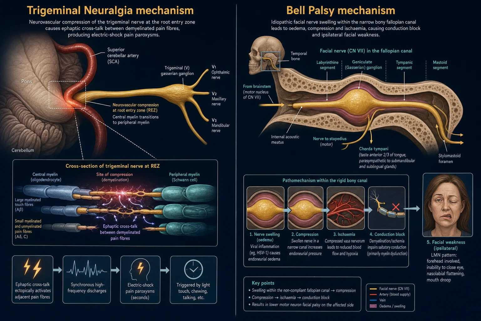
Classical trigeminal neuralgia — neurovascular conflict and ephaptic transmission. A blood vessel — most often the superior cerebellar artery, less often the anterior inferior cerebellar artery, a petrosal vein, or rarely a persistent primitive artery — compresses the trigeminal nerve at the root-entry zone. This is the point where central myelin (maintained by oligodendrocytes) meets peripheral myelin (maintained by Schwann cells), and it is therefore mechanically and metabolically vulnerable to pulsatile injury. Chronic pulsatile compression produces focal demyelination of the nerve root. Demyelinated A-beta (low-threshold mechanoreceptor, light-touch) fibres then lie physically adjacent to demyelinated A-delta and C (nociceptive, pain) fibres, allowing ephaptic (direct, non-synaptic electrical) cross-activation: a benign touch stimulus is thereby perceived as an electric-shock pain paroxysm. The nerve also develops ectopic firing — spontaneous action potentials arising at sites of demyelination — and the so-called ignition hypothesis describes a self-sustaining positive-feedback burst of ectopic discharges that constitutes the paroxysm, followed by a refractory period after each attack during which further triggers cannot fire the nerve. This refractory period is the explanation for the clinical observation that several attacks may cluster, then pause, then resume. Mechanistically, this is why an anticonvulsant that dampens ectopic firing and stabilises neuronal membranes (carbamazepine) works, and why surgically decompressing the vessel (microvascular decompression, MVD) can be curative by removing the source of demyelination.[1]
Secondary TN results not from a blood vessel but from a demyelinating plaque of MS directly involving the trigeminal root or root-entry zone, or from a tumour (meningioma, vestibular or trigeminal schwannoma, epidermoid) compressing or infiltrating the nerve root or Gasserian ganglion. These patients more often have sensory loss in the trigeminal distribution, bilateral symptoms, other cranial-nerve deficits, or objective neurological signs — any of which excludes classical TN and mandates imaging. [1]
Bell palsy — compression neuropathy of CN VII in the fallopian canal. The facial nerve's course through the temporal bone — the fallopian canal — is the longest bony canal traversed by any cranial nerve, and its labyrinthine segment and the meatal foramen at the geniculate ganglion constitute the narrowest point and the site of entrapment. Inflammation and oedema of the facial nerve (most likely viral, with herpes simplex virus type 1 reactivation in the geniculate ganglion the leading candidate, and varicella-zoster virus causing Ramsay Hunt) within this narrow segment produces compression, venous congestion, ischaemia and conduction block (neuropraxia). If the insult is severe and prolonged, the cycle of swelling and ischaemia progresses to Wallerian axonal degeneration, which carries a far worse prognosis and a longer, incomplete recovery. The swollen nerve is constrained by unyielding bone, so even modest oedema raises intra-neural pressure and impairs perfusion — a vicious cycle that corticosteroids (reducing oedema) interrupt if given early, within the 72-hour window of predominantly neuropraxic (conduction-block) injury.[2][6]
Facial-nerve branch anatomy localises the lesion level and explains the constellation of deficits. Within the fallopian canal the facial nerve gives off three branches in order: the greater petrosal nerve (carrying parasympathetic fibres for lacrimation — tested by the Schirmer test, which measures tear production), the nerve to stapedius (which dampens the stapes and the ossicular chain; its loss causes the painful hypersensitivity to sound that is hyperacusis), and the chorda tympani (carrying taste to the anterior two-thirds of the tongue and parasympathetic salivation). A lesion proximal to all three — at the meatal foramen or labyrinthine segment — therefore reduces lacrimation, causes hyperacusis and impairs taste, the worst-case constellation. A lesion distal to the chorda tympani produces pure motor weakness with preserved taste, hearing and tears. This branch-by-branch logic is summarised at the bedside as 'tears, tastes and taps', and the presence of all three deficits marks a proximal, severe lesion with a poorer outlook.[2]
Clinical Presentation
Trigeminal neuralgia
The pain is severe, paroxysmal, electric-shock, stabbing or shooting in quality, lasting a fraction of a second to under two minutes (rarely longer). Attacks are strictly unilateral and follow one or more trigeminal divisions: V2 and V3 combined in about 60 percent, V3 alone in about 20 percent, V2 alone in about 20 percent, and V1 alone in fewer than 5 percent. Bilateral disease is rare in classical TN and strongly suggests MS. The patient can usually point to a discrete trigger zone on the face — commonly the nasolabial fold, lip, or gum — and attacks are precipitated by innocuous stimuli: washing the face, shaving, brushing the teeth, eating, talking, swallowing, a cold wind, or even a light breeze. A refractory period of minutes to hours follows each cluster of attacks, during which triggers cannot provoke another paroxysm. [1]
Between attacks the patient is neurologically normal and pain-free. Any persisting sensory loss, allodynia, or other cranial-nerve deficit between attacks excludes classical TN and points to secondary disease. The secondary morbidity is substantial: patients lose weight and become dehydrated from food avoidance (eating triggers the pain), many undergo unnecessary dental extraction for misdiagnosed toothache before the correct diagnosis is made, and depression and social isolation are common as the unpredictable attacks dominate daily life.[1]
Bell palsy
The hallmark is acute onset, over hours to less than 72 hours, of unilateral facial weakness. The weakness involves the whole ipsilateral hemiface: the forehead (the patient cannot wrinkle it or raise the eyebrow), the eye (the patient cannot close it — lagophthalmos — and on attempted closure the globe rolls upward, the Bell phenomenon, exposing the cornea), and the mouth (drooping of the angle, drooling, dysarthria for labial consonants, and food trapping in the buccal sulcus). Associated features reflecting proximal nerve involvement include reduced taste on the anterior two-thirds of the tongue (chorda tympani), hyperacusis (nerve to stapedius), reduced lacrimation and a dry eye (greater petrosal), and sometimes a dull retro-auricular pain that may precede the weakness by a day or two. Crucially, there is no limb weakness, no sensory loss on the face, and no other cranial-nerve abnormality in an uncomplicated case — their presence redefines the diagnosis.[2][3]
Atypical presentations that mandate reclassification rather than reassurance: TN with sensory loss, onset under 40, or bilaterality (secondary TN — MS/tumour, MRI mandatory); TN with a continuous background ache beneath the paroxysms (TN type 2, harder to treat, poorer prognosis); Bell palsy with complete paralysis at onset (House-Brackmann V/VI, worse prognosis and the group most likely to benefit from aggressive treatment); Ramsay Hunt syndrome (ear/palate vesicles, severe otalgia, facial palsy ± hearing loss, tinnitus or vertigo); and bilateral facial palsy (GBS, sarcoidosis, Lyme, HIV — never Bell palsy).[1][2]
Differential Diagnosis
For trigeminal neuralgia, the differential is broad and the consequences of mislabelling range from unnecessary dental work to a missed tumour:
- Dental disease (pulpitis, apical abscess) — the pain is continuous and localised to a tooth or gum, with dental signs on examination; it is not paroxysmal, not triggerable by light touch elsewhere, and not in a full trigeminal-division distribution.
- Cluster headache — strictly unilateral periorbital/temporal pain lasting 15 to 180 minutes, with prominent ipsilateral autonomic signs (lacrimation, conjunctival injection, rhinorrhoea, ptosis), recurring in bouts; not triggered by light touch to the face.
- SUNCT / SUNA (short-lasting unilateral neuralgiform headache with conjunctival injection and tearing) — very brief attacks (seconds) but many per day (often dozens) with prominent conjunctival injection and tearing; distinct from TN in its autonomic prominence and frequency.
- Post-herpetic neuralgia — a constant, burning pain in a zoster-scarred dermatome with a clear history of a preceding vesicular rash; not electric, not triggerable in the same way.
- Glossopharyngeal neuralgia — paroxysmal throat or ear pain triggered by swallowing, talking or coughing, in the distribution of CN IX rather than V.
- Giant-cell (temporal) arteritis — in the over-50 patient: continuous temporal headache, jaw claudication (pain on prolonged chewing that eases with rest), scalp tenderness, visual disturbance, and a markedly raised ESR/CRP; never call it TN, and never miss it.
- Atypical facial pain / persistent idiopathic facial pain — continuous, ill-defined, often bilateral, not confined to a nerve distribution, not triggerable, frequently with psychological overlay; and temporomandibular joint dysfunction (pain on chewing and jaw movement, joint tenderness, limited mouth opening).[1]
For an acute facial palsy, the causes of a secondary LMN facial palsy must be excluded before labelling it Bell palsy:
- Ramsay Hunt syndrome (herpes zoster oticus) — vesicles in the ear canal, on the pinna, or on the palate, with severe otalgia and facial palsy ± sensorineural hearing loss; worse prognosis, antivirals essential.
- Otitis media / mastoiditis — ear discharge, fever, systemic features, otoscopic signs.
- Lyme disease — tick exposure, erythema migrans, often bilateral facial palsy in endemic regions.
- HIV seroconversion — a mononucleosis-like illness with facial palsy at seroconversion.
- Cholesteatoma — chronic otorrhoea, conductive hearing loss, otoscopic mass.
- Parotid tumour — a palpable parotid mass invading the facial nerve (a peripheral branch lesion).
- Trauma — temporal bone fracture, with Battle sign, CSF otorrhoea, or hearing loss. [1]
The single most important distinction remains forehead-sparing UMN weakness (stroke) from forehead-involving LMN weakness (Bell palsy) — a fork that decides whether the patient is fast-tracked to thrombolysis or sent home with steroids and eye care.[2]
Clinical & Bedside Assessment
In suspected TN, the history is the diagnosis. Confirm the pain quality (electric, stabbing, shooting), the duration (seconds), the distribution (a trigeminal division, strictly unilateral), and the trigger pattern (innocuous stimuli, a trigger zone). On examination, map the affected division(s) by testing light touch and pinprick in V1 (forehead), V2 (cheek, upper lip) and V3 (lower lip, chin). Examine for a trigger zone (typically the nasolabial fold or gum) and, critically, for any inter-paroxysm sensory loss or other cranial-nerve deficit — these exclude classical TN. Inspect the teeth, ears and temporomandibular joint to exclude dental, otological and TMJ causes, and screen for weight loss and mood, since depression and malnutrition are common sequelae of untreated TN.[1]
In suspected Bell palsy, examine all four facial-nerve functions systematically:
- Motor — forehead (wrinkle/raise eyebrow), eye closure (can the lashes be buried?), and mouth (show teeth, puff out cheeks, whistle). Look for lagophthalmos and the Bell phenomenon.
- Taste — test the anterior two-thirds of the tongue with salt or sugar on the affected then the normal side (reduced on the affected side if chorda tympani involved).
- Lacrimation — perform the Schirmer test (reduced tearing on the affected side indicates a proximal, greater-petrosal lesion).
- Stapedial reflex — ask about and demonstrate hyperacusis (painful loudness on the affected side). [1]
Then inspect the ear canal, pinna and palate for vesicles (Ramsay Hunt — examine this in every facial palsy, since missing it changes both treatment and prognosis), examine the parotid for a mass, and perform a full cranial-nerve and peripheral neurological examination to exclude limb weakness or other signs that would redefine the diagnosis. Always check the forehead first — it decides LMN from UMN, and it is the single bedside observation that an examiner will reward.[2][6]
Grade the severity of any facial palsy with the House-Brackmann system (see Investigations): a higher grade at onset predicts a poorer recovery and identifies the subgroup that may warrant more aggressive therapy and closer follow-up. [1]
Investigations
Both TN and Bell palsy are clinical diagnoses. Routine blood tests and imaging are not required for a typical presentation — over-investigating a classical case is itself a pitfall. The role of investigation is to confirm the diagnosis by excluding the secondary causes that masquerade as the common syndrome.[1][6]
Indications for MRI in TN — any atypical feature mandates imaging: sensory loss in the trigeminal distribution, onset under 40, bilaterality, non-response to an adequate carbamazepine trial, deafness or any other cranial-nerve deficit, or progression to continuous pain. MRI with gadolinium contrast, using high-resolution CISS/FIESTA (constructive interference in steady state / fast imaging employing steady-state acquisition) cisternal sequences, demonstrates the neurovascular conflict (the offending vessel abutting and deforming the root) and excludes an MS plaque (high signal in the trigeminal root entry zone on T2/FLAIR, plus periventricular white-matter lesions) or a CPA tumour (meningioma, schwannoma). MRI is also essential before any surgical intervention.[1]
Indications for imaging or bloods in facial palsy — the AAO-HNS guideline recommends no routine imaging or electrophysiology for a typical Bell palsy. Imaging (MRI with contrast of the internal auditory meatus and parotid) and bloods are reserved for: slow progression beyond three weeks, no recovery by three to six months, recurrent or bilateral palsy, other cranial-nerve signs, a parotid mass, severe otalgia with vesicles (Ramsay Hunt), or clinical suspicion of a secondary cause. Targeted bloods include Lyme serology (in endemic regions), glucose/HbA1c (diabetes screening), HIV serology, ACE level (sarcoidosis) and an autoimmune screen; check ESR/CRP if giant-cell arteritis is in the differential.[6]
Electroneurography (ENoG) and EMG assess the severity and prognosis of a Bell palsy by comparing the compound muscle action potential amplitude on the affected and normal sides. Degeneration exceeding 90 percent within the first two weeks (a severe, axonal rather than neuropraxic injury) predicts a poor outcome and may inform discussion of facial-nerve decompression surgery. ENoG is not needed for a typical mild palsy that begins to recover within three weeks.[2]
House-Brackmann facial-nerve grading (reproduced verbatim)
The House-Brackmann scale is the international standard for grading facial-nerve function and must be reproduced accurately: [1]
- Grade I — Normal: normal facial function in all areas.
- Grade II — Mild dysfunction: slight asymmetry on close inspection; at rest, normal symmetry and tone; forehead movement moderate to good; eye closure complete with minimum effort; slight mouth asymmetry.
- Grade III — Moderate dysfunction: obvious but not disfiguring asymmetry; symmetrical at rest; forehead movement slight to moderate; eye closure with effort; noticeable but not disfiguring mouth movement.
- Grade IV — Moderately severe dysfunction: obvious disfiguring asymmetry; symmetrical at rest; no forehead movement; incomplete eye closure; asymmetric mouth movement.
- Grade V — Severe dysfunction: only barely perceptible movement; asymmetry at rest; no forehead movement, no eye closure, slight mouth movement.
- Grade VI — Total paralysis: no movement. [1]
A higher grade predicts a poorer recovery, and grades IV to VI at onset define the severe subgroup in which early, aggressive treatment and close ophthalmic follow-up matter most.[2][6]
Management — Resuscitation
Neither TN nor Bell palsy is a time-critical ABC resuscitation emergency, but several acute priorities must be recognised at first contact, and missing them causes preventable harm.[1][2]
- Eye protection in Bell palsy — institute immediately at first contact, before the patient leaves the room. Lubricating drops hourly by day, ointment at night, and eyelid taping or a moisture chamber/pad overnight prevent exposure keratopathy, corneal ulceration and blindness — the single most preventable complication of Bell palsy. Refer urgently to ophthalmology if there is corneal exposure, ulceration, or a Bell phenomenon that risks exposure. This is the highest-yield, lowest-cost intervention in the entire topic.
- Forehead sparing or any limb weakness — treat as possible acute stroke: fast-track to stroke imaging (CT/CTA or MRI) and thrombolysis pathways. Do not label it Bell palsy, do not give steroids, and do not delay.
- Ramsay Hunt syndrome — start urgent high-dose antivirals (aciclovir or valaciclovir) plus corticosteroids without waiting for confirmatory tests; delay worsens the already-poor prognosis for facial recovery and hearing.
- Carbamazepine toxicity in TN — in the unwell or elderly patient on carbamazepine, check sodium (symptomatic SIADH hyponatraemia may present as drowsiness or seizures) and look for rash (Stevens-Johnson syndrome — stop immediately and do not rechallenge; the HLA-B*1502 allele confers high risk in Han Chinese and Thai populations, where screening is recommended).
- Severe TN — may cause dehydration and weight loss from food avoidance; provide rapid medical escalation (titrate or add an agent), nutritional support, and analgesia for the constant component of TN type 2. [1]
Management — Definitive & Stepwise
Trigeminal neuralgia — the medical ladder
- First-line: carbamazepine. Start 100 to 200 mg once or twice daily and titrate every few days to a maintenance of 600 to 1200 mg/day in divided doses (maximum 1600 mg/day). About 70 percent of patients obtain initial relief, often dramatic. This response is itself nearly diagnostic. Monitor serum sodium (SIADH hyponatraemia), full blood count (leucopenia, aplastic anaemia), rash (Stevens-Johnson — stop immediately), and the clinical effects of drowsiness, ataxia, diplopia and nausea; carbamazepine is a potent CYP3A4 inducer with many drug interactions (including the oral contraceptive pill — counsel accordingly).[1]
- Second-line / add-on agents, used when carbamazepine is ineffective, poorly tolerated, or loses efficacy over time: oxcarbazepine (a related agent with less hepatic induction but its own hyponatraemia risk), lamotrigine (titrate slowly to avoid Stevens-Johnson), gabapentin and pregabalin (particularly useful in MS-related TN), baclofen (a GABA-B agonist, especially in MS and as an add-on), and phenytoin. Combination therapy is common in refractory cases, and the EAN guideline notes the limited randomised evidence for agents beyond carbamazepine and oxcarbazepine.[1]
- Monotherapy failure or intolerance is the trigger to refer to a neurologist, perform (or review) the MRI, and consider the surgical options below.
Trigeminal neuralgia — the surgical ladder
When an adequate medical trial fails (or side-effects are intolerable), three families of procedures are available, in roughly escalating invasiveness: [1]
- Microvascular decompression (MVD, Jannetta procedure) — a posterior fossa craniotomy (retrosigmoid) that physically separates the offending vessel from the trigeminal root with a small Teflon felt pad. It is the only procedure that treats the cause and gives the best and most durable long-term pain relief (about 90 percent immediate relief, roughly 70 percent pain-free at five years, 60 percent at ten years). It carries, however, the risks of any posterior-fossa craniotomy: ipsilateral hearing loss (the commonest significant complication, up to 10 percent), facial numbness or weakness, CSF leak, chemical or bacterial meningitis, cerebellar injury, stroke and death (under 1 percent). It is the procedure of choice for a fit patient with a clear neurovascular conflict on MRI.
- Gamma knife stereotactic radiosurgery — a non-invasive, single-fraction high-dose radiation targeted at the trigeminal root (typically 75 to 90 Gy to the root-entry zone). It is effective but has a delayed onset of action (weeks to months), produces facial numbness in a significant minority, and recurrence is common over years. It suits the patient who declines or cannot tolerate craniotomy.
- Percutaneous procedures on the Gasserian ganglion via the foramen ovale — three techniques, all performed under sedation and brief anaesthesia with a needle passed through the cheek into Meckel's cave: radiofrequency thermocoagulation (heating the ganglion), percutaneous balloon compression (Mullan) (a Fogarty-type balloon inflated to compress the ganglion), and glycerol rhizolysis (injecting anhydrous glycerol). These are chosen for the frail and elderly or those with limited life expectancy; they give immediate relief but reliably produce facial numbness and have a higher recurrence rate than MVD.[1]
Trigeminal neuralgia — escalation ladder
Carbamazepine
First-line; 100-200 mg BD titrated to 600-1200 mg/day; ~70 percent respond. Check sodium, FBC, rash.
Add or switch drug
Oxcarbazepine, lamotrigine, gabapentin, pregabalin, baclofen (esp. MS), phenytoin. Combine if needed.
MRI with contrast
Confirm neurovascular conflict (CISS/FIESTA); exclude MS plaque or tumour. Mandatory before surgery.
Surgery — MVD (Jannetta)
Fit patient, clear conflict. Best long-term relief (~70 percent at 5 years). Risk: hearing loss, CSF leak, under 1 percent mortality.
Surgery — gamma knife OR percutaneous
Gamma knife (non-invasive, delayed, may recur); percutaneous RF/balloon/glycerol (frail/elderly, immediate, numbness, recurrence).
Bell palsy — the evidence-based bundle
- Oral prednisolone is the cornerstone. Give 60 mg once daily for 5 days, then taper over 5 days (for example, 50 mg, then 40 mg, 30 mg, 20 mg, 10 mg on successive days), started within 72 hours of symptom onset. This improves the rate of complete recovery from about 80 percent (untreated) to about 94 percent, confirmed in the Scottish SCOPE trial (Sullivan, NEJM 2007), the Swedish trial (Engstrom, Lancet Neurology 2008), and the Cochrane review (Madhok, 2016).[2][3][4]
- Antivirals (aciclovir or valaciclovir) — alone are no better than placebo, and the added benefit over steroids alone is small and not statistically robust in pooled analyses (Gagyor Cochrane, 2015). The AAO-HNS guideline therefore recommends that antivirals not be offered as sole therapy, but they may be added to steroids for severe palsy (House-Brackmann IV to VI), Ramsay Hunt syndrome (where high-dose antivirals such as valaciclovir 1 g three times daily for 7 to 10 days are essential), or immunocompromise.[4][5][6]
- Eye protection — mandatory, non-negotiable, for every patient. Lubricating drops hourly by day (preservative-free where possible for frequent use), ointment at night, and taping the eyelid shut or a moisture chamber/pad overnight; for severe lagophthalmos, a temporary tarsorrhaphy or botulinum-toxin-induced ptosis (chemodenervation of the levator) protects the cornea until recovery. Neglect of eye care is the classic, preventable cause of corneal blindness.[6]
- Supportive measures: facial physiotherapy and massage/exercises to maintain muscle tone and reduce contracture; reassurance that the outlook is good; and psychological support for the disfigurement and anxiety of an acute facial palsy.
Specific Subtypes & Scenarios
- TN in multiple sclerosis — patients are younger, the TN may be bilateral, and MRI shows a demyelinating plaque at the trigeminal root-entry zone. Carbamazepine remains first-line, but the response is often less durable, and the surgical preference shifts toward percutaneous procedures and gamma knife rather than MVD (since the conflict is a plaque, not a vessel). The EAN guideline specifically addresses this subgroup.[1]
- Ramsay Hunt syndrome (herpes zoster oticus) — reactivation of varicella-zoster virus in the geniculate ganglion, producing vesicles in the ear canal, on the pinna or on the palate, severe otalgia, an LMN facial palsy, and sometimes sensorineural hearing loss, tinnitus and vertigo. It is a more severe and proximal injury than Bell palsy. Treat with high-dose antivirals (aciclovir 800 mg five times daily, or valaciclovir 1 g three times daily for 7 to 10 days) plus corticosteroids, started as early as possible. The prognosis is worse than Bell palsy (about 70 percent recovery of facial function, lower if treatment is delayed).[2][6]
- Bilateral facial palsy — this is never Bell palsy and mandates investigation. Think Guillain-Barre syndrome (ascending weakness, areflexia, CSF albuminocytologic dissociation — high protein with normal cell count), sarcoidosis (Heerfordt syndrome / uveoparotid fever — uveitis, parotitis, fever, raised ACE), Lyme disease (the commonest cause of bilateral facial palsy in endemic regions — tick exposure, erythema migrans), and HIV seroconversion. Investigate rather than treat empirically with steroids alone.[2]
- Bell palsy in pregnancy — commoner in the third trimester and the first postpartum week, often more severe than in the non-pregnant. Treat with prednisolone within 72 hours (the benefit outweighs the late-pregnancy risk; avoid the first trimester where possible on teratogenicity grounds), institute strict eye protection, and screen for hypertension/pre-eclampsia and gestational diabetes, which co-segregate with pregnancy-related Bell palsy.[2]
- TN with concomitant continuous pain (TN type 2) — a constant background ache beneath the paroxysms; harder to treat medically, with a poorer response to surgery. The differential includes atypical facial pain and persistent idiopathic facial pain, both of which should not be sent for destructive surgery.
- Recurrent facial palsy / Melkersson-Rosenthal syndrome — a triad of recurrent facial palsy, recurrent facial/lip swelling (cheilitis granulomatosa) and a fissured (scrotal) tongue; a granulomatous disorder that may respond to steroids and, in recurrent cases, to prophylaxis. Familial forms of recurrent Bell palsy also exist.[2]
Complications & Pitfalls
TN complications arise from both the disease and its treatment. The disease causes depression, weight loss, dehydration from food avoidance, and social isolation; rarely, the severe, unremitting pain of TN is a risk factor for suicide, which is why refractory TN is a neurosurgical urgency. Carbamazepine carries the risks of SIADH hyponatraemia, drowsiness, ataxia, leucopenia, aplastic anaemia, and Stevens-Johnson syndrome; oxcarbazepine its own hyponatraemia. The surgical risks of MVD — hearing loss, facial numbness or weakness, CSF leak, meningitis, cerebellar injury, stroke and death (under 1 percent) — must be discussed in consent. Percutaneous procedures reliably cause facial numbness (sometimes deafferentation pain, worse than the original TN) and have higher recurrence; gamma knife causes numbness and has delayed, sometimes recurring, efficacy.[1]
Bell palsy complications centre on the eye and on aberrant regeneration. Exposure keratopathy and corneal ulceration are preventable with eye care and are a recognised, avoidable cause of blindness. Residual weakness may persist, especially after a severe (House-Brackmann V/VI) palsy. Synkinesis and aberrant regeneration — misdirection of regrowing axons to the wrong branches — produce crocodile tears (the eye waters when eating, as salivary fibres reach the lacrimal gland) and mouth-eye synkinesis (the mouth corner moves on blinking or eye closure). Hemifacial spasm and permanent contracture are late sequelae.[2]
Classic pitfalls — the errors examiners probe and that cause real harm: labelling dental pain or atypical facial pain as TN (leading to unnecessary dental extraction and delayed correct treatment); missing MS or a tumour in young-onset or bilateral TN (delayed imaging); labelling a UMN (forehead-sparing) weakness as Bell palsy and missing a stroke (the most dangerous error — delays thrombolysis); missing Ramsay Hunt by not examining the ear (denying the patient essential antivirals); calling bilateral facial palsy 'Bell palsy' and missing GBS, Lyme or sarcoid; giving antivirals without steroids, or delaying steroids beyond 72 hours, in Bell palsy; and neglecting eye care at any point. In TN specifically, stopping carbamazepine abruptly (risk of seizure exacerbation if used for epilepsy, and pain rebound) or escalating to surgery without an adequate medical trial and an MRI are pitfalls.[1][2][3]
Prognosis & Disposition
Bell palsy prognosis. About 70 to 85 percent of patients recover fully without any treatment. Early prednisolone within 72 hours raises complete recovery to about 94 percent. Recovery typically begins within three weeks (early recovery is a favourable sign) and is complete by three to six months. The poor prognostic factors — the facts an examiner will ask for — are: complete paralysis at onset (House-Brackmann V/VI), age over 60, diabetes mellitus, pregnancy, severe retro-auricular pain, hyperacusis, complete loss of taste (marking a proximal lesion), no recovery by three weeks, and ENoG showing more than 90 percent degeneration within the first two weeks (a severe axonal injury rather than a conduction block).[2][3][4]
Ramsay Hunt syndrome has a worse prognosis than Bell palsy — about 70 percent recovery of facial function, and lower still if treatment is delayed beyond 72 hours; hearing loss may be permanent. Trigeminal neuralgia is chronic and relapsing-remitting: medical control is initially good but efficacy often wanes over years (tachyphylaxis), and many patients eventually need surgery. Microvascular decompression gives the most durable relief (about 70 percent pain-free at five to ten years); percutaneous procedures and gamma knife have higher recurrence.[1]
Disposition and follow-up. Review Bell palsy at one to two weeks (assess for eye complications and early recovery) and again at three months — refer to a specialist (neurology, ENT, or facial reanimation) if there is no recovery by three months, as the diagnosis should then be reconsidered and further imaging obtained. For TN, titrate the medication against pain and side-effects, and refer to a neurologist or a neurosurgical MDT for refractory or atypical disease; counsel on eye care and medication side-effects at every contact. Refer all Ramsay Hunt patients urgently to ENT and ophthalmology. [1]
Special Populations
- Pregnancy — Bell palsy is commoner in the third trimester and early postpartum, often more severe. Treat with prednisolone within 72 hours (benefit outweighs risk in late pregnancy; avoid the first trimester where possible on teratogenicity grounds), institute strict eye protection, and screen for pre-eclampsia and gestational diabetes. TN in pregnancy is managed medically (carbamazepine has teratogenic concerns — discuss with neurology and obstetrics; lamotrigine may be preferred).
- Diabetes mellitus — confers both a higher incidence and a worse recovery in Bell palsy. Monitor blood glucose when prescribing corticosteroids (they may precipitate hyperglycaemia). TN management is unchanged, but comorbidity affects surgical risk stratification for MVD.
- Children — Bell palsy occurs in children and is treated with steroids in the same 72-hour window per the AAO-HNS guideline. Always consider Lyme disease in endemic paediatric cases (a common cause in children in endemic regions). TN is rare in children and should prompt an MS evaluation and MRI.
- Elderly — Bell palsy recovery is slower and less complete; favour less-invasive TN procedures (percutaneous, gamma knife) over craniotomy where comorbidity is high. Screen for giant-cell arteritis in the older patient presenting with facial or jaw pain — it mimics TN and is treatable with steroids, not surgery.
- Immunocompromised (HIV) — facial palsy may occur at seroconversion or with opportunistic infection (CMV, Ramsay Hunt, toxoplasmosis); investigate the cause and add antiviral cover more readily.
- Resource-limited settings — carbamazepine and generic prednisolone are cheap and universally available, and are the backbone of management. Eye-protection education is the single highest-yield low-cost intervention (a preventable cause of blindness). Access to MRI, microvascular decompression and gamma knife is limited, so medical management predominates and surgery is reserved for the few refractory cases who can access it.[1][2][6]
Evidence, Guidelines & Regional Differences
Trigeminal neuralgia is guided by the European Academy of Neurology (EAN) guideline (Bendtsen, 2019), which is the contemporary reference standard. It distinguishes classical TN (neurovascular conflict with morphological change) from secondary TN (MS or tumour) and idiopathic TN, recommends carbamazepine or oxcarbazepine first-line, notes the limited randomised evidence for add-on agents (lamotrigine, baclofen, gabapentinoids), and places MVD, gamma knife and percutaneous procedures for refractory disease, with MVD offering the most durable relief where a clear conflict is demonstrated.[1]
Bell palsy evidence rests on three landmark randomised trials and two Cochrane reviews. The Scottish SCOPE trial (Sullivan, NEJM 2007) — a factorial double-blind trial of prednisolone, aciclovir, both, and placebo in 551 patients — showed that early prednisolone within 72 hours improves recovery and that antivirals alone do not. The Swedish trial (Engstrom, Lancet Neurology 2008) replicated the benefit of prednisolone and found valaciclovir added no significant benefit. The Cochrane reviews (Madhok, 2016, for corticosteroids; Gagyor, 2015, for antivirals) confirmed that steroids help (NNT about 11 for one additional complete recovery) and that antivirals add at most marginal benefit (a small pooled effect of uncertain clinical significance). The AAO-HNS clinical practice guideline (Baugh, 2013) synthesises these into recommendations: oral steroids within 72 hours for new-onset Bell palsy; antivirals not as sole therapy (may be added, benefit uncertain); no routine imaging or electrophysiology for typical cases; and eye protection for all.[2][3][4][5][6]
SCOPE trial — early prednisolone in Bell palsy
Sullivan FM, et al. NEJM 2007;357:1598
Multicentre, randomised, double-blind, placebo-controlled 2x2 factorial trial; n=551 patients with Bell palsy within 72 hours of onset. Arms: prednisolone 60 mg/day for 5d then taper, aciclovir, both, or double placebo.
Key finding
Prednisolone significantly increased complete facial recovery at 3 and 9 months; aciclovir alone did not; combination not superior to prednisolone alone.
Practice change
Corticosteroids within 72 hours became the evidence-based standard of care for Bell palsy; antivirals add little.
Regional practice diverges by resources. In high-income settings, MRI to confirm neurovascular conflict and access to MVD and gamma knife are routine for refractory TN; in India and other low/middle-income countries, cost and access favour long-term medical therapy (carbamazepine and generic prednisolone are cheap and ubiquitous), with surgery reserved for the few, and eye-protection education prioritised for Bell palsy as the highest-yield low-cost intervention. [1]
Controversies that an examiner may probe: whether antivirals add meaningful benefit to steroids in Bell palsy (the pooled effect is small and of uncertain clinical significance); the role and timing of facial-nerve decompression surgery in severe Bell palsy (some advocate middle-fossa decompression for ENoG-greater-than-90-percent degeneration within 14 days, but it remains controversial and not standard); the optimal surgical choice and timing in TN (MVD vs gamma knife vs percutaneous, by patient fitness and preference); the place of botulinum toxin for synkinesis and hemifacial spasm; and whether atypical TN / TN type 2 should be managed differently from classical TN.[1][6]
Exam Pearls
[1]Facial-nerve branches in the canal — Three Ts
TTT
greater petrosal nerve — lacrimation (Schirmer test); lost if the lesion is proximal
nerve to stapedius — dampens the stapes; its loss causes hyperacusis
chorda tympani — taste to the anterior two-thirds of the tongue and salivation
When facial palsy is NOT Bell palsy — SAVE
SAVE
forehead spared = UMN weakness; urgent imaging
Ramsay Hunt (herpes zoster oticus) — ear/palate vesicles; urgent antivirals
giant-cell arteritis in the over-50 with jaw/face pain — check ESR/CRP
bilateral palsy (GBS, sarcoid, Lyme, HIV), parotid mass, otitis media — investigate
The high-yield one-liners an examiner rewards: [1]
- TN = electric-shock facial pain (V2/V3), seconds, triggered by touch; carbamazepine first-line; MVD (Jannetta) if refractory.
- Forehead spared = UMN = stroke; forehead involved = LMN = Bell palsy. This single bedside fact decides the answer.
- Bell palsy: prednisolone 60 mg for 5 days within 72 hours + mandatory eye protection; 94 percent recovery with early steroids.
- Ramsay Hunt: ear/palate vesicles + facial palsy → urgent high-dose antivirals + steroids; worse prognosis (~70 percent).
- TN with sensory loss, onset under 40, or bilateral → MRI for MS/tumour.
- Bilateral facial palsy → GBS, sarcoidosis (Heerfordt), Lyme, HIV — never label as Bell palsy.
- Carbamazepine: SIADH hyponatraemia, Stevens-Johnson (HLA-B*1502), drowsiness, ataxia — monitor sodium and FBC.
- Trigeminal divisions: V1 forehead, V2 cheek, V3 jaw; V2+V3 is the commonest TN distribution (~60 percent).
- Facial-nerve branches in the canal: greater petrosal (tears), nerve to stapedius (hyperacusis), chorda tympani (taste) — 'tears, tastes and taps'. [1]
Exam application bank (NEET-PG / INICET)
One-line answer
Trigeminal neuralgia (TN, tic douloureux) is severe, paroxysmal, electric-shock-like facial pain in the distribution of the trigeminal nerve (usually V2/V3), lasting seconds and provoked by light touch (washing, eating, cold air). It is usually caused by neurovascular compression of the trigeminal root at the root-entry zone, producing focal demyelination and ephaptic transmission. Carbamazepine is first-line; refractory cases need microvascular decompression (Jannetta), gamma knife or percutaneous procedures. Bell palsy is an acute, unilateral lower motor neurone (LMN) facial nerve (CN VII) palsy of idiopathic (likely viral) cause producing facial asymmetry with forehead involvement, inability to close the eye, drooping mouth and altered taste. Treatment is oral prednisolone 60 mg daily for 5 days then taper, started within 72 hours, plus mandatory eye protection; antivirals add limited [1]
Worked stems (answer without another resource)
Stem 1 — Classic presentation. Map symptoms to mechanism; name the first investigation and first treatment step with dose/route if drug therapy is standard. [1]
Stem 2 — Unstable / complicated. List red flags that force immediate resuscitation, theatre, ICU, antidote, or reperfusion — and what you do in the first 15 minutes. [1]
Stem 3 — Atypical group. Elderly, pregnancy, child, or immunocompromised: how presentation and thresholds change. [1]
Stem 4 — Differential trap. Name the three closest mimics and one discriminator for each. [1]
Stem 5 — Disposition. Who goes home with safety-netting, who is admitted, who needs HDU/ICU/theatre, and what follow-up is mandatory. [1]
Rapid viva checklist
- Definition + classification
- Pathophysiology chain
- Bedside signs / criteria
- Score with exact components (if any)
- Emergency bundle
- Definitive therapy with doses
- Complications of disease and of treatment
- Special populations
- Guideline/trial name if classic
- Three exam traps
Coverage self-check
If you cannot answer any stem above from this page alone, re-read the matching section — the page is intended to be self-sufficient for final-prof and NEET-PG/INICET questions on Trigeminal Neuralgia & Bell Palsy.
References
- [1]Bendtsen L, Zakrzewska JM, Abbott J, et al. European Academy of Neurology guideline on trigeminal neuralgia Eur J Neurol, 2019.PMID 30860637
- [2]Madhok VB, Gagyor I, Daly FD, et al. Corticosteroids for Bell's palsy (idiopathic facial paralysis) Cochrane Database Syst Rev, 2016.PMID 27428352
- [3]Sullivan FM, Swan IRC, Donnan PT, et al. Early treatment with prednisolone or acyclovir in Bell's palsy N Engl J Med, 2007.PMID 17942873
- [4]Engstrom M, Berg T, Stjernquist-Desatnik A, et al. Prednisolone and valaciclovir in Bell's palsy: a randomised, double-blind, placebo-controlled, multicentre trial Lancet Neurol, 2008.PMID 18849193
- [5]Gagyor I, Madhok VB, Daly F, et al. Antiviral treatment for Bell's palsy (idiopathic facial paralysis) Cochrane Database Syst Rev, 2015.PMID 26559436
- [6]Baugh RF, Basura GJ, Ishii LE, et al. Clinical practice guideline: Bell's palsy Otolaryngol Head Neck Surg, 2013.PMID 24189771