Obstetrics & Gynaecology · Obstetrics & Gynaecology
Antepartum Haemorrhage
Also known as Antepartum haemorrhage · APH · Placenta praevia · Placental abruption · Vasa praevia
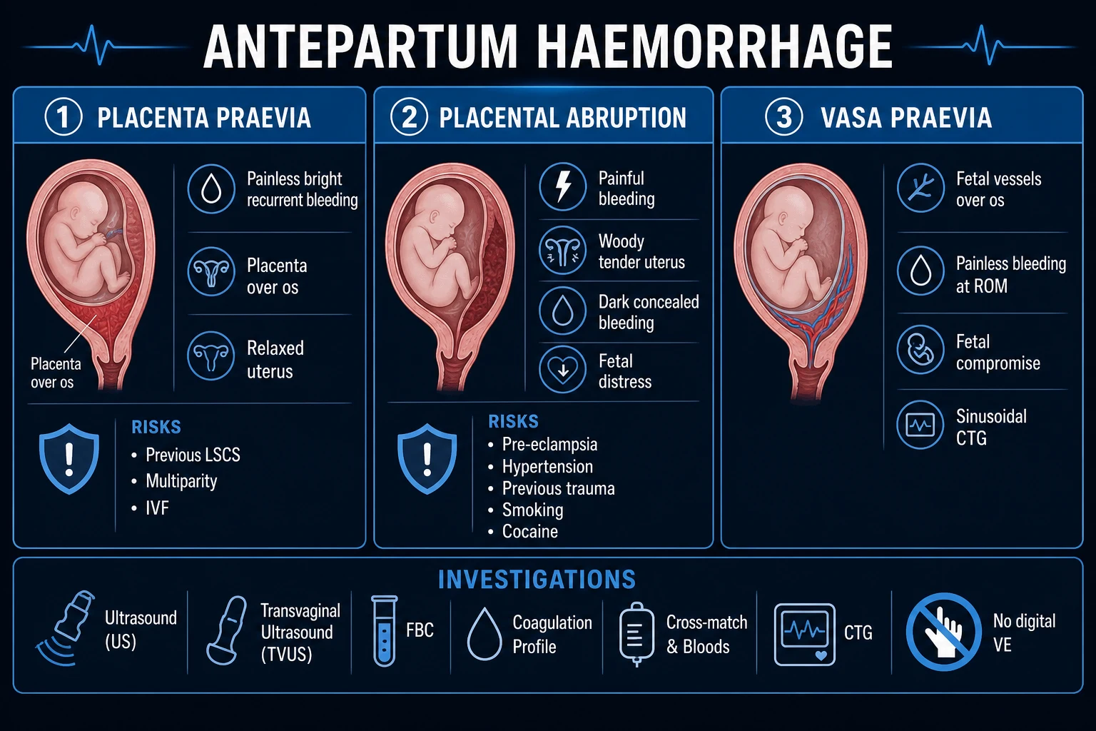
Antepartum haemorrhage (APH) is vaginal bleeding from the genital tract from 24 weeks of gestation until the birth of the baby. The three placental causes are placenta praevia (low placenta over/near the internal os — classically painless, bright-red, recurrent bleeding, soft relaxed non-tender uterus, often abnormal lie — diagnosed by transvaginal ultrasound, never digital vaginal examination until praevia excluded, caesarean if major), placental abruption (premature separation of a normally-sited placenta — painful, tense tender woody-hard uterus, dark or concealed bleeding, fetal distress, shock disproportionate to visible loss; risks hypertension/pre-eclampsia, previous abruption, trauma, smoking, cocaine; resuscitate and deliver), and vasa praevia (fetal vessels running over the membranes across the internal os — painless bleeding at rupture of membranes with sudden fetal compromise (sinusoidal CTG) in a haemodynamically normal mother; emergency caesarean). Always resuscitate, group-and-save/cross-match, FBC/coagulation/fibrinogen, continuous CTG, anti-D to Rh-negative, tranexamic acid 1 g IV if major, and deliver when there is maternal or fetal compromise.
On this page & tools
Your progress
Saved locally on this device.
Exam tags
Red flags
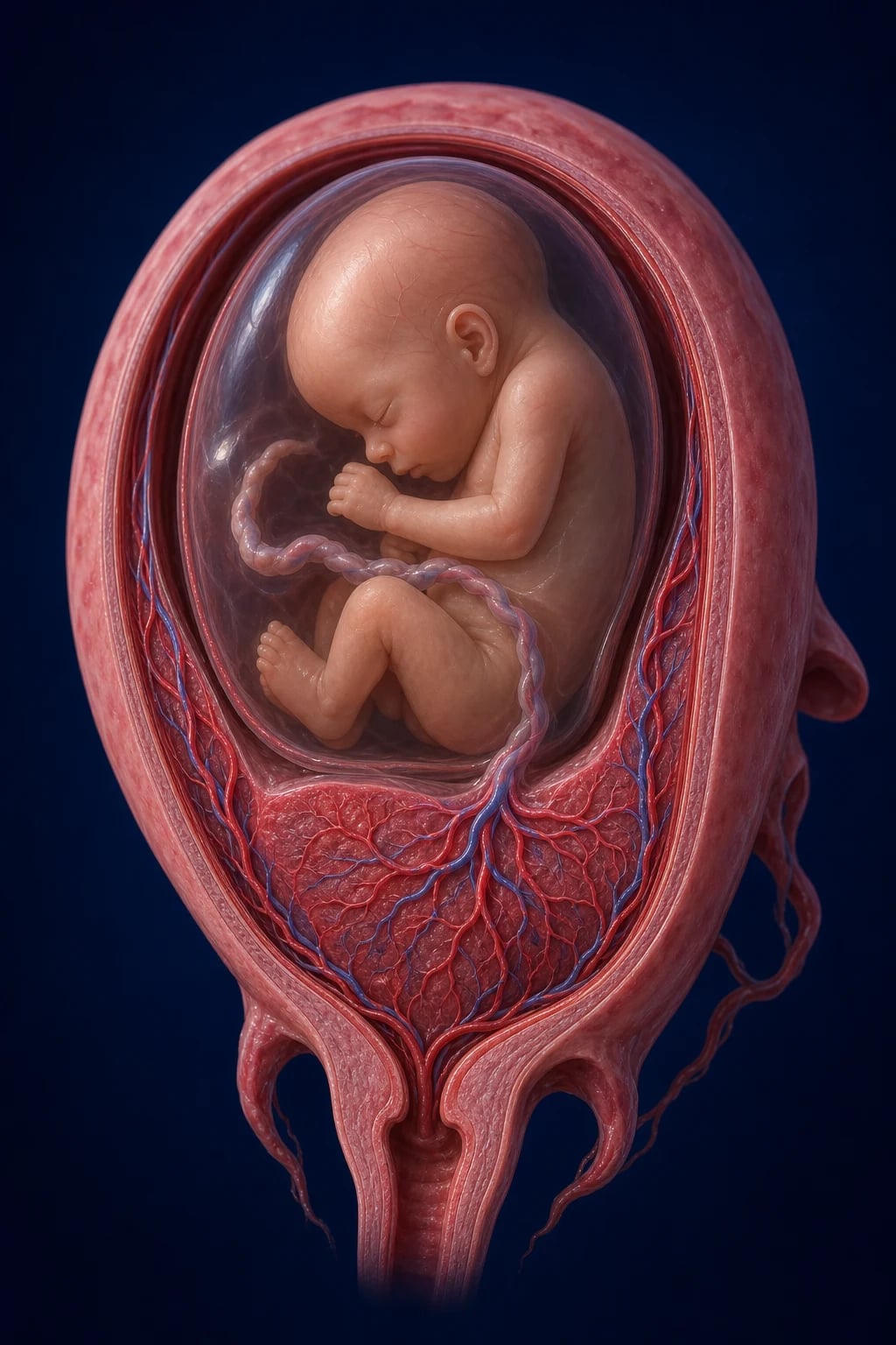
Overview & Definition
Antepartum haemorrhage (APH) is defined as vaginal bleeding from the genital tract occurring after 24 weeks of gestation and until the birth of the baby.[1] The 24-week cut-off is the lower threshold of fetal viability in most settings; bleeding before this gestation is classified as a threatened or inevitable miscarriage, which has a different cause, prognosis, and management pathway. Some textbooks and historical systems use a 20- or 28-week cut-off, so the exact threshold should be stated in any exam answer, but 24 weeks is the operational definition used by RCOG and most modern guidelines.[3]
APH matters because it is common (3 to 5% of pregnancies), it is a leading reason for emergency antenatal admission, and two of its causes — placental abruption and vasa praevia — can kill the fetus within minutes and the mother within hours. The clinical skill it tests is triage: every woman who bleeds in the third trimester must be assumed to have a praevia until ultrasound proves otherwise, must be resuscitated in parallel with assessment, and must be delivered when there is maternal or fetal compromise.[1]
The praevia-versus-abruption clinical contrast — painless vs painful, bright vs dark, relaxed vs woody uterus, mother vs fetus compromised — is one of the highest-yield discriminator pairs in all of obstetrics, and examiners return to it in MCQ, SAQ, viva, and OSCE alike. Around it sits a smaller, equally examinable third player, vasa praevia, plus the surgical emergencies uterine rupture and placenta accreta spectrum, and the broader non-placental causes that must be excluded by a speculum examination once praevia is ruled out.[2]
Classification
APH is classified in two complementary ways — by cause and by severity. [1]
By cause (the examiner's framework): [1]
Placenta praevia
- Placenta implanted low, over or within 2 cm of the internal cervical os
- Painless, bright-red, recurrent bleeding
- Soft, relaxed, non-tender uterus
- Incidence about 0.3 to 0.5%
Placental abruption
- Premature separation of a normally-sited placenta
- Painful, tender, tense woody-hard uterus
- Dark or concealed bleeding with fetal distress
- Incidence about 0.5 to 1%
Vasa praevia
- Fetal vessels running unprotected through the membranes across the internal os
- Painless bleeding at ROM with sudden fetal compromise
- Mother haemodynamically normal (fetal blood)
- Incidence about 1 in 1200 to 1 in 5000 (much higher in IVF)
Uterine rupture
- Full-thickness disruption of the uterine wall, usually through a previous scar
- Sudden severe tearing pain, cessation of contractions
- Fetal bradycardia, loss of station, haemoperitoneum
Placenta accreta spectrum
- Abnormal placental invasion into/through the myometrium
- Catastrophic haemorrhage at attempted separation
- Strongly linked to previous caesarean plus low/anterior placenta
Local (non-placental) causes
- Cervical ectropion or polyp, cervicitis
- Vulval varicosities, cervical cancer
- Post-coital or post-examination bleeding, trauma
Unexplained APH
- Bleed of unknown origin after praevia, abruption, vasa praevia, local causes excluded
- The largest single group
- Behaves like a mild abruption with elevated adverse-outcome risk
By severity (drives the resuscitate-vs-expectant decision): [1]
Placenta praevia itself is graded twice over. The traditional 4-grade system (Macafee–Johnson, based on the relationship of the placenta to the internal os) is still asked in vivas: Grade I low-lying (placenta in lower segment but not reaching the os); Grade II marginal (placenta reaches the edge of the os but does not cover it); Grade III partial (placenta covers the internal os asymmetrically); Grade IV complete (placenta covers the os symmetrically, central). The modern two-category system used in clinical practice collapses these into minor praevia (placental edge more than 2 cm from the internal os — usually safe for vaginal delivery if no other indication) and major praevia (placental edge covering or within 2 cm of the os — caesarean required).[3]
Epidemiology & Risk Factors
APH affects about 3 to 5% of pregnancies and is the commonest reason for emergency antenatal admission in obstetrics. The three placental causes together account for roughly half of all cases; the remainder are local causes and the substantial unexplained group.[1]
Risk factors for placenta praevia: previous caesarean section (dose-response with the number of scars, which also drives accreta), multiparity, advanced maternal age (over 35), multiple gestation, IVF/intracytoplasmic sperm injection, smoking, prior uterine surgery (myomectomy, curettage), and a previously affected pregnancy (recurrence 4 to 8%).[3]
Risk factors for placental abruption were quantified in the Chen 2025 systematic review and meta-analysis:[2] hypertensive disorder of pregnancy (pre-eclampsia or chronic hypertension — the strongest modifiable risk, OR around 2 to 4), previous abruption (RR 10 to 15, rising to 25 after two), maternal trauma (road traffic accident, fall, domestic violence), cigarette smoking (OR around 1.6 to 2.5, dose-dependent and modifiable), cocaine use, advanced maternal age, multiparity, preterm premature rupture of membranes, chorioamnionitis, abrupt decompression of polyhydramnios, short umbilical cord, thrombophilias (factor V Leiden, antithrombin deficiency, antiphospholipid syndrome), and intrauterine growth restriction.
Risk factors for vasa praevia: a low-lying placenta or praevia (especially with a subsequent upward migration), a bilobed or succenturiate-lobed placenta, IVF pregnancy, a second-trimester low placenta, and velamentous cord insertion (where the cord inserts into the membranes rather than the placental disc).[5]
Pathophysiology
The three placental causes of APH have distinct mechanisms, and examiners reward the candidate who can explain why each produces its characteristic clinical picture. [1]
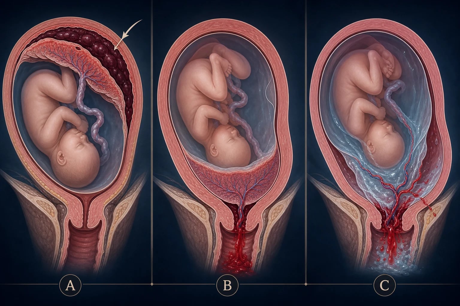
Placenta praevia. In the third trimester the lower uterine segment progressively forms, lengthens, and thins, and the cervix effaces and begins to dilate. The placenta is inelastic and cannot stretch with this thinning tissue, so the placental edge shears away from the underlying decidua. The torn decidual sinus vessels bleed — and because the blood has a short, direct path through the cervix and vagina, it is fresh, bright-red, and painless. The lower segment is poorly contractile, so it cannot compress the bleeding vessels (unlike the fundus postpartum), which is why praevia bleeding tends to be recurrent and to escalate.[3] Because the praevia mass sits low in the pelvis, the presenting part cannot engage, producing the classic unstable, transverse, or breech lie that is the bedside clue.
Placental abruption. The initiating event is a rupture of a maternal decidual spiral artery, producing a haematoma at the decidua–placental interface. As the retroplacental clot expands, it progressively strips the placenta off the uterine wall, shearing further vessels and destroying placental tissue. The patient perceives this as a sudden, constant, severe pain; the uterus is irritated into hypertonus and is tender and tense, woody-hard on palpation. The blood may track down and escape through the cervix (revealed abruption, the more common form) or be retained behind the placenta (concealed abruption). Concealed abruption is the more dangerous form: the separation is more extensive, the myometrium becomes infiltrated with extravasated blood producing the purplish, copper-bruised Couvelaire uterus (uteroplacental apoplexy), and the patient may go into shock disproportionate to the visible blood loss.[2] Couvelaire uterus is contracted but atonic — after delivery it fails to retract and is a cause of postpartum haemorrhage.
Disseminated intravascular coagulation in abruption. The disrupted placenta and decidua release large quantities of tissue factor (thromboplastin) into the maternal circulation, which ignites the extrinsic coagulation cascade systemically. Platelets, fibrinogen, and clotting factors are consumed faster than the liver and bone marrow can replace them, and secondary fibrinolysis is activated. The result is a consumptive coagulopathy (DIC) that converts a surgical bleed into a haematological one. Clinically this presents as oozing from venepuncture sites and gums, prolonged PT and APTT, a low fibrinogen (normal pregnancy fibrinogen is 4 to 6 g/L; abruption drops it below 2 g/L), thrombocytopenia, and raised D-dimer. The trigger (the retained dead placenta and clot) must be removed by delivery for the DIC to resolve.[2]
Vasa praevia. In a velamentous cord insertion or with a bilobed placenta, some fetal vessels run unprotected through the chorionic membranes (no Wharton's jelly) on their way between cord and placenta. When these vessels traverse the membranes over the internal cervical os, they are termed vasa praevia. They are fetal vessels, so they carry fetal (not maternal) blood at fetal blood pressure. The catastrophic event is rupture of the membranes — either spontaneous or artificial — which tears the vessels. The fetus bleeds directly into the amniotic fluid and vagina. The average term fetus has a circulating volume of only about 80 to 100 mL/kg (around 250 to 300 mL total), so the loss of even 50 to 100 mL is a major haemorrhage: the fetus exsanguinates within minutes, producing the classic sudden fetal heart rate abnormality (sinusoidal pattern, then bradycardia) at the very moment the membranes rupture. The mother, by contrast, is haemodynamically normal because no maternal blood has been lost.[5]
Placenta accreta spectrum. Normally the decidua basalis forms a plane of cleavage (Nitabuch's layer) that allows the placenta to separate cleanly at the third stage. When the decidua is deficient — most often beneath a previous caesarean scar where a low anterior placenta has implanted — the chorionic villi invade directly into the myometrium without that cleavage plane. Invasion limited to the inner myometrium is placenta accreta, deeper into the myometrium is placenta increta, and penetration through the serosa to the bladder or broad ligament is placenta percreta. At attempted placental separation the placenta fails to come away, and catastrophic haemorrhage ensues from the uncontrolled open maternal sinus bed.[4]
Uterine rupture. A full-thickness disruption of the uterine wall — almost always through a previous classical or lower-segment scar, but occasionally de novo after trauma, oxytocin hyperstimulation, or grand multiparity. The fetus and placenta may extrude into the peritoneal cavity, the presenting part recoils (loss of station), contractions characteristically cease as the uterine muscle retracts, and the mother develops haemoperitoneum and shock with referred shoulder-tip pain from diaphragmatic irritation. [1]
Clinical Presentation
The clinical presentation is defined by the underlying cause, and the praevia-vs-abruption-vs-vasa praevia contrast is the spine of the assessment. [1]
[1]Placenta praevia presents with painless, causeless, bright-red, recurrent vaginal bleeding, typically after 28 weeks and most commonly around 30 to 34 weeks as the lower segment forms. The bleeding is fresh because the blood has a short transit; it stops spontaneously (the clot tamponades the venous oozing) and then recurs, often more heavily each time. The uterus is soft, relaxed, and non-tender; the lie is often transverse or oblique and the head fails to engage because the praevia sits in the way; fetal heart tones are normal unless there has been major maternal exsanguination. A warning bleed is the small first episode; a major bleed follows days to weeks later.[3]
Placental abruption presents with the sudden onset of constant, severe abdominal or low back pain (described as a tearing or crushing pain that does not radiate in waves like a contraction). The uterus is tender, tense, and woody-hard with poorly relaxing hypertonus between contractions; the fundus may be high if there is concealed blood or a retroplacental clot. Vaginal bleeding, when present, is dark red (it has been in contact with the clot and is partially defibrinated). Fetal distress (recurrent late or variable decelerations, bradycardia) or intrauterine death is common. Maternal shock is disproportionate to the visible blood loss in concealed abruption — the concealed retroplacental clot can hold 1 to 2 L of blood that never reaches the perineum.[2]
Atypical presentations of abruption are deliberately tested: a concealed abruption with no visible bleeding that presents as preterm labour or unexplained fetal distress; a constant backache that the patient (and the candidate) mistakes for musculoskeletal pain; a patient who does not yet look shocked despite extensive placental separation; or rupture of membranes with blood-stained liquor. Abruption is fundamentally a clinical diagnosis — ultrasound is poor at seeing a fresh retroplacental clot, and a normal ultrasound does not exclude it.[2]
Vasa praevia presents at the moment of membrane rupture (spontaneous or artificial amniotomy) with painless vaginal bleeding of fresh or dark blood mixed with amniotic fluid, accompanied by rapid onset fetal heart rate abnormalities — classically a sinusoidal pattern (reflecting fetal anaemia and hypoxia) or sudden bradycardia. The mother is haemodynamically normal because only fetal blood has been lost — this is the cardinal clue.[5]
Uterine rupture presents as sudden, severe, tearing abdominal pain during labour (or rarely antenatally after trauma), cessation of previously painful contractions (the uterus has retracted away from the defect), loss of the presenting part (the head that was engaged recoils), fetal bradycardia, vaginal bleeding, and maternal shock with shoulder-tip pain from haemoperitoneum. A rupture may also present more insidiously as scar tenderness, persistent tachycardia, or non-reassuring CTG in a woman labouring after a previous caesarean.[1]
Local (non-placental) causes present with lighter bleeding, often post-coital or after a vaginal examination, and a normal fetus and uterus. Cervical ectropion (common in pregnancy and with the pill), cervical polyp, cervicitis, vulval varicosities, or — importantly — cervical cancer must be considered. Even when a local cause is suspected, praevia must be excluded by ultrasound before any speculum or digital examination, and any visible cervical lesion must be referred for biopsy. [1]
A 'show' (the blood-tinged mucus plug expelled at the onset of labour) is mucus mixed with a small amount of blood and is not a true APH — but a candidate must be able to distinguish it, and any heavier bleeding in labour must be managed as possible praevia or abruption until proven otherwise. [1]
Differential Diagnosis
The differential of APH is the list of causes above, and the task is to distinguish them rather than enumerate them. [1]
Placenta praevia vs abruption
- Praevia: painless, bright-red, recurrent, soft relaxed uterus, abnormal lie, mother well
- Abruption: painful, dark/concealed, woody-hard tender uterus, fetal distress, shock disproportionate
- Ultrasound localises the placenta (TVS safe and accurate)
Abruption vs uterine rupture
- Both painful and shocking
- Rupture: previous scar, cessation of contractions, loss of station, haemoperitoneum, shoulder-tip pain, often during labour
- Abruption: contractions continue (often hypertonic), uterus tender and woody, usually hypertensive or trauma history
Vasa praevia vs praevia/abruption
- Vasa: bleeding AT rupture of membranes, mother haemodynamically normal
- Fetal compromise disproportionate (sinusoidal CTG), Apt test positive (fetal blood)
- Praevia and abruption compromise the mother; vasa compromises the fetus
Local cause vs placental
- Local (ectropion, polyp, cancer): light bleeding, often post-coital, normal uterus and fetus, visible lesion on speculum
- Placental: heavier, with characteristic pain/tone/fetal-pattern findings
- Always exclude praevia by ultrasound first
Show vs APH
- Show = blood-tinged mucus plug at term with onset of labour
- Small, self-limiting, no clots
- APH = heavier, with clots and/or pain; managed as APH until proven otherwise
Unexplained APH
- Bleed of unknown origin after praevia, abruption, vasa praevia, local causes excluded
- Behaves like a mild abruption: higher rates of IUGR, preterm birth, stillbirth
- Warrants enhanced surveillance in the rest of the pregnancy
The can't-miss mimic is vasa praevia masquerading as a normal third-trimester bleed — missing it converts a 95% survival (antenatally diagnosed) into a 50%-or-worse mortality. The classic examiner's trap is attributing fetal distress in an abruption to "prematurity" and delaying delivery: the perinatal mortality of abruption is dominated by delay, not by the bleed itself.[2]
Clinical & Bedside Assessment
The focused assessment of APH runs in parallel with resuscitation (the two do not happen in sequence). The history establishes onset, volume (in sanitary pads or clots), colour, pain, relation to ROM, fetal movements, trauma, drug use (cocaine, anticoagulants), obstetric history (gestational age, parity, previous scars, previous abruption or praevia), and risk factors. [1]
Abdominal examination is the bedside discriminator: [1]
Praevia
- Uterus soft, relaxed, non-tender; normal tone between Braxton-Hicks
- Lie often unstable/transverse/oblique; presenting part high and unengaged
- Fundal height appropriate; fetal heart normal unless major maternal loss
Abruption
- Uterus tense, tender, woody-hard; hypertonus between contractions
- Fundus may be high (concealed clot/bleed); lie usually longitudinal
- Severe abdominal or back pain on palpation; fetal distress or absent heart
Vasa praevia (post-ROM)
- Uterus and mother normal
- Only the CTG is abnormal (sinusoidal pattern or bradycardia)
Rupture
- Loss of uterine contour; tender abdomen with guarding/peritonism from haemoperitoneum
- Absent or altered fetal parts
- Shock out of proportion to vaginal loss
A speculum examination is used (after ultrasound) to assess the cervix and vagina for a local cause, to visualise the source of bleeding, and to take swabs — but never digital. The speculum may be left in place to observe ongoing loss. [1]
Assess shock in four quadrants. The young pregnant woman compensates remarkably: tachycardia, tachypnoea, and a narrowed pulse pressure (the diastolic rises with sympathetic drive) appear before hypotension. Hypotension is a late, pre-terminal sign — by the time the systolic drops, more than 30 to 40% of the blood volume is already lost. Cool peripheries, delayed capillary refill, anxiety, and oliguria signal decompensated shock. [1]
Named signs and manoeuvres. The woody-hard uterus of concealed abruption feels like a board — the fundus is tender, the uterus does not relax between Braxton-Hicks, and the patient winces on light palpation. A high fundus for dates suggests concealed blood or a retroplacental clot. Loss of station of the presenting part during labour signals uterine rupture. The Apt test (see Investigations) is the bedside laboratory test that confirms fetal blood in suspected vasa praevia. [1]
Investigations
Investigations in APH serve four purposes: to localise the placenta (praevia), to identify vasa praevia, to support (not exclude) abruption, to diagnose accreta antenatally, and to prepare for delivery and transfusion. [1]
Bloods (every APH): [1]
- Full blood count — haemoglobin, haematocrit, platelets (thrombocytopenia in DIC); note that Hb at presentation underestimates the loss because haemodilution takes hours.
- Group and save for minor APH; cross-match 4 to 6 units for major APH; activate the massive haemorrhage protocol if shock.
- Coagulation screen — PT, APTT, fibrinogen (the single most useful coagulation test in obstetric haemorrhage; aim to keep above 2 g/L, some guidelines 4 g/L in pregnancy), D-dimer.
- Urea and electrolytes, liver function tests — baseline and to detect pre-eclampsia/HELLP as a comorbidity.
- Blood gas — base deficit and lactate quantify shock severity.
- Kleihauer-Betke test or flow cytometry — to size the fetomaternal haemorrhage in Rh-negative mothers (to dose anti-D) and after maternal trauma. [1]
Ultrasound — localise the placenta. The single most important diagnostic step. Transabdominal ultrasound is performed first; if the placenta is low or the view is unclear, transvaginal ultrasound (TVS) is performed — it is safe in praevia (it does not provoke bleeding) and is more accurate because the internal os is seen directly. The placental edge is measured relative to the internal os: major praevia if the placenta covers or lies within 2 cm; minor praevia if it is more than 2 cm from the os (and vaginal delivery is usually feasible).[3] A praevia diagnosed at the 20-week anomaly scan commonly resolves (the lower segment forms and the placenta appears to 'migrate' away from the os), so the diagnosis is confirmed by a repeat scan at 32 weeks (and again at 36 if still low).
Ultrasound — vasa praevia. Transvaginal ultrasound with colour and pulsed-wave Doppler is the diagnostic test: it demonstrates fetal vessels crossing the internal os, with the characteristic arterial waveform matching the fetal heart rate. Universal screening is debated but targeted screening in high-risk women (low placenta, bilobed or succenturiate placenta, IVF pregnancy, velamentous cord insertion) is recommended and converts the perinatal mortality from over 50% to under 5%.[5]
Apt test (Singer test). A bedside chemical test on vaginal blood that distinguishes fetal from maternal blood. The blood is mixed with sodium hydroxide (an alkali): adult haemoglobin denatures and turns brown/pinkish-brown, whereas fetal haemoglobin (which resists alkali denaturation) stays pink. A pink result = fetal blood = vasa praevia until proven otherwise. Modern alternatives include the Kleihauer-Betke test and flow cytometry on the vaginal blood. [1]
Ultrasound and MRI — placenta accreta spectrum. Ultrasound features of accreta include: loss of the normal retroplacental 'clear space' (sonolucent zone), placental lacunae (swiss-cheese appearance), myometrial thinning to under 1 mm, bladder wall interruption (loss of the hyperechoic bladder–uterine interface), and increased vascularity on colour Doppler (abnormal placental bed vascularity). MRI is used as an adjunct for suspected deep invasion (increta, percreta) and to map the anatomy pre-operatively, particularly the relationship to the bladder.[3][4]
Placental abruption is a clinical diagnosis — ultrasound is insensitive for an acute retroplacental clot (sensitivity around 25 to 50%). A normal ultrasound does not exclude abruption. When seen, a retroplacental clot appears as a heterogeneous, hyperechoic then hypoechoic mass behind the placenta that compresses the placenta; an elevated fundal height and a tender uterus are more reliable than the scan.[2]
Continuous cardiotocography (CTG) is mandatory in any APH of concern. Recurrent late decelerations, bradycardia, reduced variability, or a sinusoidal pattern suggest abruption or vasa praevia. A sinusoidal pattern (smooth, undulating baseline of 5 to 15 cycles per minute, normal variability absent) in the context of bleeding at ROM is vasa praevia until proven otherwise.[5]
Speculum examination (after ultrasound has excluded praevia) inspects the cervix and vagina for a local cause and to visualise the bleeding source; cervical polyps, ectropion, and suspicious lesions can be biopsied. [1]
Management — Resuscitation
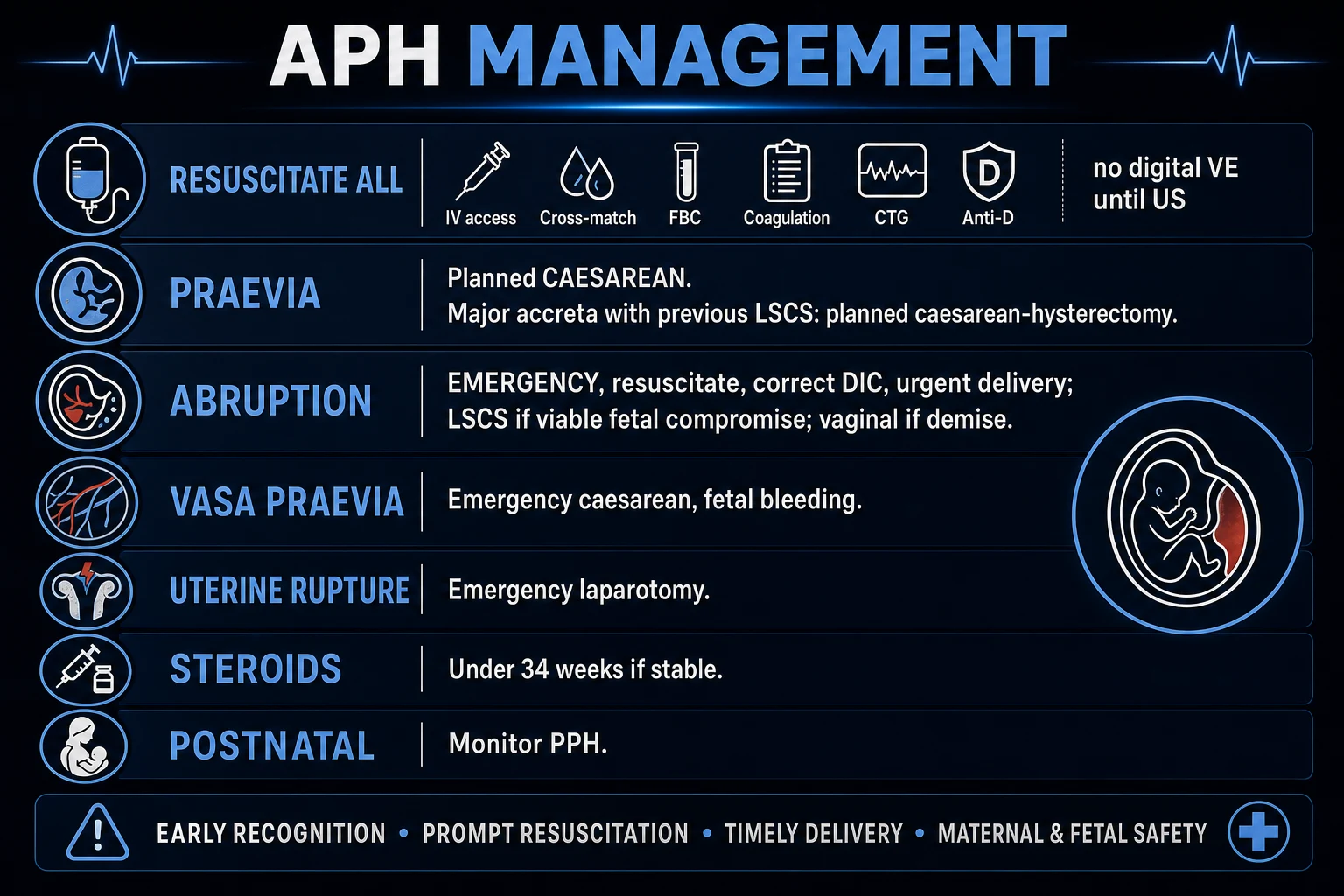
Resuscitation and assessment run in parallel, not in sequence. The aim is to restore circulating volume, correct coagulopathy, monitor the fetus, and prepare for delivery while the obstetric team gathers. [1]
RAPID
ABC, oxygen, 2 large-bore IV cannulae (14–16 G), warm crystalloid bolus (Hartmann's or 0.9% saline 1 to 2 L) in aliquots, left lateral tilt to relieve aortocaval compression
Call for help: senior obstetrician, anaesthetist, haematology, neonatal team, alert theatre; activate massive haemorrhage protocol for major bleed
FBC, group and save or cross-match 4–6 units, coagulation incl. fibrinogen, U&E, LFTs, Kleihauer-Betke (Rh-negative or trauma); blood gas for base deficit
Continuous CTG, ultrasound to localise placenta, urinary catheter (hourly output); do NOT perform a digital vaginal exam
Anti-D to Rh-negative within 72 h; tranexamic acid 1 g IV if major bleed; betamethasone if under 34 weeks and stable; magnesium sulphate for neuroprotection if under 32 weeks
Fluid resuscitation: warm isotonic crystalloid (Hartmann's solution is preferred over 0.9% saline because it does not cause hyperchloraemic acidosis) 1 to 2 L as a bolus in shocked patients, in aliquots of 250 to 500 mL, titrated to response. Blood transfusion with red cells, fresh-frozen plasma, platelets, and cryoprecipitate in a near-1:1:1 ratio for massive haemorrhage (mimicking whole blood). Keep fibrinogen above 2 g/L with cryoprecipitate; this is the single most important coagulation target in obstetric haemorrhage because pregnancy fibrinogen is normally high (4 to 6 g/L) and a low value is the earliest marker of DIC. Keep the patient warm (avoid hypothermia, acidosis, and hypocalcaemia — the lethal triad of trauma that worsens coagulopathy).[1]
Anti-D immunoglobulin 500 IU intramuscularly within 72 hours to every Rh-negative, non-sensitised woman with any APH (because even small bleeds can cause fetomaternal haemorrhage); the dose is sized up using the Kleihauer-Betke test to detect large-volume fetomaternal haemorrhage (additional 500 IU per 4 mL of fetal bleed or 25 mL of fetal cells detected, per local protocol). [1]
Tranexamic acid 1 g intravenously over 10 minutes (repeat once after 30 minutes if bleeding continues) — an antifibrinolytic that stabilises clot; given early, extrapolated from the WOMAN-trial evidence in postpartum haemorrhage and recommended in major APH with coagulopathy.[1]
Corticosteroids for fetal lung maturation: betamethasone 12 mg IM, two doses 24 hours apart, or dexamethasone 6 mg IM, four doses 12 hours apart, if the gestational age is under 34 weeks (some guidelines under 35 weeks) and the mother is stable enough to wait. The aim is to buy 24 to 48 hours for surfactant production before delivery. [1]
Magnesium sulphate for fetal neuroprotection (4 g IV loading over 20 minutes, then 1 g/hour infusion) if delivery is anticipated before 32 weeks — reduces the risk of cerebral palsy in the surviving preterm infant. [1]
[1]Management — Definitive & Stepwise
Definitive management is dictated by the cause and the gestational age and condition of mother and fetus. [1]
Placenta praevia. The principle is no digital vaginal examination and caesarean delivery for major praevia. Planned caesarean section is performed at 36 to 37 weeks (before labour, to avoid an emergency presentation with the cervix dilating and the praevia edge shearing).[3] A praevia with the edge more than 2 cm from the os may be allowed a trial of vaginal delivery if there is no other indication for caesarean and the patient consents. The Macafee–Johnson expectant regimen is used when the fetus is immature and bleeding has settled: inpatient bed rest, cross-matched blood held at all times, no vaginal examination, serial growth and wellbeing scans, and corticosteroids — to gain gestational age until 36 to 37 weeks, balancing the risk of a catastrophic recurrent bleed against the risk of prematurity. Anti-D is given to Rh-negative mothers after each bleed.
Placental abruption. The principle is resuscitate, correct coagulopathy, and deliver — delivery is the definitive treatment because it removes the trigger for DIC and stops the bleed. Vaginal delivery (amniotomy plus oxytocin infusion to augment) is appropriate if the fetus is dead (intrauterine death), or the fetus and mother are stable and vaginal delivery is achievable in a reasonable time; the dead fetus still needs prompt delivery because the retained dead placenta perpetuates DIC. Emergency caesarean section is performed if the fetus is alive with compromise, or if vaginal delivery is not feasible or would be unsafe (e.g. severe pre-eclampsia, prior classical scar).[2] Correct coagulopathy before and during surgery with cryoprecipitate, FFP, and platelets — operating with an INR over 1.5 or fibrinogen under 2 g/L courts disaster. The uterus may be Couvelaire and atonic — be ready for postpartum haemorrhage (see uterotonic ladder below).
Vasa praevia. Planned caesarean section at 34 to 35 weeks (earlier than praevia because of the risk of spontaneous ROM and catastrophic fetal bleeding), with antenatal counselling that the rationale is to deliver before the membranes rupture spontaneously. Emergency caesarean if presenting with acute bleeding at ROM and a live fetus. The Apt test confirms fetal blood. If the fetus is already dead, the mode of delivery follows obstetric indications.[5]
Placenta accreta spectrum. Multidisciplinary planned caesarean at 34 to 36 weeks in a centre of excellence, with the consultant obstetrician, anaesthetist, haematologist, urologist (if percreta into the bladder), and interventional radiologist present. Options are elective caesarean–hysterectomy (the traditional definitive procedure, leaving the placenta in situ within the removed uterus) or conservative management (delivering the baby, tying the cord short, and leaving the placenta in situ to involute over months, accepting the risk of infection, delayed haemorrhage, and need for methotrexate). Pre-operative balloon catheters in the internal iliac arteries (inflated at delivery to reduce pelvic inflow), cell salvage (now considered acceptable in obstetrics with a separate suction line for amniotic fluid), and a massive haemorrhage protocol are standard.[4]
Uterine rupture. Immediate laparotomy under general anaesthesia. The fetus is delivered, the rupture is repaired if feasible, or hysterectomy is performed if the tear is irreparable or bleeding is uncontrollable. Aggressive blood and clotting-factor replacement is essential. Neonatal resuscitation is anticipated. [1]
The uterotonic and surgical ladder for refractory bleeding at caesarean (accreta, atonic Couvelaire uterus, or concurrent PPH): [1]
- Uterine massage and bimanual compression.
- Oxytocin 5 IU IV bolus (over 1 minute), then infusion 40 IU in 500 mL Hartmann's at 125 mL/hour.
- Ergometrine 0.25 mg IM (or 0.25 mg IV slowly) — avoid in hypertension/pre-eclampsia (it is a vasopressor and precipitates hypertensive crisis).
- Carboprost (15-methyl PGF2-alpha) 250 micrograms IM every 15 minutes, maximum 8 doses (2 mg total) — avoid in asthma (it is a bronchoconstrictor).
- Misoprostol 800 micrograms sublingual or rectally — safe in asthma and hypertension.
- Tranexamic acid 1 g IV (repeat once at 30 minutes).
- Uterine balloon tamponade (Bakri balloon) inflated with warm saline.
- Compression sutures (B-Lynch, Cho).
- Vessel ligation — uterine artery (stepwise devascularisation), then internal iliac (hypogastric) artery ligation (bilateral).
- Hysterectomy — the life-saving last resort; do not delay if the patient is exsanguinating. [1]
Specific Subtypes & Scenarios
Minor / unexplained APH. After excluding praevia, abruption (clinically), vasa praevia (in high-risk cases), and local causes by speculum, a residual 'unexplained' bleed is the commonest single label. It is not benign: it behaves like a mild abruption with higher rates of intrauterine growth restriction, preterm birth, and stillbirth, so the rest of the pregnancy warrants enhanced surveillance (growth scans, umbilical artery Doppler, weekly CTG or reduced fetal movements review, and patient warning about recurrent bleeding).[2]
APH with a viable fetus vs a previable fetus. Management is modified by gestational age. Under 34 weeks with a stable mother and fetus, the aim is to gain gestational age (corticosteroids, expectant inpatient care, careful monitoring) — the Macafee–Johnson regimen for praevia, inpatient observation for abruption that has settled. At 34 weeks and beyond, or with compromise at any gestation, deliver. Before 24 weeks the bleed is classified and managed as a threatened miscarriage.[3]
APH with trauma (road traffic accident, fall, domestic violence). Direct abdominal trauma carries a significant risk of abruption (from shearing forces or seat-belt injury) and rarely of uterine rupture. Assess the mother (ABCDE), then the fetus (CTG for 4 to 6 hours minimum, or 24 hours if any abnormality); perform a Kleihauer-Betke test to size fetomaternal haemorrhage (especially in Rh-negative women); ultrasound for placenta, fetal wellbeing, and retroplacental clot. Deliver for maternal or fetal compromise, non-reassuring CTG, or major abruption. Domestic violence screening is mandatory — APH may be its presentation.[6]
The pregnant woman on anticoagulation. A woman on therapeutic low-molecular-weight heparin (e.g. for a mechanical heart valve or thrombophilia) presenting with APH is balanced between bleeding (the presenting problem) and thrombosis (the indication). Withhold the next LMWH dose, consider protamine sulphate reversal (1 mg per 100 IU of LMWH given in the previous 8 hours, partial reversal), and involve haematology; once bleeding is controlled, restart prophylactic LMWH as soon as safe. Warfarin is reversed with vitamin K and prothrombin complex concentrate. [1]
APh with preterm labour. Abruption often presents as preterm labour (the retroplacental clot irritates the uterus). Tocolysis is generally contraindicated in significant abruption (it may mask the diagnosis and worsen the bleed) — the management is delivery, not tocolysis. Steroids for fetal lung maturation are given if time allows. [1]
Complications & Pitfalls
Maternal complications of APH: haemorrhagic shock, disseminated intravascular coagulation, acute kidney injury (acute tubular or cortical necrosis from prolonged hypotension), Sheehan syndrome (anterior pituitary infarction from profound hypovolaemia — failure to lactate, amenorrhoea, secondary hypothyroidism and adrenal insufficiency), postpartum haemorrhage (from atonic Couvelaire uterus or accreta), peripartum hysterectomy and loss of fertility, infection (chorioamnionitis, wound infection), thromboembolism (the hypercoagulable state plus immobility), psychological trauma, and Rh sensitisation in the unprotected mother.[1]
Fetal and neonatal complications: prematurity (iatrogenic and spontaneous), intrauterine growth restriction, hypoxic ischaemic encephalopathy, intrauterine death, fetal anaemia and exsanguination (vasa praevia), birth asphyxia, and neonatal coagulopathy.[2]
Couvelaire uterus (uteroplacental apoplexy). In concealed abruption the blood extravasates through the myometrium to the serosa, giving a purplish, copper-bruised appearance. The muscle is contracted but functionally atonic — after delivery it fails to retract and is a cause of refractory postpartum haemorrhage, often forcing hysterectomy. [1]
[1]Recurrence: abruption recurs in 10 to 15% (rising to 25% after two previous episodes); praevia and accreta recur with the number of caesareans; aspirin 75 to 150 mg daily from 12 weeks reduces recurrent abruption and pre-eclampsia in high-risk women.[2]
Prognosis & Disposition
Maternal outcome is determined by the cause, severity, gestational age, presence of coagulopathy, and the speed of resuscitation and delivery. In well-resourced settings with prompt care, maternal mortality is low; in low-resource settings with delayed access, APH remains a leading direct cause of maternal death.[1]
Perinatal outcome by cause: [1]
Disposition: a woman with major APH is managed in a tertiary obstetric unit with on-site theatre, haematology, neonatology, and (for accreta) interventional radiology. After a minor, settled, explained bleed, she may be discharged with a clear safety-net: return immediately for recurrent or heavier bleeding, abdominal pain, reduced fetal movements, rupture of membranes, or fever; anti-D follow-up; and a plan for enhanced antenatal surveillance.[3]
Follow-up: review at the next antenatal visit; arrange growth scans and Doppler for unexplained APH (which behaves like a mild abruption); address modifiable risk factors (smoking cessation, blood pressure optimisation); offer psychological support; counsel on future pregnancy (risk of recurrence, the role of aspirin, surveillance for praevia/accreta, mode of future delivery). [1]
Special Populations
Previous caesarean scar plus low/anterior placenta (placenta accreta spectrum). The highest-risk constellation in modern obstetrics. A woman with one or more previous caesareans and a low anterior placenta should have a formal accreta assessment by ultrasound at 32 weeks, with MRI if sonographic features are present, and planned delivery at 34 to 36 weeks in a centre of excellence with multidisciplinary input and consent for possible hysterectomy.[4]
Pre-eclampsia or chronic hypertension. These women are at markedly elevated risk of abruption (the strongest modifiable risk factor). Optimise blood pressure, aspirin 75 to 150 mg daily from 12 weeks (reduces both pre-eclampsia and abruption), and counsel them to report abdominal pain or bleeding urgently with a low threshold to admit and assess.[2]
IVF, multiple pregnancy, and high-risk placental anatomy. These women are at elevated risk of praevia and vasa praevia. Routine transvaginal ultrasound with colour Doppler at the 20-week anomaly scan (with targeted screening in those with a low placenta, bilobed placenta, or velamentous cord insertion) is the standard of care.[5]
Anticoagulated woman. See Specific Subtypes: withhold the next dose, consider reversal, balance bleeding against thrombosis, involve haematology early. [1]
Previable fetus (under 24 weeks). Bleeding is classified as threatened miscarriage (or ectopic until intrauterine pregnancy confirmed); management is expectant with anti-D to Rh-negative women. The 24-week cut-off is regional (some settings use 20 or 28 weeks); state the definition you are using.[3]
Jehovah's Witness patient. Refusal of blood products markedly increases maternal mortality from major APH. Management must include early multidisciplinary discussion, a documented management plan, cell salvage (often acceptable), tranexamic acid, erythropoietin, iron, and explicit consent about which components are and are not acceptable. Mortality rises about 6-fold if blood is refused in a major bleed.[1]
Evidence, Guidelines & Regional Differences
The principal guidelines are RCOG Green-top Guideline No. 27a (placenta praevia) and 27b (placenta accreta), updated by Jauniaux and colleagues in 2019;[3] ACOG Committee Opinion on placenta accreta spectrum in the United States; RANZCOG guidance in Australia and New Zealand; and FOGSI guidance in India. They are broadly concordant but differ in detail:
The diagnosis of praevia has converged on the 2 cm rule: a placental edge within 2 cm of the internal os at term is managed as major praevia (caesarean); an edge more than 2 cm away may attempt vaginal delivery. The cut-off was previously 2 cm but some older texts and exam questions may cite 2 cm vs a 'safe' distance — answer the 2 cm threshold.[3]
Landmark evidence: [1]
- Silver et al (2015) defined the centre of excellence for placenta accreta — multidisciplinary planning, consultant presence, and pre-operative planning reduce maternal morbidity.[4]
- Jauniaux et al (2019) integrated praevia and accreta guidance in Green-top Guideline No. 27, formalising the 2 cm rule, the role of MRI, and the conservative-management option.[3]
- Chen et al (2025) quantified the independent risk factors for placental abruption in a systematic review and meta-analysis, confirming hypertension, prior abruption, smoking, and trauma as the dominant drivers.[2]
- Oyelese et al (2004) established that antenatal diagnosis of vasa praevia dramatically improves perinatal survival, underpinning the case for targeted screening.[5]
- The WOMAN trial (2017) established tranexamic acid 1 g IV early as standard in postpartum haemorrhage, and its principles are extrapolated to major APH with coagulopathy.
Controversies. Whether to screen universally for vasa praevia (cost-effectiveness debated, but growing consensus for at least targeted screening). The optimal timing of delivery in accreta (balancing iatrogenic prematurity against emergency presentation with bleeding). The role of interventional radiology balloon catheters (helpful but with their own complications). The acceptability and safety of cell salvage in obstetrics (now widely accepted with a separate suction line for amniotic fluid). The conservative (placenta-in-situ) management of accreta — increasingly offered in selected cases but carries real risks of infection, re-bleeding, and delayed hysterectomy. [1]
Exam Pearls
APH = PAVE
Painless, bright-red, recurrent; placenta over os; previous LSCS; NO digital VE; caesarean if major
Painful, woody, dark/concealed, fetal distress, shock disproportionate; risks HTN/PE, trauma, smoking, cocaine; deliver
Painless bleeding AT ROM, sinusoidal CTG, mother well, fetal blood (Apt test); emergency caesarean
Uterine rupture (sudden pain, cessation of contractions, loss of station); accreta (previous LSCS + low placenta, catastrophic at separation)
- APH = bleeding from 24 weeks to birth. Below 24 weeks it is threatened miscarriage.[1]
- Praevia: painless, bright-red, recurrent; soft relaxed uterus; abnormal lie; previous LSCS. Never digital VE until ultrasound excludes praevia. Abruption: painful, woody-hard, dark/concealed, fetal distress, shock disproportionate. Risks HTN/PE, previous, trauma, smoking, cocaine.[2]
- Vasa praevia: painless bleeding at rupture of membranes + sudden fetal compromise (sinusoidal CTG); mother haemodynamically normal because it is fetal blood. Emergency caesarean. Apt test positive (fetal Hb resists alkali denaturation, stays pink).[5]
- Concealed abruption = Couvelaire uterus + DIC + shock out of proportion; classical hard board-like abdomen; high fundus.
- Resuscitate every APH: ABC, 2 large-bore IV, cross-match 4 to 6 units, FBC/coagulation/fibrinogen, continuous CTG, anti-D to Rh-negative, tranexamic acid 1 g IV, betamethasone if under 34 weeks and stable, magnesium sulphate for neuroprotection if under 32 weeks.[1]
- Planned delivery timing: praevia 36 to 37 weeks, vasa praevia 34 to 35 weeks, accreta 34 to 36 weeks.[3]
- Accreta: previous LSCS + low/anterior placenta; ultrasound features — loss of retroplacental clear space, placental lacunae, myometrium under 1 mm, bladder wall interruption.[4]
- Ergometrine: avoid in hypertension/pre-eclampsia (vasopressor). Carboprost: avoid in asthma (bronchoconstrictor). Misoprostol: safe in both.
- Sheehan syndrome: anterior pituitary infarction from profound hypovolaemic shock — failure to lactate, amenorrhoea, hypothyroidism, adrenal insufficiency.
- Aspirin 75 to 150 mg daily from 12 weeks reduces recurrent abruption and pre-eclampsia in high-risk women.[2]
- Macafee–Johnson regimen: expectant inpatient care (bed rest, cross-matched blood at all times, no VE, serial scans) for immature praevia with settled bleeding to gain gestational age.
- Abruption is a clinical diagnosis — ultrasound is insensitive for retroplacental clot; a normal scan does not exclude it. Deliver the live compromised fetus by emergency caesarean; the dead fetus by prompt vaginal delivery (with amniotomy and oxytocin) to stop DIC.
- 2 cm rule: placental edge within 2 cm of internal os at term = caesarean; more than 2 cm = may trial vaginal.
Exam application bank (NEET-PG / INICET)
One-line answer
Antepartum haemorrhage (APH) is vaginal bleeding from the genital tract from 24 weeks of gestation until the birth of the baby. The three placental causes are placenta praevia (low placenta over/near the internal os — classically painless, bright-red, recurrent bleeding, soft relaxed non-tender uterus, often abnormal lie — diagnosed by transvaginal ultrasound, never digital vaginal examination until praevia excluded, caesarean if major), placental abruption (premature separation of a normally-sited placenta — painful, tense tender woody-hard uterus, dark or concealed bleeding, fetal distress, shock disproportionate to visible loss; risks hypertension/pre-eclampsia, previous abruption, trauma, smoking, cocaine; resuscitate and deliver), and vasa praevia (fetal vessels running over the membranes across the internal os — painless bleeding at rupture of membranes with sudden fetal compromise
Worked stems (answer without another resource)
Stem 1 — Classic presentation. Map symptoms to mechanism; name the first investigation and first treatment step with dose/route if drug therapy is standard. [1]
Stem 2 — Unstable / complicated. List red flags that force immediate resuscitation, theatre, ICU, antidote, or reperfusion — and what you do in the first 15 minutes. [1]
Stem 3 — Atypical group. Elderly, pregnancy, child, or immunocompromised: how presentation and thresholds change. [1]
Stem 4 — Differential trap. Name the three closest mimics and one discriminator for each. [1]
Stem 5 — Disposition. Who goes home with safety-netting, who is admitted, who needs HDU/ICU/theatre, and what follow-up is mandatory. [1]
Rapid viva checklist
- Definition + classification
- Pathophysiology chain
- Bedside signs / criteria
- Score with exact components (if any)
- Emergency bundle
- Definitive therapy with doses
- Complications of disease and of treatment
- Special populations
- Guideline/trial name if classic
- Three exam traps
Coverage self-check
If you cannot answer any stem above from this page alone, re-read the matching section — the page is intended to be self-sufficient for final-prof and NEET-PG/INICET questions on Antepartum Haemorrhage.
[1]References
- [1]Young JS, Jeelani L, Green BS, Mahendru A, Khan R. Vaginal Bleeding in Late Pregnancy Emerg Med Clin North Am, 2019.PMID 30940370
- [2]Chen D, Li X, Wang Y, et al. Independent risk factors for placental abruption: a systematic review and meta-analysis BMC Pregnancy Childbirth, 2025.PMID 40140972
- [3]Jauniaux E, Alfirevic Z, Bhide AG, et al. Placenta Praevia and Placenta Accreta: Diagnosis and Management: Green-top Guideline No. 27a BJOG, 2019.PMID 30260097
- [4]Silver RM, Fox KA, Barton JR, et al. Center of excellence for placenta accreta Am J Obstet Gynecol, 2015.PMID 25460838
- [5]Oyelese Y, Catanzarite V, Prefumo F, et al. Three-dimensional sonographic diagnosis of vasa previa Ultrasound Obstet Gynecol, 2004.PMID 15287065
- [6]Wright GL, Geller BS, Goh W, et al. Placental Abruption: Temporal Trends, Risk Factors, and Associated Adverse Maternal Outcomes Am J Perinatol, 2026.PMID 40940025