obstetrics-gynaecology · obstetrics-gynaecology
Endometriosis
Also known as Endometriosis · Endometrioma · Chocolate cyst · Deep infiltrating endometriosis · DIE
Endometriosis is the presence of functional endometrial glands and stroma outside the uterine cavity, producing a chronic, oestrogen-dependent, inflammatory condition. It classically presents with the triad of secondary dysmenorrhoea, deep dyspareunia and chronic pelvic pain, with infertility a frequent sole presentation. The ovary is the commonest site; the three phenotypes are superficial peritoneal, ovarian endometrioma ('chocolate cyst') and deep infiltrating endometriosis (DIE). Diagnosis is clinical plus transvaginal ultrasound; MRI maps DIE; laparoscopy with histology is the gold standard. Management is NSAIDs plus hormonal suppression (combined OCP, progestogens, GnRH analogues/antagonists) and surgery (excision/ablation, cystectomy) for refractory disease, DIE and selected infertility.
On this page & tools
Your progress
Saved locally on this device.
Exam tags
Red flags
Overview & Definition
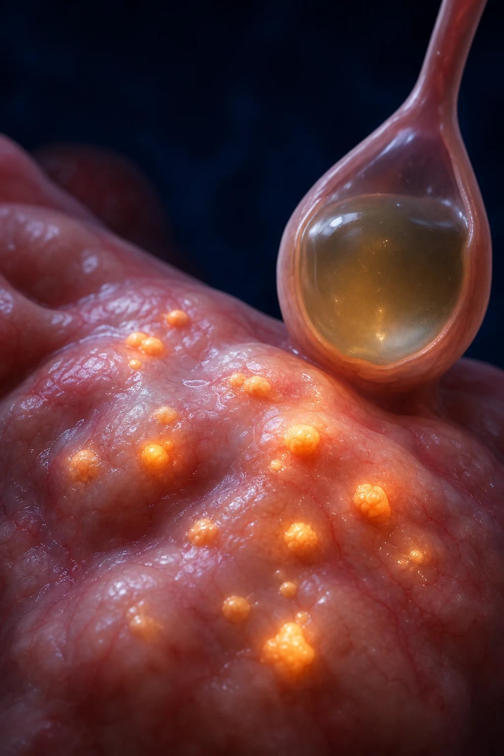
Endometriosis is defined as the presence of functional endometrial glands and stroma outside the uterine cavity — tissue that, like the eutopic endometrium, responds to cyclic ovarian steroids and therefore menstruates, proliferates and secretes in each cycle. Because this blood has no exit, it provokes a chronic, oestrogen-dependent inflammatory reaction: implants become surrounded by macrophages, cytokines and adhesions, producing pain, anatomical distortion and infertility.[1][2]
It must be distinguished from adenomyosis (endometriosis within the myometrium — a separate but related condition producing an enlarged, boggy, tender uterus with menorrhagia).[3]
The clinical skill in endometriosis is not making a tissue diagnosis at the bedside (only laparoscopy can) but recognising the cyclical symptom pattern, knowing when to start empiric hormonal treatment versus when to refer for laparoscopy, and managing the two goals that can pull in opposite directions — pain control versus fertility preservation. The single biggest process lever is to stop attributing severe dysmenorrhoea in young women to 'normal periods': endometriosis carries an average diagnostic delay of 6 to 10 years, driven by normalisation of menstrual pain.[2][7]
Classification
Endometriosis is classified three ways — by phenotype (macroscopic appearance), by surgical stage (rASRM), and by location/severity of deep disease (ENZIAN). The three phenotypes coexist in many patients and behave biologically differently.[2][9]
By macroscopic phenotype (the clinically most useful division): [1]
- Superficial peritoneal endometriosis (SPE) — the commonest form. Lesions on the peritoneal, ovarian or visceral surface: classic red flame-like lesions, black 'powder-burn' / 'matchstick' lesions, or white opacified scars. Often the source of pain out of proportion to visible bulk.
- Ovarian endometrioma ('chocolate cyst') — a cystic mass within the ovary filled with old, degraded blood (hence chocolate-coloured fluid). Pathognomonic TVUS appearance: ground-glass echogenic content, no internal papillary projections, thick wall.
- Deep infiltrating endometriosis (DIE) — solid, fibrotic nodules infiltrating more than 5 mm beneath the peritoneal surface, typically into the uterosacral ligaments, rectovaginal septum, vagina, bowel (rectosigmoid), bladder or ureter. The most symptomatic and surgically challenging form.[9]
Superficial peritoneal
- Commonest phenotype; peritoneal surface lesions
- Red, black (powder-burn) or white opacified lesions at laparoscopy
- Pain often disproportionate to visible disease
- First-line: hormonal suppression; ablation if surgical
Ovarian endometrioma
- Commonest site overall; 'chocolate cyst' of old blood
- TVUS: ground-glass echogenic content, no papillary projections
- Cystectomy preferred over drainage/ablation (lower recurrence)
- Small but real risk of malignant transformation (clear-cell / endometrioid)
Deep infiltrating (DIE)
- Invasion more than 5 mm beneath peritoneum
- Uterosacral ligaments, rectovaginal septum, bowel, bladder, ureter
- Cyclical dyschezia, dyspareunia, dysuria/haematuria, rectal bleeding
- MRI for mapping; multidisciplinary excisional surgery
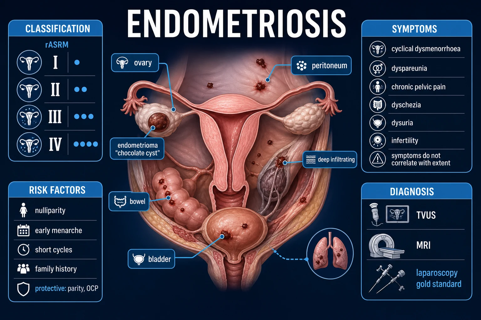
By surgical stage — the revised American Society for Reproductive Medicine (rASRM) score: a point-based system tallying the size and depth of peritoneal implants, the size of endometriomas, and the extent and density of adhesions, to give: [1]
- Stage I — minimal (1 to 5 points)
- Stage II — mild (6 to 15 points)
- Stage III — moderate (16 to 40 points)
- Stage IV — severe (over 40 points) [1]
The central limitation — examiners reward knowing this — is that rASRM stage correlates poorly with pain severity: a patient with Stage IV disease may be nearly pain-free, while a few deep nodules (Stage I to II) can cause excruciating symptoms. The rASRM score does, however, correlate reasonably with infertility prognosis and is required to compute the Endometriosis Fertility Index.[2][5]
ENZIAN score — used specifically to classify deep infiltrating endometriosis, grading disease in three compartments (A = rectosigmoid/bowel; B = vagina/uterus/sacrouterine ligament; C = ureter/bladder; plus F = pouch of Douglas obliteration and FA = adenomyosis and other distant sites) with severity grades 1 to 3. It captures what rASRM cannot — the depth and compartment of deep disease — and is used for surgical planning and outcome comparison.[9]
By location: pelvic (ovary, peritoneum, uterosacral ligaments, pouch of Douglas, rectovaginal septum, bladder) is overwhelmingly commonest; extrapelvic endometriosis (abdominal wall — including scar endometriosis after caesarean/episiotomy; umbilical; thoracic causing catamenial pneumothorax/haemoptysis; intestinal; and, rarely, brain/skin) is rare but clinically characteristic. [1]
Epidemiology & Risk Factors
Endometriosis affects roughly 10 per cent of women of reproductive age worldwide — about 190 million women globally. The prevalence rises dramatically in selected populations: 30 to 50 per cent of women with infertility and 45 to 70 per cent of women with chronic pelvic pain have endometriosis.[2][7]
Risk factors (all share the theme of increased menstrual exposure / retrograde flow):[2]
| Risk factor | Mechanism |
|---|---|
| Nulliparity | More cumulative menstrual cycles, no pregnancy-induced amenorrhoea |
| Early menarche | Earlier onset of cyclic hormonal stimulation |
| Short menstrual cycles (under 27 days) | More frequent retrograde menstruation |
| Heavy / prolonged menses (menorrhagia) | Greater volume of retrograde debris |
| Low BMI | Lower peripheral oestrogen antagonism; less adipose aromatisation |
| Nulliparity with delay in first pregnancy | Prolonged uninterrupted cycling |
| Müllerian anomalies (e.g. obstructed hemivagina, cervical atresia) | Mechanical obstruction amplifies retrograde flow |
| Family history | Genetic component; 6 to 9 times higher risk if an affected first-degree relative |
Protective factors: multiparity, prolonged lactation, combined oral contraceptive use, regular exercise, and late menarche — all reduce lifetime menstrual exposure and ovulatory cycles.[2]
Diagnostic delay: the average time from symptom onset to a confirmed diagnosis is 6 to 10 years, longer in adolescents and in low-resource settings. The delay is driven by (1) normalisation of dysmenorrhoea by patients and clinicians alike, (2) the non-specific nature of pelvic pain, (3) normal examination and imaging in early/superficial disease, and (4) requirement for laparoscopy for definitive histology in many settings. Endometriosis is the second commonest benign gynaecological condition after fibroids.[7]
Heritability: twin and family studies estimate heritability at around 50 per cent; however, the inheritance is polygenic — over 20 genome-wide significant loci identified, many involving ovarian steroidogenesis and WNT signalling — so a single 'endometriosis gene' does not exist. A first-degree relative of an affected woman has a roughly 6-fold higher relative risk.[2]
Pathophysiology
No single theory explains every case; endometriosis is best understood as a multifactorial disease in which ectopic endometrial tissue survives, implants, attracts a blood supply and resists immune clearance in a pro-oestrogen, pro-inflammatory peritoneal milieu.[1][9]
The classical theories of origin: [1]
- Retrograde menstruation (Sampson, 1927) — the leading theory. Menstrual debris flows backward through the fallopian tubes into the pelvic cavity during menses, where viable endometrial fragments implant on peritoneal/ovarian surfaces. Retrograde menstruation occurs in up to 90 per cent of menstruating women, yet only about 10 per cent develop endometriosis — so retrograde flow is necessary but not sufficient. Additional defects in immune clearance, attachment and angiogenesis must allow the fragments to persist.[1]
- Coelomic metaplasia (Meyer) — the peritoneal mesothelium (of coelomic origin, like endometrium) differentiates (metaplasias) into endometrial-type tissue under hormonal/inflammatory stimulation. This explains endometriosis in women without a uterus or without menstruation (e.g. pre-menarche, after hysterectomy, and in rare male cases on oestrogen therapy) where retrograde menstruation is impossible.
- Müllerian remnants / embryonic rest — residual Müllerian duct cells persist and are activated at puberty by oestrogen.
- Lymphatic / vascular spread (Halban) — endometrial cells disseminate via lymphatic or haematogenous routes, explaining extrapelvic disease (lung, brain, abdominal wall, umbilicus, scars) remote from the pelvis.
- Induction theory — retrograde endometrial cells release factors that induce the surrounding peritoneal mesothelium to metaplasia, unifying the Sampson and Meyer theories.
Molecular hallmarks — the four that examiners ask about:[1]
- Oestrogen dependence. Endometriotic implants overexpress aromatase and 17-beta-hydroxysteroid dehydrogenase type 1, producing and failing to inactivate oestradiol locally — they make their own oestrogen. They also overexpress oestrogen receptor beta, amplifying oestrogen signalling. This oestrogen dependence is the therapeutic rationale for all hormonal suppression (it starves the implants of mitogenic drive).
- Progesterone resistance. Eutopic endometrium of endometriosis patients shows a blunted progesterone response — reduced progesterone receptor (PR-B) expression and failure to induce the 17-beta-HSD type 2 enzyme that normally inactivates oestradiol. The molecular basis is aberrant HOXA10 / Steroidogenic factor-1 (SF-1) / GATA6 signalling and epigenetic promoter methylation. Progesterone resistance explains why some patients respond poorly to progestogens and why a single mechanism drives both endometriosis and part of infertility (impaired decidualisation, defective implantation).
- Chronic inflammation. Ectopic implants bleed cyclically into a closed cavity; the haemoglobin/iron debris activates peritoneal macrophages, which release pro-inflammatory cytokines — IL-1, IL-6, IL-8, TNF-alpha — and prostaglandins (PGE2). PGE2 in turn upregulates aromatase, creating a positive-feedback loop (PGE2 to aromatase to oestradiol to COX-2 to more PGE2). This inflammatory milieu is the direct source of pain (via cytokine sensitisation of nociceptors) and contributes to infertility (impaired oocyte quality, sperm transport, fertilisation, implantation).
- Impaired immune clearance. In health, natural killer cells and macrophages clear refluxed endometrial debris. In endometriosis there is reduced NK-cell cytotoxicity and altered macrophage function, allowing implants to evade surveillance. There is also increased angiogenesis (VEGF) so implants can recruit a blood supply — they cannot grow beyond 1 to 2 mm without neovascularisation. [1]
Why does it hurt and why does it cause infertility?[1][3]
- Pain arises from inflammation of peritoneal implants, traction on adhesions, deep infiltration of nerves (DIE lesions invading the rectovaginal septum are richly innervated), and prostaglandin sensitisation of pain fibres. Over time, chronic nociceptive input produces central sensitisation — the pain becomes partly independent of ongoing tissue injury, explaining why pain can persist after complete excision.
- Infertility is multifactorial: anatomical distortion (adhesions, tubal blockage, ovarian distortion), ovulatory dysfunction (luteinised unruptured follicle), peritoneal inflammation (cytokines toxic to gametes/embryos), impaired oocyte quality, defective sperm-oocyte interaction, and impaired implantation (progesterone resistance). Importantly, the severity of anatomical distortion correlates with the severity of infertility. [1]
Clinical Presentation
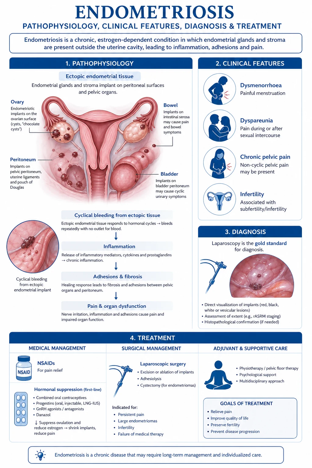
The hallmark is cyclical, progressive symptoms beginning years after menarche, classically the triad of secondary dysmenorrhoea, deep dyspareunia and chronic pelvic pain, frequently accompanied by subfertility.[1][2]
Symptom-by-symptom: [1]
- Secondary dysmenorrhoea — pain beginning before menses (unlike primary dysmenorrhoea, which is confined to menses) and continuing through it, often refractory to NSAIDs and progressive over months to years. The cardinal clue is dysmenorrhoea severe enough to disrupt daily activity (school, work) that is not relieved by standard analgesia.
- Deep dyspareunia — pain on deep penetration (not superficial, which suggests vestibulitis/vaginitis), typically worse premenstrually; localises disease in the pouch of Douglas, uterosacral ligaments or rectovaginal septum (DIE).
- Chronic pelvic pain — non-cyclical lower abdominal/pelvic pain present for at least 6 months, often with sacroiliac/low-back radiation.
- Dyschezia — painful defecation, cyclical, worse perimenstrually, often with the patient describing tenesmus or a sense of rectal fullness; indicates rectal/rectosigmoid DIE.
- Cyclic dysuria and/or haematuria — indicates bladder DIE; classically catamenial (occurring only with menses).
- Cyclic rectal bleeding — perimenstrual rectal bleeding indicates bowel mucosal invasion by DIE.
- Heavy menstrual bleeding and premenstrual spotting — common; up to half of patients. [1]
Atypical presentations (high-yield):[2]
- Asymptomatic woman presenting with infertility — endometriosis may be the only finding; up to 25 to 50 per cent of infertile women have endometriosis and may have no pain at all.
- Adolescent with severe dysmenorrhoea refractory to NSAIDs / OCP — endometriosis is found in nearly half of adolescents with chronic pelvic pain refractory to first-line treatment; suspect it and do not dismiss the pain as behavioural.
- Catamenial pneumothorax / haemoptysis / haemothorax — thoracic endometriosis, the commonest extrapelvic form; cyclical chest pain and pneumothorax within 72 hours of menses.
- Scar endometriosis — a painful, cyclical, growing nodule in a caesarean or episiotomy scar, swelling and becoming tender with each period.
- Umbilical or inguinal cyclical nodule, cyclic sciatica (sciatic nerve involvement), cyclic rectal/urinary symptoms. [1]
Key clinical principle — extent of disease does NOT correlate with pain severity. A few deep nodules (Stage I to II) can produce excruciating pain, while extensive superficial disease (Stage III to IV) may be silent. This is the single most-tested clinical pearl and the reason laparoscopic stage never predicts symptom burden.[2]
Differential Diagnosis
Chronic cyclical pelvic pain and an adnexal mass have a wide differential. Distinguish each by its distinguishing features:[2][3]
Adenomyosis
- Endometrium WITHIN myometrium (same tissue, wrong layer)
- Diffusely enlarged, boggy, tender uterus; menorrhagia and secondary dysmenorrhoea
- TVUS: junctional zone over 12 mm, myometrial cysts, asymmetric walls; MRI: junctional zone thickening
- Not associated with endometrioma or deep nodules
PID / tubo-ovarian abscess
- Acute febrile illness, vaginal discharge, cervical motion + adnexal tenderness
- Risk factors: multiple partners, STI, IUD; raised WCC/CRP
- Tender adnexal mass; TVUS shows complex tubo-ovarian collection
- Cyclical pattern absent; usually bilateral; responds to antibiotics
Pelvic congestion syndrome
- Non-cyclical dull pelvic ache, worse on standing/sitting; post-coital ache without cyclicity
- Dilated ovarian and pelvic veins on TVS/MRI/venography
- Often coexists with varices; no endometrioma
- OCPs may help; embolisation in refractory cases
Ovarian malignancy
- Solid/cystic ovarian mass WITH papillary projections, ascites, septations, colour Doppler flow
- Raised CA-125 (can be high in endometriosis too — not discriminatory)
- Older, postmenopausal; rapid growth; weight loss
- Definitive: histology; image first, do not assume a chocolate cyst
Dermoid (mature teratoma)
- Young women; the commonest ovarian germ-cell tumour
- TVUS: echogenic mass with fat-fluid level, calcification (teeth), hair
- No cyclical pain; tumour markers (AFP, hCG) negative
- Risk of torsion; surgical removal
Haemorrhagic corpus luteum
- Self-limiting; appears in luteal phase and REGRESSES over weeks
- Acute pain if rupture causes haemoperitoneum
- TVUS: irregular internal echoes, no ground-glass uniform content
- Repeat scan confirms resolution — the key discriminator from endometrioma
For cyclic rectal pain/bleeding (DIE vs bowel disease): [1]
- Inflammatory bowel disease (Crohn's, UC) — diarrhoea, blood/mucus per rectum, weight loss, extra-intestinal features; not cyclical; colonoscopy with biopsy diagnostic.
- Irritable bowel syndrome — chronic abdominal pain with altered bowel habit, no bleeding, no weight loss, normal colonoscopy and calprotectin; pain may be food- and stress-related but not menstrual.
- Colorectal cancer — older patient, weight loss, change in bowel habit, iron-deficiency anaemia; not cyclical; colonoscopy essential. [1]
Always consider adenomyosis (which coexists with endometriosis in up to a third of patients) and pelvic congestion syndrome, and exclude an acute surgical cause (ruptured cyst, torsion, ectopic) in the patient with sudden severe pain. [1]
Clinical & Bedside Assessment
Endometriosis is not ruled out by a normal examination — early and superficial disease frequently has no findings. A focused pelvic examination is performed to support the diagnosis, map deep disease and detect masses.[2][4]
Focused pelvic examination findings in endometriosis: [1]
- Tender nodularity in the posterior vaginal fornix / on the uterosacral ligaments — the single most specific bedside finding; palpated on bimanual and speculum exam, indicates DIE of the uterosacral ligaments / rectovaginal septum. The nodules are fibrotic, fixed and tender.
- Fixed, retroverted, tender uterus — adhesions in the pouch of Douglas tether the uterus posteriorly; the immobility (fixed pelvis) is the clue.
- Adnexal tenderness or a tender, fixed adnexal mass — an endometrioma (ovary is the commonest site).
- Tenderness on cervical motion — non-specific inflammation (also present in PID).
- Visualised bluish nodules in the posterior fornix/vagina — rare but pathognomonic when seen.
- On speculum and rectovaginal examination, assess for rectovaginal septum nodules, vaginal disease, and exclude other pathology (discharge, cervix appearance). [1]
Why is the exam often normal in early disease? Superficial peritoneal implants are flat, beneath the surface and produce no palpable mass or nodularity; retrograde-flow volume and depth of invasion have not yet generated adhesions or deep nodules. Hence a normal pelvic exam never excludes endometriosis — a symptomatic patient still warrants imaging and, if indicated, laparoscopy.[2]
Bedside tools: a structured pain and symptom history (cyclicality, relationship to menses, NSAID response, impact on school/work/sex, dyspareunia localisation, bladder and bowel symptoms), a pain diary across at least two cycles, and a validated quality-of-life tool (e.g. the Endometriosis Health Profile-30). Always take a sexual/reproductive and family history and screen for depression and anxiety, which are common and compound the pain experience. [1]
Investigations
The diagnostic pathway is stepwise: clinical suspicion then transvaginal ultrasound first-line, MRI for DIE mapping, and laparoscopy with histology as the gold standard — but laparoscopy is no longer required before starting empiric hormonal treatment in a typical case.[4][5]
Serum CA-125: frequently mildly to moderately elevated in endometriosis (especially advanced/disseminated disease and endometrioma), but it is non-specific and NOT diagnostic — it is also raised in ovarian cancer, PID, fibroids, pregnancy and menstruation. Its role is limited: it cannot screen for or diagnose endometriosis; it may help monitor treatment response in known disease, and it is part of the work-up of an adnexal mass (with RMI) to exclude malignancy. A normal CA-125 does not exclude endometriosis; a very high CA-125 (hundreds) in a cystic ovarian mass raises concern for malignancy rather than a simple endometrioma.[2]
Transvaginal ultrasound (TVUS) — first-line imaging. Inexpensive, widely available, and highly accurate for the ovarian endometrioma. The classic appearance: [1]
- Ground-glass, homogeneous, low-level internal echogenicity (diffuse, low-level echoes from old, degraded blood)
- No internal papillary projections or solid components (their presence raises suspicion of malignancy)
- Thick, hyperechoic wall, often with multilocularity
- Absent or scant internal colour Doppler flow
- Often bilateral; does not regress on serial scans (unlike a haemorrhagic corpus luteum). [1]
Endometrioma vs other adnexal masses — TVUS discriminators:[2]
| Feature | Endometrioma | Dermoid | Malignancy | Haemorrhagic CL |
|---|---|---|---|---|
| Internal echoes | Ground-glass, uniform | Echogenic fat, hair, calcification | Solid/papillary, mixed | Irregular, retracting clot |
| Papillary projections | Absent | Absent | Present | Absent |
| Doppler flow | Little/none | Little | Increased | Peripheral |
| Course over weeks | Persistent | Persistent | Growing | Resolves |
TVUS can also detect bladder and rectal DIE (especially when combined with saline infusion sonovaginography or bowel preparation) but is insensitive for superficial peritoneal disease.[4]
MRI pelvis — for DIE mapping and surgical planning. MRI shows the depth of infiltration of deep disease and detects ureteric, bladder, bowel, rectovaginal and extrapelvic involvement that ultrasound may miss. Endometriomas appear as T1-hyperintense (bright) and T2-hypointense (shading sign) cysts — the shading sign reflects concentrated blood products and is highly characteristic. MRI with intravenous contrast is used when TVUS cannot characterise a mass or when malignancy is suspected. Recommended before complex DIE surgery to plan the multidisciplinary team (gynaecology, colorectal, urology).[2][9]
Other investigations: [1]
- Hydrosonography / saline infusion — to assess the endometrial cavity and adnexal masses.
- CT urography / renal function — when ureteric DIE or hydronephrosis is suspected (silent hydronephrosis with loss of renal function is the feared complication of ureteric DIE).
- Colonoscopy / cystoscopy — if bowel or bladder mucosal involvement suspected, or to exclude IBD/malignancy.
- Laparoscopy with histology — the gold standard. Direct visualisation of the three phenotypes (red flame lesions, black powder-burn lesions, white opacified peritoneum, endometriomas, deep nodules) plus biopsy/histology (endometrial glands and stroma with haemosiderin-laden macrophages) confirms the diagnosis. Laparoscopy is indicated when imaging is negative but clinical suspicion remains, when malignancy cannot be excluded, in refractory disease, or when surgical treatment is planned. The ESHRE guidance now endorses a clinical-plus-imaging diagnosis permitting empiric hormonal treatment without laparoscopy in a typical case — laparoscopy is no longer a prerequisite for starting medical therapy.[4][5]
ESHRE/algorithm-based diagnostic approach (reproduced):[4]
- Suspect endometriosis on the basis of cyclical pelvic pain, dyspareunia, dyschezia/dysuria or infertility.
- Examine the pelvis and arrange transvaginal ultrasound (and/or MRI if DIE suspected).
- If imaging confirms an endometrioma or DIE, the clinical diagnosis is established — begin treatment or refer for surgery as indicated.
- If imaging is negative but suspicion is high, start empiric hormonal treatment (no laparoscopy required).
- Reserve laparoscopy for refractory disease, suspected malignancy, definitive diagnosis when needed, or when surgery is the planned treatment.
- Exclude mimics (pregnancy, infection, malignancy) at every step. [1]
Management — Resuscitation
Endometriosis is a chronic condition, but several scenarios are acute and time-critical and require emergency surgical assessment.[2]
Acute, time-critical scenarios: [1]
- Ruptured endometrioma — sudden-onset severe lower abdominal pain with peritonism, often mid-cycle or perimenstrual, in a woman with a known ovarian cyst. Leakage of old blood into the peritoneum is intensely irritant. Immediate management: ABCDE, IV access, analgesia (IV opioid), bloods (CBC, group and save, beta-hCG to exclude ectopic), urgent pelvic ultrasound, and emergency laparoscopy/laparotomy if haemodynamically unstable or peritonitis; many stable cases settle with conservative management and analgesia.
- Ovarian torsion — sudden severe unilateral pain with vomiting; emergency laparoscopy within 6 hours to detorse and salvage the ovary (oophoropexy if recurrent).
- Acute urinary obstruction from bladder/ureteric DIE — silent hydronephrosis progressing to acute kidney injury; urgent stent or nephrostomy then definitive DIE surgery. Ureteric involvement may be silent — always check renal function and imaging in known DIE.
- Bowel obstruction from rectosigmoid DIE — crampy abdominal pain, distension, vomiting; acute large-bowel obstruction is a surgical emergency (resection with primary anastomosis or stoma). Most DIE causes chronic partial obstruction rather than acute, but acute presentation mandates laparotomy. [1]
ED approach to severe acute pelvic pain with a suspected ruptured endometrioma: resuscitate (ABC, IV fluids), analgesia, exclude pregnancy (beta-hCG) and infection, image (pelvic ultrasound ± CT if peritonitis), consult gynaecology urgently, and take to emergency laparoscopy if unstable or if peritonitis/haemoperitoneum is significant. [1]
Management — Definitive & Stepwise
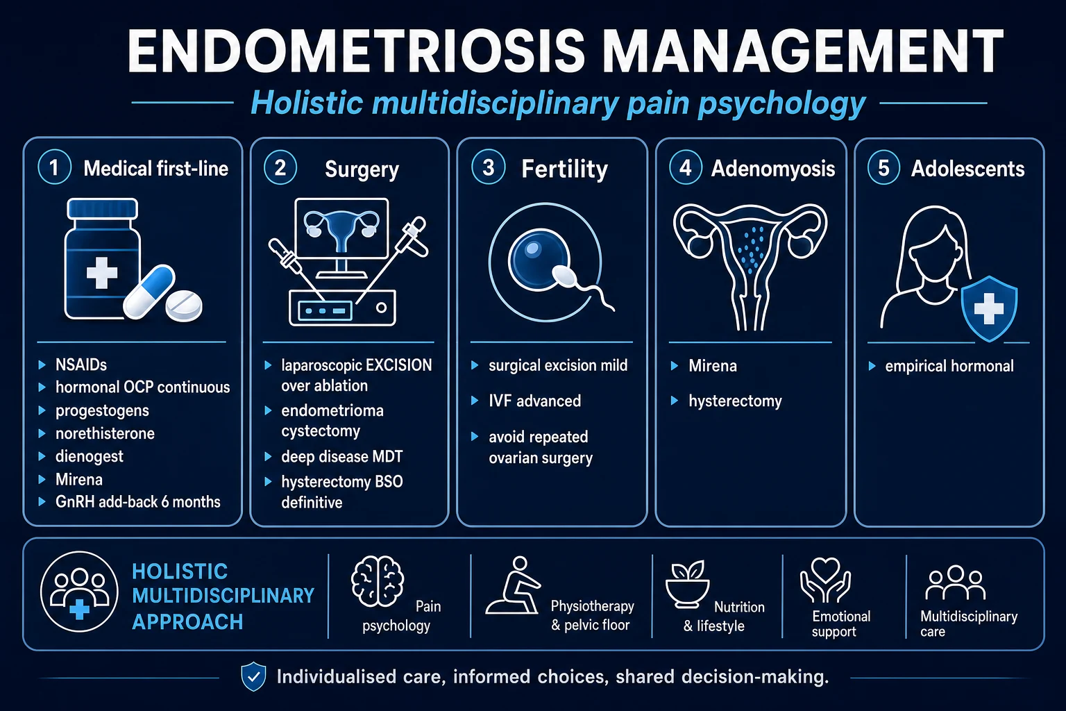
Treatment is goal-directed: the two goals — pain control versus fertility — pull in opposite directions, because most effective medical therapies suppress ovulation. Decide the primary goal first, then choose the therapy.[2][4]
Principles: [1]
- For pain in a woman not actively trying to conceive: empiric hormonal suppression + analgesia is first-line, without requiring laparoscopy.
- For infertility: surgery has a role in selected stages; medical suppression does NOT improve spontaneous fertility (it only suppresses ovulation) — refer to assisted reproduction appropriately.
- For DIE, refractory pain, large endometrioma, or suspected malignancy: surgery.
- Multidisciplinary care (gynaecology, pain specialist, colorectal/urology as needed, psychology) is recommended, especially for DIE and chronic pain. [1]
Medical management — drug, dose, route, rationale, monitoring
1. Analgesia (first-line symptom control):[2]
- NSAIDs — ibuprofen 400 mg PO TDS, naproxen 500 mg PO BD, or mefenamic acid 500 mg PO TDS, started before/at onset of menses. Rationale: inhibit prostaglandin synthesis, the proximate mediator of dysmenorrhoea. Monitor for GI, renal and cardiovascular side effects; avoid in peptic ulcer and renal impairment. Do not use in women trying to conceive (may impair ovulation/implantation).
- Paracetamol 1 g PO QDS as adjunct. [1]
2. Combined hormonal contraception — first-line hormonal suppression ('pseudo-pregnancy'):[2][4]
- Combined oral contraceptive pill (COC) — e.g. ethinylestradiol 30 to 35 micrograms + a progestogen (levonorgestrel, drospirenone, norethisterone), one tablet daily, given continuously or cyclically (continuous preferred for pain, to induce amenorrhoea). Rationale: suppresses ovulation and the hypothalamic-pituitary-ovarian axis, decidualises and atrophies ectopic endometrium. Monitoring: VTE risk, BP, migraine with aura (contraindication). Long-term, well tolerated and cheap — the default first-line hormonal option. [1]
3. Progestogens — second-line / first-line in many guidelines:[4][8]
- Dienogest 2 mg PO once daily — a 19-nortestosterone progestogen with marked endometrial selectivity; the best-studied and recommended oral progestogen for endometriosis. Rationale: induces atrophy of ectopic endometrium without inducing hypo-oestrogenism. Long-term efficacy and safety (up to 5 years and beyond) shown; side effects — irregular bleeding, headache, breast tenderness, mood change. Monitor: bleeding pattern, mood, bone density with very long-term use.[8]
- Norethisterone (acetate) 5 mg PO BD to TDS (up to 15 mg/day) — cheap, widely available alternative.
- Medroxyprogesterone acetate (DMPA) — 150 mg IM every 3 months, or 104 mg SC every 3 months. Highly effective; side effects — weight gain, delayed return to fertility (9 to 12 months), bone density loss, irregular bleeding. Avoid in women desiring imminent fertility.
- Levonorgestrel intrauterine system (LNG-IUS, 'Mirena') — 52 mg levonorgestrel, 5-year device, inserted into the uterine cavity. Provides local high-dose progestogen with low systemic levels; reduces dysmenorrhoea and dyspareunia, and (importantly) reduces recurrence after endometrioma surgery. Rationale: local endometrial atrophy, reduced menstrual flow. Side effects — irregular bleeding (first 6 months), expulsion, perforation. Particularly useful when adenomyosis coexists.
4. GnRH agonists — 'pseudo-menopause' (second-line, supervised):[2][4]
- Goserelin 3.6 mg SC every 4 weeks (or 10.8 mg SC every 12 weeks); leuprorelin (leuprolide) 3.75 mg IM every 4 weeks (or 11.25 mg IM every 12 weeks); triptorelin 3.75 mg IM every 4 weeks (or 11.25 mg IM every 12 weeks). Rationale: initial flare then down-regulation of pituitary GnRH receptors, producing a reversible, hypo-oestrogenic (menopausal) state that starves implants of oestrogen. Highly effective for pain, but limited to 6 months without add-back because of bone density loss and menopausal side effects (hot flushes, vaginal dryness, mood, headache). The 'flare' in the first 1 to 2 weeks may transiently worsen pain.
- Add-back therapy — tibolone 2.5 mg PO once daily, norethisterone 5 mg PO once daily, or a combined OCP — given from the start alongside the GnRH agonist to counter hypo-oestrogenic side effects and bone loss, allowing treatment to continue up to 12 months or longer. Add-back does not negate efficacy. [1]
5. GnRH antagonists — the modern oral option:[4][6]
- Relugolix combination — relugolix 40 mg + estradiol 1 mg + norethisterone acetate 0.5 mg, one tablet PO once daily. Rationale: immediate (no flare) dose-dependent suppression of gonadotrophins and ovarian oestrogen, with built-in add-back to limit bone loss. Supported by the SPIRIT 1 and 2 phase 3 trials, which showed significant reduction in dysmenorrhoea and non-menstrual pelvic pain versus placebo over 24 weeks (and sustained to 52 weeks in the SPIRIT open-label extension).[6]
- Elagolix — 150 mg PO once daily (lower dose) or 200 mg PO BD (higher dose). The higher dose more effective for pain but carries greater bone-density loss; add-back recommended for the 200 mg BD dose. Approved in the US for moderate-to-severe endometriosis-associated pain.
- Limitations: cost and availability — relugolix and elagolix are not universally available (regional access varies widely); hypo-oestrogenic side effects; duration typically limited to 24 months pending long-term bone-safety data.
Surgical management
Indications for surgery:[2][4]
- Definitive diagnosis (when imaging is negative or malignancy cannot be excluded)
- Refractory pain despite adequate medical therapy
- Deep infiltrating endometriosis with bowel/bladder/ureteric involvement (especially with obstruction or hydronephrosis)
- Large (over 4 cm) or symptomatic endometrioma
- Infertility with treatable disease (selected patients — see Evidence)
- Suspected malignant transformation [1]
Two surgical philosophies — ablation vs excision:[2]
- Ablation (coagulation/vaporisation) — destroys the lesion surface using diathermy/laser. Suitable for superficial peritoneal disease; quick but does not remove deep disease, so higher recurrence in DIE.
- Excision — the lesion is cut out to clear margins (the nodules are often deeper than they appear). Preferred for DIE and most surgeons' standard; lower recurrence, better pain outcomes, but technically harder and more risk of damaging bowel/bladder/ureter/ovary. Excision is the operation of choice for deep infiltrating disease. [1]
Ovarian endometrioma — cystectomy vs drainage/ablation:[2][4]
- Laparoscopic ovarian cystectomy (stripping) — the cyst wall is stripped from the ovary. Lower recurrence (around 15 to 30 per cent vs higher with drainage) and better pain and fertility outcomes, but removes some healthy ovarian tissue with measurable loss of ovarian reserve (AMH falls).
- Drainage + ablation of the cyst wall — faster, less ovarian tissue removed, better ovarian reserve preservation, but higher recurrence.
- Practical rule: cystectomy is preferred for symptomatic, recurrent or larger endometriomas; drainage/ablation or expectant management when ovarian reserve must be maximally preserved (e.g. before IVF). This is an active controversy (see Evidence). [1]
Endometriosis-associated infertility:[2][3]
- Surgical excision/ablation of early-stage (rASRM I to II) disease modestly improves spontaneous pregnancy rates compared with diagnostic laparoscopy alone.
- Medical ovarian suppression before IVF does NOT improve live birth rates and is not recommended solely to improve IVF outcomes (it simply delays treatment). However, 3 to 6 months of GnRH agonist down-regulation before IVF may improve outcomes in advanced (Stage III to IV) disease in some guidelines.
- Surgery for advanced-stage disease / endometrioma: cystectomy can improve spontaneous conception but must be balanced against ovarian reserve loss — in a woman with low AMH, bilateral endometriomas, or prior ovarian surgery, direct IVF may be preferable to surgery.
- IVF is the most effective treatment for endometriosis-associated infertility when surgery fails or disease is severe; pregnancy rates are somewhat lower than in tubal infertility but still substantial.
- Endometriosis Fertility Index (EFI) — the best-validated surgical staging tool for fertility prognosis, combining the rASRM score with the least function (LF) score of the fallopian tubes, fimbriae and ovaries at surgery, plus age, duration of infertility and prior pregnancy. The EFI (0 to 10) predicts cumulative pregnancy probability and helps decide between expectant management, surgery, and IVF. [1]
Definitive surgery — hysterectomy with bilateral salpingo-oophorectomy (BSO): reserved for women who have completed their family with severe, refractory disease failing conservative surgery and medical therapy. Removing the ovaries (BSO) is what produces the sustained hypo-oestrogenic state that drives endometrial implants to regress; hysterectomy alone may be insufficient if ovarian function continues. Excise all visible DIE at the same operation. HRT may be required post-BSO — combined continuous HRT is generally used (oestrogen + progestogen if uterus remains); note that oestrogen replacement can reactivate residual disease, so progestogen component or close surveillance is advised. [1]
Non-pharmacological / supportive
- Patient education, pain psychology, CBT for chronic pain, physiotherapy for pelvic-floor dysfunction, and lifestyle (exercise, anti-inflammatory diet — limited evidence) — part of the multidisciplinary package, especially for central sensitisation.
- Complementary therapies (acupuncture, TENS) have limited evidence but may help individual patients. [1]
Specific Subtypes & Scenarios
Superficial peritoneal (SPE)
- Commonest phenotype; red/black/white peritoneal lesions
- Pain often out of proportion to visible disease
- First-line: NSAIDs + continuous COC or dienogest
- Surgery: laparoscopic ablation/excision if refractory
Ovarian endometrioma
- 'Chocolate cyst'; ground-glass TVUS; ovary is commonest site
- Cystectomy (stripping) preferred — lower recurrence; weigh reserve loss
- Persistent, does not regress (vs haemorrhagic CL)
- Malignant transformation risk (clear-cell / endometrioid) — small but real
Deep infiltrating (DIE)
- Invasion more than 5 mm; uterosacral, rectovaginal, bowel, bladder, ureter
- Cyclical dyschezia, deep dyspareunia, dysuria/haematuria, rectal bleeding
- MRI for mapping; check ureters and renal function
- Multidisciplinary excisional surgery (gynae + colorectal + urology)
Extrapelvic
- Scar (caesarean/episiotomy), umbilical, abdominal wall
- Thoracic: catamenial pneumothorax, haemoptysis
- Cyclical symptoms localised to the site — the diagnostic clue
- Excision + hormonal suppression; thoracic may need pleurodesis
Adolescent
- Severe dysmenorrhoea refractory to NSAIDs/OCP; school absence
- Empiric hormonal treatment WITHOUT laparoscopy first
- Avoid repeat surgery; preserve fertility
- Pain psychology and school support essential
Pregnancy
- Pain usually IMPROVES (high progesterone, amenorrhoea)
- Lesions shrink; rare complications — ruptured endometrioma, haemoperitoneum
- Discontinue medical therapy (COC, GnRH, dienogest)
- Surgical management only for acute complications
- Superficial peritoneal endometriosis — managed medically first; laparoscopic ablation or excision if refractory. The lesion most likely to be missed on imaging (TVUS and MRI may be normal) — laparoscopy is the only way to confirm.
- Ovarian endometrioma — see Management. Malignant transformation risk is small but real (clear-cell and endometrioid ovarian carcinoma); suspect in an endometrioma with rapid growth, solid/papillary components on imaging, ascites, or markedly raised CA-125, especially in a postmenopausal woman.
- DIE — definition: invasion more than 5 mm beneath the peritoneal surface; classic sites uterosacral ligaments, rectovaginal septum, vagina, rectosigmoid colon, bladder, ureter. Requires MRI for mapping and multidisciplinary surgical excision; the morbidity (bowel resection, ureteric re-implantation) is significant, so surgery is reserved for symptomatic, obstructing, or progressive disease. Ureteric involvement may be silent and cause irreversible renal loss — always image the upper tract.[9]
- Extrapelvic — abdominal wall (especially scar endometriosis after caesarean/episiotomy — a cyclical, growing, tender nodule in the scar), umbilical, inguinal, thoracic (catamenial pneumothorax/haemoptysis — typically within 72 hours of menses), intestinal, and very rarely CNS. The cyclicality is the diagnostic clue. Treatment: wide surgical excision + hormonal suppression; thoracic disease may need pleurodesis.
- Adolescent endometriosis — found in nearly half of teenagers with chronic pelvic pain refractory to NSAIDs/OCP. Empiric hormonal treatment without laparoscopy is now standard first-line; reserve laparoscopy for refractory symptoms. Avoid repeat surgery (adhesions, reserve loss) and prioritise fertility preservation and pain psychology.
- Endometriosis in pregnancy — the high-progesterone, amenorrhoeic state usually shrinks lesions and improves pain; rare complications include ruptured endometrioma (acute abdomen) and spontaneous haemoperitoneum. All endocrine therapy is contraindicated in pregnancy.
Complications & Pitfalls
- Infertility — via anatomic distortion (adhesions, tubal blockage), ovulatory dysfunction (luteinised unruptured follicle), peritoneal inflammation (cytokines toxic to gametes/embryos), impaired oocyte quality, and impaired implantation (progesterone resistance).
- Chronic pelvic pain and central sensitisation — chronic nociceptive input restructures central pain processing, so pain can persist after complete surgical excision; this is the rationale for pain psychology and neuromodulators (e.g. amitriptyline, gabapentin) in chronic disease.
- Reduced quality of life and work productivity — endometriosis is a leading cause of lost work and school days in young women; psychological morbidity (depression, anxiety, relationship strain) is high.[7]
- Malignant transformation — endometriosis carries a small but real risk of malignant transformation, almost exclusively to ovarian clear-cell or endometrioid carcinoma (and rarely adenosarcoma); estimated at under 1 per cent of women with endometriosis, but the relative risk of ovarian cancer is roughly doubled. Suspect in an endometrioma that changes character (rapid growth, solid component, marked CA-125 rise) especially postmenopause.
- Loss of ovarian reserve after endometrioma cystectomy (measurable AMH decline; bilateral disease and prior surgery compound this — counsel women planning IVF).
- Ureteric injury, bowel and bladder injury in DIE surgery — major morbidity; the reason for multidisciplinary teams and preoperative ureteric stents in selected cases.
- Adhesion formation after any pelvic surgery — may itself cause pain and infertility, paradoxically the disease it was meant to treat.
- Conversion to laparotomy, bleeding, infection, VTE. [1]
- Hypo-oestrogenism with GnRH analogues — bone density loss (limit to 6 months without add-back), hot flushes, vaginal dryness, mood, headache; reversible on stopping but bone recovery may be incomplete.
- Progestogen side effects — irregular bleeding, weight change, mood, breast tenderness; DMPA causes delayed return to fertility and bone loss.
- VTE risk of combined hormonal contraception and high-dose progestogens — avoid in smokers over 35, migraine with aura, prior VTE. [1]
Classic pitfalls: [1]
- Normalising severe dysmenorrhoea in adolescents and delaying diagnosis by years.
- Over-investigating / over-operating — multiple repeat laparoscopies cause adhesions and reserve loss; a clinical-plus-imaging diagnosis permits empiric treatment.
- Assuming a complex adnexal mass is an endometrioma — always consider and exclude ovarian malignancy (papillary projections, solid components, ascites, very high CA-125).
- Operating on bilateral endometriomas in a woman with low AMH desiring fertility without counselling on reserve loss — IVF may be the better first option.
- Missing ureteric DIE — silent hydronephrosis with irreversible renal loss; always check renal function and upper-tract imaging in known DIE.
- Using hormonal suppression to 'improve fertility' — it suppresses ovulation and does not improve spontaneous conception. [1]
Prognosis & Disposition
Endometriosis is a chronic, relapsing disease — it is not curable, but it is manageable. The natural history is one of fluctuation driven by hormonal status.[2][4]
Recurrence:[2]
- After medical therapy alone, recurrence of pain after stopping treatment is high (around 50 per cent at 5 years) — because therapy suppresses, not eradicates, the implants.
- After surgery, recurrence rates are roughly 20 to 40 per cent at 5 years; lower after complete excision and higher after ablation.
- Post-operative hormonal suppression (especially LNG-IUS or COC) reduces recurrence and is recommended after surgery for symptomatic disease. [1]
Predictors of recurrence: incomplete excision, younger age at diagnosis, severe (Stage III to IV) disease, presence of an ovarian endometrioma, and no subsequent hormonal suppression.[2]
Natural history by hormonal status: [1]
- Pregnancy — high progesterone and amenorrhoea usually improve symptoms and shrink lesions (transient effect; symptoms may recur postpartum).
- Menopause — loss of ovarian oestrogen generally leads to regression of disease; pain usually resolves. However, women on HRT may experience reactivation of residual disease (oestrogen drives it); combined HRT (oestrogen + progestogen) is preferred over oestrogen-only. [1]
Fertility prognosis:[3]
- After surgery for early-stage (Stage I to II) disease, spontaneous cumulative pregnancy rates are modestly but significantly improved versus no surgery.
- For advanced disease, IVF achieves pregnancy rates somewhat below those for pure tubal infertility but still substantial (live birth per cycle typically 30 to 40 per cent in younger women, lower with age).
- The Endometriosis Fertility Index (EFI) is the best-validated tool to predict spontaneous pregnancy probability and to guide the choice between expectant management, surgery and IVF. [1]
Disposition: endometriosis is managed ambulatory / outpatient by gynaecology with a multidisciplinary team; admit only for acute complications (rupture, torsion, obstruction) or for elective surgery. Early referral to a specialist endometriosis centre for DIE and complex disease improves outcomes. [1]
Special Populations
Adolescents:[4]
- Suspect endometriosis in any teenager with severe dysmenorrhoea refractory to NSAIDs and OCP, school absenteeism, or chronic pelvic pain.
- Empiric hormonal treatment (continuous COC or progestogen) WITHOUT laparoscopy is first-line — diagnostic laparoscopy is reserved for refractory symptoms.
- Avoid repeat surgery: each operation creates adhesions and risks ovarian reserve loss; prioritise fertility preservation and pain psychology.
- Adolescents with Müllerian anomalies causing outflow obstruction (e.g. obstructed hemivagina, imperforate hymen with didelphys uterus) are at very high risk of endometriosis — early surgical correction of the obstruction may prevent or reverse disease. [1]
Women desiring fertility:[2]
- Minimise ovarian reserve damage — avoid unnecessary endometrioma surgery, especially bilateral or repeat; counsel on AMH decline after cystectomy.
- Medical suppression does NOT improve spontaneous fertility — do not use hormonal suppression to 'treat' infertility; it suppresses ovulation.
- Refer to assisted reproduction when disease is severe, surgery has failed, or other infertility factors coexist; IVF is most effective.
- Use the Endometriosis Fertility Index to guide the strategy. [1]
Pregnancy:[2]
- Pain usually improves in pregnancy (high progesterone, amenorrhoea); lesions shrink.
- Rare complications: ruptured endometrioma (acute abdomen, haemoperitoneum), torsion.
- Discontinue all endocrine therapy (COC, progestogens, GnRH agonists/antagonists, dienogest) — contraindicated in pregnancy.
- Surgery only for acute complications; defer elective surgery until postpartum. [1]
Perimenopausal and menopausal:[2]
- Pain typically regresses after menopause as ovarian oestrogen falls; persistent or worsening pain postmenopause raises suspicion of malignant transformation.
- HRT (especially oestrogen-only) can reactivate residual disease — use combined continuous HRT and maintain surveillance.
- Definitive surgery (hysterectomy + BSO) is reasonable for women who have completed their family with refractory disease. [1]
Müllerian anomalies: outflow tract obstruction (obstructed hemivagina, cervical/vaginal atresia, non-communicating horn) markedly increases retrograde flow and endometriosis risk; early surgical correction of the obstruction is both treatment and prevention. [1]
Immunocompromised / on hormonal therapy: no specific change in management, but be alert to malignancy mimicking endometriosis in these groups. [1]
Evidence, Guidelines & Regional Differences
ESHRE 2022 guideline (Becker et al.) — key recommendations:[4][5]
- A clinical diagnosis based on symptoms and imaging is sufficient to start treatment — laparoscopy is not required before empiric therapy.
- Transvaginal ultrasound is the first-line imaging investigation; MRI for DIE and characterisation.
- Empiric hormonal treatment (COC, progestogens) ± analgesia is first-line for endometriosis-associated pain.
- GnRH agonists and antagonists are effective second-line for pain.
- Surgery for refractory pain, DIE, large endometrioma, and selected infertility; excision preferred over ablation for DIE.
- Endometrioma cystectomy preferred to drainage/ablation for symptom control and recurrence (with counselling on ovarian reserve).
- Centralised care in specialist centres recommended for DIE.
- Adolescents should receive empiric hormonal therapy; laparoscopy reserved for refractory disease. [1]
SPIRIT 1 and SPIRIT 2 (Giudice et al., 2022) — relugolix combination:[6]
- Design: two identical multinational, phase 3, randomised, double-blind, placebo-controlled trials of once-daily oral relugolix combination (relugolix 40 mg + estradiol 1 mg + norethisterone acetate 0.5 mg) in women with endometriosis-associated pain.
- Results: both trials met their primary endpoint — a significantly greater proportion of relugolix-treated women had a clinically meaningful reduction in dysmenorrhoea and non-menstrual pelvic pain at week 24 versus placebo, with sustained benefit to week 52 in the extension. Bone mineral density changes were small and similar to placebo with the built-in add-back. The trials established oral relugolix combination as an effective, well-tolerated, no-flare medical option for moderate-to-severe endometriosis pain. [1]
Surgery for endometriosis-associated infertility:[2][3]
- A randomised trial comparing operative laparoscopy (ablation/excision) vs diagnostic laparoscopy in early-stage (Stage I to II) disease showed a modest but significant increase in cumulative pregnancy with surgery — supporting surgical treatment of early-stage disease at the time of diagnostic laparoscopy.
- For advanced-stage disease, surgery has not been shown to improve spontaneous fertility as clearly, and IVF is often more effective — especially where ovarian reserve is already compromised. [1]
Controversy — cystectomy vs drainage/ablation for endometrioma:[4]
- Cystectomy has lower recurrence and better pain/fertility outcomes, but removes ovarian tissue and reduces reserve (AMH) — problematic in women desiring fertility, especially with bilateral or recurrent disease.
- Drainage/ablation preserves reserve but recurs more.
- Practical consensus: cystectomy for symptomatic/recurrent/larger endometriomas; drainage/ablation or expectant management when maximal reserve preservation is required (e.g. before IVF, low AMH, bilateral). Consider AMH measurement before and counselling before operating. [1]
- Access to laparoscopy varies widely — in many low- and middle-income countries, laparoscopy and specialist DIE surgery are scarce, increasing diagnostic delay and reliance on empiric hormonal therapy.
- Drug availability: relugolix (Ryeqo) and elagolix (Orilissa) are not universally available (approved in US, UK/EU, Japan, some others; often limited by cost) — GnRH agonists + add-back remain the practical second-line option in much of the world.
- Surgical sub-specialisation for DIE is concentrated in specialist centres; outcomes (complications, recurrence, fertility) are better with high-volume, multidisciplinary teams.
- Dienogest is more widely and cheaply available in Asia (including India) than the newer GnRH antagonists, making it a practical mainstay. [1]
Exam Pearls
- Endometriosis = functional endometrial glands AND stroma outside the uterus, responsive to cyclic hormones. Adenomyosis is endometriosis within the myometrium (enlarged, boggy, tender uterus).
- Ovary is the commonest site. Classic triad: dysmenorrhoea + deep dyspareunia + chronic pelvic pain; infertility may be the only feature.
- 'Chocolate cyst' is not a true cyst wall — it is old, degraded blood in an endometrioma; the pathognomonic TVUS appearance is ground-glass echogenic content, no papillary projections, thick wall.
- Laparoscopy is the gold standard: the three phenotypes — superficial 'powder-burn'/'matchstick' lesions, ovarian endometriomas, and deep nodules.
- Treatment = hormonal suppression — induce pseudo-pregnancy (continuous COC, progestogens) or pseudo-menopause (GnRH agonists with add-back; GnRH antagonists relugolix/elagolix) — plus surgery (excision for DIE, cystectomy for endometrioma) for definitive diagnosis and refractory disease.
- Pain severity does NOT correlate with disease stage — a few deep nodules can cause excruciating pain while extensive superficial disease is silent. This is the single most-tested clinical pearl.
- rASRM stage correlates with fertility prognosis, NOT with pain; use the ENZIAN score for DIE, and the Endometriosis Fertility Index (EFI) for fertility prognosis.
- Cyclical symptoms are the clue to extrapelvic/scar endometriosis — catamenial pneumothorax/haemoptysis (thoracic), cyclical dyschezia/rectal bleeding (bowel DIE), cyclical dysuria/haematuria (bladder DIE), cyclical scar nodule (caesarean/episiotomy scar endometriosis).
- DIE = invasion more than 5 mm beneath the peritoneum; classic sites uterosacral ligaments, rectovaginal septum, bowel, bladder, ureter. Always check the ureters — ureteric DIE may be silent and cause irreversible renal loss.
- Progesterone resistance (low PR-B, low 17-beta-HSD type 2) is the molecular lynchpin — explains oestrogen dominance, partial progestogen resistance, and infertility via defective implantation.
- Endometrioma carries a small but real risk of malignant transformation — almost always to clear-cell or endometrioid ovarian carcinoma.
- Diagnostic delay averages 6 to 10 years — never dismiss severe dysmenorrhoea, especially in adolescents.
- Medical suppression does NOT improve spontaneous fertility; IVF is most effective for advanced disease. Surgery improves fertility only in early-stage disease. [1]
Sites of endometriosis — mnemonic
OUTSIDE
Commonest site overall — 'chocolate cyst' / endometrioma
Tender nodularity on vaginal exam; classic DIE site
Adnexal adhesions, distortion, infertility
Superficial peritoneal — red/black/white lesions, 'powder-burn'
DIE — cyclic dyschezia/rectal bleeding, cyclic dysuria/haematuria
Deep dyspareunia; DIE nodule
Catamenial pneumothorax/haemoptysis; scar endometriosis
Endometriosis — key numbers
Exam application bank (NEET-PG / INICET)
One-line answer
Endometriosis is the presence of functional endometrial glands and stroma outside the uterine cavity, producing a chronic, oestrogen-dependent, inflammatory condition. It classically presents with the triad of secondary dysmenorrhoea, deep dyspareunia and chronic pelvic pain, with infertility a frequent sole presentation. The ovary is the commonest site; the three phenotypes are superficial peritoneal, ovarian endometrioma ('chocolate cyst') and deep infiltrating endometriosis (DIE). Diagnosis is clinical plus transvaginal ultrasound; MRI maps DIE; laparoscopy with histology is the gold standard. Management is NSAIDs plus hormonal suppression (combined OCP, progestogens, GnRH analogues/antagonists) and surgery (excision/ablation, cystectomy) for refractory disease, DIE and selected infertility.
Worked stems (answer without another resource)
Stem 1 — Classic presentation. Map symptoms to mechanism; name the first investigation and first treatment step with dose/route if drug therapy is standard. [1]
Stem 2 — Unstable / complicated. List red flags that force immediate resuscitation, theatre, ICU, antidote, or reperfusion — and what you do in the first 15 minutes. [1]
Stem 3 — Atypical group. Elderly, pregnancy, child, or immunocompromised: how presentation and thresholds change. [1]
Stem 4 — Differential trap. Name the three closest mimics and one discriminator for each. [1]
Stem 5 — Disposition. Who goes home with safety-netting, who is admitted, who needs HDU/ICU/theatre, and what follow-up is mandatory. [1]
Rapid viva checklist
- Definition + classification
- Pathophysiology chain
- Bedside signs / criteria
- Score with exact components (if any)
- Emergency bundle
- Definitive therapy with doses
- Complications of disease and of treatment
- Special populations
- Guideline/trial name if classic
- Three exam traps
Coverage self-check
If you cannot answer any stem above from this page alone, re-read the matching section — the page is intended to be self-sufficient for final-prof and NEET-PG/INICET questions on Endometriosis.
References
- [1]Bulun SE. Endometriosis N Engl J Med, 2009.PMID 19144942
- [2]Zondervan KT, Becker CM, Missmer SA. Endometriosis N Engl J Med, 2020.PMID 32212520
- [3]Giudice LC, Kao LC. Endometriosis Lancet, 2004.PMID 15541453
- [4]Becker CM, Bokor A, Heikinheimo O, et al. ESHRE guideline: endometriosis Hum Reprod Open, 2022.PMID 35350465
- [5]Dunselman GA, Vermeulen N, Becker C, et al. ESHRE guideline: management of women with endometriosis Hum Reprod, 2014.PMID 24435778
- [6]Giudice LC, As-Sanie S, Marret H, et al. Once daily oral relugolix combination therapy versus placebo in patients with endometriosis-associated pain: two replicate phase 3, randomised, double-blind, studies (SPIRIT 1 and 2) Lancet, 2022.PMID 35717987
- [7]Nnoaham KE, Hummelshoj L, Webster P, et al. Impact of endometriosis on quality of life and work productivity: a multicenter study across ten countries Fertil Steril, 2011.PMID 21718982
- [8]Petraglia F, Hornung D, Seitz C, et al. Reduced pelvic pain in women with endometriosis: efficacy of long-term dienogest treatment Arch Gynecol Obstet, 2012.PMID 21681516
- [9]Gordts S, Koninckx P, Brosens I. Pathogenesis of deep endometriosis Fertil Steril, 2017.PMID 29100623
- [10]Horne AW, Saunders PTK, Abokhrais IM, Hogg L; ENDOMETRIOSIS Priority Setting Partnership. Pathophysiology, diagnosis, and management of endometriosis BMJ, 2022.PMID 36375827