Obstetrics & Gynaecology · Obstetrics & Gynaecology
Gestational Diabetes Mellitus
Also known as Gestational diabetes mellitus · GDM · Gestational diabetes · Diabetes in pregnancy
Gestational diabetes mellitus (GDM) is any degree of glucose intolerance with onset or first recognition during pregnancy (typically 24 to 28 weeks), excluding overt diabetes detectable at booking. Pathogenesis: placental anti-insulin hormones (hPL, cortisol, progesterone, prolactin, growth hormone, oestrogen) plus TNF-alpha drive progressive insulin resistance; pancreatic beta-cells fail to compensate. Risk factors: BMI over 30, age over 35, South Asian/Black/Hispanic ethnicity, previous GDM or macrosomia, family history, PCOS. Diagnosis: universal 75 g 2-hour OGTT at 24 to 28 weeks. Management: lifestyle (medical nutrition therapy, exercise) first; metformin then insulin to targets (fasting under 5.3 mmol/L, 1-h post-meal under 7.8); fetal growth surveillance; delivery at 38 to 39 weeks; postpartum glucose testing and lifelong T2DM screening.
On this page & tools
Your progress
Saved locally on this device.
Exam tags
Red flags
Overview & Definition
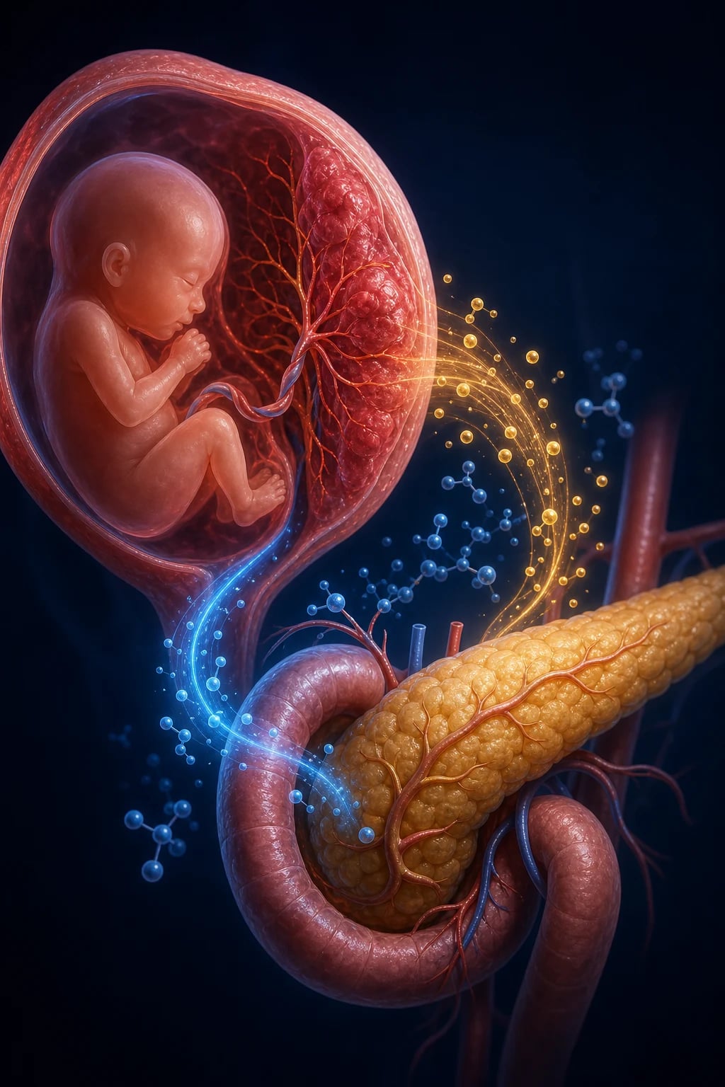
Gestational diabetes mellitus (GDM) is defined as any degree of glucose intolerance with onset or first recognition during pregnancy, irrespective of whether it persists after delivery.[1] The definition is operational — it is a label applied to hyperglycaemia discovered through routine antenatal screening, not a statement about underlying aetiology. By convention it excludes overt (pre-gestational) diabetes detectable at the booking visit, which is why every pregnant woman should have a booking HbA1c or fasting glucose so that pre-existing type 1 or type 2 diabetes is identified and managed as the higher-risk condition it is.[4]
The condition typically declares itself between 24 and 28 weeks of gestation, the window when placental anti-insulin hormone output peaks and the gap between insulin demand and beta-cell supply becomes clinically apparent. The 24 to 28 week window is not arbitrary: it balances two needs — screening early enough to modify fetal growth trajectory before the third trimester, yet late enough that the diabetogenic signal has become detectable. First-trimester screening misses most GDM because placental hormone output is still low, while third-trimester screening may leave too little time to reverse macrosomia. Most women are completely asymptomatic; the diagnosis is a laboratory one, and its clinical importance lies almost entirely in the preventable fetal and maternal harms it produces when untreated — macrosomia, shoulder dystocia, neonatal hypoglycaemia, pre-eclampsia, caesarean delivery — and in the powerful signal it sends about the woman's future type 2 diabetes risk.[3]
The definition also carries a subtle but examinable point: GDM is not a single disease entity but a syndrome defined by the timing of detection. Some women labelled GDM have pre-existing, undiagnosed type 2 diabetes unmasked by pregnancy; others have true, pregnancy-limited beta-cell inadequacy. The practical distinction is made at the booking visit — an HbA1c over 6.5 percent or fasting glucose over 7.0 mmol/L at booking signals overt diabetes, not GDM, no matter when the label would otherwise be applied. This operational definition is why every woman should have glucose status assessed at the first antenatal contact, and why the postpartum glucose test is needed to see who truly has persistent diabetes. [1]
GDM is high-yield for examinations for four reasons. First, the pathophysiology (placental hormones driving insulin resistance) explains the timing, the postpartum resolution, and the molecular targets of therapy. Second, the diagnostic thresholds of the 75 g oral glucose tolerance test are routinely examined verbatim and differ between guideline bodies (IADPSG/WHO/FIGO versus NICE versus Carpenter-Coustan). Third, the management ladder — lifestyle first, metformin next, insulin for failures — is a textbook stepwise algorithm with explicit glucose targets and escalation triggers. Fourth, the postpartum pathway (exclude overt diabetes, then lifelong annual screening, with breastfeeding and weight loss to prevent T2DM) is a model of preventive medicine and a common viva theme.[2]
Classification
GDM is sub-classified by the treatment required to maintain glycaemic targets, which correlates closely with fetal risk and the intensity of surveillance needed. The two widely used groups are A1 (diet-controlled) and A2 (pharmacotherapy-required), expanded from Priscilla White's classification of diabetes in pregnancy. [1]
A1 GDM (diet-controlled)
medical nutrition therapy only
- Glycaemic targets met with **diet + exercise alone**
- Lower fetal risk; lower-intensity surveillance
- ~70 to 85 percent of GDM cases
- Outcome closely parallels uncomplicated pregnancy
A2 GDM (pharmacotherapy)
metformin and/or insulin required
- Targets NOT met within 1 to 2 weeks of lifestyle
- Higher risk of macrosomia, polyhydramnios, neonatal hypoglycaemia
- **Insulin** if fetal AC over 90th centile or metformin inadequate
- Closer fetal growth surveillance, intrapartum glucose control
Overt diabetes in pregnancy
NOT GDM — pre-gestational or unmasked
- Booking **HbA1c over 6.5 percent** OR fasting glucose over 7.0 mmol/L
- Higher anomaly, miscarriage, stillbirth risk
- Managed as pre-gestational DM: insulin, anomaly scan, retinal/renal review
- Does NOT classify as or behave like GDM
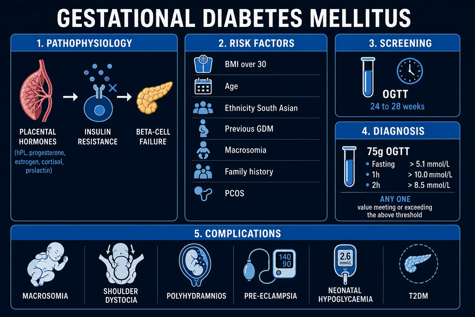
The White classification (older, still examined in some curricula) extends through classes B, C, D, F, R, H, T according to age of onset, duration, and vascular complications — these classes denote overt diabetes in pregnancy, not GDM. The pragmatic A1/A2 split is what governs day-to-day obstetric decisions and what examiners expect at MBBS level. [1]
Epidemiology & Risk Factors
Reported GDM prevalence varies enormously with population, obesity, and the diagnostic criteria used — from under 1 percent in lean East Asian populations screened by older criteria to over 14 percent in South Asian and Middle Eastern cohorts screened by the inclusive IADPSG one-step 75 g OGTT.[9] Adoption of the IADPSG thresholds (any one positive on a 75 g OGTT) increased diagnosed GDM by roughly 30 to 50 percent compared with older two-step strategies, with the increase concentrated in mild, previously undetected disease. Globally, GDM prevalence is rising, tracking obesity, older maternal age, sedentary lifestyle, and migration of high-risk ethnic groups. The condition is now the most common metabolic disorder of pregnancy and a major contributor to the global type 2 diabetes epidemic, because up to half of women with GDM will develop T2DM within a decade.
The prevalence figures are not merely academic — they determine the cost-effectiveness of universal versus risk-based screening. In low-prevalence populations, a risk-based approach (screen only those with risk factors) may appear efficient, but it misses 10 to 30 percent of cases, including many women from high-risk ethnic groups who do not meet BMI thresholds. In high-prevalence populations such as India and the Middle East, universal screening is clearly superior and is the basis of the DIPSI and FIGO recommendations. The choice of diagnostic criteria also changes prevalence: applying the NICE thresholds (fasting over 5.6 or 2-hour over 7.8 mmol/L) diagnoses fewer women than the IADPSG thresholds (fasting over 5.1, 1-hour over 10.0, or 2-hour over 8.5 mmol/L), because the IADPSG fasting threshold is lower and the 1-hour value is included. [1]
A useful viva point: IADPSG was deliberately designed to identify women at the glucose level where fetal overgrowth risk begins to climb, not to identify every woman with any glucose abnormality. The trade-off is more labelled disease, more clinic visits, and more pharmacotherapy, but with the benefit of fewer macrosomic births and shoulder dystocias. [1]
GDM also acts as a cardiovascular risk marker for the mother. Even after glucose normalises postpartum, women with a history of GDM have a higher lifetime risk of hypertension, dyslipidaemia, and myocardial infarction. This is why postpartum follow-up is not simply about diabetes screening — it is an entry point into lifelong cardiometabolic prevention. [1]
GDM — key numbers
The classical risk factors fall into four groups. Maternal anthropometry and age: BMI over 30 kg/m2 (or over 23 in South Asian populations, who develop insulin resistance at lower adiposity), age over 35 years, and excessive gestational weight gain. Reproductive and obstetric history: previous GDM (recurrence 30 to 60 percent), previous macrosomia (birthweight over 4.5 kg) or unexplained stillbirth, PCOS, multiparity (parity 2 or more), and multiple pregnancy or IVF conception (higher placental hormone load). Family and genetic background: a first-degree relative with diabetes, and high-risk ethnicity — South Asian, Black African-Caribbean, Hispanic, Middle Eastern, Polynesian/Indigenous, Native American — each conferring a 3 to 7 fold elevated risk. Biochemical markers at booking: glycosuria on antenatal dipstick (especially 2+ on more than one occasion) and elevated booking HbA1c in the pre-diabetic range (5.7 to 6.4 percent).[1]
Pathophysiology
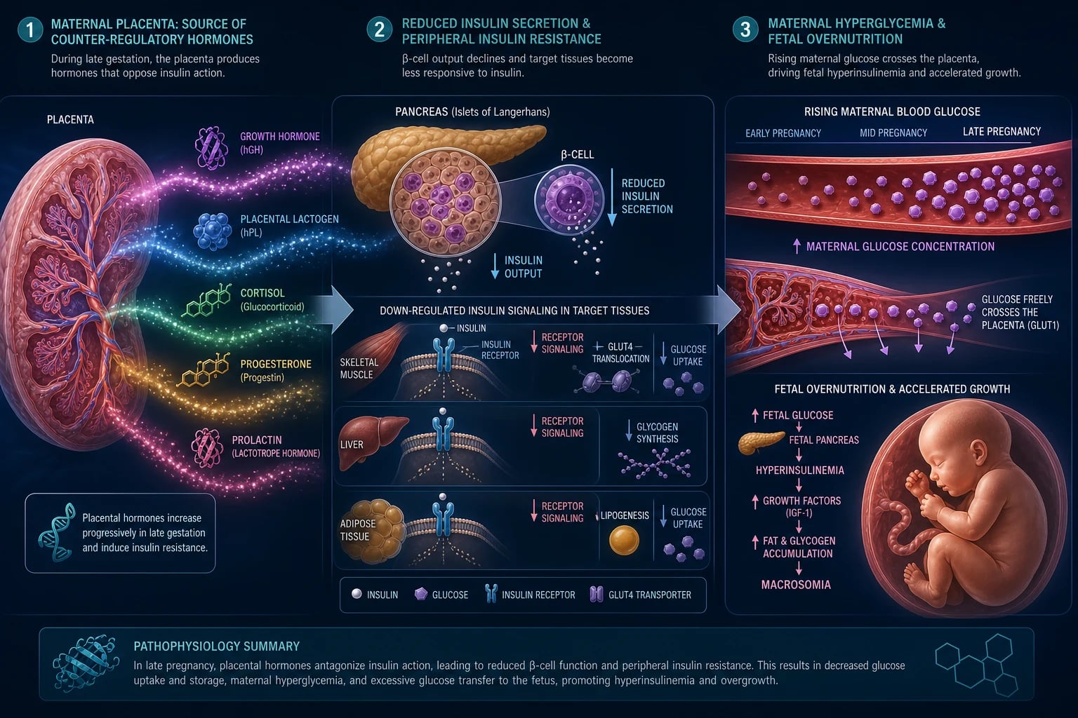
Pregnancy is, in metabolic terms, a progressively diabetogenic state. From the late second trimester onward the placenta secretes a cocktail of counter-regulatory (anti-insulin) hormones: human placental lactogen (also called human chorionic somatomammotropin — the dominant anti-insulin signal), a placental growth hormone variant, cortisol, progesterone, prolactin, and oestrogen. Together with cytokines — notably TNF-alpha — these remodel maternal metabolism to divert glucose and amino acids to the growing fetus.[2]
At the molecular level the resistance is largely post-receptor: insulin binds its receptor normally, but downstream signalling through insulin receptor substrate-1 (IRS-1) is blunted by serine phosphorylation, so GLUT-4 translocation to the sarcolemmal and adipocyte membrane is reduced and glucose uptake falls. Progesterone and cortisol additionally increase hepatic gluconeogenesis, opposing insulin's normal suppression of glucose output. The placenta itself contributes insulinase, which degrades maternal insulin and increases the secretory demand further.[2]
In a normal pregnancy the pancreas meets this challenge by beta-cell hyperplasia and amplified glucose-stimulated insulin secretion, raising circulating insulin two- to threefold. GDM develops when this compensation is inadequate. The woman who develops GDM carries a sub-clinical predisposition to beta-cell failure — a mix of genetic susceptibility (variants in TCF7L2, GCK and other loci shared with type 2 diabetes), chronic insulin resistance, low-grade inflammation, and adipokine dysregulation (low adiponectin, high leptin and resistin from visceral adiposity). Adiponectin normally enhances insulin sensitivity through AMP-activated protein kinase (AMPK) and PPAR-gamma pathways; its fall in visceral obesity removes a brake on insulin resistance. Leptin and resistin, conversely, promote insulin resistance and hepatic glucose output. The clinical result is postprandial hyperglycaemia first (insulin-mediated peripheral glucose disposal fails), followed by fasting hyperglycaemia as hepatic glucose output escapes suppression. [1]
This sequence has a direct clinical implication: the 1-hour post-load glucose is usually the first value to become abnormal, and postprandial hyperglycaemia is the dominant driver of fetal overgrowth. It is also why management targets postprandial glucose and why rapid-acting bolus insulin is so effective when required. The fasting glucose, by contrast, reflects hepatic insulin resistance and is often the last to deteriorate; when fasting glucose is elevated, beta-cell compensation has usually failed substantially.[2]
The fetal consequences are the clinical point of the disease. Maternal glucose crosses the placenta by facilitated diffusion; maternal insulin does not cross. Fetal hyperglycaemia stimulates fetal pancreatic beta-cell hypersecretion, producing fetal hyperinsulinaemia — an anabolic, growth-driving state that Pedersen called fuel-mediated teratogenesis in late gestation. The result is macrosomia disproportionate in the shoulder and trunk (the head/pelvis mismatch that produces shoulder dystocia), organomegaly, polycythaemia (erythropoietin response to chronic hyperglycaemia), and — after cord clamping — neonatal hypoglycaemia, because the now-withdrawn maternal glucose supply cannot match the still-high fetal insulin. Insulin also antagonises surfactant maturation, raising the risk of neonatal respiratory distress syndrome even at term. [1]
The long-term significance extends beyond the neonatal period. Fetal hyperinsulinaemia and nutrient excess produce metabolic programming of the offspring's pancreas, adipose tissue, and hypothalamus. Children born to hyperglycaemic pregnancies have higher rates of childhood obesity, impaired glucose tolerance, and early-onset type 2 diabetes — a transgenerational cycle in which maternal GDM begets metabolic disease in the next generation. This is why GDM is considered a window of opportunity: treating it not only protects the current pregnancy but may reduce the lifetime cardiometabolic risk of both mother and child. The PedMet and related cohorts continue to follow metformin-exposed offspring to determine whether specific pharmacotherapies modify this programming trajectory.[3][10]
The HAPO study demonstrated that these harms rise continuously with maternal glucose across the range previously considered normal — there is no sharp threshold — and that the strongest single predictor of fetal overgrowth is the 1-hour post-load glucose.[3] This continuous relationship is what underpinned the IADPSG decision to lower diagnostic thresholds.
Clinical Presentation
GDM is, in the great majority of cases, clinically silent and is detected by the routine 24 to 28 week OGTT. Symptomatic hyperglycaemia — polyuria, polydipsia, fatigue, recurrent vulvovaginal candidiasis — suggests either severe GDM or, more often, overt diabetes misclassified as GDM (which the booking HbA1c should have caught). [1]
The clinical picture more often presents as the consequences of GDM detected on antenatal assessment: macrosomia on ultrasound (abdominal circumference over the 90th centile, estimated fetal weight above the 90th centile), polyhydramnios (fetal polyuria from hyperglycaemia), an accelerated symphysis-fundal height, or the co-occurrence of gestational hypertension or pre-eclampsia (with which GDM shares insulin-resistance and endothelial-dysfunction biology). Atypical presentations worth flagging include: the South Asian woman at a BMI as low as 23 (where ethnicity lowers the screening threshold); the teenager with obesity; the IVF or multiple pregnancy with a heavier placental hormone load; and the woman with glycosuria 2+ on more than one antenatal dipstick who should be screened earlier than 24 weeks.[1]
Atypical and scenario-based presentations
GDM detected on ultrasound. Many women have no symptoms and are first flagged because a routine 28-week growth scan shows fetal abdominal circumference over the 90th centile or estimated fetal weight above the 90th centile. In this scenario, the first question is whether the OGTT was already done; if not, it should be performed urgently. If the OGTT was normal but growth is accelerated, consider repeating glucose testing or reviewing the dating scan, because GDM can evolve after 28 weeks and severe postprandial spikes may be missed by a single test. [1]
GDM presenting with polyhydramnios. A sudden increase in amniotic fluid index (AFI over 24 cm or deepest vertical pocket over 8 cm) in the third trimester should prompt glucose testing even if the OGTT was normal. Fetal polyuria from hyperglycaemia is an under-recognised presentation; once GDM is treated, the polyhydramnios often improves. [1]
GDM presenting with recurrent vulvovaginal candidiasis. Candida thrives in glycosuric, hyperglycaemic environments. Recurrent episodes after treatment, especially in the second trimester, should trigger a glucose check even if the routine OGTT is not yet due. [1]
Steroid-induced hyperglycaemia. Women receiving antenatal corticosteroids for threatened preterm labour (betamethasone or dexamethasone) may show transient hyperglycaemia that peaks 12 to 48 hours post-dose. If glucose remains abnormal after the steroid effect has waned, true GDM is likely. Conversely, a woman with known GDM receiving steroids needs intensified glucose monitoring and often a temporary increase in insulin. [1]
GDM presenting in late third trimester. Occasionally, a woman presents with polyhydramnios, accelerated growth, or incidental hyperglycaemia after 32 weeks. If the OGTT at 24 to 28 weeks was normal but suspicion is high, repeat testing is reasonable. Late-diagnosed GDM still benefits from management, though there is less time to modify fetal growth. These women are more likely to need insulin and earlier delivery. [1]
Type 2 diabetes unmasked in late pregnancy. A woman with a normal booking HbA1c but marked, persistent hyperglycaemia in the third trimester may have pre-existing type 2 diabetes revealed by the insulin resistance of pregnancy. The postpartum glucose test is critical: failure to normalise confirms that the label should change from GDM to diabetes. [1]
Differential Diagnosis
The differential of hyperglycaemia first detected in pregnancy is narrow but consequential, because the management differs sharply between entities. The pivotal distinction is GDM from overt (pre-gestational) diabetes, which carries higher anomaly, miscarriage, and stillbirth risk and requires insulin, anomaly scanning, and organ review. [1]
Overt diabetes in pregnancy
not GDM
- **Booking HbA1c over 6.5 percent** OR fasting glucose over 7.0 OR random over 11.1 mmol/L
- First-trimester anomaly risk (neural tube, cardiac) is elevated
- Insulin mandatory, folic acid 5 mg, retinal/renal review
- Outcome and counselling are those of pre-gestational diabetes
Type 1 diabetes presenting in pregnancy
autoimmune
- Ketosis, weight loss, marked symptoms; may present as DKA
- **Anti-GAD, anti-IA-2, anti-ZnT8 antibodies** positive; C-peptide low
- Insulin from diagnosis; risk of rapid decompensation
- Autoimmune clustering (thyroid, Addison)
Renal glycosuria
benign
- Glucose in urine from low renal threshold (physiological in pregnancy from increased GFR)
- **Normal blood glucose and normal OGTT**
- No fetal consequences, no treatment
- Reassure; distinguished by a normal OGTT
Steroid-induced hyperglycaemia
transient
- Spike after **betamethasone or dexamethasone** for fetal lung maturation
- Usually peaks 12 to 48 hours post-dose
- Treat if sustained; re-evaluate off steroid
- May unmask true GDM needing follow-through
Gestational impaired glucose tolerance
borderline
- Abnormal OGTT not meeting GDM threshold
- Most contemporary guidelines fold into GDM (IADPSG)
- Same lifestyle management; lower fetal risk
- Watch for progression
The other distinction worth making explicitly is glycosuria without hyperglycaemia (renal glycosuria, normal in pregnancy) versus true GDM with glycosuria — resolved by the OGTT, never by dipstick alone. [1]
Clinical & Bedside Assessment
The focused assessment of a woman with suspected or confirmed GDM has four aims: confirm glycaemic status, characterise fetal risk, screen for co-morbidities, and engage the woman in self-management. [1]
History captures the risk-factor profile (BMI, age, ethnicity, previous GDM, previous macrosomia or stillbirth, first-degree family history of diabetes, PCOS), the gestational age, any symptoms of hyperglycaemia, current medications (especially corticosteroids, antipsychotics), and the woman's understanding of and attitude towards insulin. Bedside examination includes weight and BMI (with appropriate screening thresholds for ethnicity), blood pressure (to detect co-existing gestational hypertension or pre-eclampsia, which shares insulin-resistance biology), symphysis-fundal height (macrosomia, polyhydramnios), and urine dipstick for glucose, protein, and ketones. Look specifically for vulvovaginal candidiasis, which is over-represented in GDM.[1]
Named signs and manoeuvres at the bedside
Symphysis-fundal height (SFH) is measured with a tape from the top of the symphysis pubis to the uterine fundus. In GDM, an SFH above the gestational age by more than 3 cm or a sudden increase in SFH suggests macrosomia or polyhydramnios. A fundal height of 33 cm at 28 weeks, for example, demands an ultrasound. The measurement is unreliable in obesity; in such women, rely more on ultrasound. [1]
Leopold's manoeuvres are used to assess fetal lie, presentation, engagement, and estimated fetal size. In a woman with GDM and suspected macrosomia, the examiner may feel a large, ballotable head and a broad, fleshy trunk; the fetus may feel larger than dates. However, clinical estimation of fetal weight is inaccurate and should not override ultrasound. [1]
Blood pressure must be recorded at every visit. GDM and pre-eclampsia share an insulin-resistance, endothelial-dysfunction substrate; co-occurrence raises the risk of severe pre-eclampsia and influences delivery timing. A reading of 140/90 mmHg or more on two occasions, or proteinuria 1+ or more, should trigger a full pre-eclampsia work-up. [1]
Urine dipstick is informative for three reasons: glycosuria (supports hyperglycaemia but may be renal), ketones (inadequate calorie intake or undertreated hyperglycaemia), and protein (pre-eclampsia). Ketonuria in a woman on MNT usually means her carbohydrate restriction is too severe; it should prompt a dietary review rather than intensification of therapy. [1]
The decisive bedside skill is capillary glucose self-monitoring: teaching the woman to use a calibrated glucometer to check fasting and 1-hour post-meal glucose, since these two values drive every escalation decision in management. Fetal assessment at the bedside combines symphysis-fundal height with scheduled ultrasound growth scans and, when indicated, cardiotocography. [1]
Counselling and shared decision-making
From the first visit, the woman needs a clear explanation of what GDM is, why it matters, and what she can do. Anxiety about insulin is common; framing insulin as a safe, effective tool that does not cross the placenta improves acceptance. Discuss the realistic probability of each step: most women control GDM with lifestyle alone; if metformin is needed, it is safe and convenient; insulin is reserved for those who need it, not a punishment for non-adherence. Document her preferences and ensure she knows how to contact the diabetes team. This counselling is not optional — it drives adherence, reduces no-shows, and is the single best way to ensure postpartum follow-up and long-term health. [1]
Investigations
Investigations in GDM serve three purposes: screen and diagnose (OGTT), monitor glycaemic control (capillary glucose, HbA1c), and survey fetal and maternal complication risk (growth scans, bloods).
Booking visit (first antenatal contact). Every woman should have an HbA1c at booking to detect overt diabetes (over 6.5 percent = overt diabetes; 5.7 to 6.4 percent is pre-diabetes and flags high risk for GDM). In high-risk women a booking fasting glucose is also reasonable — over 7.0 mmol/L denotes overt diabetes that must not be mislabelled as GDM.[4]
Universal screening at 24 to 28 weeks. All pregnant women not already known to have diabetes undergo a 75 g 2-hour oral glucose tolerance test (IADPSG/WHO/FIGO one-step). NICE (UK) historically used a two-step approach — a 50 g glucose challenge test followed by a diagnostic 75 g OGTT in screen-positive women — but most units now offer a one-step 75 g OGTT to all. [1]
Performing the OGTT correctly
The 75 g 2-hour OGTT is deceptively simple and frequently performed incorrectly. The woman should be fasting for at least 8 hours (water allowed). A fasting baseline venous plasma glucose is drawn; she then drinks 75 g anhydrous glucose dissolved in 250 to 300 mL water over 5 minutes. A second venous sample is taken at 2 hours (and a 1-hour sample in many centres using IADPSG/WHO/FIGO). She should remain seated, not smoke, and not eat or drink anything except water during the test. Acute illness, bed rest, or recent major dietary change can invalidate the result. If the woman vomits the glucose load, the test should be repeated another day. [1]
The threshold values are for plasma glucose, not whole blood or capillary glucose. A laboratory plasma glucose is the diagnostic standard; capillary glucometer values are used for monitoring, not diagnosis. If a woman is on corticosteroids or has acutely reduced her carbohydrate intake, interpretation must be adjusted. [1]
[1]The HAPO study provided the data: each threshold was set at the glucose value conferring a 1.75-fold odds of an adverse outcome (birthweight over 90th centile, cord C-peptide over 90th centile, or neonatal adiposity) relative to the population mean.[3][4]
On treatment. Capillary glucose targets (NICE): fasting under 5.3 mmol/L and 1-hour post-meal under 7.8 mmol/L (or 2-hour under 6.7 mmol/L). HbA1c is monitored intermittently but is less useful in GDM than in overt diabetes because of the shortened red-cell survival and expanded red-cell mass of pregnancy (erythropoietic rate lowers HbA1c). Urine should be checked for ketones — ketonuria signals inadequate calorie intake or undertreated hyperglycaemia. Baseline bloods include renal function, liver function, and TSH (thyroid disease clusters with autoimmune diabetes).[1]
Continuous glucose monitoring and other tools
Continuous glucose monitoring (CGM) is increasingly used in selected GDM cases, particularly for women on insulin or those with erratic glucose profiles. CGM provides a 24-hour glucose curve and reveals postprandial spikes that intermittent capillary testing may miss. However, CGM is not yet a universal standard in GDM; capillary glucose monitoring remains the cost-effective backbone. Time-in-range targets (e.g. over 70 percent of readings between 3.5 and 7.8 mmol/L) are being studied but are not guideline-standard. [1]
Ketone testing should be performed if the woman reports reduced oral intake, nausea, or has persistent hyperglycaemia. Small ketonuria from calorie restriction is common and usually benign; large ketonuria or ketonaemia requires urgent review to exclude diabetic ketoacidosis, especially in insulin-treated women. [1]
Fetal investigations. Serial ultrasound growth scans every 3 to 4 weeks from 28 weeks, measuring abdominal circumference, estimated fetal weight, and amniotic fluid index. Abdominal circumference over the 90th centile is the earliest marker of fetal hyperinsulinaemic overgrowth. Cardiotocography is reserved for indications (reduced movements, postdates, A2 disease). Doppler is added only if growth restriction co-exists. A detailed fetal echocardiogram is not routinely needed in GDM (unlike overt diabetes, where congenital heart disease risk is higher), but may be considered if control has been poor or if there are other risk factors. [1]
Management — Resuscitation
GDM is not usually a resuscitation emergency — the acute threats are fetal and neonatal and are managed by the structured antepartum pathway below. Two exceptions deserve mention because they appear in vivas and on call. [1]
Severe maternal hyperglycaemia or diabetic ketoacidosis is more characteristic of overt or type 1 diabetes but can occur in poorly controlled GDM under stress (infection, corticosteroids). The bundle is: intravenous access, fluid resuscitation with normal saline, a fixed-rate intravenous insulin infusion at 0.1 unit/kg/hour, potassium replacement (insulin drives potassium intracellularly), and hourly monitoring of blood glucose, blood ketones (beta-hydroxybutyrate), and potassium, with treatment of the precipitant. Targets are a fall in glucose of 3 mmol/L/hour and a fall in ketones of 0.5 mmol/L/hour.[1]
Pregnancy-specific points for DKA: the fetal heart must be monitored once viable; maternal acidosis can cause fetal distress even if the fetus is not directly diabetic. DKA can occur at lower glucose levels in pregnancy because of the relative insulin resistance and accelerated starvation ketogenesis; therefore, do not wait for a very high glucose to suspect DKA if the woman has acidosis, ketonaemia, and an anion-gap metabolic acidosis. Bicarbonate is rarely needed and is reserved for severe acidosis (pH under 7.0) with haemodynamic compromise. Once the woman is stabilised, obstetric review for delivery timing is essential if the fetus is compromised. [1]
Neonatal hypoglycaemia after delivery is the predictable, anticipatable emergency. Fetal hyperinsulinaemia persists after cord clamping while the maternal glucose supply is withdrawn. Prevention is by intrapartum maternal glucose control (target 4 to 7 mmol/L), early feeding within 30 minutes, and neonatal blood glucose measurement at 2 to 4 hours of age, with 200 mg/kg (0.2 mL/kg) of 40 percent dextrose gel massaged into the buccal mucosa for asymptomatic hypoglycaemia and intravenous 10 percent dextrose (a 2 to 5 mL/kg bolus then an infusion) for symptomatic or persistent hypoglycaemia.[1]
Neonatal hypoglycaemia algorithm
- At risk: infant of mother with GDM, especially insulin-treated or poorly controlled.
- Prevention: feed within 30 minutes; check glucose at 2 to 4 hours of age and then before feeds until stable.
- Asymptomatic hypoglycaemia (glucose under 2.6 mmol/L): feed or encourage breastfeed; apply 40% dextrose gel 200 mg/kg (0.2 mL/kg) to the buccal mucosa and massage; recheck in 30 minutes. Repeat once if still low.
- Symptomatic hypoglycaemia or persistent despite gel: transfer to NICU; give 10% dextrose 2 to 5 mL/kg IV bolus followed by an infusion to maintain glucose; investigate and treat sepsis if indicated.
- Thresholds vary by unit and by gestational age, but the principle is universal: early feeding and glucose monitoring prevent seizures and brain injury. [1]
Management — Definitive & Stepwise
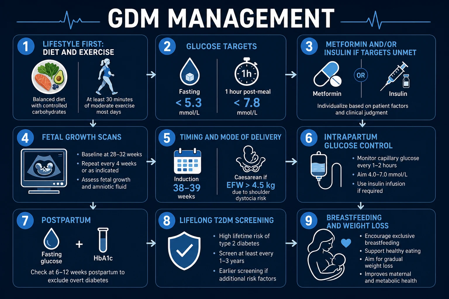
Definitive management is a stepwise ladder with explicit glucose targets, escalation triggers, fetal surveillance, a delivery plan, and a defined postpartum pathway. The aim is to keep fasting glucose under 5.3 mmol/L and 1-hour post-meal under 7.8 mmol/L, because both the ACHOIS and the Landon (MFMU) trials proved that doing so reduces serious perinatal outcomes (shoulder dystocia, death, fracture, nerve palsy) and maternal pre-eclampsia.[5][6]
Definitive management is a stepwise ladder with explicit glucose targets, escalation triggers, fetal surveillance, a delivery plan, and a defined postpartum pathway. The aim is to keep fasting glucose under 5.3 mmol/L and 1-hour post-meal under 7.8 mmol/L, because both the ACHOIS and the Landon (MFMU) trials proved that doing so reduces serious perinatal outcomes (shoulder dystocia, death, fracture, nerve palsy) and maternal pre-eclampsia.[5][6]
Step 1 — Lifestyle (medical nutrition therapy), for all women
Medical nutrition therapy (MNT), ideally dietitian-led, is the foundation and the only treatment needed in 70 to 85 percent of women. The prescription is: a low-glycaemic-index diet favourishing complex carbohydrates at 40 to 45 percent of energy, lean protein and unsaturated fats, distributed across three meals and two to three snacks (a bedtime snack reduces overnight ketogenesis); calorie restriction of 25 to 30 percent in women with BMI over 30; and 30 minutes of moderate exercise daily (walking after meals is particularly effective — it disposes of postprandial glucose). Weight management targets modest, gestation-appropriate gain. Self-monitoring of fasting and 1-hour post-meal glucose is taught and reviewed at 1 to 2 weeks.[1]
A common error is to restrict carbohydrates so severely that ketonuria develops. The goal is glucose control, not ketosis. If ketones appear, carbohydrate intake should be redistributed rather than slashed. The bedtime snack is important: it provides overnight carbohydrate and reduces lipolysis and ketogenesis. [1]
Step 2 — Pharmacotherapy if targets unmet within 1 to 2 weeks
If lifestyle alone fails to achieve targets, metformin is added first in most guidelines (NICE, FIGO). Metformin is oral, safe in pregnancy and breastfeeding, reduces insulin resistance (hepatic gluconeogenesis, peripheral uptake), is preferred by women, and is supported by the MiG trial which demonstrated non-inferiority to insulin for perinatal outcomes (with supplemental insulin needed in about half).[7][8]
Metformin — oral, start 500 mg once daily with breakfast, titrate over days to 500 mg twice daily, then 1 g twice daily, up to 1 g three times daily (maximum 2 to 2.5 g/day). Gastrointestinal side-effects (nausea, diarrhoea) are common but usually settle; the extended-release formulation improves tolerance. Metformin crosses the placenta; the PedMet follow-up reports mixed but largely reassuring long-term offspring cardiometabolic outcomes, and ongoing studies continue to follow exposed children.[7][10]
Insulin is added when metformin is contraindicated or refused, when targets remain unmet on maximum metformin, or when there is fetal abdominal circumference over the 90th centile or estimated fetal weight over 4.5 kg (where insulin is preferred from the outset because of its tighter control). The regimen is basal-bolus to mimic physiological secretion: [1]
- Basal (long-acting) — insulin detemir (preferred — category A in pregnancy) or isophane (NPH), started at 0.1 to 0.2 unit/kg at bedtime, titrated to fasting glucose.
- Bolus (rapid-acting) — insulin aspart or insulin lispro, given immediately before each main meal, started at 4 to 6 units per meal and titrated to the 1-hour post-meal glucose.
- Review and titrate every 1 to 2 days based on the glucose diary. Insulin requirements rise progressively across the third trimester and fall precipitously after delivery. [1]
Practical insulin titration
A simple titration algorithm is: increase the relevant insulin by 2 units if the corresponding glucose value is above target on two consecutive days. For fasting hyperglycaemia, increase the basal insulin. For post-breakfast hyperglycaemia, increase the breakfast bolus; for post-lunch, the lunch bolus; for post-dinner, the dinner bolus. Hypoglycaemia (under 4.0 mmol/L) requires a reduction of 2 to 4 units in the insulin responsible. Women are taught to recognise and treat hypoglycaemia with fast-acting carbohydrate (15 g glucose tablets or juice), followed by a snack. Severe hypoglycaemia is rare but is more likely with tight targets and escalating insulin; glucagon should be available for insulin-dependent women. [1]
Glibenclamide (glyburide), a sulfonylurea, is offered in some guidelines (ACOG) as an alternative oral agent. It is less effective than insulin, crosses the placenta, and carries a higher rate of neonatal hypoglycaemia; the Balsells meta-analysis showed metformin superior to glibenclamide for glycaemic control, and NICE does not recommend glibenclamide.[8]
Step 3 — Fetal surveillance and escalation triggers
Serial ultrasound growth scans every 3 to 4 weeks from 28 weeks assess abdominal circumference and estimated fetal weight. Escalation triggers are: persistent fasting glucose over 5.3 mmol/L or 1-hour post-meal over 7.8 on lifestyle plus metformin; fetal AC over the 90th centile or EFW over 4.5 kg (move to insulin); or persistent ketonuria (calorie intake too low).[1]
Step 4 — Delivery
With good glycaemic control and normal fetal growth, offer induction of labour at 38 to 39 weeks, or expectant management up to 40+6 weeks with ongoing surveillance. Elective caesarean should be discussed where the estimated fetal weight exceeds 4.5 kg, because the risk of shoulder dystocia rises steeply above this weight and is poorly predicted by clinical assessment. In women with poor control, macrosomia, or polyhydramnios, earlier delivery may be warranted. Intrapartum glucose control targets a maternal glucose of 4 to 7 mmol/L: women on insulin use a variable-rate intravenous insulin and dextrose infusion with hourly glucose checks; women diet-controlled or on metformin alone usually maintain normoglycaemia but should still have glucose checked hourly. Active management of the third stage reduces postpartum haemorrhage.[1]
Intrapartum management detail
The variable-rate intravenous insulin infusion is usually prepared as 50 units of short-acting insulin in 50 mL normal saline (1 unit/mL), running at a rate determined by a sliding scale based on hourly capillary glucose. A concurrent 5% or 10% dextrose infusion (often 125 mL/hour) provides glucose substrate and prevents starvation ketosis while the insulin infusion controls the level. The aim is to keep maternal glucose between 4 and 7 mmol/L; readings outside this range trigger rate adjustments. All women with insulin-treated GDM should have intrapartum glucose monitoring; those on diet or metformin may still need hourly checks if labour is prolonged or if they receive steroids. [1]
For mode of delivery, the EFW over 4.5 kg threshold is lower than the 5.0 kg sometimes quoted for non-diabetic macrosomia, because diabetic macrosomia is disproportionately truncal and shoulder-heavy, making shoulder dystocia more likely at any given weight. A caesarean does not eliminate the risk entirely, but it reduces it substantially. For EFW between 4.0 and 4.5 kg, a careful discussion of risks, maternal preferences, and obstetric factors is needed; some guidelines suggest caesarean at EFW over 4.5 kg and shared decision-making below that. [1]
Step 5 — Postpartum pathway
Insulin and metformin are stopped at delivery in nearly all women (insulin requirements collapse within hours as placental hormones clear). Neonatal blood glucose is monitored 2 to 4 hourly for the first 24 hours, with early feeding and dextrose gel as above. The mother undergoes a fasting plasma glucose at 6 to 13 weeks postpartum (NICE) — or an HbA1c at 3 months — to exclude persistent overt diabetes. Breastfeeding is strongly encouraged (it reduces maternal T2DM risk and improves neonatal metabolic outcomes). Thereafter she enters lifelong annual screening for type 2 diabetes with fasting glucose or HbA1c, supported by structured weight-loss and exercise advice (5 to 10 percent weight loss halves subsequent T2DM risk, as shown in the Diabetes Prevention Program).[1]
GDM management ladder — 'L-M-I-M-D-P'
LMIMDP
first-line for ALL; targets fasting under 5.3, 1-h post-meal under 7.8
500 mg PO OD titrate to 1 to 2 g/day if lifestyle fails in 1 to 2 weeks
basal detemir/NPH + bolus aspart/lispro; if metformin inadequate or fetal AC over 90th centile
EFW over 4.5 kg → discuss elective caesarean (shoulder dystocia)
offer induction at 38 to 39 weeks; intrapartum glucose 4 to 7 mmol/L
stop insulin/metformin; fasting glucose at 6 to 13 weeks; LIFELONG annual T2DM screening
Specific Subtypes & Scenarios
A1 (diet-controlled) GDM carries fetal risk closer to uncomplicated pregnancy; routine antenatal care with growth scans suffices, and most women deliver at term without pharmacotherapy. These women still need serial growth scans and a delivery plan, because a minority will develop macrosomia or fail targets later in the third trimester. [1]
A2 GDM (on metformin and/or insulin) requires closer fetal growth surveillance, intrapartum glucose control, and neonatal hypoglycaemia vigilance — the group that drives the perinatal morbidity signal in trials. Any woman escalating to insulin should have a clear intrapartum plan documented before labour. [1]
Overt diabetes in pregnancy (booking HbA1c over 6.5 percent or fasting glucose over 7.0) is not GDM and must be managed as pre-gestational diabetes: pre-conception optimisation where possible (HbA1c under 6.5 percent, folic acid 5 mg daily from pre-conception, medication review — stop ACE inhibitors, statins, and potentially teratogenic agents and switch to insulin), first-trimester anomaly scan with detailed cardiac views, retinal and renal review each trimester, and insulin throughout. The anomaly, miscarriage, stillbirth, and pre-eclampsia risks are all substantially higher than in GDM.[4]
Previous GDM in a current pregnancy warrants early booking assessment (HbA1c ± early OGTT), lifestyle intervention from the first trimester, and consideration of metformin prophylaxis in selected high-risk women. Recurrence is 30 to 60 percent; counselling about this and about inter-pregnancy weight loss is part of the consultation. [1]
GDM with macrosomia (AC over 90th centile or EFW over 4.5 kg) drives a move to insulin (for tighter control), careful discussion of mode and timing of delivery, and a neonatal team alert at birth. [1]
GDM with pre-eclampsia. Co-occurrence is common and intensifies both maternal and fetal risk. Delivery timing may need to be earlier than 38 to 39 weeks, balancing the risks of preterm birth against those of continuing a hypertensive, hyperglycaemic pregnancy. Antihypertensive and magnesium sulphate protocols follow standard pre-eclampsia care. [1]
GDM requiring steroids for fetal lung maturation. Betamethasone or dexamethasone transiently worsen hyperglycaemia. Women on insulin usually need a temporary 20 to 40 percent increase in insulin for 24 to 48 hours; women on metformin or diet may need short-acting insulin during this window. Close glucose monitoring (every 1 to 2 hours) is required until values return to baseline. [1]
Complications & Pitfalls
The complications of GDM are predominantly fetal and neonatal but extend meaningfully into maternal and long-term territory. The HAPO study established that maternal and neonatal morbidity rise continuously across the glucose spectrum that GDM occupies.[3]
Maternal complications. Pre-eclampsia and gestational hypertension (roughly twofold excess), polyhydramnios (fetal polyuria), caesarean delivery (twofold excess, partly driven by macrosomia and failed induction), operative vaginal delivery, birth injury, and postpartum haemorrhage (macrosomic fetus, polyhydramnios). Long term: a 30 to 50 percent risk of type 2 diabetes over 10 to 15 years (the single most important long-term consequence of GDM), increased cardiovascular disease, and recurrent GDM in 30 to 60 percent of subsequent pregnancies. [1]
Fetal and neonatal complications. Macrosomia (with the asymmetric shoulder/trunk adiposity that produces shoulder dystocia, clavicular fracture, and brachial plexus injury), neonatal hypoglycaemia (rebound from fetal hyperinsulinaemia), polycythaemia (with hyperviscosity and a need for partial exchange transfusion in severe cases), hyperbilirubinaemia (from polycythaemia breakdown), hypocalcaemia and hypomagnesaemia, respiratory distress syndrome (insulin delays surfactant maturation), NICU admission, and — in poorly controlled disease — stillbirth (rare with modern management). The PedMet and related cohorts report that offspring of hyperglycaemic pregnancies carry higher rates of childhood obesity and adolescent glucose intolerance — a metabolic programming effect across generations.[10]
Shoulder dystocia: recognition and response
Shoulder dystocia is the most feared acute complication of diabetic macrosomia. The classic scenario is delivery of the head followed by retraction and failure of the shoulders to descend (turtle sign). The HELPERR mnemonic guides management: Help (call for assistance), Evaluate for episiotomy, Legs (McRoberts position — flex maternal thighs onto abdomen), Pressure (suprapubic pressure to displace the anterior shoulder), Enter the vagina for internal manoeuvres (Rubin, Woods' screw, delivery of posterior arm), Remove the posterior arm, and Roll the patient to all fours. Immediate neonatal team presence is essential; fractures and brachial plexus injury (Erb's palsy) are recognised complications. The best management is prevention: elective caesarean when EFW exceeds 4.5 kg in a diabetic pregnancy. [1]
Neonatal hypoglycaemia and metabolic adaptation
After delivery, the abrupt withdrawal of maternal glucose leaves the neonate with high circulating insulin and low glucose substrate. This mismatch can produce seizures if not anticipated. The threshold for treatment varies by unit, but any symptomatic hypoglycaemia (jitteriness, apnoea, seizures, poor feeding) requires immediate intravenous dextrose. Asymptomatic hypoglycaemia is managed with feeding and buccal dextrose gel as described above. Neonatal hypoglycaemia is the single commonest reason for NICU admission in infants of diabetic mothers. [1]
Polycythaemia and hyperbilirubinaemia
Chronic fetal hyperglycaemia stimulates erythropoietin release, producing polycythaemia (venous haematocrit over 65 percent). Hyperviscosity can cause thrombosis, renal vein thrombosis, and poor feeding. The breakdown of excess red cells leads to hyperbilirubinaemia and jaundice. Severe polycythaemia may require partial exchange transfusion. [1]
Pitfalls. Common and avoidable errors include: missing overt diabetes at booking (treating pre-gestational diabetes as if it were GDM, with under-treatment and missed anomaly prevention); failing to titrate insulin to post-prandial glucose (the strongest predictor of macrosomia); over-reliance on glibenclamide (less effective, more neonatal hypoglycaemia); not checking neonatal glucose after delivery; and — the most common real-world failure — losing women to postpartum glucose testing, so that the lifelong T2DM risk goes unmonitored. Another pitfall is ketonuria from over-restricted calories, which the mother (and some clinicians) misread as good control; the correct response is to relax calorie restriction, not intensify it. [1]
Prognosis & Disposition
GDM resolves within hours to days of delivery in the great majority of women, as the placental anti-insulin signal is removed and maternal insulin resistance falls rapidly. The postpartum fasting glucose (or 3-month HbA1c) defines who has persistent overt diabetes — a small but important minority — versus persistent pre-diabetes versus complete resolution.[1]
The recurrence rate in subsequent pregnancy is 30 to 60 percent, and the long-term maternal type 2 diabetes risk is 30 to 50 percent over 10 to 15 years. This risk is modifiable: breastfeeding, a 5 to 10 percent body-weight reduction, regular exercise, and — in high-risk selected women — metformin prophylaxis each substantially lower subsequent T2DM incidence (the Diabetes Prevention Program showed lifestyle intervention reducing progression by 58 percent, metformin by 31 percent).[1]
Perinatal mortality with modern detection and management is low; the principal residual morbidities are macrosomia-related birth injury and neonatal hypoglycaemia, both reduced by the management ladder above. Disposition: routine antenatal care for well-controlled A1 disease; obstetric-led care with serial growth scans and a delivery plan for A2 disease, macrosomia, or poor control; primary-care-led postpartum glucose testing and lifelong annual T2DM screening for all. [1]
Interpreting the postpartum glucose test
The 6 to 13 week fasting plasma glucose is the NICE-recommended test. Interpretation follows standard thresholds: under 6.0 mmol/L = normal; 6.0 to 6.9 mmol/L = impaired fasting glucose (pre-diabetes, high risk); 7.0 mmol/L or more = diabetes — these women need ongoing diabetes care. If HbA1c is used at 3 months, the threshold is 6.5 percent or more for diabetes and 5.7 to 6.4 percent for pre-diabetes. The postpartum test is also a counselling moment: even a normal result carries a substantially elevated lifetime risk, so lifestyle advice and annual screening remain essential. [1]
Preventing progression to type 2 diabetes
The postpartum period is a teachable moment with strong evidence for intervention. A 5 to 10 percent reduction in body weight, achieved through diet and 150 minutes per week of moderate exercise, reduces progression to T2DM by roughly 50 percent. Breastfeeding for at least 6 to 12 months further reduces maternal risk. For selected high-risk women, metformin prophylaxis (typically 500 to 850 mg twice daily) reduces progression by approximately 30 percent, though lifestyle remains superior and should be first-line. Annual fasting glucose or HbA1c screening should continue lifelong; some guidelines suggest more frequent testing (every 6 months) in the first year or two after delivery. [1]
Special Populations
South Asian and other high-risk ethnicities. South Asian, Black African-Caribbean, Hispanic, Middle Eastern, Polynesian, and Indigenous populations develop GDM at lower BMI and higher prevalence — screening thresholds should be lowered (BMI over 23 rather than 30 in South Asians), and postpartum T2DM prevention should be aggressive given the steeply elevated lifetime risk.[9]
Previous GDM. Early screening, lifestyle intervention from the first trimester, metformin prophylaxis in selected women, and explicit recurrence counselling are the cornerstones. Many units offer an early OGTT at 16 to 18 weeks or even at booking, because recurrence is common and early treatment may improve outcomes. [1]
Teenage pregnancy with obesity. Insulin resistance is compounded by adolescent physiology; dietitian-led management and contraception counselling inter-pregnancy are important. Adherence to self-monitoring can be challenging and requires extra support. [1]
Multiple pregnancy, IVF conception, and PCOS. All carry a heavier placental hormone load and intrinsic insulin resistance, lowering the threshold for insulin and intensifying surveillance. Women with PCOS also have a higher baseline risk of GDM independent of obesity; some evidence supports metformin prophylaxis from the first trimester in high-risk PCOS pregnancies, though this is not universal standard care. [1]
Pre-existing (overt) diabetes — a different entity. Pre-conception optimisation (HbA1c under 6.5 percent, folic acid 5 mg, medication review), first-trimester anomaly scan, retinal and renal review each trimester, and insulin throughout pregnancy distinguish this population from GDM. Confusing the two is a high-stakes error. [1]
Obesity (BMI over 35). These women are more likely to need insulin, less likely to achieve glycaemic targets with lifestyle alone, and have higher caesarean and wound-infection rates. Calorie restriction must be cautious enough to avoid ketosis; dietitian input is essential. Exercise recommendations should be realistic and tailored to fitness level. [1]
GDM after bariatric surgery. A growing population. Dumping syndrome can mimic hypoglycaemia; glucose metabolism is altered after gastric bypass or sleeve. OGTT interpretation is complicated by reactive hypoglycaemia; some units use capillary glucose monitoring or HbA1c instead of a formal OGTT. These women need specialist multidisciplinary care. [1]
Breastfeeding. Reduces maternal T2DM risk, improves offspring metabolic outcomes, and is safe with metformin and insulin — strongly encouraged.[1]
Evidence, Guidelines & Regional Differences
GDM is one of the best-evidenced areas of obstetric medicine; the current diagnostic thresholds and management ladder rest on a clear chain of landmark trials. [1]
Landmark trials and what they changed
The HAPO study (NEJM 2008; PMID 18463375) recruited over 25,000 pregnant women across nine countries and showed a strong, continuous association of maternal glucose (across the range below overt diabetes) with birthweight over the 90th centile, cord C-peptide over the 90th centile, neonatal adiposity, and pre-eclampsia — with no inflection threshold. This continuous association drove the IADPSG recommendation.[3]
The IADPSG consensus (Diabetes Care 2010; PMID 20190296) translated HAPO into the one-step 75 g 2-hour OGTT with diagnostic thresholds set at the glucose value conferring 1.75-fold odds of adverse outcome, and any one positive value diagnosing GDM. Adopted by WHO (2013), FIGO (2018), and ADA. The Sacks et al. analysis (Diabetes Care 2012; PMID 22355019) reported that applying IADPSG criteria increased GDM prevalence by roughly 30 to 50 percent at participating centres.[4][9]
Two RCTs proved that treating GDM changes outcomes. ACHOIS (Crowther, NEJM 2005; PMID 15951574) showed treatment of GDM reduced the composite of serious perinatal outcomes (shoulder dystocia, death, bone fracture, nerve palsy) from 4 to 1 percent and reduced pre-eclampsia. Landon (MFMU Network, NEJM 2009; PMID 19797280) extended this to mild GDM, showing reduced fetal overgrowth, shoulder dystocia, pre-eclampsia, and caesarean delivery.[5][6]
The MiG trial (Rowan, NEJM 2008; PMID 18463376) established that metformin (alone or with supplemental insulin) is non-inferior to insulin for composite perinatal outcome and is preferred by women; about 46 percent of metformin-treated women needed supplemental insulin. The Balsells meta-analysis (BMJ 2015; PMID 25609400) confirmed that metformin improves maternal glycaemic control and reduces weight gain versus insulin, and is more effective than glibenclamide; glibenclamide had more neonatal hypoglycaemia.[7][8]
The PedMet study (Lancet Child Adolesc Health 2019; PMID 30704873) followed offspring of the PregMet trial at 5 to 10 years, reporting mixed but overall reassuring cardiometabolic outcomes from intrauterine metformin exposure; longer follow-up is ongoing.[10]
Regional guideline differences and controversies
The metformin versus insulin debate is largely resolved in favour of metformin as first-line pharmacotherapy, but three caveats remain. First, metformin crosses the placenta, and long-term offspring data, while reassuring, are incomplete. Second, roughly half of metformin-treated women need supplemental insulin, so insulin must remain available. Third, in some countries (notably the US under ACOG), glibenclamide is still used as an oral alternative despite inferior evidence. [1]
The diagnostic criteria controversy centres on whether the IADPSG one-step approach over-diagnoses mild disease. Critics argue that the increase in GDM prevalence strains services and exposes many women to unnecessary treatment; proponents cite the continuous HAPO relationship and the ACHOIS/Landon trials showing benefit even in mild disease. The pragmatic compromise in many countries is to use IADPSG/WHO thresholds but to risk-stratify intensity of follow-up. [1]
[1] [1] [1]FIGO (2018) endorses universal one-step 75 g OGTT with IADPSG thresholds as the global standard, with regional adaptation permitted.
Exam Pearls
- GDM = glucose intolerance FIRST recognised in pregnancy, excluding overt diabetes at booking.[1]
- Pathophysiology: placental anti-insulin hormones (hPL, cortisol, progesterone, prolactin, GH, oestrogen) + TNF-alpha → post-receptor insulin resistance; beta-cell compensation FAILS.[2]
- Diagnosis IADPSG/WHO/FIGO (75 g 2-h OGTT, ANY ONE positive): fasting over 5.1, 1-hour over 10.0, 2-hour over 8.5 mmol/L.[4]
- Diagnosis NICE: fasting over 5.6 OR 2-hour over 7.8 mmol/L.
- Screen ALL at 24 to 28 weeks; booking HbA1c to exclude overt diabetes (over 6.5 percent = NOT GDM).[4]
- Targets (NICE): fasting under 5.3 mmol/L; 1-h post-meal under 7.8 mmol/L.
- Management ladder: lifestyle (MNT + exercise) → metformin (500 mg titrate to 1 to 2 g/day) → insulin (basal detemir/NPH + bolus aspart/lispro). Glibenclamide NOT recommended by NICE.[7][8]
- Macrosomia = EFW over 4.5 kg → caesarean discussion (shoulder dystocia).[1]
- Insulin does NOT cross the placenta; maternal GLUCOSE does → fetal hyperinsulinaemia → macrosomia + neonatal hypoglycaemia.
- Offer induction 38 to 39 weeks; intrapartum glucose 4 to 7 mmol/L (variable-rate IV insulin-dextrose if insulin-treated).[1]
- Postpartum: fasting glucose at 6 to 13 weeks OR HbA1c at 3 months; LIFELONG annual screening — T2DM risk 30 to 50 percent over 10 to 15 years.[1]
- Mnemonic 'BIG BABIES, BIG PANCREAS': fetal hyperinsulinaemia drives macrosomia, organomegaly, polycythaemia, RDS, hypoglycaemia.
- Evidence chain: HAPO (continuous glucose–outcome link) → IADPSG thresholds → ACHOIS + Landon (treat to change outcome) → MiG (metformin safe) → Balsells (metformin preferred over glibenclamide).[3][5][6][7][8]
- South Asian BMI threshold over 23, not over 30, for heightened screening.[9]
- HELPERR for shoulder dystocia if it occurs despite prevention.
- Ketonuria on diet usually means carbohydrate restriction is too severe, not that control is good.
Exam application bank (NEET-PG / INICET)
One-line answer
Gestational diabetes mellitus (GDM) is any degree of glucose intolerance with onset or first recognition during pregnancy (typically 24 to 28 weeks), excluding overt diabetes detectable at booking. Pathogenesis: placental anti-insulin hormones (hPL, cortisol, progesterone, prolactin, growth hormone, oestrogen) plus TNF-alpha drive progressive insulin resistance; pancreatic beta-cells fail to compensate. Risk factors: BMI over 30, age over 35, South Asian/Black/Hispanic ethnicity, previous GDM or macrosomia, family history, PCOS. Diagnosis: universal 75 g 2-hour OGTT at 24 to 28 weeks. Management: lifestyle (medical nutrition therapy, exercise) first; metformin then insulin to targets (fasting under 5.3 mmol/L, 1-h post-meal under 7.8); fetal growth surveillance; delivery at 38 to 39 weeks; postpartum glucose testing and lifelong T2DM screening. [1]
Worked stems (answer without another resource)
Stem 1 — Classic presentation. Map symptoms to mechanism; name the first investigation and first treatment step with dose/route if drug therapy is standard. [1]
Stem 2 — Unstable / complicated. List red flags that force immediate resuscitation, theatre, ICU, antidote, or reperfusion — and what you do in the first 15 minutes. [1]
Stem 3 — Atypical group. Elderly, pregnancy, child, or immunocompromised: how presentation and thresholds change. [1]
Stem 4 — Differential trap. Name the three closest mimics and one discriminator for each. [1]
Stem 5 — Disposition. Who goes home with safety-netting, who is admitted, who needs HDU/ICU/theatre, and what follow-up is mandatory. [1]
Rapid viva checklist
- Definition + classification
- Pathophysiology chain
- Bedside signs / criteria
- Score with exact components (if any)
- Emergency bundle
- Definitive therapy with doses
- Complications of disease and of treatment
- Special populations
- Guideline/trial name if classic
- Three exam traps
Coverage self-check
If you cannot answer any stem above from this page alone, re-read the matching section — the page is intended to be self-sufficient for final-prof and NEET-PG/INICET questions on Gestational Diabetes Mellitus.
References
- [1]ACOG. ACOG Practice Bulletin No. 190: Gestational Diabetes Mellitus Obstet Gynecol, 2018.PMID 29370047
- [2]Plows JF, Stanley JL, Baker PN, Reynolds CM, Vickers MH. The Pathophysiology of Gestational Diabetes Mellitus Int J Mol Sci, 2018.PMID 30373146
- [3]The HAPO Study Cooperative Research Group (Metzger BE, Lowe LP, Dyer AR, et al.). Hyperglycemia and adverse pregnancy outcomes N Engl J Med, 2008.PMID 18463375
- [4]Metzger BE, Gabbe SG, Persson B, Buchanan TA, et al. (IADPSG Consensus Panel). International association of diabetes and pregnancy study groups recommendations on the diagnosis and classification of hyperglycemia in pregnancy Diabetes Care, 2010.PMID 20190296
- [5]Crowther CA, Hiller JE, Moss JR, McPhee AJ, Jeffries WS, Robinson JS (ACHOIS Trial Group). Effect of treatment of gestational diabetes mellitus on pregnancy outcomes N Engl J Med, 2005.PMID 15951574
- [6]Landon MB, Spong CY, Thom E, Carpenter MW, et al. (Eunice Kennedy Shriver NICHD MFMU Network). A multicenter, randomized trial of treatment for mild gestational diabetes N Engl J Med, 2009.PMID 19797280
- [7]Rowan JA, Hague WM, Gao W, Battin MR, Moore MP (MiG Trial). Metformin versus insulin for the treatment of gestational diabetes N Engl J Med, 2008.PMID 18463376
- [8]Balsells M, Garcia-Patterson A, Sola I, Roque M, Corcoy R. Glibenclamide, metformin, and insulin for the treatment of gestational diabetes: a systematic review and meta-analysis BMJ, 2015.PMID 25609400
- [9]Sacks DA, Hadden DR, Maresh M, et al. Frequency of gestational diabetes mellitus at collaborating centers based on IADPSG consensus panel-recommended criteria: the Hyperglycemia and Adverse Pregnancy Outcome (HAPO) Study Diabetes Care, 2012.PMID 22355019
- [10]Hanem LGE, et al. (PedMet Study). Intrauterine metformin exposure and offspring cardiometabolic risk factors (PedMet study): a 5-10 year follow-up of the PregMet randomised controlled trial Lancet Child Adolesc Health, 2019.PMID 30704873